Báo cáo y học: "Year in review 2007: Critical Care - nephrology" ppsx
Bạn đang xem bản rút gọn của tài liệu. Xem và tải ngay bản đầy đủ của tài liệu tại đây (110.17 KB, 7 trang )
Page 1 of 7
(page number not for citation purposes)
Available online />Abstract
We summarize original research in the field of critical care
nephrology that was accepted for publication or published in
2007 in Critical Care and, when considered relevant or directly
linked to this research, in other journals. Four main topics were
identified for a brief overview. The first of these was the definition
of acute kidney injury and recent evidence showing the validity of
RIFLE (Risk, Injury, Failure, Loss and End-stage kidney disease)
criteria and the recent Acute Kidney Injury Network review of the
same criteria. Second, we cover the clinical and experimental
utilization of novel biomarkers for diagnosis of acute kidney injury,
giving special attention to neutrophil gelatinase-associated
lipocalin protein. The third area selected for review is outcomes
of acute kidney injury during the past 10 years, described by a
recent Austrailian epidemiological study. Finally, specific
technical features of renal replacement therapies were examined
in 2007, specifically regarding anticoagulation and vascular
access.
Introduction
Original research in the field of critical care nephrology has
produced in 2007 interesting results on different aspects of
acute kidney injury (AKI), that are expected to provide
relevant information in the next years. The definition of AKI
and the validation of severity of disease criteria have been
exstensively evaluated and revised. The research on clinical
and experimental utilization of novel biomarkers for the
diagnosis of AKI and especially of neutrophil gelatinase-
associated lipocalin protein is promising early diagnosis of
kidney injury. The outcome of acute kidney injury in the last
10 years has not significantly changed, but this finding must
be evaluated with great attention. New approaches to
manage circuit patency and extracorporporeal circuits blood
flow rate have been scarcely studied in the last years and
new experimental and clinical trials are welcome.
Definition
Reminiscent of the acute lung injury/acute respiratory
distress syndrome consensus criteria [1], the epidemiology of
AKI, as defined by RIFLE criteria (Risk, Injury, Failure, Loss
and End-stage kidney disease), is becoming clearer. It now
appears that kidney dysfunction is often under-recognized,
although its severity is clearly associated with outcome. AKI is
now considered the correct nomenclature for the clinical
disorder formerly termed ‘acute renal failure’ (ARF). This new
taxonomy underscores the fact that AKI exists along a
continuum, recognizing that the greater the severity of injury,
the more likely it is that the overall outcome will be
unfavourable. AKI is not acute tubular necrosis and it is not
ARF. Rather, it includes both as well as other, less severe
conditions that are not necessarily ‘structural damage’ but
also include ‘dysfunction’ (slight, apparently innocuous
increases in serum creatinine; decreases in urine output due
to volume depletion, generally defining prerenal ARF, with the
implied and flawed meaning of a benign and reversible form of
renal dysfunction). Instead of focusing exclusively on patients
with severe and established renal failure, those who are
receiving dialysis and those who have a specific clinical
syndrome, the robust association of all AKI classes with
hospital mortality behoves us to raise the profile of this
disorder as a matter of urgency [2].
We recently reviewed 13 studies in which patient-level data
on mortality were available for Risk, Injury and Failure patients
(RIFLE classification), as well as those without AKI (non-AKI
patients) [3]. Our objective was to calculate a pooled
estimate of risk ratio for mortality in patients of Risk, Injury, or
Failure classes as compared with non-AKI patients. More
than 71,000 patients were included in the analysis of
Review
Year in review 2007:
Critical Care
- nephrology
Zaccaria Ricci
1
and Claudio Ronco
2
1
Department of Pediatric Cardiosurgery, Bambino Gesù Hospital, Piazza S. Onofrio 4, 00100, Rome, Italy
2
Department of Nephrology, Dialysis and Transplantation, S. Bortolo Hospital, Viale Rodolfi 37, 36100, Vicenza, Italy
Corresponding author: Zaccaria Ricci,
Published: 14 October 2008 Critical Care 2008, 12:230 (doi:10.1186/cc6952)
This article is online at />© 2008 BioMed Central Ltd
AKI = acute kidney injury; AKIN = Acute Kidney Injury Network; ARF = acute renal failure; AUC = area under the curve; CBP = cardiopulmonary
bypass; CVVH = continuous venovenous haemofiltration; HIT = heparin-induced thrombocytopenia; ICU = intensive care unit; IL = interleukin;
NGAL = neutrophil gelatinase-associated lipocalin; Qa = access flow; RIFLE = Risk, Injury, Failure, Loss and End-stage kidney disease; RRT =
renal replacement therapy.
Page 2 of 7
(page number not for citation purposes)
Critical Care Vol 12 No 5 Ricci and Ronco
published reports. Relative to non-AKI patients, there
appeared to be a step-wise increase in risk ratio for death
going from Risk class (risk ratio = 2.40) to Injury class (risk
ratio = 4,15) and then to Failure class (risk ratio = 6.37;
P < 0.0001 for all comparisons; Figure 1).
The Acute Kidney Injury Network (AKIN), formed by members
representing key societies in critical care and nephrology
along with additional experts in adult and paediatric AKI,
participated in a 2-day conference in Amsterdam, The
Netherlands, in September 2005 in order to review the RIFLE
criteria [4,5]. AKIN redefined AKI as ‘An abrupt (within
48 hours) reduction in kidney function currently defined as an
absolute increase in serum creatinine of more than or equal to
0.3 mg/dl, a percentage increase in serum creatinine of more
than or equal to 50% (1.5-fold from baseline), or a reduction
in urine output (documented oliguria of less than 0.5 ml/kg
per hour for more than six hours).’ Several specifications were
provided by the workgroup to this updated definition and
were followed by a new staged classification of AKI severity
(Table 1). To summarize, the time constraint of 48 hours for
diagnosis was selected based on evidence of adverse
outcomes associated with small changes in creatinine when
the creatinine elevation occurred within 24 to 48 hours [6]
and to ensure that the process was acute and representative
of events within a clinically relevant time period. In a landmark
study conducted by Chertow and colleagues [7], the odds
ratio for mortality with a change in creatinine of 0.3 mg/dl
(25 μmol/l) was 4.1 (confidence interval 3.1 to 5.5), adjusting
for chronic kidney disease. The time period of 48 hours was
designed to eliminate situations in which the increase in
serum creatinine by 0.3 mg/dl (25 μmol/l) is very slow and
therefore is not ‘acute’. The authors did not regard it
necessary to wait 48 hours to diagnose AKI or to initiate
appropriate treatment. Furthermore, the need to include urine
output as a diagnostic criterion was based on knowledge that
in critically ill patients this parameter often emerges as a sign
of renal dysfunction before serum creatinine increases. It was
recognized that a urine output reduction of less than
0.5 ml/kg per hour over 6 hours is not specific enough to lead
one to diagnose AKI confidently, because hydration status,
use of diuretics and obstruction can also influence urine
volume; hence, the clinical context must be considered.
Additionally, means to obtain accurate measurement of urine
output may not be readily available in all cases, particularly in
patients who are not admitted directly to the intensive care
unit (ICU). Despite these limitations, it was felt that the
evaluation of urine output might offer a sensitive way to
identify AKI; its value as an independent criterion for
diagnosing AKI remains to be established.
The goal of adopting these sets of explicit diagnostic criteria is
to increase clinical awareness and diagnosis of AKI. Both RIFLE
and AKIN criteria may cause an increase in false positives, such
that some patients regarded as having AKI will not have the
condition. Nonetheless, the decision to adopt less inclusive
criteria should raise the index of suspicion for AKI, allow its
earlier identification during the course of critical illness, and
enhance opportunities to prevent or manage kidney damage.
Recently, Barrantes and coworkers [8], in their retrospective
study conducted in 120 patients who satisfied the AKIN
criteria, showed that the mortality rate in this cohort of
patients was significantly greater than that in patients who did
not have AKI (45.8% versus 16.4%; P < 0.01). Interestingly,
in multivariate logistic regression analyses, AKI was an
independent predictor of mortality (adjusted odds ratio = 3.7,
95% confidence interval = 2.2 to 6.1) and it was a better
predictor of in-hospital mortality than was Acute Physiology
and Chronic Health Evaluation II score, advanced age, or
presence of nonrenal organ failures. Median hospital stay was
twice as long in patients with AKI (14 days versus 7 days;
P < 0.01). Finally, the oliguria criterion of AKI included in the
AKIN criteria did not affect the odds of in-hospital mortality.
However, results of studies employing the AKIN criteria
should be analyzed with caution. Several studies comparing
the RIFLE and AKIN classifications were recently published
and many others are currently under review. The largest of
these is a retrospective reanalysis of the Australian and New
Zealand Intensive Care Society database [9], which
compared RIFLE versus AKIN during the first 24 hours after
admission to the ICU. Estimates of prevalence and crude
mortality were similar between the two classification
schemes. The authors concluded that, compared with the
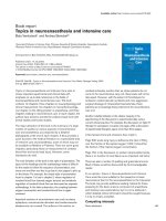
Figure 1
Risk for death among patients with AKI. The data were obtained in a
recent review of 13 studies that used RIFLE criteria where patient level
data on mortality were available for Risk, Injury and Failure patients, as
well as those without AKI [3]. The review was conducted to establish a
pooled estimate of risk ratio (RR) for mortality for patients of Risk,
Injury or Failure classes compared with non-AKI patients. More than
71,000 patients were included in the analysis of published reports.
With respect to non-AKI patients, there appeared to be a step-wise
increase in RR for death going from Risk class (RR = 2.40) to Injury
class (RR = 4.15) and to Failure class (RR = 6.37; P < 0.0001 for all).
AKI, acute kidney injury; RIFLE, Risk, Injury, Failure, Loss and End-
stage kidney disease.
Page 3 of 7
(page number not for citation purposes)
RIFLE criteria, the AKIN criteria did not substantially improve
the sensitivity, robustness and predictive ability of the
definition and classification of AKI. However, this study
looked only at the first ICU day, and therefore the 48-hour
time frame required by AKIN was totally neglected. The
design employed by that study was suboptimal in terms of
comparing the two classifications, but new evidence
regarding the clinical validity of the AKIN criteria (and
compared with RIFLE) is expected in the coming year.
Biomarkers
AKI has been defined and detected by measuring surrogates
of kidney function such as serum creatinine and urine output,
but these markers are acknowledged as being insensitive and
nonspecific for both acute changes to kidney function and
kidney injury. They also increase late in the injury process.
Consequently, they may not allow detection of an acute insult
or potentially ongoing injury to the kidney. Alternative markers
such as urinary analysis and novel biomarkers have been
extensively examined and reviewed during the past 2 years.
Two recent reviews [10,11] recently concluded that the
scientific basis for use of urinary biochemistry, indices and
microscopy in patients with septic AKI is weak. More
research is required to describe the accuracy, pattern and
time course of these parameters in patients with septic AKI.
Another review of 14 articles on the value of urinary bio-
markers in the specific setting of septic AKI [12] showed that
a promising future is apparently reserved for urinary
biomarkers, although most of the reviewed studies were
small, single-centre and included mixed populations of
medical/surgical adult patients. Retrieved articles included
data on low-molecular-weight proteins (β
2
-microglobulin,
α
1
-microglobulin, adenosine deaminase binding protein,
retinol binding protein, cystatin C and renal tubular epithelial
antigen-1), enzymes (N-acetyl-β-glucosaminidase, alanine
aminopeptidase, alkaline phosphatase, lactate dehydroge-
nase, α/π-glutathione-S-transferase, and γ-glutamyl trans-
peptidase), cytokines (platelet-activating factor and IL-18)
and other biomarkers (kidney injury molecule-1 and Na/H
exchanger isoform-3). Increased levels of platelet-activating
factor, IL-18 and Na/H exchanger isoform-3 were detected
early in septic AKI and predicted kidney failure. Several
additional biomarkers were evident early in AKI, but their
diagnostic value in sepsis remains unknown. In one study,
IL-18 excretion was greater in septic than in nonseptic AKI.
IL-18 also predicted deterioration in kidney function, with
increased values preceding clinically significant kidney failure
by 24 to 48 hours. Detection of cystatin C, α
1
-microglobulin
and IL-18 predicted need for renal replacement therapy
(RRT). The authors concluded that that selected biomarkers
may be promising in early detection of AKI in sepsis and may
have value for predicting subsequent deterioration in kidney
function.
Neutrophil gelatinase-associated lipocalin (NGAL) is among
the most upregulated genes in the kidney soon after
ischaemic injury [13] and is easily detected in plasma and
Available online />Table 1
Classification/staging system for acute kidney injury
System Class/stage Serum creatinine criteria Urine output criteria
RIFLE Class R Serum creatinine increase to 1.5-fold or GFR decrease >25% <0.5 ml/kg/hour for 6 hours
from baseline
Class I Serum creatinine increase to 2-fold or GFR decrease >50% <0.5 ml/kg/hour for 12 hours
from baseline
Class F Serum creatinine to 3-fold, GFR decrease >75% from baseline Anuria for 12 hours
or serum creatinine ≥4 mg/dl (≥ 354 μmol/l) with an acute increase of
at least 0.5 mg/dl (44 μmol/l)
AKIN Stage 1 Serum creatinine increase ≥0.3 mg/dl (≥26.4 μmol/l) or increase <0.5 ml/kg per hour for 6 hours
to 1.5-fold to 2-fold from baseline
Stage 2 Serum creatinine increase >2-fold to 3-fold from baseline <0.5 ml/kg per hour for 12 hours
Stage 3 Serum creatinine increase >3-fold from baseline or serum <0.3 ml/kg per hour for 24 hours or
creatinine ≥4.0 mg/dl (≥354 μmol/l) with an acute increase of at anuria for 12 hours
least 0.5 mg/dl (44 μmol/l)
Need for RRT
Synopsis of RIFLE and AKIN criteria for AKI classification/staging. Small but significant changes can be identified between the two definitions. A
time constraint of 48 hours for diagnosis (creatinine or urine output modifications) is required in AKIN criteria. GFR decreases for diagnosis are
specified only by RIFLE. In both cases, only one criterion - creatinine or urine output - must be fulfilled to qualify for a class/stage. Classes L (loss of
function) and E (end-stage kidney disease) of the RIFLE criteria are not reported. Given the wide variation in indications and timing of initiation of
RRT, individuals who receive RRT are considered to have met the criteria for AKIN stage 3 irrespective of the stage they are in at the time of RRT.
From Bellomo and coworkers [4] and Mehta and colleagues [5]. AKI, acute kidney injury; AKIN, Acute Kidney Injury Network; GFR, glomerular
filtration rate; RIFLE, Risk, Injury, Failure, Loss and End-stage kidney disease; RRT, renal replacement therapy.
urine in animal models of ischaemic and nephrotoxic AKI [14].
Expression of NGAL protein is also dramatically increased in
kidney tubules of humans with ischaemic, septic, and post-
transplant AKI [15]. Importantly, NGAL in plasma was found
to be an early predictive biomarker of AKI in a variety of acute
clinical settings in pilot studies. In a cohort of 20 patients who
developed AKI 2 to 3 days after cardiac surgery, plasma
NGAL was elevated within 2 to 6 hours after cardiopulmonary
bypass (CBP) [16]. Preliminary results using the research-
based assay also suggested that plasma NGAL measure-
ments predict AKI after contrast administration [17].
A recent prospective study was conducted to validate the
Triage
®
NGAL test (Biosite, Inc., San Diego, CA, USA), a
point-of-care, fluorescence-based immunoassay for the rapid
quantitative measurement of NGAL concentration in EDTA-
anticoagulated whole blood or plasma specimens [18]. The
study had the double aim of determining whether a rapid,
standardized point-of-care NGAL assay correlated with the
research-based assay. The second objective was to
determine the utility of the point-of-care NGAL assay as a
predictive biomarker of AKI after CPB in a large prospective
paediatric cohort. During the cross-sectional analysis of 40
plasma samples (NGAL range 60 to 730 ng/ml) and 12
calibration standards (NGAL range 0 to 1,925 ng/ml), NGAL
measurements by enzyme-linked immunosorbent assay and
by Triage
®
NGAL test were highly correlated (r = 0.94).
Then, 120 children undergoing CPB were enrolled in a
subsequent prospective and uncontrolled cohort study [18].
Plasma was collected at baseline and at frequent intervals for
24 hours after CPB and analyzed for NGAL using the Triage
®
NGAL test. The primary outcome was AKI, defined as a 50%
or greater increase in serum creatinine. AKI developed in 45
patients (37%), but diagnosis using serum creatinine was
delayed by 2 to 3 days after CPB. In contrast, mean plasma
NGAL levels increased threefold within 2 hours of CPB, and
remained significantly elevated for the duration of the study.
By multivariate analysis, plasma NGAL at 2 hours after CPB
was the most powerful independent predictor of AKI
(P < 0.0001). For the 2-hour plasma NGAL measurement,
the area under the curve (AUC) was 0.96, sensitivity was
0.84, and the specificity was 0.94 for prediction of AKI using
a cut-off value of 150 ng/ml. The 2-hour postoperative plasma
NGAL levels correlated strongly with change in creatinine
(r = 0.46, P < 0.001), duration of AKI (r = 0.57, P < 0.001)
and length of hospital stay (r = 0.44, P < 0.001). The 12-hour
plasma NGAL strongly correlated with mortality (r = 0.48,
P = 0.004) and all measures of morbidity mentioned above.
The study is interesting and results are significant. The
authors enrolled a large cohort of paediatric cardiac surgery
patients, correctly utilizing the RIFLE criteria in order to
achieve results that were comparable with current literature
and similar trials. Nonetheless, it must be noted that paedia-
tric postcardiosurgical AKI may have particular characteristics
(starting from an AKI incidence of more than 30%) that
require these results to be confirmed in a large adult cohort
before the assay can be regarded to be definitively validated.
Furthermore, additional prospective studies are needed to
describe the diagnostic and prognostic values of NGAL
accurately in septic AKI.
Interestingly, in a cohort of 140 paediatric critically ill patients
(aged between 1 month and 21 years who were mechanically
ventilated and had a bladder catheter), Zappitelli and co-
workers [19] correlated urinary NGAL values with AKI
severity diagnosed with pRIFLE (a paediatric modified version
of the RIFLE criteria described previously [20]). Thirty-four
(24.3%) patients never sustained AKI and served as control
patients. A total of 106 (75.7%) patients developed AKI.
Mean and peak urinary NGAL concentrations increased with
worsening pRIFLEmax status (P < 0.05). Urinary NGAL
concentrations rose (at least sixfold higher than in control
patients) in AKI, 2 days before and after a 50% or greater rise
in serum creatinine, without change in control urinary NGAL.
This marker performed as a good diagnostic marker for AKI
development (AUC = 0.78) and persistent AKI for 48 hours
or longer (AUC = 0.79). Urinary NGAL exhibited a worse
performance in terms of diagnosing AKI, when it was
measured after a rise in serum creatinine had occurred
(AUC = 0.63). Furthermore, despite the known association
between NGAL and inflammation [21], an association
between pRIFLEmax and increasing urinary NGAL concentra-
tions in patients with sepsis and in those without sepsis was
observed. Urinary NGAL can be considered an early AKI
marker in a heterogeneous group of critically ill paediatric
patients with unknown timing of kidney injury.
If the promising value of novel urinary and serum biomarkers
is confirmed by large-scale studies conducted in different
populations of critically ill patients, then it might be expected
that these molecules will be utilized as an alternative to
creatinine for AKI definition, diagnosis, prognosis and
therapeutic timing [22].
Epidemiology
An Australian survey of data of all adult admissions to 20
Australian ICUs for 24 hours or more from 1 January 1996 to
31 December 2005 was conducted to assess trends in
incidence and mortality for ICU admissions associated with
early AKI [23]. The authors analyzed 91,254 patient
admissions, including 4,754 cases of AKI, for an estimated
crude cumulative incidence of 5.2%. The incidence of AKI
increased during the study period, with an estimated annual
increment of 2.8%. The crude hospital mortality was
significantly higher in patients with AKI than in those without it
(42.7% versus 13.4%). There was also a decrease in AKI
crude mortality (annual percentage change: -3.4%), however,
which was not observed in patients without AKI. After
covariate adjustment, AKI remained associated with a higher
mortality (odds ratio = 1.23; P < 0.001), but there was a
declining trend in the odds ratio for hospital mortality.
Critical Care Vol 12 No 5 Ricci and Ronco
Page 4 of 7
(page number not for citation purposes)
Such retrospective studies and many other observational
ones suggest that critically ill patients with AKI are
increasingly older, have more co-morbid disease, have a
higher incidence of sepsis, and have greater severity of
illness and organ failure [24]. It is possible, however, that
outcomes in patients treated about 10 years ago is not
meaningful when compared with prospective data from
multinational and multicentre assessment of patients who are
all critically ill, treated in an ICU, and typically receiving
mechanical ventilation and vasopressor therapy [25]. Despite
the much greater severity of illness in patients treated today,
the mortality of AKI has not increased and has perhaps
slightly decreased, the duration of treatment has clearly
decreased in terms of need for dialysis; also, the time spent in
the ICU and in hospital, and artificial renal support techniques
have also changed markedly (significant findings have been
reported for the subgroup of AKI patients who have suffered
trauma [23]). It is likely, hence, that the 50% to 60% crude
mortality associated with AKI will not change dramatically in
the coming years, because as therapeutic (and diagnostic)
capabilities improve, the health care system will admit and
treat progressively sicker patients with AKI. Comparisons that
do not take into account this continuing adjustment and that
do not appreciate the continuing change in illness severity
will present a misleading picture of achievements associated
with improvements in technology and medications.
Danaparoid and vascular access for renal
replacement therapy
Heparin-induced thrombocytopenia (HIT) is caused by a
heparin-induced antibody that binds to the heparin-PF-4
complex on the platelet surface. In patients receiving heparin
continuous infusion, this may or may not lead to platelet
activation and consumption, thrombocytopenia, and both
arterial and venous thrombosis [26]. Depending on the dose
and type of heparin, the population and the criteria used, 1%
to 5% of treated patients develop HIT. Platelet count typically
decreases rapidly by more than 50% after approximately
1 week or earlier after use of heparin. Diagnosis depends on
a combination of clinical and laboratory results. Reliable
diagnosis is complicated by the fact that the incidence of
false-positive enzyme-linked immunosorbent assay findings is
high. Unfortunately, the more precise carbon 14-serotonin
release assay is not routinely available [27]. While awaiting a
final diagnosis, all forms of heparin should be discontinued
and an alternative anticoagulant administered. There are no
randomized controlled trials to show which anticoagulant is
best for HIT. The choice depends on local availability and
monitoring experience. Inhibition of thrombin generation can
be achieved via direct inhibition of factor IIa (r-hirudin,
argatroban, or dermatan sulphate), factor Xa (danaparoid or
fondaparinux), or both (nafamostat) [23].
In a recent study, the pharmacokinetics and pharmaco-
dynamics of danaparoid during continuous venovenous
haemofiltration (CVVH) were examined in patients with sus-
pected HIT [28]. During CVVH, danaparoid can be removed
only by means of a polyarylethersulphone membrane, with a
sieving coefficient of 0.78 ± 0.03 [25]. Therefore, treatment
with a continuous infusion of danaparoid may carry a risk for
accumulation in patients with AKI who are dependent on
CVVH. Using the recommended dosing scheme, patients
may experience bleeding, especially when peak anti-Xa levels
exceed 1.0 anti-Xa U/ml. Therefore, a safer dosing scheme
for danaparoid was tested, based on a mathematical model
aiming to achieve a peak anti-Xa level of less than 1.0 and a
maintenance level of between 0.5 and 0.7 anti-Xa U/ml. This
dosing scheme was prospectively tested in the first CVVH
run in a cohort of five patients with suspected HIT. CVVH
with a blood flow rate of 150 ml/minute and a substitution
rate of 2,000 ml/hour was performed using a cellulose
triacetate membrane. Based on the mathematical model,
danaparoid was administered as a continuous infusion of 100
anti-Xa U/hour after a loading dose of 3,500 anti-Xa U. Serial
measurements of anti-Xa activity and prothrombin fragment
F
1+2
were performed at baseline; at 5, 15 and 30 minutes;
and at 1, 2, 4, 8, 16 and 24 hours after the danaparoid
loading dose. The median anti-Xa activity reached a maximum
of 1.02 (0.66 to 1.31) anti-Xa U/ml after 15 minutes and
gradually declined to 0.40 (0.15 to 0.58) anti-Xa U/ml over
24 hours. Target anti-Xa levels were reached 2 to 12 hours
after the loading dose. Median prothrombin fragment F
1+2
gradually decreased from 432 (200 to 768) to 262 (248 to
317) pmol/l after 24 hours. A more than adequate median
circuit survival time of 50.2 (20 to 89) hours was achieved.
No bleeding or thromboembolic events occurred throughout
the described treatment period. These interesting results
require confirmation in a clinical trial using standard heparin
as control anticoagulation aiming to evaluate the efficacy and
safety of danaparoid ‘low-dose’ infusion.
Unger and colleagues [29] conducted an original experi-
mental study to verify a strategy of blood flow rate
optimization in a extracorporeal circuit via common dual-
lumen catheters. The authors treated 34 ventilated supine
pigs with CVVH using traditional axial dual-lumen catheter
(11-Fr, 20 cm long, side holes) placed in the femoral vein (19
animals) or dual-lumen catheter plus an 8.5-Fr sheath (15
animals), after determination of the highest values for access
flow (Qa). High Qa rates (>300 ml/minute) were allocated to
the dual-lumen catheter group; low Qa rates were switched
to a ‘dual-vein’ approach, placing a second catheter (8.5-Fr
sheath) for separate blood delivery. The authors aimed to
demonstrate whether hypovolaemia, pathological haemo-
rheology (obtained with blood substitution by normal saline,
different 6% hydroxyethyl starches, human albumin or gelatin
polysuccinate) and/or low cardiac output reduced available
blood flow rates with the two approaches. Haemodynamics
(cardiac output and central venous pressure) and blood
composition (blood cell counts, plasma proteins and colloid
osmotic pressure) were measured. Catheter tip positions and
vessel diameters were studied using computed tomography.
Available online />Page 5 of 7
(page number not for citation purposes)
Interestingly, neither haemorheologically relevant aspects nor
cardiac output and central venous pressure correlated with
the achievable Qa via the femoral vein access. Even though
the catheter tip of the alternative catheter provided common
iliac vein but not caval vein access, this catheter type allowed
greater Qa than the dual-lumen catheter positioned in the
caval vein. None of the evaluated reasons for limited Qa via
the arterial line of an axial dual-lumen catheter could be
confirmed. The 8.5-Fr sheath performed quite well as an
alternative catheter, and the authors believe that clinical
studies to re-evaluate the dual-vein approach, with separate
blood delivery via a tip-hole catheter in order to provide high-
volume haemofiltration, are justified.
Similarly, Di Carlo and coworkers [30] proposed use of an
analogous approach in paediatric critically ill patients
undergoing continuous RRT. In order to establish non-
obstructive access for haemofiltration in a newborn, whose
femoral vein is about 4.5 mm diameter and is unlikely to
accommodate a 7-Fr catheter without near-total occlusion
and subsequent stasis, two single-lumen thin-walled vascular
‘introducer’ sheaths can be used in two separate veins.
During insertion over a guidewire, the thin wall of the sheath is
supported by a removable tapered dilator. A haemostasis valve
is in position immediately above the proximal entry point to the
sheath, and must be fully secured with an obturator to prevent
air embolus. Animal data suggest that polyurethane catheters
are less likely than silastic to encourage the growth of bacteria
in the presence of a fibrin sheath. The traditional sheath is
composed of Teflon, however, which is stiffer than polyurethane.
New generation sheaths will be released that are composed
of a polyurethane blend with improved kink resistance.
Another innovation might be a method for integrating wound-
wire reinforcement within the wall of the catheter.
It will be interesting to observe how these new approaches
may be used to manage the issues of circuit patency and
adequate blood flow rate during continuous RRT, which are
generally left to operator experience and nurse expertise.
Increased awareness of the importance of CRRT session
length and downtime is progressively leading to more
detailed research into the field of anticoagulation and
vascular access. However, in routine clinical practice
(especially in the paediatric setting), continuous treatments
with low-dose or no heparin and double-lumen catheters
requiring a single venipuncture are still ‘gold standards’ and
(thus far) represent the safest and simplest strategy.
Conclusion
In conclusion, the reported manuscripts showed how RIFLE
criteria were widely accepted by scientific community,
whereas AKIN criteria are still under validation. NGAL assay
and other biomarkers for early AKI diagnosis, when certified
for routine use in clinical practice, will certainly be a major
progress for prevention and therapy of AKI, and we will might
assist to a significant improvement in AKI outcome in the next
10 years. Finally, if original and safe anticoagulation strategies
are important for CRRT circuit patency, optimization of
vascular access is definitely one of the most important targets
in order to increase filter lifespan: new original studies on
these fields are strongly needed.
Competing interests
The authors declare that they have no competing interests.
References
1. Bernard GR, Artigas A, Brigham KL, Carlet J, Falke K, Hudson L,
Lamy M, Legall JR, Morris A, Spragg R. The American-European
Consensus Conference on ARDS. Definitions, mechanisms,
relevant outcomes, and clinical trial coordination. Am J Respir
Crit Care Med 1994, 149:818-824.
2. Kellum JA: Acute kidney injury. Crit Care Med 2008, 36:S141-
S145.
3. Ricci Z, Cruz D, Ronco C: The RIFLE criteria and mortality in
acute kidney injury: A systematic review. Kidney Int 2008, 73:-
538-546.
4. Bellomo R, Ronco C, Kellum JA, Mehta RL , Palevsky P and the
ADQI workgroup: Acute renal failure - definition, outcome
measures, animal models, fluid therapy and information tech-
nology needs: the Second International Consensus Confer-
ence of the Acute Dialysis Quality Initiative (ADQI) Group. Crit
Care 2004, 8:R204-R212.
5. Mehta RL, Kellum JA, Shah SV, Molitoris BA, Ronco C, Warnock
DG, Levin A and the Acute Kidney Injury Network: Acute Kidney
Injury Network: report of an initiative to improve outcomes in
acute kidney injury. Critical Care 2007, 11:R31.
6. Lassnigg A, Schmidlin D, Mouhieddine M, Bachmann LM, Druml
W, Bauer P, Hiesmayr M. Minimal changes of serum creatinine
predict prognosis in patients after cardiothoracic surgery: a
prospective cohort study. J Am Soc Nephrol 2004, 15:1597-
1605.
7. Chertow GM, Burdick E, Honour M, Bonventre JM, Bates DW:
Acute kidney injury, mortality, length of stay, and costs in hos-
pitalized patients. J Am Soc Nephrol 2005, 16:3365-3370.
8. Barrantes F, Tian J, Vazquez R, Amoateng-Adjepong Y, Manthous
CA: Acute kidney injury criteria predict outcomes of critically
ill patients. Crit Care Med 2008, 36:1397-1403.
9. Bagshaw SM, George C, Bellomo R; ANZICS Database Manage-
ment Committee: A comparison of the RIFLE and AKIN criteria
for acute kidney injury in critically ill patients. Nephrol Dial
Transplant 2008, 23:1569-1574.
10. Bagshaw SM, Langenberg C, Wan L, May CN, Bellomo R: A sys-
tematic review of urinary findings in experimental septic acute
renal failure. Crit Care Med 2007, 35:1592-1598.
11. Bagshaw SM, Langenberg C, Bellomo R: Urinary biochemistry
and microscopy in septic acute renal failure: a systematic
review. Am J Kidney Dis 2006, 48:695-705.
12. Bagshaw SM, Langenberg C, Haase M, Wan L, May CN, Bellomo
R: Urinary biomarkers in septic acute kidney injury. Intensive
Care Med 2007, 33:1285-1296.
13. Mishra J, Ma Q, Prada A, Mitsnefes M, Zahedi K, Yang J, Barasch
J, Devarajan P: Identification of NGAL as a novel urinary bio-
marker for ischemic injury. J Am Soc Nephrol 2003, 4:
2534-
2543.
14. Mishra J, Mori K, Ma Q, Kelly C, Yang J, Mitsnefes M. Barasch J.
Devarajan P. Amelioration of ischemic acute renal injury by
NGAL. J Am Soc Nephrol 2004, 15:3073-3082.
15. Mishra J, Ma Q, Kelly C, Mitsnefes M, Mori K, Barasch J, Devara-
jan P: Kidney NGAL is a novel early marker of acute injury fol-
lowing transplantation. Pediatr Nephrol 2006, 21:856-863.
16. Mishra J, Dent C, Tarabishi R, Mitsnefes MM, Ma Q, Kelly C, Ruff
SM, Zahedi K, Shao M, Bean J, Mori K, Barasch J, Devarajan P:
Neutrophil gelatinase-associated lipocalin (NGAL) as a bio-
marker for acute renal injury after cardiac surgery. Lancet
2005, 365:1231-1238.
17. Hirsch R, Dent C, Pfriem H, Allen J, Beekman RH, Ma Q, Bennett
M, Mitsnefes M, Devarajan P: NGAL is an early predictive bio-
marker of contrast-induced nephropathy in children. Pediatr
Nephrol 2007, 22:2089-2095.
Critical Care Vol 12 No 5 Ricci and Ronco
Page 6 of 7
(page number not for citation purposes)
18. Dent CL, Ma Q, Dastrala S, Bennett M, Mitsnefes MM, Barasch J,
Devarajan P: Plasma neutrophil gelatinase-associated
lipocalin predicts acute kidney injury, morbidity and mortality
after pediatric cardiac surgery: a prospective uncontrolled
cohort study. Crit Care 2007, 11:R127.
19. Zappitelli M, Washburn KK, Arikan AA, Loftis L, Ma Q, Devarajan
P, Parikh CR, Goldstein SL. Urine neutrophil gelatinase-associ-
ated lipocalin is an early marker of acute kidney injury in criti-
cally ill children: a prospective cohort study. Crit Care 2007,
11:R84.
20. Akcan-Arikan A, Zappitelli M, Loftis LL, Washburn KK, Jefferson
LS, Goldstein SL: Modified RIFLE criteria in critically ill children
with acute kidney injury. Kidney Int 2007, 71:1028-1035.
21. Xu SY, Pauksen K, Venge P: Serum measurements of human
neutrophil lipocalin (HNL) discriminate between acute bacter-
ial and viral infections. Scand J Clin Lab Invest 1995, 55:125-
131.
22. Ronco C: N-GAL: diagnosing AKI as soon as possible. Crit
Care 2007, 11:173.
23. Bagshaw SM, George C, Bellomo R; the ANZICS Database Man-
agement Committee: Changes in the incidence and outcome
for early acute kidney injury in a cohort of Australian intensive
care units. Crit Care 2007, 11:R68.
24. de Mendonca A, Vincent JL, Suter PM, Moreno R, Dearden NM,
Antonelli M, Takala J, Sprung C, Cantraine F: Acute renal failure
in the ICU: risk factors and outcome evaluated by the SOFA
score. Intensive Care Med 2000, 26:915-921.
25. Bellomo R: The epidemiology of acute renal failure: 1975
versus 2005. Curr Opin Crit Care 2006, 12:557-560.
26. Joannidis M, Oudemans-van Straaten HM: Clinical review:
patency of the circuit in continuous renal replacement
therapy. Crit Care 2007, 11:218.
27. Warkentin TE, Greinacher A: Heparin-induced thrombocytope-
nia: recognition, treatment, and prevention: the Seventh ACCP
Conference on Antithrombotic and Thrombolytic Therapy.
Chest 2004, 126:311S-337S.
28. de Pont AC, Hofstra JJ, Pik DR, Meijers JC, Schultz MJ: Pharma-
cokinetics and pharmacodynamics of danaparoid during con-
tinuous venovenous hemofiltration: a pilot study. Crit Care
2007, 11:R102.
29. Unger JK, Pietzner K, Francis RC, Birnbaum J, Theisen MM,
Lemke AJ, Niehues SM: Dual-lumen catheters for continuous
venovenous hemofiltration: limits for blood delivery via
femoral vein access and a potential alternative in an experi-
mental setting in anesthetized pigs. Crit Care 2007, 11:R18.
30. DiCarlo JV, Auerbach SR and Alexander SR: Clinical review:
alternative vascular access techniques for continuous
hemofiltration. Critical Care 2006, 10:230.
Available online />Page 7 of 7
(page number not for citation purposes)