Báo cáo y học: "Comparison of functional residual capacity and static compliance of the respiratory system during a positive end-expiratory pressure (PEEP) ramp procedure in an experimental model of acute respiratory distress syndrome" potx
Bạn đang xem bản rút gọn của tài liệu. Xem và tải ngay bản đầy đủ của tài liệu tại đây (319.57 KB, 6 trang )
Open Access
Available online />Page 1 of 6
(page number not for citation purposes)
Vol 12 No 4
Research
Comparison of functional residual capacity and static compliance
of the respiratory system during a positive end-expiratory
pressure (PEEP) ramp procedure in an experimental model of
acute respiratory distress syndrome
Bernard Lambermont
1,2
, Alexandre Ghuysen
1,3
, Nathalie Janssen
1,3
, Philippe Morimont
1,2
,
Gary Hartstein
3
, Paul Gerard
1,4
and Vincent D'Orio
1,3
1
Hemodynamic Research Center, HemoLiege, University of Liege, Belgium
2
Medical Intensive Care Unit, Department of Medicine, University Hospital of Liege, Belgium
3
Emergency Care Department, University Hospital of Liege, Belgium
4
Department of Statistics, University of Liege, Belgium
Corresponding author: Bernard Lambermont,
Received: 11 Apr 2008 Revisions requested: 11 Jun 2008 Revisions received: 25 Jun 2008 Accepted: 16 Jul 2008 Published: 16 Jul 2008
Critical Care 2008, 12:R91 (doi:10.1186/cc6961)
This article is online at: />© 2008 Lambermont et al.; licensee BioMed Central Ltd.
This is an open access article distributed under the terms of the Creative Commons Attribution License ( />),
which permits unrestricted use, distribution, and reproduction in any medium, provided the original work is properly cited.
Abstract
Introduction Functional residual capacity (FRC) measurement
is now available on new ventilators as an automated procedure.
We compared FRC, static thoracopulmonary compliance (Crs)
and PaO
2
evolution in an experimental model of acute
respiratory distress syndrome (ARDS) during a reversed,
sequential ramp procedure of positive end-expiratory pressure
(PEEP) changes to investigate the potential interest of
combined FRC and Crs measurement in such a pathologic
state.
Methods ARDS was induced by oleic acid injection in six
anesthetised pigs. FRC and Crs were measured, and arterial
blood samples were taken after induction of ARDS during a
sequential ramp change of PEEP from 20 cm H
2
O to 0 cm H
2
O
by steps of 5 cm H
2
O.
Results ARDS was responsible for significant decreases in
FRC, Crs and PaO
2
values. During ARDS, 20 cm H
2
O of PEEP
was associated with FRC values that increased from 6.2 ± 1.3
to 19.7 ± 2.9 ml/kg and a significant improvement in PaO
2
. The
maximal value of Crs was reached at a PEEP of 15 cm H
2
O, and
the maximal value of FRC at a PEEP of 20 cm H
2
O. From a
PEEP value of 15 to 0 cm H
2
O, FRC and Crs decreased
progressively.
Conclusion Our results indicate that combined FRC and Crs
measurements may help to identify the optimal level of PEEP.
Indeed, by taking into account the value of both parameters
during a sequential ramp change of PEEP from 20 cm H
2
O to 0
cm H
2
O by steps of 5 cm H
2
O, the end of overdistension may
be identified by an increase in Crs and the start of derecruitment
by an abrupt decrease in FRC.
Introduction
In acute respiratory distress syndrome (ARDS) the setting of
positive end-expiratory pressure (PEEP) is determined using
several methods, including FiO
2
requirement, measurement of
either static (Crs) or dynamic thoracopulmonary compliance
[1-4], generation of pressure-volume curves [2,5,6] and,
rarely, using computed tomography (CT) scan analysis [5,7-
10]. During a decremental PEEP manoeuvre, the point of max-
imal Crs has been shown to correspond to the minimum open
lung positive end-expiratory pressure preventing end-expira-
tory collapse of those alveoli which are inflated at end inspira-
tion [1]. The volume recruited by PEEP is usually assessed by
a method based on the static pressure-volume curve of the
respiratory system. Alveolar recruitment leads to an upward
shift along the volume axis of the pressure-volume curve with
PEEP, compared to the curve with zero end-expiratory pres-
sure, and is quantified as the volume increase with PEEP at the
same elastic pressure [11]. Functional residual capacity
(FRC), which reflects the amount of gas present in the lungs,
has been suggested to be a better indicator than Crs to
assess the state of recruitment and derecruitment caused by
ARDS = acute respiratory distress syndrome; Crs = static thoracopulmonary compliance of the respiratory system; FRC = functional residual capac-
ity, PEEP = positive end-expiratory pressure; Pexp = expiratory plateau airway pressure; Pins = inspiratory plateau airway pressure; Vt = tidal volume.
Critical Care Vol 12 No 4 Lambermont et al.
Page 2 of 6
(page number not for citation purposes)
PEEP manipulations, because it directly measures the lung
volume increase induced by PEEP, mainly due to the recruit-
ment of collapsed alveoli [12]. However, FRC measurement is
not usually performed at the bedside because of technical lim-
itations. More recently, an automated procedure for FRC
measurement has become available and is incorporated into
the software of specific intensive care ventilators [13]. There-
fore, we compared FRC, Crs and PaO
2
evolution in an experi-
mental model of ARDS during a reversed sequential ramp
procedure of PEEP changes to investigate the potential inter-
est of combining FRC and Crs measurements in such a path-
ologic state.
Materials and methods
All experimental procedures and protocols used in this inves-
tigation were reviewed and approved by the Ethics Committee
of the Medical Faculty of the University of Liege. The investiga-
tion conforms with guidelines on laboratory animals published
by the US National Institutes of Health.
Six pigs weighing 26 ± 2 kg were premedicated with tiletamin/
zolazepam 5 mg/kg and subsequently anaesthetised by a con-
tinuous infusion of sufentanil 0.5 μg/kg/h, pentobarbital 5 mg/
kg/h and cisatracurium 2 mg/kg/h. They were ventilated
through a tracheotomy in volume control mode at a fraction of
inspired oxygen (FiO
2
) of 0.5 with a tidal volume of 10 ml/kg,
an inhalation/exhalation (I:E) ratio of 1:2, a rate of 20 breath/
min, and 5 cm H
2
O PEEP (Engström CareStation, Datex, Gen-
eral Electric, Finland).
Systemic arterial pressure was measured by a catheter (Sen-
tron pressure-measuring catheter, Cordis, Miami, FL, USA)
introduced in the abdominal aorta through the right femoral
artery. Heart rate was obtained from one derivation continuous
electrocardiogram monitoring.
FRC was calculated using an automated procedure available
on the ventilator based on the nitrogen washout method with
a FiO
2
step change of 0.1, as previously described by Olegard
et al. [13]. Using sidestream gas analysing technology, calcu-
lation of FRC values was obtained by applying the following
equations.
The fractions of inspired and end-tidal nitrogen were calcu-
lated from:
F
I
N
2
= 1 - F
I
O
2
ETN
2
= 1 - ETO
2
- ETCO
2
Expired and inspired alveolar tidal volumes were calculated
using energy expenditure measurements for VO
2
and VCO
2
where VO
2
= (VCO
2
/RQ):
Nitrogen volumes associated with expiration and inspiration
for a single breath were:
The changes during one breath equalled:
Before making the step change in F
I
O
2
, a baseline condition
was determined. This involved the determination of VO
2
,
VCO
2
and ETN
2
baseline
. VO
2
anv VCO
2
were assumed to be
constant throughout the measurement. After a step response
the FRC was calculated as:
Where the ETN
2
was the last recorded value after the step
change:
Airway pressure values were measured by the ventilator at the
level of the Y piece just before the tracheostomy tube. Crs was
measured by holding a 10 s inspiratory pause to obtain the
value of the inspiratory plateau airway pressure (Pins) and a 10
s expiratory pause to obtain the end-expiratory airway pressure
(Pexp). The value of Crs was obtained by dividing tidal volume
(Vt) by the difference between inspiratory plateau airway pres-
sure and end-expiratory airway pressure:
Crs = Vt/(Pins - Pexp)
Materials and methods
After a 30-min period of stabilisation, measurements were
obtained at a PEEP of 5 cm H
2
O (basal). Then, ARDS was
induced by administration of 0.12 ml/kg of oleic acid over 30
min.
At 120 min after the beginning of oleic acid injection, a set of
parameters was obtained at a PEEP level of 5 cm H
2
O
(ARDS). Subsequently, PEEP was increased to 20 cm H
2
O
and then reduced by steps of 5 cm H
2
O to 0 cm H
2
O
V
VC O
2
ETCO
2
.RR
t
alvE()
=
VV
VO
2
VC O
2
RR
t
alvI
t
alvE() ( )
=+
−
VETNV
E
N
t
alvE
2
2
= .
()
VFNV
I
N
It
alvI
2
2
= .
()
ΔVVV
N
E
N
I
N
2
22
=−
FR C
V
N
ETN
=
Δ
Δ
2
2
FR C
V
N
breaths
ETN
baseline
ETN
=
∑
Δ
2
2
2
−
Available online />Page 3 of 6
(page number not for citation purposes)
(ARDS20, ARDS15, ARDS10, ARDS5, ARDS0). Each PEEP
level was maintained for 15 min before a set of measurements
to allow for haemodynamic and respiratory stabilisation.
Arterial blood samples were taken during the basal condition
(basal), 120 min after oleic acid injection, and at each PEEP
level during ARDS.
Animals received neither vasoactive nor inotrope drugs during
the procedure.
Statistics
Data are presented as mean ± standard error of the mean. Sta-
tistical comparison of data over time was conducted by a two-
way analysis of variance (ANOVA) for repeated measure-
ments, followed by Scheffe's multiple comparisons test if the
analysis of variance resulted in p value < 0.05 (Statistica,
Statsoft Inc., Tulsa, OK, USA).
Correlations between FRC, static compliance, and PaO
2
were
evaluated by a Pearson's linear correlation test (Statistica).
Difference between correlations was evaluated by an equality
of dependent correlations test [14]. A p value < 0.05 was con-
sidered statistically significant.
Results
ARDS was responsible for significant decreases in both FRC
and Crs, from 16 ± 2 to 6.2 ± 1.3 ml/kg and from 28 ± 2 to
17 ± 1 ml/cm H
2
O, respectively. Values of PaO
2
changed
from 201 ± 7 to 52 ± 5 mmHg 120 min after oleic acid
administration.
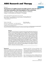
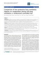
During ARDS, 20 cm H
2
O of PEEP was associated with FRC
values that increased from 6.2 ± 1.3 to 19.7 ± 2.9 ml/kg. This
change was associated with a significant improvement in
PaO
2
, which reached 172 ± 15 mmHg. The point of maximum
Crs during ARDS was reached at a PEEP of 15 cm H
2
O. From
a PEEP value of 15 to 0 cm H
2
O, FRC and Crs decreased pro-
gressively (Figure 1).
The time course of haemodynamic data, arterial blood gases,
and inspiratory plateau airway pressure during ARDS is pre-
sented in Table 1. Inspiratory plateau pressure increased sig-
nificantly (p < 0.05) from 14 ± 0.8 (basal) to 20 ± 0.5 cm H
2
O
(ARDS) after oleic acid injection. During ARDS, inspiratory
plateau airway pressure was significantly increased at a PEEP
of 20 and 15 cm H
2
O (p < 0.05). Maximal oxygenation was
obtained at a PEEP of 20 and 15 cm H
2
O. PaCO
2
was 42 ±
2 mmHg during basal condition and 54 ± 5 mmHg after oleic
acid injection. During ARDS, PaCO
2
increased significantly
from 54 ± 5 mmHg (ARDS) to 62 ± 3 at a PEEP of 0 cm H
2
O
(p < 0.05).
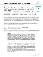
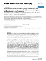
Correlation between PaO
2
and FRC (Figure 2), and between
PaO
2
and Crs (Figure 3) were significant (p < 0.05) but weak
(r
2
= 0.53 and 0.4, respectively); the difference between the
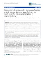
two correlations was not significant (p = 0.41). Correlation
between FRC and Crs was also significant but weak (p <
0.05, r
2
= 0.26) (Figure 4).
Discussion
In this experimental model of ARDS, FRC and Crs values
obtained during mechanical ventilation were correlated to the
changes in PaO
2
obtained during a sequential ramp change of
PEEP from 20 cm H
2
O to 0 cm H
2
O by steps of 5 cm H
2
O.
The maximal value of Crs was reached immediately before
FRC began to decrease.
Our results are in accordance with those of Suarez-Sipman et
al., who showed that maximal dynamic compliance of the res-
piratory system immediately preceded the beginning of alveo-
lar collapse after lung recruitment as shown by computed
tomography (CT) scan studies [4]. Previous studies have
already demonstrated a strong and inverse correlation
between arterial oxygenation and the amount of collapsed lung
mass in multislice CT scans [15]. Rylander et al. suggested
that FRC was a more sensitive indicator of PEEP-induced aer-
ation and recruitment of lung tissue than Crs [12]. However,
an increase in FRC may be due to alveolar recruitment, but
may also be secondary to alveolar overdistension. To distin-
guish between these possibilities, use of thoracopulmonary
compliance has been suggested. Indeed, a parallel increase in
FRC and thoracopulmonary compliance suggests alveolar
recruitment while a decrease in thoracopulmonary compliance
together with increasing FRC would tend to indicate alveolar
overdistension [16]. Our results strengthen this suggestion:
Figure 1
Time course of functional residual capacity (FRC) and static compli-ance of the respiratory system (Crs) during a decremental positive end-expiratory pressure (PEEP) trial after acute respiratory distress syn-drome (ARDS) induction by oleic acid injectionTime course of functional residual capacity (FRC) and static compli-
ance of the respiratory system (Crs) during a decremental positive end-
expiratory pressure (PEEP) trial after acute respiratory distress syn-
drome (ARDS) induction by oleic acid injection. Measurements were
obtained 120 min after the oleic acid injection (ARDS) at a PEEP of 5
cm H
2
O and during a decremental PEEP trial from 20 to 0 cm H
2
O by
steps of 5 cm H
2
O (ARDS20, ARDS15, ARDS10, ARDS5, ARDS0).
Each PEEP level was maintained for 15 min before a set of measure-
ments to allow for haemodynamic and respiratory stabilisation. § p <
0.05 vs ARDS (Crs); * p < 0.05 vs ARDS (FRC).
Critical Care Vol 12 No 4 Lambermont et al.
Page 4 of 6
(page number not for citation purposes)
the fact that FRC did not decrease, while Crs increased, when
PEEP decreased from 20 and 15 cm H
2
O suggests that alve-
olar overdistension was present at a PEEP of 20 cm H
2
O.
In this study, the optimal level of PaO
2
was obtained at a PEEP
of 20 and 15 cm H
2
O. Because one of the goals of PEEP is
to reach an arterial saturation greater than 90% at the lowest
possible FiO
2
, the monitoring of arterial oxygenation could be
considered as the unique gold standard for optimising PEEP.
However, because alveolar recruitment and lung overinflation
can be simultaneously observed in different parts of the lung,
changes in PaO
2
cannot be considered sensitive enough to
detect the risk of ventilator induced lung injury [10]. Increasing
the level of PEEP can be more harmful than beneficial since it
will serve also to increase inflation of lung regions that are
already open, increasing the stress and strain on these regions
[7]. Two studies showed a significant positive correlation
between PEEP-related recruitment and arterial oxygenation
[8,17]. The correlations found in these two studies are unfor-
tunately relatively weak, suggesting that arterial oxygenation
cannot be used reliably to predict the amount of recruitment
induced by a given level of PEEP [9]. Our results confirm these
data, since correlations between either FRC or Crs and PaO
2
are also significant and weak without a difference between the
two correlations.
As suggested by our results, such an assessment is a valuable
tool to help to identify the optimal level of PEEP. It can also be
used for trend analysis, as a decrease in FRC can be the first
sign of derecruitment and may help the clinician to understand
the pathophysiological mechanism worsening blood oxygena-
tion. Finally, this parameter might provide practical help in ther-
apeutic decision making [18].
To our knowledge, this study is the first to measure FRC by
using the automated procedure available on the Engstrom
Care Station ventilator in a porcine model of ARDS. This
method has been validated by Olegard et al. [13]. They have
shown that FRC measurement with high precision can be
obtained using a N
2
multiple breath washout technique based
on standard gas monitoring equipment and an FiO
2
step
change as little as 0.1. As the calculation of FRC is based on
the values of VCO
2
, end-tidal O
2
and end-tidal CO
2
, all these
values need to be valid to result in acceptable results. The con-
ditions that may cause invalid data include: rapid and/or irreg-
ular respiratory rates, large variations in tidal volumes, high
fevers, agitation, neurological conditions that alter respiration.
Constant breathing patterns are required to achieve valid
VCO
2
measurements; this was the case in the experimental
Table 1
Time course of haemodynamic parameters, inspiratory plateau airway pressure, arterial blood pH, PaO
2
and PaCO
2
after oleic acid
injection and during a decremental PEEP trial from 20 to 0 cm H
2
O.
HR (beats/min) Mean AP (mmHg) pH PaO
2
(mmHg) PaCO
2
(mmHg) Inspiratory plateau pressure (cm H
2
O)
ARDS 129 ± 3 75 ± 16 7.32 ± 0.06 52 ± 5 54 ± 5 20 ± 1
ARDS20 132 ± 4 66 ± 13 7.33 ± 0.04 172 ± 15* 54 ± 3 36 ± 3*
ARDS15 134 ± 4 73 ± 13 7.38 ± 0.02 175 ± 17* 54 ± 2 26 ± 1*
ARDS10 145 ± 3* 75 ± 9 7.28 ± 0.05 89 ± 19* 56 ± 5 23 ± 2
ARDS5 165 ± 10* 118 ± 9* 7.33 ± 0.02 52 ± 4* 59 ± 2 22 ± 2
ARDS0 153 ± 7* 101 ± 5* 7.3 ± 0.03 52 ± 4* 62 ± 3* 21 ± 2
Data are presented as mean ± standard error of the mean. Measurements were obtained 120 min after oleic acid injection at a PEEP of 5 cm H
2
O
(ARDS) and during a decremential PEEP trial (ARDS20, ARDS15, ARDS10, ARDS5, ARDS0). Each PEEP level was maintained for 15 min
before each measurement to allow for haemodynamic and respiratory stabilisation. ARDS, acute respiratory distress syndrome; HR, heart rate;
mean AP, mean systemic arterial pressure; PEEP, positive end-expiratory pressure. * p < 0.05 versus ARDS
Figure 2
Correlation between PaO
2
and functional residual capacity (FRC)Correlation between PaO
2
and functional residual capacity (FRC).
Available online />Page 5 of 6
(page number not for citation purposes)
conditions of the present study, but may not be warranted in
the clinical setting.
It could be argued that the effects of PEEP changes observed
during the ramp procedure were due to a lack of stability of this
ARDS model. However, The porcine oleic acid model of
ARDS used in this study has been extensively studied and
used to represent the early, exudative phase of ARDS. We
allowed a 120-min period of stabilisation after oleic acid injec-
tion before initiating the PEEP ramp procedure in order to pro-
vide a stable condition. This is more time than is actually
necessary since stable conditions can be generally reached in
this model after 30 to 60 min, according to several previous
studies [12,19,20].
No specific evaluation of the FRC technique was performed in
this study. FRC measurements were performed during a dec-
remental PEEP trial without specific evaluation of the tech-
nique, which was out of the scope of the study. However, it
would be interesting to determine, in a complementary study,
how reproducible the measurement is, and how this method of
determining FRC compares to other techniques for absolute
lung volume measurements as well as with techniques that
measures lung volume changes. Finally, since the findings of
this study were obtained using volume control mode ventila-
tion with a tidal volume of 10 ml/kg, the efficacy of this method
remains to be demonstrated in other ventilatory modes (such
as pressure control) and also with the lower tidal volumes usu-
ally used during ARDS.
Conclusion
Our results indicate that a combination of FRC and Crs meas-
urements obtained in this porcine model of ARDS may help to
identify the optimal level of PEEP. Indeed, by taking into
account the value of both parameters, during a sequential
ramp change of PEEP from 20 cm H
2
O to 0 cm H
2
O by steps
of 5 cm H
2
O, the end of overdistension may be identified by
an increase in Crs, and the start of derecruitment by an abrupt
decrease in FRC. Using this approach to find the best value of
PEEP should allow for the tidal excursion to be positioned
between derecruitment and overdistension on the pressure-
volume curve.
Competing interests
The authors declare that they have no competing interests.
Authors' contributions
BL, AG, NJ, PM participated in the design of the study, and
collected the data during the experiments. BL and PG ana-
lysed the data and performed the statistical analysis. VD par-
ticipated in the design and the coordination of the study and
helped to draft the manuscript. BL, AG, NJ, PM, GH, PG, VD
Figure 3
Correlation between PaO
2
and static compliance of the respiratory sys-tem (Crs)Correlation between Pa
and static compliance of the respiratory system (Crs).
Figure 4
Correlation between functional residual capacity (FRC) and static com-pliance of the respiratory system (Crs)Correlation between functional residual capacity (FRC) and static com-
pliance of the respiratory system (Crs).
Key messages
● Functional residual capacity measurement is now availa-
ble on new ventilators as an automated procedure.
● Combined measurement of thoracopulmonary static
compliance and functional residual capacity may help to
identify the optimal level of PEEP in ARDS.
Critical Care Vol 12 No 4 Lambermont et al.
Page 6 of 6
(page number not for citation purposes)
have been involved in drafting the manuscript or revising it crit-
ically and have given final approval of the version to be pub-
lished. All authors read and approved the final manuscript.
Acknowledgements
The authors thank Veronique Mommens for collecting the data. This
study was funded by a Grant of Fondation Léon Frédericq. The ventilator
used in the study was loaned by GE Healthcare.
References
1. Hickling KG: Best compliance during a decremental, but not
incremental, positive end-expiratory pressure trial is related to
open-lung positive end-expiratory pressure – A mathematical
model of acute respiratory distress syndrome lungs. Am J
Respir Crit Care Med 2001, 163:69-78.
2. Jonson B, Richard JC, Straus C, Mancebo J, Lemaire F, Brochard
L: Pressure-volume curves and compliance in acute lung
injury – evidence of recruitment above the lower inflection
point. Am J Respir Crit Care Med 1999, 159:1172-1178.
3. Stahl CA, Moller K, Schumann S, Kuhlen R, Sydow M, Putensen
C, Guttmann J: Dynamic versus static respiratory mechanics in
acute lung injury and acute respiratory distress syndrome. Crit
Care Med 2006, 34:2090-2098.
4. Suarez-Sipmann F, Bohn SH, Tusman G, Pesch T, Thamm O,
Reissmann H, Reske A, Magnusson A, Hedenstierna G: Use of
dynamic compliance for open lung positive end-expiratory
pressure titration in an experimental study. Crit Care Med
2007, 35:214-221.
5. Lu Q, Constantin JM, Nieszkowska A, Elman M, Vieira S, Rouby JJ:
Measurement of alveolar derecruitment in patients with acute
lung injury: computerized tomography versus pressure-vol-
ume curve. Critical Care 2006, 10:R95.
6. Ranieri VM, Giuliani R, Fiore T, Dambrosio M, Milicemili J: Volume-
pressure curve of the respiratory system predicts effects of
PEEP in ARDS – occlusion versus constant flow technique.
Am J Respir Crit Care Med 1994, 149:19-27.
7. Gattinoni L, Caironi P, Cressoni M, Chiumello D, Ranieri VM, Quin-
tel M, Russo S, Patroniti N, Cornejo R, Bugedo G: Lung recruit-
ment in patients with the acute respiratory distress syndrome.
N Eng J Med 2006, 354:1775-1786.
8. Malbouisson LM, Muller JC, Constantin JM, Lu Q, Puybasset L,
Rouby JJ: Computed tomography assessment of positive end-
expiratory pressure-induced alveolar recruitment in patients
with acute respiratory distress syndrome. Am J Respir Crit
Care Med 2001, 163:1444-1450.
9. Richard JC, Maggiore SM, Mercat A: Clinical review: Bedside
assessment of alveolar recruitment. Critical Care 2004,
8:163-169.
10. Rouby JJ, Lu Q, Goldstein I: Selecting the right level of positive
end-expiratory pressure in patients with acute respiratory dis-
tress syndrome. Am J Respir Crit Care Med 2002,
165:1182-1186.
11. Maggiore SM, Richard JC, Brochard L: What has been learnt
from P/V curves in patients with acute lung injury/acute respi-
ratory distress syndrome. Eur Respir J Suppl 2003, 42:22s-26s.
12. Rylander C, Hogman M, Perchiazzi G, Magnusson A, Hedenstierna
G: Functional residual capacity and respiratory mechanics as
indicators of aeration and collapse in experimental lung injury.
Anesth Analg 2004, 98:782-789.
13. Olegard C, Sondergaard S, Houltz E, Lundin S, Stenqvist O: Esti-
mation of functional residual capacity at the bedside using
standard monitoring equipment: a modified nitrogen wash-
out/washin technique requiring a small change of the inspired
oxygen fraction. Anesth Analg 2005, 101:206-212.
14. Neil JJ, Dunn OJ: Equality of dependent correlations. Biometrics
1975, 32:531-543.
15. Borges JB, Okamoto VN, Matos GFJ, Caramez MPR, Arantes PR,
Barros F, Souza CE, Victorino JA, Kacmarek RM, Barbas CSV,
Carvalho CRR, Amato MBP: Reversibility of lung collapse and
hypoxemia in early acute respiratory distress syndrome. Am J
Respir Crit Care Med 2006, 174:268-278.
16. Lichtwarck-Aschoff M, Hedlund AJ, Nordgren KA, Wegenius GA,
Markstrom AM, Guttmann J, Sjostrand UH: Variables used to set
PEEP in the lung lavage model are poorly related. Br J Anaesth
1999, 83:890-897.
17. Maggiore SM, Jonson B, Richard JC, Jaber S, Lemaire F, Brochard
L: Alveolar derecruitment at decremental positive end-expira-
tory pressure levels in acute lung injury – comparison with the
lower inflection point, oxygenation, and compliance. Am J
Respir Crit Care Med 2001, 164:795-801.
18. Hedenstierna G: The recording of FRC – is it of importance and
can it be made simple. Intensive Care Med 1993, 19:365-366.
19. Grotjohan HP, Heijde RM van der, Jansen JR, Wagenvoort CA,
Versprille A: A stable model of respiratory distress by small
injections of oleic acid in pigs. Intensive Care Med 1996,
22:336-344.
20. Sum-Ping ST, Symreng T, Jebson P, Kamal GD: Stable and
reproducible porcine model of acute lung injury induced by
oleic acid. Crit Care Med 1991, 19:405-408.