Báo cáo y học: "Association between inflammatory mediators and response to inhaled nitric oxide in a model of endotoxin-induced lung injury" docx
Bạn đang xem bản rút gọn của tài liệu. Xem và tải ngay bản đầy đủ của tài liệu tại đây (170.66 KB, 8 trang )
Open Access
Available online />Page 1 of 8
(page number not for citation purposes)
Vol 12 No 5
Research
Association between inflammatory mediators and response to
inhaled nitric oxide in a model of endotoxin-induced lung injury
Sebastien Trachsel
1,2
, Ginette Deby-Dupont
3,4
, Edwige Maurenbrecher
1
, Monique Nys
3,4
,
Maurice Lamy
3,4
and Göran Hedenstierna
1
1
Department of Medical Sciences, Clinical Physiology, Uppsala University, S-75185 Uppsala, Sweden
2
Department of Anesthesiology, University Hospital, Inselspital Bern, CH-3010 Bern, Switzerland
3
Department of Anaesthesia and Intensive Care Medicine, University Hospital of Liège, Domaine du Sart Tilman – B35, B-4000, Liège, Belgium
4
Centre for Oxygen Research and Development, Institute of Chemistry, B6a, University of Liège, Sart Tilman, Belgium University of Liège, B-4000
Liege, Belgium
Corresponding author: Göran Hedenstierna,
Received: 25 Jul 2008 Revisions requested: 21 Aug 2008 Revisions received: 16 Sep 2008 Accepted: 27 Oct 2008 Published: 27 Oct 2008
Critical Care 2008, 12:R131 (doi:10.1186/cc7099)
This article is online at: />© 2008 Trachsel et al.; licensee BioMed Central Ltd.
This is an open access article distributed under the terms of the Creative Commons Attribution License ( />),
which permits unrestricted use, distribution, and reproduction in any medium, provided the original work is properly cited.
Abstract
Introduction Inhaled nitric oxide (INO) allows selective
pulmonary vasodilation in acute respiratory distress syndrome
and improves PaO
2
by redistribution of pulmonary blood flow
towards better ventilated parenchyma. One-third of patients are
nonresponders to INO, however, and it is difficult to predict who
will respond. The aim of the present study was to identify, within
a panel of inflammatory mediators released during endotoxin-
induced lung injury, specific mediators that are associated with
a PaO
2
response to INO.
Methods After animal ethics committee approval, pigs were
anesthetized and exposed to 2 hours of endotoxin infusion.
Levels of cytokines, prostanoid, leucotriene and endothelin-1
(ET-1) were sampled prior to endotoxin exposure and hourly
thereafter. All animals were exposed to 40 ppm INO: 28 animals
were exposed at either 4 hours or 6 hours and a subgroup of
nine animals was exposed both at 4 hours and 6 hours after
onset of endotoxin infusion.
Results Based on the response to INO, the animals were
retrospectively placed into a responder group (increase in PaO
2
≥ 20%) or a nonresponder group. All mediators increased with
endotoxin infusion although no significant differences were seen
between responders and nonresponders. There was a mean
difference in ET-1, however, with lower levels in the
nonresponder group than in the responder group, 0.1 pg/ml
versus 3.0 pg/ml. Moreover, five animals in the group exposed
twice to INO switched from responder to nonresponder and had
decreased ET-1 levels (3.0 (2.5 to 7.5) pg/ml versus 0.1 (0.1 to
2.1) pg/ml, P < 0.05). The pulmonary artery pressure and ET-1
level were higher in future responders to INO.
Conclusions ET-1 may therefore be involved in mediating the
response to INO.
Introduction
Despite years of research and efforts for specific treatments of
acute respiratory distress syndrome (ARDS), mortality remains
significant [1]. A symptomatic approach aimed at fluid restric-
tion, diuresis, reducing pulmonary hypertension and improving
arterial oxygenation are the goals of therapy. The use of intra-
venous vasodilators to reduce pulmonary hypertension is lim-
ited because of deleterious side effects. Arterial oxygenation
may worsen because of increased blood flow to nonventilated
areas of the lung and systemic effects that can result in hypo-
tension [2]. Inhaled nitric oxide (INO) allows selective pulmo-
nary vasodilation and improves arterial oxygenation by
redistribution of blood flow towards better ventilated paren-
chyma [3]. The clinical application of INO in ARDS and septic
shock is still not definitive, however, and fails to show an
improved outcome in ARDS [4-6]. Moreover, septic shock
ARDS: acute respiratory distress syndrome; ET-1: endothelin-1; IL: interleukin; INO: inhaled nitric oxide; 6-keto-PGF
1α
: 6-keto-prostaglandin F 1
alpha; LTB
4
: leukotriene B
4
; MPAP: mean pulmonary arterial pressure; NO: nitric oxide; PaCO
2
: arterial carbon dioxide partial pressure; PaO
2
: arterial
oxygen partial pressure; PaO
2
/FiO
2
: ratio of arterial oxygen partial pressure to inspired oxygen fraction; PGF
2α
: prostaglandin F 2 alpha; TNFα: tumor
necrosis factor alpha; TXB
2
: thromboxane B
2
.
Critical Care Vol 12 No 5 Trachsel et al.
Page 2 of 8
(page number not for citation purposes)
appears to be a condition associated with blunted response to
INO [7] and nonresponse to INO occurs in about one-third of
patients with ARDS [8].
Mechanisms of nonresponse to nitric oxide (NO) are proposed
but remain inconclusive [9]. No indepth studies have been per-
formed focusing on the systemic release of vasoactive inflam-
matory mediators and subsequent INO administration. We
previously developed an experimental model of endotoxin infu-
sion in pigs and tested the degree of response to INO 4 hours
and 6 hours after onset of an endotoxin infusion [10]: a posi-
tive response, defined as a 20% PaO
2
increase, was observed
in most animals at 4 hours but not at 6 hours. This present
report includes results from 28 animals. The aim of the study
was to compare physiological and biochemical events to try to
elucidate the mechanisms of response and nonresponse to
INO in an endotoxin-induced animal lung injury model.
Materials and methods
Animals
After approval of the local Animal Research Ethical committee,
30 pathogen-free pigs (mixed Hampshire, Yorkshire and land
race breeds) of either sex submitted to regular health testing
were studied. Two pigs died before completion of the study,
making a total of 28 pigs weighing 26.2 ± 1.0 kg.
Experimental protocol
Anesthesia and catheterization
The protocol has been described previously [10]. After induc-
tion of anesthesia and tracheal intubation, mechanical ventila-
tion (volume-cycled mode, Servo 900C; Siemens-Elema AB,
Lund, Sweden) was performed with the following baseline set-
tings: tidal volume, 10 ml/kg at 20 breaths/minute; inspiration
to expiration ratio, 1:2; FiO
2
, 0.5; positive end-expiratory pres-
sure, 5 cmH
2
O. The tidal volume was adjusted hourly to main-
tain normoventilation using the end-tidal carbon dioxide level
as a guide (38.3 ± 0.5 mmHg).
Anesthesia was maintained by continuous infusion of clome-
thiazole (400 mg/hour, Heminevrin; Astra, Södertälje, Swe-
den), fentanyl (150 μg/hour) and pancuronium (2.5 mg/hour).
Ringer acetate solution (1,000 ml; Pharmacia, Stockholm,
Sweden) was infused before baseline measurements, in con-
ditions to obtain a stable systemic pressure and a stable
hemoglobin concentration (84 ± 1.1 g/l). Results on oxygena-
tion are presented as the PaO
2
/FiO
2
.
A left carotid arterial line was inserted, and a Swan–Ganz
catheter was introduced into the right jugular vein. The bladder
was catheterized (balloon catheter Ch 20; Rüsch AG, Kernen,
Germany) and peritoneal fluid drained via a multihole catheter.
Endotoxin infusion and nitric oxide challenges
After a stabilization period of 1 hour and baseline measure-
ments, lung injury was induced by an endotoxin infusion (30
μg/kg/hour, Escherichia coli lipopolysaccharide 0111:B4;
Sigma-Aldrich, Stockholm, Sweden) via a peripheral venous
line over 2 hours. The animals were then given INO for a period
of 10 minutes. A single exposure to INO was given to 20 ani-
mals at 4 hours and to another eight animals at 6 hours after
onset of lung injury. Nine out of the 20 animals exposed to INO
at 4 hours received a second INO challenge at 6 hours after
the onset of lung injury, with the purpose of observing whether
an animal changes its response to INO over time [10].
Nitric oxide (1,000 ppm; AGA Gas AB, Lidingö, Sweden) was
delivered in an air/oxygen mixture from a low-flow air–oxygen
blender (AGA AB, Sundbyberg, Sweden) into the low-pres-
sure gas-flow inlet of the ventilator. The NO level was adjusted
to an inspiratory concentration of 40 ppm, as measured by an
NO chemiluminescence analyzer (9841 NOx; Lear Siegler
Measurement Controls Corporation, Englewood, CO, USA).
All measurements and blood gas sampling were collected
after 10 minutes of NO inhalation. A positive response to INO
was defined as a 20% increase in PaO
2
compared with pre-
treatment levels [7].
At the end of the experiment the pigs were killed by an intrave-
nous injection of potassium chloride (40 mmol).
Physiological parameters
The systemic mean arterial pressure, the mean pulmonary arte-
rial pressure (MPAP) and the central venous pressure were
continuously displayed and recorded (series 7010 Tram; Mar-
quette Electronics, Milwaukee, WI, USA). The pulmonary cap-
illary wedge pressure was measured intermittently, and the
systemic vascular resistance and the pulmonary vascular
resistance calculated. Cardiac output was determined by ther-
modilution using an injection of 8 ml cold 5% glucose solution.
Arterial and mixed venous blood gases (oxygen partial pres-
sure, carbon dioxide partial pressure, hemoglobin oxygen sat-
uration), pH, and hemoglobin were analyzed by
spectrophotometry with the analyzer calibrated for porcine
blood (ABL 300 and OSM 3; Radiometer, Copenhagen, Den-
mark).
Blood sampling
Arterial blood samples were taken at baseline (T0) and every
hour thereafter until 4 hours (T1 to T4) or 6 hours (T1 to T6);
the blood samples at 4 hours and 6 hours were drawn just
before NO inhalation. The total leukocyte count with the
respective percentages of neutrophils and macrophages were
obtained as well as the percentages of proteins, endotoxin (for
control of the efficacy of the endotoxin infusion and evolution
over time), cytokines (TNFα, IL-8), prostanoids (thromboxane
B
2
(TXB
2
), 6-keto-prostaglandin F 1 alpha (PGF
1α
) and pros-
taglandin F 2 alpha (PGF
2α
)), leucotriene B
4
(LTB
4
), endothe-
lin-1 (ET-1) and nitrates.
Available online />Page 3 of 8
(page number not for citation purposes)
Biochemical parameters measurements
For endotoxin measurements, blood was drawn into pyrogen-
free Chromogenix tubes and analyzed using a quantitative
endpoint chromogenic method (Coatest; Chromogenix AB,
Mölndal, Sweden). The E. coli 0111:B4 reference endotoxin
was the standard curve performed in pig serum or in sterile
pyrogen-free water. The endotoxin value was expressed in
picograms per milliliter and the lowest limit of detection was 5
pg/ml [11].
Cytokines (TNFα, IL-8) were measured in duplicate using
commercially available cytokine-specific ELISA kits (Quantik-
ine
®
; R&D Systems, Oxon, UK). The limits of sensitivity were
4.4 pg/ml for TNFα and 10 pg/ml for IL-8.
Prostanoids (TXB
2
, 6-keto-PGF
1α
, PGF
2α
) and LTB
4
were
measured by competitive enzyme immunoassay using com-
mercially available kits (Cayman, Ann Arbor, MI, USA), after
extraction on a C-18 reverse phase cartridge (Sep-Pak-C18
cartridges; Pharmacia). The limits of sensitivity were 13 pg/ml,
11 pg/ml, 8 pg/ml and 4 pg/ml for TXB
2
, 6-keto-PGF
1α
, PGF
2α
and LTB
4
, respectively.
ET-1 was measured by an immunometric assay using a com-
mercially available kit (Cayman), after extraction on C-18
reverse phase cartridges. The limit of sensitivity was 1.5 pg/ml.
The kits used for cytokines and ET-1 measurements were valid
for humans and pigs. The kits used for prostanoids and LTB
4
are not species specific. Nitrates were measured by the
Griess reaction in the presence of nitrate reductase. Proteins
were measured by the Folin–Ciocalteu technique.
Statistical analysis
Data are presented as the mean ± standard deviation or as the
median (25th percentile to 75th percentile) when not normally
distributed. One-way analysis of variance with Bonferroni cor-
rection was used for multiple comparisons. For comparison of
two groups of values between responders and nonresponders
or between two sampling times, we used the Wilcoxon test or
the t test without correction for multiple comparisons. Com-
parisons between selected physiological and biochemical
parameters not normally distributed were made by nonpara-
metric correlation using the Spearman ρ coefficient (SPSS
14.0 for Windows; SPSS Inc., Chicago, IL, USA). Statistical
significance was considered P < 0.05.
Results
Physiological events
There were no differences in any hemodynamic or gas
exchange variable between 4 hours and 6 hours after induc-
tion of lung damage. The data for the single exposure to INO
at 4 hours and 6 hours were therefore pooled for analysis.
Effect of endotoxin
Endotoxin exposure caused an increase in the MPAP and the
pulmonary vascular resistance, whereas the cardiac output
remained unaltered. There was a mean decrease in the mean
arterial pressure. The systemic vascular resistance fell as well,
but the decrease was only significant in nonresponders. Arte-
rial oxygenation (PaO
2
/FiO
2
) was reduced and the PaCO
2
increased with endotoxin infusion (Table 1).
Effect of inhaled nitric oxide
Inhalation of NO caused an increase in the PaO
2
/FiO
2
of 50
mmHg (+22% of pre-INO PaO
2
/FiO
2
) when all pigs were
pooled (n = 28). A decrease in the MPAP was seen with a
mean of 8 mmHg (P < 0.05). There were 60% responders
when data from all pigs were pooled. When the pigs were
divided into a responder (20% increase in the PaO
2
/FiO
2
) and
a nonresponder group, 65% of animals at 4 hours and 32% of
animals at 6 hours after onset of endotoxin exposure were
assigned the responder group. The PaO
2
/FiO
2
increased from
215 mmHg to 316 mmHg in the responder group (P < 0.05),
and the MPAP decreased from 40 mmHg to 30 mmHg (P <
0.05) (Table 1). The MPAP was significantly higher in the
future responders and the decrease in MPAP during INO was
twice as marked compared with the nonresponder group
(Table 1). The venous admixture was reduced in the responder
group during INO whereas it tended to increase in the nonre-
sponder group (Table 1).
Of those nine animals exposed to a second NO challenge,
seven pigs were responders at 4 hours and only two pigs were
considered responders at 6 hours. Five animals had therefore
become nonresponders at 6 hours after being considered
responders at 4 hours. These five pigs increased the PaO
2
/
FiO
2
by 75% (P < 0.05) at the first exposure to INO but had
no change in the PaO
2
/FiO
2
at the second exposure.
Blood cells and protein
Effect of endotoxin
The total leukocyte count decreased early, at T1 (1 hour after
onset of endotoxin infusion) and on endotoxin exposure, and
remained low until the end of the experiment (T4 to T6). Initially
the neutrophils decreased as a fraction of the total leukocyte
count, whereas macrophages increased. By the end of the
experiment, the fraction of neutrophils had increased above
baseline and macrophages were lowered. Platelets were
decreased until T6. The blood protein concentration
decreased until 3 hours after endotoxin administration and
then remained low throughout the study period (Table 2).
Response to inhaled nitric oxide
In responders to INO the total leukocyte count and the fraction
of neutrophils were higher compared with nonresponders (P <
0.05 for both comparisons) (Table 3). Thrombocytes and pro-
teins were not different in the two groups.
Critical Care Vol 12 No 5 Trachsel et al.
Page 4 of 8
(page number not for citation purposes)
Biochemical variables
Effect of endotoxin
The endotoxin concentration in plasma increased after 1 hour
of endotoxin infusion, slowly decreased after the cessation of
the 2-hour infusion period and was no longer different from
baseline at T6 (Table 2).
ET-1, TXB
2
, PGF
2α
, and TNFα all increased after 1 hour of
endotoxin infusion and all remained elevated until the 6 hour
measurement – except TNFα, which was no longer different
from baseline at T2. 6-Keto-PGF
1α
and IL-8 differed from base-
line after 2 hours of endotoxin infusion, and only 6-keto-PGF
1α
remained elevated at T4 and T6 (Table 2). The LTB
4
levels did
not differ from baseline.
The nitrate concentration in blood decreased initially but was
not different from baseline in samples taken before the INO
challenges at 4 hours and 6 hours (Table 2).
Positive correlation was seen between plasma concentrations
of endotoxin and IL-8 (
ρ
= 0.62, P < 0.01). Further positive
correlations were seen between MPAP on one hand and IL-8
(
ρ
= 0.72, P < 0.01) and ET-1 (
ρ
= 0.68, P < 0.01), on the
other. Protein in plasma showed negative correlation with all
parameters except 6-keto-PGF
1α
and TNFα.
Response to inhaled nitric oxide
The five animals that switched from being responders at 4
hours to become nonresponders at 6 hours showed less
endothelin (3.0 (2.5 to 7.5) pg/ml versus 0.1 (0.1 to 2.1) pg/
ml, P < 0.05) and less IL-8 (27 (16 to 28) ng/ml versus 1.5 (0
to 3.25) ng/ml;, P < 0.05) at the later occasion. The PaO
2
/
FiO
2
increase or decrease is plotted against the concentration
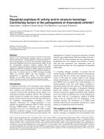
of ET-1 and IL-8 in Figure 1a, b.
Less ET-1 in blood was seen in nonresponders in the total
material (28 pigs) (0.1 (0.1 to 8) pg/ml versus 3.0 (1.8 to 8)
pg/ml in responders), but this difference did not reach signifi-
cance.
Prostanoids (PGF
2α
, TXB
2
, 6-keto-PGF
1α
) and LTB
4
did not
differ between responders and nonresponders, and neither
did TNFα or nitrate concentrations (Table 3).
Discussion
Endotoxin has dramatic and complex effects on the structure
and function of the lungs in intact animals and also on isolated
lung cells [12]. The 2-hour endotoxin infusion in the present
model resulted in a marked lung dysfunction with a PaO
2
/FiO
2
around 200 mmHg and pulmonary hypertension around 35 to
40 mmHg. Histological evidence of endotoxin-induced lung
injury was previously performed and is described in a work by
Da and colleagues [13]. The impairment remained stable over
Table 1
Hemodynamic parameters of responders and nonresponders
Parameter Responder (n = 15) Nonresponder (n = 13)
Baseline T0 Endotoxin Inhaled nitric oxide Baseline T0 Endotoxin Inhaled nitric oxide
PaO
2
/FiO
2
(mmHg) 502 ± 42 215 ± 118* 316 ± 141
†
462 ± 62 234 ± 119* 225 ± 126
PaCO
2
(mmHg) 37 ± 3.0 50 ± 11* 49 ± 11 39 ± 4.8 47 ± 7.1* 51 ± 9.2
pH 7.51 ± 0.03 7.29 ± 0.10* 7.22 ± 0.29 7.50 ± 0.04 7.34 ± 0.08* 7.30 ± 0.08
CO (l/min) 4.2 ± 1.2 4.4 ± 0.9 4.6 ± 1.2 4.1 ± 0.9 5.0 ± 1.5 5.4 ± 1.7
MPAP (mmHg) 16 ± 1.6 40 ± 7.7* 30 ± 7.5
†
15 ± 2.3 33 ± 5.5*
‡
28 ± 5.0
†
PVR (dyne/s/cm
5
) 222 ± 77 617 ± 231* 425 ± 179
†
186 ± 38 407 ± 172*
‡
296 ± 125
CVP (mmHg) 4.0 ± 1.6 7.0 ± 3.0* 7.6 ± 3.9 4.8 ± 1.8 8.4 ± 3.2* 7.8 ± 2.8
MAP (mmHg) 85 ± 8.3 80 ± 17 77 ± 15 81 ± 9.8 78 ± 21 75 ± 22
SVR (dyne/s/cm
5
) 1,700 ± 436 1,380 ± 288 1,314 ± 341 1,541 ± 339 1,128 ± 256*
‡
1,032 ± 292
‡
Qs/Qt (%) 9.4 ± 2.8
‡
30 ± 20* 23 ± 17 12 ± 4.1
‡
24 ± 9.0* 29 ± 12
Crs (ml/cmH
2
O) 27 ± 3.8 12 ± 3.2* Not measured 26 ± 4.0 13 ± 3.2* Not measured
Rrs (cmH
2
O·s/l) 15 ± 4.0 29 ± 8.8* Not measured 15 ± 2.8 29 ± 8.6* Not measured
Parameters were recorded at baseline T0, 4 hours after the start of endotoxin infusion, and after 15 minutes of nitric oxide inhalation. Data
represent the mean ± standard deviation. CO, cardiac output; MPAP, mean pulmonary arterial pressure; PVR, pulmonary vascular resistance;
CVP, central venous pressure; MAP, mean arterial pressure; SVR, systemic vascular resistance; Qs/Qt, intrapulmonary shunt; Crs, compliance of
the respiratory system; Rrs, resistance of the respiratory system. *P < 0.05, endotoxin versus baseline;
†
P < 0.05, inhaled nitric oxide versus
endotoxin;
‡
P < 0.05, responder versus nonresponder.
Available online />Page 5 of 8
(page number not for citation purposes)
the 4-hour or 6-hour study period and 28 animals survived the
whole experiment. Sixty percent of the animals were respond-
ers to a brief period of inhaled NO, similar to clinical observa-
tions in ARDS and sepsis [8].
The analysis of the animal group exposed twice to INO distin-
guished responders from nonresponders in terms of ET-1 and
IL-8 levels. The responders had higher ET-1 and IL-8 levels.
Inflammatory response to endotoxin
There was no difference in the endotoxin concentration in
plasma at 4 hours between responders and nonresponders,
but the rapid decrease in endotoxin concentration in the non-
responder group during the last 2-hour period suggests a
faster metabolism of endotoxin in nonresponders. Endotoxin
also binds to the endothelium, proteins and circulating cells,
and this reduces the plasma concentration; however, whether
this interacts with the vasodilating effect of inhaled NO is not
clear.
The endotoxin infusion caused an early and severe leucopenia
that may be explained by cell trapping and adhesion to the
endothelium as well as dilution by edema formation. As shown
earlier the fraction of neutrophils increased at the expense of
the fraction of monocytes [10]. The higher neutrophil and mac-
rophage count in responders than in nonresponders may illus-
trate a different inflammatory process due to the endotoxin
infusion; however, this requires further study to be resolved.
Table 2
Cells, protein and inflammatory mediators from T0 (baseline) to T6
Parameter T0 (n = 28) T1 (n = 18)T2 (n = 18)T3 (n = 18) T4 (n = 28) T5 (n = 9) T6 (n = 9)
Leucocytes (10
6
/ml) 9.38 ± 3.85 2.02 ± 0.5* 1.44 ± 0.89* 0.97 ± 0.19* 1.13 ± 0.49* 1.25 ± 0.41* 1.86 ± 0.79
Neutrophils (%) 53 ± 11 27 ± 10* 34 ± 8.9* 43 ± 15 54 ± 15 73 ± 16* 75 ± 11*
Macrophage (%) 46 ± 11 71 ± 11* 65 ± 9.3* 55 ± 15 44 ± 16 27 ± 17* 23 ± 11*
Thrombocytes (10
6
/ml) 384 ± 73 245 ± 51* 208 ± 48* 172 ± 41* 177 ± 55* 178 ± 65* 191 ± 52*
Proteins
(mg/ml)
47 (42 to 52) 39
(34 to 46)*
33 (29 to 36)* 28 (23 to 34)* 35 (26 to 38)* 27 (22 to 31)* 34 (29 to 35)*
Endotoxin
(pg/ml)
0 (0 to 0.04) 758
(542 to 1,008)*
783
(525 to 1,004)*
200
(125 to 755)*
85 (27 to 413)* 12 (5 to 73)* 2.3 (0.3 to 11)
Endothelin-1 (pg/ml) 0.1 5.0 (4.0 to 6.0)* 5.0
(5.0 to 6.5)*
5.0
(4.5 to 6.5)*
3.0 (2.0 to 7.5)* 4.5 (4 to 6.5)* 0.6 (0.1 to 8.8)*
PGF
2α
(pg/ml) 225
(167 to 344)
1,011
(859 to 1,216)*
1,301
(766 to 1,690)*
1,439
(971 to 1,708)*
387
(355 to 1513)*
1,028
(868 to 1,531)*
1,224
(333 to 1,562)*
TXB
2
(pg/ml) 687
(581 to 793
3,110
(2,617 to 3,546)*
2,824
(2,120 to 3,287)*
2,409
(2,129 to 3,110)*
4,103
(1,521 to 4,986)*
2,107
(1,648 to 2,430)*
3,150
(891 to 4,002)*
6-keto-PGF
1α
(pg/ml) 294
(260 to 518)
749
(548 to 1778)
1,301
(899 to 1,658)*
732
(613 to 1341)
973
(716 to 1,504)*
464
(332 to 771)
877
(534 to 1,747)*
LTB
4
(pg/ml) 36
(23 to 46)
42 (22 to 210) 69 (24 to 225) 48 (29 to 266) 52 (42 to 423) 70 (43 to 187) 240
(42 to 463)
TNFα (pg/ml) 17 (11 to 22) 53 (21 to 120)* 29 (5 to 52) 34 (14 to 69) 15 (10 to 38) 26 (0.5 to 60) 3.5 (2.0 to 9.5)
IL-8 (ng/ml) 0 2.0 (0 to 63) 117 (108 to 127)* 116 (104 to 126)* 31 (27 to 46) 39 (19 to 52) 6 (2 to 21)
Nitrates
(nmol/ml)
211
(162 to 309)
151 (139 to 174)* 135 (114 to 172)* 133 (119 to 149)* 189
(147 to 237)
140 (120 to 157)* 190
(173 to 240)
Data presented as the mean ± standard deviation when normally distributed, or as the median (25th percentile to 75th percentile). PGF
2α
,
prostaglandin F 2 alpha; TXB
2
, thromboxane B
2
; 6-keto-PGF
1α
, 6-keto-prostaglandin F 1 alpha; LTB
4
, leukotriene B
4
. *P < 0.05 when different to
baseline.
Critical Care Vol 12 No 5 Trachsel et al.
Page 6 of 8
(page number not for citation purposes)
Endotoxin also caused a rapid release of inflammatory media-
tors and vasoactive substances. Prostanoids, LTB
4
, ET-1,
nitrates and cytokines increased in the blood. The vasocon-
strictor TXB
2
(a metabolite of thromboxane A
2
) also increased,
and its concentration was always higher than that of the
vasodilator 6-keto-PGF
1α
(metabolite of prostacycline). The
levels of the proinflammatory cytokine IL-8 followed the endo-
toxin evolution in blood.
Endothelin-1 and inhaled nitric oxide
The ET-1 in plasma rose rapidly and markedly already after 1
hour and paralleled the increase in blood endotoxin. There was
a correlation between ET-1 levels and the pulmonary artery
pressure. The higher MPAP in responders before INO chal-
lenge may therefore be explained by their higher ET-1 levels
than in nonresponders. It may be argued that a general pulmo-
nary vasoconstriction caused by an increased plasma concen-
tration of ET-1 will facilitate or promote a positive response to
INO and will improve oxygenation. This oxygenation improve-
ment occurs because NO inhalation will cause vasodilation
solely or preferentially in ventilated parenchyma, whereas the
circulating ET-1 will promote vasoconstriction both in venti-
lated and nonventilated parenchyma. This mechanism may be
comparable with the combination of almitrine with INO
[14,15]. The stronger the pulmonary vasoconstriction, there-
fore, the more likely there will be a positive response to INO.
This conclusion is also supported by the findings in the limited
number of pigs exposed to INO at 4 hours and at 6 hours that
switched from response to nonresponse, with lower ET-1 con-
centrations when no longer responding to INO. A continuous
endotoxin infusion may have led to further responders since
endothelin correlates with endotoxin levels [16]. The high ini-
tial dose of endotoxin (60 μg/kg) produced severe physiologic
dysfunction that did not allow a prolongation of the endotoxin
infusion.
It is still a matter of debate whether INO increases ET-1, which
would accentuate vasoconstriction in nonventilated paren-
chyma [17]. In contrast, there is evidence that INO decreases
ET-1 secretion [18]. Rebound hypertension after withdrawal
from INO is attributed to ET-1 in an endotoxin lung injury model
[19]. The role of ET-1 during INO may be selective vasocon-
striction in nonventilated parenchyma, whereas ET-1 induces
vasoconstriction in the entire pulmonary vascular bed after
withdrawal of INO.
In isolated-perfused lungs from endotoxin-challenged rats,
when nitric oxide synthase 2 is inhibited, responsiveness to
INO improved [20]. This could be explained by the predomi-
nant vasoconstrictive effects induced by the suppression of
endogenous NO. We were not able, however, to separate
Table 3
Cells and inflammatory parameters at baseline and before inhaled nitric oxide (INO) for responders and nonresponders
Parameter Responders (n = 15) Nonresponders (n = 13)
Baseline Before INO P value Baseline Before INO P value
Leucocytes (10
6
/ml) 10.4 ± 4.4 1.6 ± 1.0
†
≤ 0.01 8.8 ± 2.4 0.9 ± 0.3*
†
≤ 0.01
Neutrophils (%) 56 ± 12 65 ± 14 0.074 53 ± 10 48 ± 17* 0.45
Macrophage (%) 44 ± 12 34 ± 15 0.064 47 ± 10 51 ± 17* 0.45
Thrombocytes (10
6
/ml) 408 ± 77 185 ± 54
†
≤ 0.01 355 ± 61 175 ± 68
†
≤ 0.01
Proteins (mg/ml) 47 ± 6.0 33 ± 5.4
†
≤ 0.01 46 ± 6 34 ± 5
†
≤ 0.01
Endotoxin (pg/ml) 0 45 (3.8 to 296)
†
≤ 0.01 0 (0 to 4.5) 32 (0 to 392)
†
0.011
Endothelin-1 (pg/ml) 0 3.0 (1.8 to 8)
†
≤ 0.01 0 0.1 (0.1 to 8) 0.31
PGF
2α
(pg/ml) 265 (236 to 327) 759 (356 to 1498)
†
≤ 0.01 218 (171 to 512) 380 (342 to 1632)
†
≤ 0.01
TXB
2
(pg/ml) 692 (530 to 778) 2881 (860 to 4504)
†
≤ 0.01 643 (574 to 821) 3883 (2286 to 5051)
†
≤ 0.01
6-keto-PGF
1α
(pg/ml) 291 (232 to 323) 1023 (810 to 1560)
†
≤ 0.01 518 (274 to 899) 603 (557 to 1099) 0.33
LTB
4
(pg/ml) 28 (16 to 41) 372 (41 to 486)
†
≤ 0.01 38 (9 to 48) 274 (46 to 414)
†
≤ 0.01
TNFα (pg/ml) 15 (11 to 22) 18 (10 to 39) 0.083 12 (8 to 21) 10 (5 to 27) 1.0
IL-8 (ng/ml) 0 24(7.2 to 32)
†
≤ 0.01 0 4 (1.5 to 47)
†
0.03
Nitrates (nmol/ml) 212 (173 to 352) 183 (147 to 236)
†
≤ 0.01 183 (133 to 255) 173 (150 to 212) 0.58
Data present as the mean ± standard deviation when normally distributed or as the median (25th percentile to 75th percentile). PGF
2α
,
prostaglandin F 2 alpha; TXB
2
, thromboxane B
2
; 6-keto-PGF
1α
, 6-keto-prostaglandin F 1 alpha; LTB
4
, leukotriene B
4
. *P < 0.05 between
responder and nonresponder,
†
P < 0.05 between baseline and before INO.
Available online />Page 7 of 8
(page number not for citation purposes)
responders from nonresponders by different nitrate levels in
this in vivo model.
Severity of pulmonary damage
The severity of pulmonary dysfunction in terms of gas
exchange and respiratory mechanics (compliance) did not dif-
fer between responders and nonresponders. The hemody-
namics differed, however, with higher MPAP, pulmonary
vascular resistance and systemic vascular resistance in the
responders before INO challenge.
The degree of pulmonary damage separating responders from
nonresponders may be an explanation for the varying
responses to INO. Besides, a limitation may include the intra-
variability and intervariability of the animal lung injury model.
On the contrary, most parameters of the inflammatory media-
tors measured here did not differ between responders and
nonresponders – although IL-8 was, on an average, higher in
the responder group [21]. This suggests that the severity of
lung damage was much the same in the two groups. ET-1 and
IL-8 were higher in responders, however, and ET-1 correlated
to the MPAP. This observation may, as said above, explain
higher values of the MPAP in responders. The separation
between responders and nonresponders made on the basis of
only two mediators (ET-1 and IL-8) may therefore support the
hypothesis of a distinct mechanism, independent of the lung
damage, responsible for the response to INO.
The results suggest that ET-1 may be a determining factor for
a positive response to INO and for the decreased physiologic
parameters. More severe pulmonary hypertension may be
explained by higher levels of ET-1. INO, known for its antiin-
flammatory properties [22], could attenuate the effect of INO
by decreasing ET-1 levels. This may explain the attenuation on
oxygenation, when INO is administered for longer than 24
hours [4].
Conclusion
The presented endotoxin lung injury model demonstrates that
responders to INO present more severe pulmonary dysfunc-
tion at a comparable inflammatory profile. This observation can
be explained by elevated ET-1 levels correlated to the magni-
tude of pulmonary hypertension that may result in a positive
response to INO. This additionally supports the hypothesis
that INO acts by two distinct mechanisms; one is vasodilation
in ventilated lung regions, and the other is vasoconstriction in
poorly ventilated or nonventilated lung regions. Other inflam-
matory parameters did not vary between responders and non-
responders, and possibly document similar injuries to the lung
and its vasculature in the present study.
Key messages
• Elevated concentration of endothelin-1 may mediate a
positive response to inhaled nitric oxide.
• Responders to inhaled nitric oxide present more severe
pulmonary dysfunction at a comparable inflammatory
profile.
• Endothelin-1 levels correlate with the magnitude of pul-
monary hypertension.
• Further support is added to the hypothesis that inhaled
nitric oxide acts by two distinct mechanisms; one is
vasodilation in ventilated lung regions, and the other is
vasoconstriction in poorly ventilated or nonventilated
lung regions.
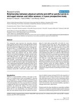
Figure 1
Levels of endothelin-1 and interleukin-8 compared with the increase or decrease of PaO
2
/FiO
2
Levels of endothelin-1 and interleukin-8 compared with the increase or
decrease of PaO
2
/FiO
2
. (a) Level of the endothelin-1 (ET-1) concentra-
tion (pg/ml) compared with the increase or decrease of PaO
2
/FiO
2
(mmHg). All five animals exposed twice to inhaled nitric oxide
decreased their ET-1 concentration level when changing from
responder to nonresponder. (b) Level of IL-8 (ng/ml) compared with
the increase or decrease of PaO
2
/FiO
2
(mmHg). Levels of IL-8
decreased from response at the first exposure to nonresponse at the
second exposure. Each symbol represents one animal; open symbol,
first inhaled nitric oxide exposure; filled symbol, second exposure of the
same animal with the same symbol.
Critical Care Vol 12 No 5 Trachsel et al.
Page 8 of 8
(page number not for citation purposes)
Competing interests
The authors declare that they have no competing interests.
Authors' contributions
ST performed the statistical analysis and interpretation of data,
edited the manuscript and acquired funding. GD-D was
involved with the biochemical analysis (immunoassays) and
with editing the manuscript. EM performed the experiments
and was involved in data acquisition. MN participated in the
biochemical analysis. ML was involved in the study design and
in revising the manuscript. GH has made a substantial contri-
bution to the design and conception of the study, and to the
interpretation of data.
Acknowledgements
The present study was supported by grants from the Swedish Medical
Research Council (No 5315), the Swedish Heart and Lung Fund and
the AGA Medical Fund and the Swiss National Science Foundation
(PIOIB – 114967/1). The work is part of the project: Influence of
endothelin activity on response to inhaled nitric oxide on ventilation per-
fusion distribution in acute lung injury, which was awarded The Alain
Harf Award on Applied Respiratoy Physiology ESICM ECCRN Awards
2006. The assistance of Marie Ekberg-Richter, Lena Almgren, Annie
Bjurebäck, Ann-Christine Linde, Hedy Magnusson and Agneta Petters-
son, as well as Kere Frey, is highly appreciated. The work was performed
at the Department of Medical Sciences, Clinical Physiology of the uni-
versity hospital in Uppsala, Sweden.
References
1. Vincent JL, Sakr Y, Ranieri VM: Epidemiology and outcome of
acute respiratory failure in intensive care unit patients. Crit
Care Med 2003, 31:S296-S299.
2. Walmrath D, Schermuly R, Pilch J, Grimminger F, Seeger W:
Effects of inhaled versus intravenous vasodilators in experi-
mental pulmonary hypertension. Eur Respir J 1997,
10:1084-1092.
3. Rossaint R, Gerlach H, Schmidt-Ruhnke H, Pappert D, Lewand-
owski K, Steudel W, Falke K: Efficacy of inhaled nitric oxide in
patients with severe ARDS. Chest 1995, 107:1107-1115.
4. Taylor RW, Zimmerman JL, Dellinger RP, Straube RC, Criner GJ,
Davis K Jr, Kelly KM, Smith TC, Small RJ: Low-dose inhaled nitric
oxide in patients with acute lung injury: a randomized control-
led trial. JAMA 2004, 291:1603-1609.
5. Dellinger RP, Zimmerman JL, Taylor RW, Straube RC, Hauser DL,
Criner GJ, Davis K Jr, Hyers TM, Papadakos P: Effects of inhaled
nitric oxide in patients with acute respiratory distress syn-
drome: results of a randomized phase II trial. Inhaled Nitric
Oxide in ARDS Study Group. Crit Care Med 1998, 26:15-23.
6. Lundin S, Mang H, Smithies M, Stenqvist O, Frostell C: Inhalation
of nitric oxide in acute lung injury: results of a European mult-
icentre study. The European Study Group of Inhaled Nitric
Oxide. Intensive Care Med 1999, 25:911-919.
7. Krafft P, Fridrich P, Fitzgerald RD, Koc D, Steltzer H: Effective-
ness of nitric oxide inhalation in septic ARDS. Chest 1996,
109:486-493.
8. Manktelow C, Bigatello LM, Hess D, Hurford WE: Physiologic
determinants of the response to inhaled nitric oxide in
patients with acute respiratory distress syndrome. Anesthesi-
ology 1997, 87:297-307.
9. Hedenstierna G, Lattuada M: Gas exchange in the ventilated
patient. Curr Opin Crit Care 2002, 8:39-44.
10. Maurenbrecher H, Lamy M, Deby-Dupont G, Frascarolo P, Heden-
stierna G: An animal model of response and nonresponse to
inhaled nitric oxide in endotoxin-induced lung injury. Chest
2001, 120:573-581.
11. Nys M, Ledoux D, Canivet JL, De Mol P, Lamy M, Damas P: Corre-
lation between endotoxin level and bacterial count in broncho-
alveolar lavage fluid of ventilated patients. Crit Care Med
2000, 28:2825-2830.
12. Brigham KL, Meyrick B: Endotoxin and lung injury. Am Rev
Respir Dis 1986, 133:913-927.
13. Da J, Chen L, Hedenstierna G: Nitric oxide up-regulates the glu-
cocorticoid receptor and blunts the inflammatory reaction in
porcine endotoxin sepsis. Crit Care Med 2007, 35:26-32.
14. Gallart L, Lu Q, Puybasset L, Umamaheswara Rao GS, Coriat P,
Rouby JJ: Intravenous almitrine combined with inhaled nitric
oxide for acute respiratory distress syndrome. The NO
Almitrine Study Group. Am J Respir Crit Care Med 1998,
158:1770-1777.
15. Papazian L, Roch A, Bregeon F, Thirion X, Gaillat F, Saux P, Fula-
chier V, Jammes Y, Auffray JP: Inhaled nitric oxide and vasocon-
strictors in acute respiratory distress syndrome. Am J Respir
Crit Care Med 1999, 160:473-479.
16. Snapper JR, Thabes JS, Lefferts PL, Lu W: Role of endothelin in
endotoxin-induced sustained pulmonary hypertension in
sheep. Am J Respir Crit Care Med 1998, 157:81-88.
17. Christou H, Adatia I, Van Marter LJ, Kane JW, Thompson JE, Stark
AR, Wessel DL, Kourembanas S: Effect of inhaled nitric oxide on
endothelin-1 and cyclic guanosine 5'-monophosphate plasma
concentrations in newborn infants with persistent pulmonary
hypertension. J Pediatr 1997, 130:603-611.
18. Kelly LK, Wedgwood S, Steinhorn RH, Black SM: Nitric oxide
decreases endothelin-1 secretion through the activation of
soluble guanylate cyclase. Am J Physiol Lung Cell Mol Physiol
2004, 286:L984-L991.
19. Chen L, He H, Fernandez Mondejar E, Freden F, Wiklund P, Alving
K, Hedenstierna G: Endothelin-1 and nitric oxide synthase in
short rebound reaction to short exposure to inhaled nitric
oxide. Am J Physiol Heart Circ Physiol 2001, 281:H124-H131.
20. Bopp C, Gust R, Taut F, Gries A, Martin E, Klein A: Responsive-
ness to inhaled NO in isolated-perfused lungs from endotoxin-
challenged rats is dependent on endogenous nitrite/nitrate
synthesis. Eur J Anaesthesiol 2007, 24:362-369.
21. Boutten A, Dehoux MS, Seta N, Ostinelli J, Venembre P, Crestani
B, Dombret MC, Durand G, Aubier M: Compartmentalized IL-8
and elastase release within the human lung in unilateral pneu-
monia. Am J Respir Crit Care Med 1996, 153:336-342.
22. Busch T, Petersen B, Deja M, Donaubauer B, Laudi S, Jaumann S,
Bercker S, Boemke W, Kaisers U: Endothelin-1 influences the
efficacy of inhaled nitric oxide in experimental acute lung
injury. Exp Biol Med (Maywood) 2006, 231:974-978.