Báo cáo y học: "Mechanical ventilation using non-injurious ventilation settings causes lung injury in the absence of pre-existing lung injury in healthy mice" pdf
Bạn đang xem bản rút gọn của tài liệu. Xem và tải ngay bản đầy đủ của tài liệu tại đây (1000.8 KB, 11 trang )
Open Access
Available online />Page 1 of 11
(page number not for citation purposes)
Vol 13 No 1
Research
Mechanical ventilation using non-injurious ventilation settings
causes lung injury in the absence of pre-existing lung injury in
healthy mice
Esther K Wolthuis
1,2,3
, Alexander PJ Vlaar
1,3
, Goda Choi
3,4
, Joris JTH Roelofs
5
,
Nicole P Juffermans
1,3
and Marcus J Schultz
1,3,6
1
Department of Intensive Care Medicine, University of Amsterdam, Meibergdreef 9, 1105 AZ Amsterdam, The Netherlands
2
Department of Anesthesiology, University of Amsterdam, Meibergdreef 9, 1105 AZ Amsterdam, The Netherlands
3
Laboratory of Experimental Intensive Care and Anesthesiology (LEICA), University of Amsterdam, Meibergdreef 9, 1105 AZ Amsterdam, The
Netherlands
4
Department of Internal Medicine, University of Amsterdam, Meibergdreef 9, 1105 AZ Amsterdam, The Netherlands
5
Department of Pathology, University of Amsterdam, Meibergdreef 9, 1105 AZ Amsterdam, The Netherlands
6
HERMES Critical Care Group, Amsterdam, The Netherlands
Corresponding author: Esther K Wolthuis,
Received: 18 Sep 2008 Revisions requested: 8 Oct 2008 Revisions received: 19 Nov 2008 Accepted: 19 Jan 2009 Published: 19 Jan 2009
Critical Care 2009, 13:R1 (doi:10.1186/cc7688)
This article is online at: />© 2009 Wolthuis et al.; licensee BioMed Central Ltd.
This is an open access article distributed under the terms of the Creative Commons Attribution License ( />),
which permits unrestricted use, distribution, and reproduction in any medium, provided the original work is properly cited.
Abstract
Introduction Mechanical ventilation (MV) may cause ventilator-
induced lung injury (VILI). Present models of VILI use
exceptionally large tidal volumes, causing gross lung injury and
haemodynamic shock. In addition, animals are ventilated for a
relative short period of time and only after a 'priming' pulmonary
insult. Finally, it is uncertain whether metabolic acidosis, which
frequently develops in models of VILI, should be prevented. To
study VILI in healthy mice, the authors used a MV model with
clinically relevant ventilator settings, avoiding massive damage
of lung structures and shock, and preventing metabolic acidosis.
Methods Healthy C57Bl/6 mice (n = 66) or BALB/c mice (n =
66) were ventilated (tidal volume = 7.5 ml/kg or 15 ml/kg;
positive end-expiratory pressure = 2 cmH
2
O; fraction of inspired
oxygen = 0.5) for five hours. Normal saline or sodium
bicarbonate were used to correct for hypovolaemia. Lung
histopathology, lung wet-to-dry ratio, bronchoalveolar lavage
fluid protein content, neutrophil influx and levels of
proinflammatory cytokines and coagulation factors were
measured.
Results Animals remained haemodynamically stable throughout
the whole experiment. Lung histopathological changes were
minor, although significantly more histopathological changes
were found after five hours of MV with a larger tidal volume. Lung
histopathological changes were no different between the
strains. In both strains and with both ventilator settings, MV
caused higher wet-to-dry ratios, higher bronchoalveolar lavage
fluid protein levels and more influx of neutrophils, and higher
levels of proinflammatory cytokines and coagulation factors.
Also, with MV higher systemic levels of cytokines were
measured. All parameters were higher with larger tidal volumes.
Correcting for metabolic acidosis did not alter endpoints.
Conclusions MV induces VILI, in the absence of a priming
pulmonary insult and even with use of relevant (least injurious)
ventilator settings. This model offers opportunities to study the
pathophysiological mechanisms behind VILI and the
contribution of MV to lung injury in the absence of pre-existing
lung injury.
Introduction
Mechanical ventilation (MV) may aggravate pre-existing lung
injury or even cause lung injury in healthy lungs, a phenomenon
frequently referred to as ventilator-induced lung injury (VILI).
BALF: broncho-alveolar lavage fluid; ELISA: enzyme-linked immunosorbent assay; H&E: haematoxylin & eosin; HV
T
: High tidal volume; IL: interleukin;
IQR: interquartile range; KC: keratinocyte-derived chemokine; LV
T
: low tidal volume; MIP: macrophage inflammatory protein; MV: mechanical ventila-
tion; PaCO
2
: partial pressure of arterial carbon dioxide; PAI: plasminogen activator inhibitor; PaO
2
: Partial pressure of arterial oxygen; PBW: predicted
bodyweight; PEEP: positive end-expiratory pressure; SD: standard deviation; TATc: thrombin-antithrombin complexes; TNF: tumour necrosis factor;
VILI: ventilator-induced lung injury; V
T
: tidal volume.
Critical Care Vol 13 No 1 Wolthuis et al.
Page 2 of 11
(page number not for citation purposes)
Present strategies at minimising VILI in critically ill patients
consist of using low tidal volumes (V
T
) [1]. However, additional
strategies to attenuate pulmonary inflammation may be useful
to further reduce VILI. Adequate animal models are also
required, to test various treatment strategies. However, exist-
ing animal models of MV have considerable disadvantages.
Most models of VILI use very high V
T
and/or inspiratory pres-
sures that are considerably higher than those used in the clin-
ical management of patients [2-6]. High V
T
may compromise
systemic circulation, eventually leading to shock. Wilson and
colleagues used an MV strategy in which mice were ventilated
with a V
T
of 34.5 ml/kg for a duration of 156 minutes until mean
blood pressure fell below 45 mmHg [5,6]. Consequently,
duration of MV is relatively short and maybe too short to draw
meaningful conclusions. In addition, most models of VILI lungs
are 'primed' before starting MV [7-11]. Indeed, animals are
challenged before onset of MV, for instance for lipopolysac-
charide causing lung injury [7,11]. Such an approach prevents
conclusions on the deleterious effects of MV in the absence of
pre-existing lung injury being drawn. One final problem may be
that infusion of saline solution to correct for low arterial blood
pressures leads to metabolic acidosis in models of VILI
[12,13], although metabolic acidosis may influence several
endpoints of VILI [14,15]. It is uncertain whether metabolic
acidosis should be corrected in models of VILI.
The aim of the present investigation was to set up a model of
VILI in healthy animals. We chose an MV strategy that closely
reflected the human setting by using clinically relevant V
T
, pre-
venting shock and gross lung histopathological changes, and
compared lower V
T
with higher V
T
with respect to several end-
points of VILI. In addition, we hypothesised preventing meta-
bolic acidosis to affect endpoints of VILI. Therefore we
compared two strategies for fluid resuscitation, using either
normal saline or sodium bicarbonate.
Materials and methods
The study was approved by the Animal Care And Use Commit-
tee of the Academic Medical Center. Animal procedures were
carried out in compliance with Institutional Standards for
Human Care and Use of Laboratory Animals.
Animals
Experiments were performed with healthy male C57Bl/6 (n =
66) and BALB/c mice (n = 66) (Charles River, Someren, the
Netherlands), aged 8 to 10 weeks, with weights ranging from
19 to 25 g. Two groups of control animals served either as
non-ventilated controls for blood gas analysis at baseline (n =
6 for each strain) or as non-ventilated controls after five hours
(n = 12 for each strain). The other animals were all mechani-
cally ventilated with two different MV-strategies and two differ-
ent fluid support strategies. Thus, five groups of animals of
each mice strain were compared.
Instrumentation and anesthesia
Throughout the experiments, rectal temperature was main-
tained between 36.5 and 37.5°C using a warming path.
Anaesthesia was achieved with intraperitoneal injection of a
mix of 100 mg/ml ketamine (Eurovet Animal Health B.V.,
Bladel, the Netherlands), 1 mg/ml medetomidine (Pfizer Ani-
mal Health B.V., Capelle a/d IJssel, the Netherlands) and 0.5
mg/ml atropine (Pharmachemie, Haarlem, the Netherlands;
KMA). Induction of anaesthesia was performed by injecting
7.5 l/g of induction KMA mix (consisting of 1.26 ml ketamine,
0.2 ml medetomidine and 1 ml atropine). To maintain anaes-
thesia, 10 l/g of maintenance KMA mix (consisting of 0.72 ml
ketamine, 0.08 ml medetomidine and 0.3 ml atropine) was
given, via an intraperitoneally placed catheter every hour.
Mechanical ventilation strategies
A Y-tube connector, 1.0 mm outer diameter and 0.6 mm inner
diameter (VBM Medizintechnik GmbH, Sulz am Neckar, Ger-
many) was surgically inserted into the trachea under general
anaesthesia. Mice were placed in a supine position and con-
nected to a ventilator (Servo 900 C, Siemens, Sweden).
Simultaneously, six mice were pressure-controlled ventilated
with either an inspiratory pressure of 10 cmH
2
O (resulting in
V
T
of about 7.5 ml/kg; low V
T
(LV
T
)) or an inspiratory pressure
of 18 cmH
2
O (resulting in V
T
of about 15 ml/kg; high V
T
(HV
T
)).
In C57Bl/6 mice, respiratory rate was set at 120 breaths/
minute and 70 breaths/minute with LV
T
and HV
T
, respectively;
in BALB/c mice, respiratory rate was set at 100 breaths/
minute and 70 breaths/minute with LV
T
and HV
T
, respectively.
Preliminary studies showed these respiratory settings resulted
in normal partial pressure of arterial carbon dioxide (PaCO
2
)
values after five hours of MV in the different mice strains. Pos-
itive end-expiratory pressure (PEEP) was set at 2 cmH
2
O with
both MV strategies. The fraction of inspired oxygen was kept
at 0.5 throughout the experiment. The inspiration to expiration
ratio was kept at 1:1 throughout the experiment.
Fluid support strategies
Mice received an intraperitoneal bolus of 1 ml normal saline
one hour before the start of MV, followed by 0.2 ml normal
saline (sodium chloride (NaCl) 0.9%) or 0.2 ml sodium bicar-
bonate (containing 200 mM sodium and bicarbonate) admin-
istered via the intraperitoneal catheter every 30 minutes.
Preliminary studies showed this fluid strategy to adequately
compensate for insensible and observed fluid loss, and to
keep the animals haemodynamically stable.
Haemodynamic and ventilatory monitoring
Systolic blood pressure and heart rate were non-invasively
monitored using a murine tail-cuff system (ADInstruments,
Spenbach, Germany). Blood pressure and pulse were meas-
ured directly after the start of MV, after 2.5 hours and 5 hours
of MV. The data were recorded on a data acquisition system
(PowerLab/4SP, ADInstruments, Spenbach, Germany). An
Available online />Page 3 of 11
(page number not for citation purposes)
average systolic blood pressure and heart rate were taken
from three consecutive measurements.
V
T
was checked hourly with a specially designed Fleisch-tube
connected to the body-plethysmograph. The flow signal was
integrated from a differential pressure transducer and data
were recorded and digitised online using a 16-channel data
acquisition program (ATCODAS, Dataq Instruments Inc,
Akron, OH) and stored on a computer for post acquisition off-
line analysis. A minimum of five consecutive breaths were
selected for analysis of the digitised V
T
signals.
Study groups
Non-ventilated control mice were selected for blood gas anal-
ysis at baseline (for both strains n = 6): animals were handled
one week before the experiment to decrease stress activation.
After induction of anaesthesia with isoflurane arterial blood
was taken from the left ventricle by heart puncture within 30
seconds.
LV
T
mice receiving either normal saline (n = 12) or sodium
bicarbonate (n = 12) and HV
T
mice receiving either saline (n =
12) or sodium bicarbonate (n = 12) were mechanically venti-
lated for five hours and then euthanased. Non-ventilated con-
trol mice (n = 12) received half the dose of induction
anaesthesia, were spontaneously breathing and then eutha-
nased after five hours.
Measurements
The first series of mice (n = 6) were euthanased and blood
was drawn from the vena cava inferior into a sterile syringe,
transferred to EDTA-coated tubes and immediately placed on
ice. Blood samples of two mice were pooled together. Bron-
choalveolar lavage fluid (BALF) was obtained from the right
lung; the left lung was used to measure the wet-to-dry ratio. In
a second series of mice (n = 6), blood was sampled from the
carotid artery for blood gas analysis. The lungs of these mice
were used for homogenate (right lung) and histopathology (left
lung).
For wet-to-dry ratios the lung was weighed and subsequently
dried for three days in an oven at 65°C. The right lung was
removed and snap frozen in liquid nitrogen. These frozen spec-
imens were suspended in four volumes of sterile isotonic
saline and subsequently lysed in one volume of lysis buffer
(150 mM NaCl, 15 mM Tris (tris(hydroxymethyl)aminometh-
ane), 1 mM MgCl.H
2
O, 1 mM CaCl
2
, 1% Triton X-100, 100
g/mL pepstatin A, leupeptin and aprotinin, pH 7.4) and incu-
bated at 4°C for 30 minutes. Homogenates were spun at
3400 rpm at 4°C for 15 minutes after which the supernatants
were stored at -20°C until assayed.
BALF was obtained by instilling three times 0.5 ml aliquots of
saline by a 22-gauge Abbocath–T catheter (Abbott, Sligo, Ire-
land) into the trachea. About 1.0 ml of BALF was retrieved per
mouse and cell counts were determined using a haemacytom-
eter (Beckman Coulter, Fullerton, CA). Subsequently, differen-
tial counts were performed on citospin preparations stained
with a modified Giemsa stain, Diff-Quick (Dade Behring AG,
Düdingen, Switzerland). Supernatant was stored at -80°C for
meausrement of total protein level, thrombin-antithrombin
complexes (TATc) and plasminogen activator inhibitor (PAI)-1.
Lung histopathology
For histopathology lungs were fixed in 4% formalin and
embedded in paraffin. Sections 4 m in diameter were stained
with H&E and analysed by a pathologist who was blinded for
group identity. To score lung injury we used a modified VILI
histopathology scoring system as previously described [2].
VILI was scored according to the following four items: alveolar
congestion; haemorrhage; infiltration or aggregation of neu-
trophils in airspace or vessel wall; and thickness of the alveolar
wall/hyaline membrane formation. A score of 0 represented
normal lungs; 1 represented mild, less than 25% lung involve-
ment; 2 represented moderate, 25 to 50% lung involvement;
3 represented severe, 50 to 75% lung involvement; and 4 rep-
resented very severe, more than 75% lung involvement. An
overall score of VILI was obtained based on the summation of
all the scores from normal or ventilated lungs (n = 12 per
group).
Assays
Total protein levels in BALF were determined using a Bradford
Protein Assay Kit (OZ Biosciences, Marseille, France) accord-
ing to the manufacturers' instructions with BSA as standard.
Cytokine levels in blood lung homogenates were measured by
ELISA according to the manufacturer's instructions. Tumour
necrosis factor (TNF) , interleukin (IL) 6, macrophage inflam-
matory protein (MIP) 2 and keratinocyte-derived chemokine
(KC) assays were all obtained from R&D Systems (Abingdon,
UK). TATc levels in BALF were measured with a mouse spe-
cific ELISA as previously described [16]. Levels of PAI-1 were
measured by means of a commercially available ELISA (Kor-
dia, Leiden, the Netherlands).
Statistical analysis
All data in the results are expressed as mean ± standard devi-
ation or median ± interquartile range (IQR), where appropriate.
To detect differences between groups the Dunnett method or
Mann Whitney U test, in conjunction with two-way analysis of
variance was performed. Haemodynamics were measured in
12 animals, all other measurements were performed in six ani-
mals. A p value of less than 0.05 was considered significantly.
All statistical analyses were carried out using SPSS 12.0.2
(SPSS, Chicago, IL).
Results
Haemodynamic and ventilatory monitoring
All animals survived five hours of MV after which they were
euthanased; control animals survived anaesthesia and were
Critical Care Vol 13 No 1 Wolthuis et al.
Page 4 of 11
(page number not for citation purposes)
also euthanased after five hours. The systolic blood pressure
and heart rate remained stable in all animals for the entire dura-
tion of the experiment, with no differences noted between
mice strains, MV strategies and fluid strategies. Although
blood gas analysis from LV
T
mice and HV
T
mice using normal
saline revealed metabolic acidosis after five hours of MV (in
C57Bl/6 mice pH with LV
T
= 7.17 ± 0.07 and pH with HV
T
=
7.23 ± 0.06, and in BALB/c mice pH with LV
T
= 7.22 ± 0.04
and pH with HV
T
= 7.11 ± 0.07, Tables 1 and 2) with the use
of sodium bicarbonate metabolic acidosis was prevented (in
C57Bl/6 mice pH with LV
T
= 7.41 ± 0.07 and pH with HV
T
=
7.49 ± 0.02, and in BALB/c mice pH with LV
T
= 7.42 ± 0.05
and pH with HV
T
= 7.37 ± 0.08). Arterial oxygenation in
C57Bl/6 mice was significantly higher in HV
T
-mice as com-
pared with LV
T
-mice (205 ± 33 vs. 141 ± 22 mmHg, p <
0.001). No differences regarding oxygenation were found
between MV-groups in BALB/c mice (partial pressure of arte-
rial oxygen (PaO
2
) for HV
T
= 167 ± 50 and PaO
2
for LV
T
= 181
± 42 mmHg).
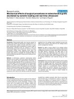
Lung histopathology scores
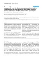
The histopathological changes were minor (Figure 1 and Table
3). For both mice strains the lung histopathology score was
higher in HV
T
mice as compared with controls. However, no
differences were noted between mice strains, MV strategies
and fluid strategies.
Wet-to-dry ratios, BALF-protein content and neutrophil
influx
In C57Bl/6 mice lung wet-to-dry ratios were significantly
higher with both MV strategies compared with controls (LV
T
mice = 4.8 ± 0.3 and HV
T
mice = 5.3 ± 0.5, as compared with
control mice = 4.2 ± 0.2; p < 0.01). Wet-to-dry ratios in HV
T
mice were also significantly higher as compared with LV
T
mice
(p = .009). For BALB/c mice higher lung wet to dry ratios were
found in HV
T
mice (5.6 ± 0.6 as compared with 4.6 ± 0.4 in
LV
T
mice (p < 0.001) and 4.5 ± 0.2 in control mice (p <
0.001), respectively). No significant differences were found
between LV
T
mice and control mice.
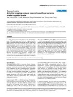
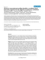
Total BALF protein levels in C57Bl/6 were significantly higher
in HV
T
mice as compared with LV
T
mice (p = .012) and control
mice (p = .008; Figure 2). No significant difference was found
between LV
T
mice and control mice. In BALB/c mice, total
BALF protein levels were significantly higher in HV
T
mice as
compared with LV
T
mice and control mice (p < .001). No sig-
nificant difference was found between LV
T
mice and control
mice.
The numbers of neutrophils in BALF were significantly higher
in HV
T
mice as compared with control mice in both mice
strains (Figure 1 and Table 3). Neutrophil counts in BALF from
HV
T
mice did not differ from LV
T
mice.
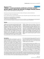
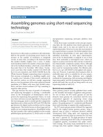
Pulmonary and plasma cytokine levels
In the HV
T
group of both mice strains, higher pulmonary levels
of TNF- were found as compared with the LV
T
group (p <
0.05) and control group (p 0.001; Figure 3). In BALBc mice
only, pulmonary levels of TNF- in LV
T
mice were higher as
compared with control mice (p = 0.018). Pulmonary levels of
IL-6 in the HV
T
group of both mice strain were higher as com-
pared with the LV
T
group and control group. Only for BALBc
mice a significant difference between LV
T
mice and control
mice were found. For pulmonary levels of MIP-2 in C57Bl/6
mice higher levels were found in HV
T
mice and LV
T
mice as
compared with control (p = 0.001). No difference was found
between LV
T
mice and HV
T
mice in this mice strain. In BALBc
mice, higher pulmonary levels of MIP-2 in the HV
T
group were
found as compared with the LV
T
group and control group, with
also a significant difference between HV
T
mice and LV
T
mice.
In both mice strain higher pulmonary levels of KC were found
in the HV
T
group as compared with the LV
T
group and control
group (p = 0.001). Only in BALBc mice, there was also a sig-
nificant difference between LV
T
mice and control mice.
Table 1
Arterial blood gas analysis in C57Bl/6 mice.
Control Low V
T
High V
T
NaCl NaHCO
3
NaCl NaHCO
3
pH 7.42 (0.04) 7.17 (0.07)‡ 7.41 (0.07) 7.23 (0.06)‡ 7.49 (0.02)
PaCO
2
(mmHg) 34.4
(32.2 to 38.3)
50.1
(36.7 to 59.6)
45.0
(38.6 to 50.0)
33.7
(32.1 to 34.0)
31.0
(27.6 to 34.4)
PaO
2
(mmHg) 133 (15) 148 (28) 186 (45) 223 (20)
HCO
3
-
(mmol/l) 21.4
(21.1 to 24.1)
16.6
(15.2 to 18.9)
28.0
(26.1 to 30.0)
14.6
(13.3 to 15.6)
24.9
(21.3 to 25.5)
BE -1.3
(-2.3 to -0.5)
-11.7
(-12.5 to -10.2)
4.1
(1.3 to 6.0)
-12.8
(-13.4 to -10.1)
2.3
(-0.4 to 2.9)
Data are mean (SD) or median [IQR]; Control = spontaneously breathing mice; Low V
T
= mice ventilated for five hours with a V
T
of 7.5 ml/kg; High
V
T
= mice ventilated for five hours with a V
T
of 15 ml/kg. n = 6 per group. *p < 0.05; ‡p < 0.001 vs. control mice.
PaCO
2
= partical pressure of arterial carbon dioxide; PaO
2
= partical pressure of arterial oxygen; BE = base excess.
Available online />Page 5 of 11
(page number not for citation purposes)
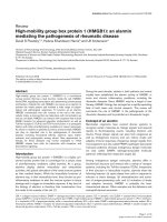
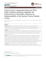
Plasma levels of IL-6 and KC were elevated in the both venti-
lation groups, with higher levels in the HV
T
group (Figure 4).
Plasma levels of TNF- and MIP-2 were below the detection
limit of the assay (data not shown).
Pulmonary coagulopathy
TATc levels in BALF were significantly higher in HV
T
mice in
both mice strain as compared with LV
T
mice and control (p <
0.001; Figure 5). No significant difference was found between
LV
T
mice and control mice in both mice strain. Levels of PAI-1
were not significantly different in C57Bl/6 mice. BALB/c mice
did show increased PAI-1 levels in the HV
T
group as compared
with the LV
T
group and control group (p < 0.001). No differ-
ences were found between LV
T
mice and control mice.
Lung injury with different fluid support strategies
The different fluid support strategies showed no difference in
endpoint of VILI, except for pulmonary MIP-2 and IL-6 levels in
C57Bl/6 mice. MIP-2 levels were significantly higher in HV
T
mice and LV
T
mice that received sodium bicarbonate as com-
pared with mice that received normal saline (p < 0.01; Figure
3). Pulmonary IL-6 levels were significantly higher in HV
T
mice
receiving sodium bicarbonate as compared with mice receiv-
ing normal saline (p = 0.026).
Discussion
We here show MV to cause VILI in healthy lungs (i.e. in the
absence of a 'priming' lung insult). VILI did not only develop in
animals ventilated with HV
T
but also in animals ventilated with
LV
T
, although to a lesser extent. We chose an MV strategy that
closely reflects the human setting by using clinically relevant
(i.e. physiological) V
T
, preventing shock and gross lung his-
topathological changes. Although we hypothesised that pre-
venting metabolic acidosis would affect the several endpoints
of VILI, we showed that correction of the acid-base balance
did not affect VILI.
We developed and tested a model of VILI in two commonly
used mice strains using clinically relevant V
T
and preventing
hypovolaemia with fluid support. By using a clinically relevant
V
T
and fluid support we prevented shock. By using sodium
bicarbonate instead of normal saline, metabolic acidosis was
prevented. We developed a model that enhances translation
of results into clinical practice and/or future studies. To our
best knowledge, this is one of the first studies that compares
more physiological V
T
then previously used in healthy lungs of
mice.
Our model has several limitations. First, V
T
in HV
T
mice are still
quite large (about 15 ml/kg). Although lung-protective ventila-
Table 2
Arterial blood gas analysis in BALB/c mice.
Control Low V
T
High V
T
NaCl NaHCO
3
NaCl NaHCO
3
PH 7.34 (0.05) 7.22 (0.04)* 7.42 (0.05) 7.11 (0.07)‡ 7.37 (0.08)
PaCO
2
(mmHg) 39.3
(31.6 to 51.3)
35.7
(31.1 to 39.5)
41.2
(35.3 to 43.6)
40.8
(37.0 to 55.6)
44.3
(36.1 to 51.7)
PaO
2
(mmHg) 193 (36) 168 (48) 173 (51) 161 (50)
HCO
3
-
(mmol/l) 21.1
(17.9 to 24.1)
14.4
(12.9 to 15.2)
25.2
(23.6 to 25.9)
13.5
(12.1 to 14.7)
24.5
(22.7 to 25.4)
BE -3.9
(-6.2 to -2.5)
-12.3
(-13.6 to -11.8)
0.15
(-1.1 to 2.2)
-15.9
(-16.7 to -14.8)
-0.7
(-2.4 to -0.1)
Data are mean (SD) or median (IQR); Control = spontaneously breathing mice; Low V
T
= mice ventilated for five hours with a V
T
of 7.5 ml/kg; High
V
T
= mice ventilated for five hours with a V
T
of 15 ml/kg. n = 6 per group. ‡p < 0.001 vs. control mice. PaCO
2
= partical pressure of arterial carbon
dioxide; PaO
2
= partical pressure of arterial oxygen; BE = base excess.
Table 3
Cell counts in lung lavage fluid and histopathological examination of lung tissue of C57Bl/6 mice.
Control LV
T
HV
T
Total cells (× 10
4
/ml BALF) 44 (30 to 45) 23 (14 to 221) 14 (10 to 20)
Neutrophils (× 10
4
/ml BALF) 0.13 (0.0 to 0.73) 1.9 (1.2 to 2.8) 4.5 (3.9 to 12.7)*
VILI–score 0.0 (0.0 to 0.5) 1.0 (0.0 to 3.0) 2.0 (1.0 to 4.5)*
Data are presented as median (IQR). Control = spontaneously breathing mice, LV
T
= low tidal volumes, HV
T
= high tidal volumes, BALF =
broncho-alveolar lavage fluid, VILI = ventilator-induced lung injury. n = 6 per group. *p < 0.05 vs. control.
Critical Care Vol 13 No 1 Wolthuis et al.
Page 6 of 11
(page number not for citation purposes)
tion with the use of LV
T
is underused in patients with acute
lung injury (ALI)/adult respiratory distress syndrome (ARDS)
[17] and patients at risk for ALI/ARDS [18], in the clinical
arena V
T
have declined gradually over the past 10 years
[19,20]. However, V
T
of as large as 15 ml/kg are still reported
to be used [21,22]. Therefore our comparison may still reveal
relevant information on lung injury caused by MV.
Second, LV
T
ventilation can promote development of atelecta-
sis. This may, in part, explain the lower oxygenation levels with
use of LV
T
in our experiments. It was recently demonstrated
that periodic recruitment with relatively frequent deep infla-
tions during ventilation with LV
T
can improve oxygenation, ven-
tilation and lung mechanical function with no evidence of lung
injury by two hours in mechanically ventilated mice [23]. There-
fore, lung injury seen in our LV
T
mice could be caused by
atelectotrauma.
Figure 1
Histological specimens from the lungs of spontaneously breathing mice and mice ventilated with low/high tidal volumesHistological specimens from the lungs of spontaneously breathing mice and mice ventilated with low/high tidal volumes. (a to c) Images of
histological specimens from the lungs of spontaneously breathing C57Bl/6 mice (control) or ventilated with low tidal volumes (LV
T
) and high V
T
(HV
T
) for five hours. H&E stain; magnification 200×. (a) Control mice; (b) LV
T
mice; (c) HV
T
mice. (d to e) Images of citospin preparations of BALF of
C57Bl/6 mice stained with Diff-Quick. (d) control mice; (e) LV
T
mice; (f) HV
T
mice.
Figure 2
Total protein level in control mice and mice ventilated with low/high tidal volumesTotal protein level in control mice and mice ventilated with low/high tidal volumes. Total protein level in control mice, and in mice ventilated
with low tidal volumes (LV
T
) and high V
T
(HV
T
) for five hours. Two fluid strategies (normal saline (white boxes) and sodium bicarbonate (grey boxes))
were compared. Data represent median and interquartile range of six mice. *p < 0.05 (HV
T
vs. LV
T
); ‡p < 0.001 (HV
T
vs. LV
T
).
Available online />Page 7 of 11
(page number not for citation purposes)
Third, our non-ventilated control animals were not sham oper-
ated, did not receive fluid resuscitation and were breathing
room air as opposed to our ventilated animals. It can be sug-
gested that the invasive surgical procedure has an influence
on the inflammatory reaction by entering endotoxins and/or
bacteria into the circulation. MV in combination with prolonged
exposure to hyperoxia (> 95% of oxygen) augmented lung
injury [24]. However, lung injury caused by 50% of oxygen, as
used in our ventilated mice, has not been previously reported.
Fourth, in accordance with previous models of murine ventila-
tion, we did not use moisture breathing gas. The problem is
that drops will obstruct the inspiratory tubing. We do realise
that this is a limitation of our and previous models of murine
ventilation.
VILI was clearly present with the use of HV
T
after five hours of
MV. For most of our endpoints of VILI significant differences
were found between HV
T
mice and LV
T
mice. Of more interest,
with LV
T
VILI also developed. This finding is in accordance with
a previous report, where low V
T
(8 ml/kg) for four hours in mice
resulted in a reversible inflammatory reaction, while preserving
tissue integrity [25]. On the other hand, Altemeier and col-
leagues demonstrated that MV with tidal volumes of 10 ml/kg
for six hours did not cause significant cytokine expression [26].
In the study of Altemeier and colleagues, cytokines were
measured in the BALF, while in our study and in the study of
Vaneker and colleagues cytokines were measured in lung
homogenate. Maybe cytokines were still in the sub-epithelium
and did not migrate further into the alveoli. Thus, even the use
of LV
T
could be considered to be potentially harmful, at least in
a murine setting. In disagreement with some reports that did
not show any effect of larger V
T
in patients with non-injured
lungs [21,22], several articles did display harmful effects of
large V
T
. In one study on postoperative MV after cardiopulmo-
nary bypass surgery, MV with tidal volumes of 6 ml/kg pre-
dicted bodyweight (PBW) resulted in significantly lower BALF
TNF- levels as compared with tidal volumes of 12 ml/kg
PBW [27]. These results were confirmed by others, who
showed that the use of large tidal volumes of 10 to 12 ml/kg
resulted in an increase of bronchoalveolar lavage fluid and
plasma IL-6 and IL-8 levels as compared with lower V
T
of 8 ml/
kg [28]. In our study, patients ventilated with HV
T
(12 ml/kg
PBW) for five hours showed upregulation of pulmonary inflam-
matory mediators as opposed to patients ventilated with LV
T
(6 ml/kg) [29]. Unrecognised differences in MV between mice
and the human setting may be responsible for this difference.
With V
T
as used in our experiments histopathological changes
were minor. In previously published studies the VILI score was
about 2 in the low V
T
or low pressure group and about 7 in the
high V
T
or high pressure group [2,30]. Worth mentioning is
that V
T
or pressures used in the high V
T
group in these former
studies were about twice as high as in our study protocol. In a
previously mentioned study in which C57Bl/6 mice were ven-
Figure 3
Pulmonary levels of tumour necrosis factor (TNF)-, interleukin (IL)-6, keratincyte-derived cytokine (KC) and macrophage inflammatory pro-tein (MIP)-2 in lung tissue homogenatePulmonary levels of tumour necrosis factor (TNF)-, interleukin
(IL)-6, keratincyte-derived cytokine (KC) and macrophage inflam-
matory protein (MIP)-2 in lung tissue homogenate. Pulmonary levels
of TNF-, IL-6, KC and MIP-2 and in lung tissue homogenate in control
mice, and in mice ventilated with low tidal volumes (LV
T
) and high V
T
(HV
T
) for five hours. Two fluid strategies (normal saline (white boxes)
and sodium bicarbonate (grey boxes)) were compared. Data represent
median and interquartile range of six mice. *p < 0.05 (LV
T
vs. control or
sodium bicarbonate vs. saline, IL-6 and MIP-2 in C57Bl/6 mice); †p <
0.01 (HV
T
vs. LV
T
or LV
T
vs. control); ‡p < 0.001 (HV
T
vs. LV
T
or LV
T
vs.
control).
Critical Care Vol 13 No 1 Wolthuis et al.
Page 8 of 11
(page number not for citation purposes)
tilated for four hours with V
T
of 8 ml/kg, electron microscopy
revealed intact epithelial cell and basement membranes with
sporadically minimal signs of partial endothelial detachment
[25].
Although it is well known that acid-base parameters are relia-
ble indicators of the general condition of the animal, these
parameters are not or only partly assessed in previous murine
models of MV [2,9,26,31]. Acid-base balance in spontane-
ously breathing mice are mainly under isoflurane anaesthesia
[12] and reported values on pH are rather acidotic [32]. It has
been suggested that mice have a considerably lower alveolar
and arterial PCO
2
than other mammals (PaCO
2
ranging from
33 to 41 mmHg). However, instrumentation of animals cannot
be completely excluded as causative [33]. Here we show nor-
mal values for pH and PaCO
2
in C57BL/6 mice and BALB/c
mice after brief anaesthesia. Our animals developed metabolic
acidosis when normal saline was used. Metabolic acidosis in
mice can be induced by isoflurane anaesthesia and/or saline
administration [12,13]. However we can not totally exclude
that metabolic acidosis was not caused by some
haemodynamic impairment, although blood pressure meas-
ured during five hours of MV was stable. Probably the effects
of anaesthetics during five hour of MV are more impressive in
terms of fluid losses. For this reason we choose a fluid resus-
citation regimen of 0.2 ml for 30 minutes intraperitoneally. In
the present study we only found subtle differences in end-
points of VILI between the two fluid therapies. Nevertheless,
we favour the use of sodium bicarbonate instead of normal
saline as fluid support therapy to prevent metabolic acidosis,
because severe acidosis may influence unmeasured end-
points of VILI.
We found higher plasma levels of KC and IL-6 as compared
with control mice and levels were higher in HV
T
mice. This find-
ing is in accordance with data from human studies. Indeed, in
patients with ALI/ARDS a lung protective MV strategy using
LV
T
and sufficient PEEP levels resulted in significantly lower
systemic inflammatory mediators as compared with ALI/ARDS
patients ventilated with a more conventional MV strategy,
using HV
T
[34].
Figure 4
Plasma levels of interleukin (IL)-6 and keratinocyte-derived chemokine (KC)Plasma levels of interleukin (IL)-6 and keratinocyte-derived chemokine (KC). Plasma levels of IL-6 and KC in control mice, and in mice venti-
lated with low tidal volumes (LV
T
) and high V
T
(HV
T
) for five hours. Data of the two fluid strategies are pooled. Data represent median and interquar-
tile range of six mice. Levels of IL-6 and KC in control mice were below the detection limit of the assay. *p < 0.05 vs. control; †p < 0.01 vs. LV
T
; ‡p
< 0.001 vs. LV
T
.
Available online />Page 9 of 11
(page number not for citation purposes)
We chose an one-hit model instead of a two-hit model to avoid
the interference of an additional source of inflammation.
Whether MV per se initiates pulmonary inflammation in
patients with non-injured lungs is still unclear, although we
have shown that a lung protective MV strategy (V
T
of 6 ml/kg
PBW and 10 cmH
2
O PEEP) attenuates pulmonary coagula-
tion caused by a more conventional MV strategy (V
T
of 12 ml/
kg and no PEEP) [35]. In addition, MV with lower V
T
and PEEP
attenuated the increase of pulmonary levels of IL-8, myeloper-
oxidase and elastase as seen with higher V
T
and no PEEP [29].
The inflammatory changes observed in healthy lungs are
merely physiological adaptations to the artificial process of
MV. Our model offers opportunities to study the pathophysio-
logical mechanisms behind VILI and the contribution of MV to
the 'multiple-hit' concept.
Several studies suggest pulmonary coagulopathy is also a fea-
ture of VILI. Indeed, we have shown that MV using high V
T
resulted in increased alveolar thrombin generation [35]. It is
likely that the alveolar epithelium can initiate intra-alveolar
coagulation by expressing active tissue factor [36]. Recently,
we also showed MV with high V
T
to attenuate fibrinolysis in
rats, in part via upregulation of PAI-1 [7,37]. These results are
in line with results from the present study. Of note, use of LV
T
also resulted in profound procoagulant changes, underlining
the fact that even a lung protective MV strategy to induce VILI
in healthy mice.
Conclusions
In this model of VILI in two commonly used mice strains we
show physiological V
T
to induce VILI in healthy mice. Lung
injury was found with both V
T
used in our experiments (i.e. also
with LV
T
VILI developed). This model offers opportunities to
study the pathophysiological mechanisms behind VILI and the
contribution of MV to lung injury in the absence of pre-existing
lung injury.
Competing interests
The authors declare that they have no competing interests.
Figure 5
Thrombin-antithrombin complexes (TATc) levels and plasminogen activator inhibitor (PAI)-1 levels in bronchoalveolar lavage fluidThrombin-antithrombin complexes (TATc) levels and plasminogen activator inhibitor (PAI)-1 levels in bronchoalveolar lavage fluid. TATc
levels and PAI-1 levels in bronchoalveolar lavage fluid in control mice, and in mice ventilated with low tidal volumes (LV
T
) and high V
T
(HV
T
) for five
hours. Two fluid strategies (normal saline (white boxes) and sodium bicarbonate (grey boxes)) were compared. Data represent median and interquar-
tile range of six mice. ‡p < 0.001 (HV
T
vs. LV
T
).
Critical Care Vol 13 No 1 Wolthuis et al.
Page 10 of 11
(page number not for citation purposes)
Authors' contributions
EW performed the experimental work, interpreted the results
and drafted the manuscript. AV and GC performed the exper-
imental work and were responsible for critical review of the
manuscript. JR performed part of the experimental work. NJ
participated in drafting and reviewing the manuscript. MS par-
ticipated in study design, interpretation of the results and draft-
ing the manuscript. All authors read and approved the final
manuscript.
Acknowledgements
MJS is supported by an unrestricted grant of the Netherlands Organiza-
tion for Health Research and Development (ZonMW); NWO-VENI grant
2004 [project number 016.056.001].
References
1. Dellinger RP, Carlet JM, Masur H, Gerlach H, Calandra T, Cohen
J, Gea-Banacloche J, Keh D, Marshall JC, Parker MM, Ramsay G,
Zimmerman JL, Vincent JL, Levy MM: Surviving Sepsis Campaign
guidelines for management of severe sepsis and septic shock.
Crit Care Med 2004, 32:858-873.
2. Belperio JA, Keane MP, Burdick MD, Londhe V, Xue YY, Li K, Phil-
lips RJ, Strieter RM: Critical role for CXCR2 and CXCR2 ligands
during the pathogenesis of ventilator-induced lung injury. J
Clin Invest 2002, 110:1703-1716.
3. Copland IB, Martinez F, Kavanagh BP, Engelberts D, McKerlie C,
Belik J, Post M: High tidal volume ventilation causes different
inflammatory responses in newborn versus adult lung. Am J
Respir Crit Care Med 2004, 169:739-748.
4. Haitsma JJ, Uhlig S, Verbrugge SJ, Goggel R, Poelma DL, Lach-
mann B: Injurious ventilation strategies cause systemic
release of IL-6 and MIP-2 in rats in vivo. Clin Physiol Funct
Imaging 2003, 23:349-353.
5. Wilson MR, Choudhury S, Goddard ME, O'Dea KP, Nicholson AG,
Takata M: High tidal volume upregulates intrapulmonary
cytokines in an in vivo mouse model of ventilator-induced lung
injury. J Appl Physiol 2003, 95:1385-1393.
6. Wilson MR, Choudhury S, Takata M: Pulmonary inflammation
induced by high-stretch ventilation is mediated by tumor
necrosis factor signaling in mice. Am J Physiol Lung Cell Mol
Physiol 2005, 288:L599-L607.
7. Dahlem P, Bos AP, Haitsma JJ, Schultz MJ, Wolthuis EK, Meijers
JC, Lachmann B: Mechanical ventilation affects alveolar fibri-
nolysis in LPS-induced lung injury. Eur Respir J 2006,
28:992-998.
8. Dhanireddy S, Altemeier WA, Matute-Bello G, O'Mahony DS,
Glenny RW, Martin TR, Liles WC: Mechanical ventilation
induces inflammation, lung injury, and extra-pulmonary organ
dysfunction in experimental pneumonia. Lab Invest 2006,
86:790-799.
9. Gurkan OU, O'Donnell C, Brower R, Ruckdeschel E, Becker PM:
Differential effects of mechanical ventilatory strategy on lung
injury and systemic organ inflammation in mice. Am J Physiol
Lung Cell Mol Physiol 2003, 285:L710-718.
10. Imai Y, Parodo J, Kajikawa O, de Perrot M, Fischer S, Edwards V,
Cutz E, Liu M, Keshavjee S, Martin TR, Marshall JC, Ranieri VM,
Slutsky AS: Injurious mechanical ventilation and end-organ
epithelial cell apoptosis and organ dysfunction in an experi-
mental model of acute respiratory distress syndrome. JAMA.
2003, 289:2104-2112.
11. Haitsma JJ, Uhlig S, Goggel R, Verbrugge SJ, Lachmann U, Lach-
mann B: Ventilator-induced lung injury leads to loss of alveolar
and systemic compartmentalization of tumor necrosis factor-
alpha. Intensive Care Med 2000, 26:1515-1522.
12. Sjoblom M, Nylander O: Isoflurane-induced acidosis depresses
basal and PGE(2)-stimulated duodenal bicarbonate secretion
in mice. Am J Physiol Gastrointest Liver Physiol 2007,
292:G899-G904.
13. Zuurbier CJ, Emons VM, Ince C: Hemodynamics of anesthetized
ventilated mouse models: aspects of anesthetics, fluid sup-
port, and strain. Am J Physiol Heart Circ Physiol 2002,
282:H2099-H2105.
14. De Smet HR, Bersten AD, Barr HA, Doyle IR: Hypercapnic acido-
sis modulates inflammation, lung mechanics, and edema in
the isolated perfused lung. J Crit Care 2007, 22:305-313.
15. Sinclair SE, Kregenow DA, Lamm WJ, Starr IR, Chi EY, Hlastala
MP: Hypercapnic acidosis is protective in an in vivo model of
ventilator-induced lung injury. Am J Respir Crit Care Med 2002,
166:403-408.
16. Sommeijer DW, van Oerle R, Reitsma PH, Timmerman JJ, Meijers
JC, Spronk HM, ten Cate H: Analysis of blood coagulation in
mice: pre-analytical conditions and evaluation of a home-
made assay for thrombin-antithrombin complexes. Thromb J
2005, 3:12.
17. Kalhan R, Mikkelsen M, Dedhiya P, Christie J, Gaughan C, Lanken
PN, Finkel B, Gallop R, Fuchs BD: Underuse of lung protective
ventilation: analysis of potential factors to explain physician
behavior. Crit Care Med 2006, 34:300-306.
18. Gillis RC, Weireter LJ Jr, Britt RC, Cole FJ Jr, Collins JN, Britt LD:
Lung protective ventilation strategies: have we applied them in
trauma patients at risk for acute lung injury and acute respira-
tory distress syndrome? Am Surg 2007, 73:347-350.
19. Weinert CR, Gross CR, Marinelli WA: Impact of randomized trial
results on acute lung injury ventilator therapy in teaching
hospitals. Am J Respir Crit Care Med 2003, 167:1304-1309.
20. Young MP, Manning HL, Wilson DL, Mette SA, Riker RR, Leiter JC,
Liu SK, Bates JT, Parsons PE: Ventilation of patients with acute
lung injury and acute respiratory distress syndrome: has new
evidence changed clinical practice? Crit Care Med 2004,
32:1260-1265.
21. Wrigge H, Zinserling J, Stuber F, von Spiegel T, Hering R, Wete-
grove S, Hoeft A, Putensen C: Effects of mechanical ventilation
on release of cytokines into systemic circulation in patients
with normal pulmonary function. Anesthesiology 2000,
93:1413-1417.
22. Wrigge H, Uhlig U, Zinserling J, Behrends-Callsen E, Ottersbach
G, Fischer M, Uhlig S, Putensen C: The effects of different ven-
tilatory settings on pulmonary and systemic inflammatory
responses during major surgery. Anesth Analg 2004,
98:775-781.
23. Allen GB, Suratt BT, Rinaldi L, Petty JM, Bates JH: Choosing the
frequency of deep inflation in mice: balancing recruitment
against ventilator-induced lung injury. Am J Physiol Lung Cell
Mol Physiol 2006, 291:L710-L717.
24. Li LF, Liao SK, Ko YS, Lee CH, Quinn DA: Hyperoxia increases
ventilator-induced lung injury via mitogen-activated protein
kinases: a prospective, controlled animal experiment. Crit
Care 2007, 11:R25.
25. Vaneker M, Halbertsma FJ, van Egmond J, Netea MG, Dijkman HB,
Snijdelaar DG, Joosten LA, Hoeven JG van der, Scheffer GJ:
Mechanical ventilation in healthy mice induces reversible pul-
monary and systemic cytokine elevation with preserved alve-
olar integrity: an in vivo model using clinical relevant
ventilation settings. Anesthesiology 2007, 107:419-426.
26. Altemeier WA, Matute-Bello G, Gharib SA, Glenny RW, Martin TR,
Liles WC: Modulation of lipopolysaccharide-induced gene
transcription and promotion of lung injury by mechanical
ventilation. J Immunol 2005, 175:3369-3376.
27. Wrigge H, Uhlig U, Baumgarten G, Menzenbach J, Zinserling J,
Ernst M, Dromann D, Welz A, Uhlig S, Putensen C: Mechanical
ventilation strategies and inflammatory responses to cardiac
surgery: a prospective randomized clinical trial. Intensive Care
Med 2005, 31:1379-1387.
28. Zupancich E, Paparella D, Turani F, Munch C, Rossi A, Massaccesi
S, Ranieri VM: Mechanical ventilation affects inflammatory
Key messages
• MV induces VILI in mice, in the absence of a priming
pulmonary insult, with use of relevant ventilator settings.
• By using sodium bicarbonate instead of normal saline
metabolic acidosis was prevented.
• Endpoints of VILI were not influenced by metabolic
acidosis.
Available online />Page 11 of 11
(page number not for citation purposes)
mediators in patients undergoing cardiopulmonary bypass for
cardiac surgery: a randomized clinical trial. J Thorac Cardio-
vasc Surg 2005, 130:378-383.
29. Wolthuis EK, Choi G, Dessing MC, Bresser P, Lutter R, Dzoljic M,
van der PT, Vroom MB, Hollmann M, Schultz MJ: Mechanical ven-
tilation with lower tidal volumes and positive end-expiratory
pressure prevents pulmonary inflammation in patients without
preexisting lung injury. Anesthesiology 2008, 108:46-54.
30. Imanaka H, Shimaoka M, Matsuura N, Nishimura M, Ohta N, Kiyono
H: Ventilator-induced lung injury is associated with neutrophil
infiltration, macrophage activation, and TGF-beta 1 mRNA
upregulation in rat lungs. Anesth Analg 2001, 92:428-436.
31. Li LF, Yu L, Quinn DA: Ventilation-induced neutrophil infiltration
depends on c-Jun N-terminal kinase. Am J Respir Crit Care
Med 2004, 169:518-524.
32. Szczesny G, Veihelmann A, Massberg S, Nolte D, Messmer K:
Long-term anaesthesia using inhalatory isoflurane in different
strains of mice-the haemodynamic effects. Lab Anim 2004,
38:64-69.
33. Schwarte LA, Zuurbier CJ, Ince C: Mechanical ventilation of
mice. Basic Res Cardiol 2000, 95:510-520.
34. Ranieri VM, Suter PM, Tortorella C, De Tullio R, Dayer JM, Brienza
A, Bruno F, Slutsky AS: Effect of mechanical ventilation on
inflammatory mediators in patients with acute respiratory dis-
tress syndrome: a randomized controlled trial. JAMA. 1999,
282:54-61.
35. Choi G, Wolthuis EK, Bresser P, Levi M, van der PT, Dzoljic M,
Vroom MB, Schultz MJ: Mechanical ventilation with lower tidal
volumes and positive end-expiratory pressure prevents alveo-
lar coagulation in patients without lung injury. Anesthesiology
2006, 105:689-695.
36. Bastarache JA, Wang L, Geiser T, Wang Z, Albertine KH, Matthay
MA, Ware LB: The alveolar epithelium can initiate the extrinsic
coagulation cascade through expression of tissue factor. Tho-
rax 2007, 62:608-616.
37. Dahlem P, Bos AP, Haitsma JJ, Schultz MJ, Meijers JC, Lachmann
B: Alveolar fibrinolytic capacity suppressed by injurious
mechanical ventilation. Intensive Care Med 2005, 31:724-732.