ABC OF INTERVENTIONAL CARDIOLOGY – PART 3 pdf
Bạn đang xem bản rút gọn của tài liệu. Xem và tải ngay bản đầy đủ của tài liệu tại đây (358.87 KB, 10 trang )
Drug eluting, coated stents
Coated stents contain drugs that inhibit new tissue growth
within the sub-intima and are a promising new option for
preventing or treating in-stent restenosis. Sirolimus (an
immunosuppressant used to prevent renal rejection which
inhibits smooth muscle proliferation and reduces intimal
thickening after vascular injury), paclitaxel (the active
component of the anticancer drug taxol), everolimus, ABT-578,
and tacrolimus are all being studied, as are other agents.
Although long term data and cost benefit analyses are not yet
available, it seems probable that coated stents will be commonly
used in the near future.
Occupation and driving
Doctors may be asked to advise on whether a patient is “fit for
work” or “recovered from an event” after percutaneous
coronary intervention. “Fitness” depends on clinical factors
(level of symptoms, extent and severity of coronary disease, left
ventricular function, stress test result) and the nature of the
occupation, as well as statutory and non-statutory fitness
requirements. Advisory medical standards are in place for
certain occupations, such as in the armed forces and police,
railwaymen, and professional divers. Statutory requirements
cover the road, marine, and aviation industries and some
recreational pursuits such as driving and flying.
Patients often ask when they may resume driving after
percutaneous coronary intervention. In Britain, the Driver and
Vehicle Licensing Agency recommends that group 1 (private
motor car) licence holders should stop driving when anginal
symptoms occur at rest or at the wheel. After percutaneous
coronary intervention, they should not drive for a week. Drivers
holding a g roup 2 licence (lorries or buses) will be disqualified
from driving once the diagnosis of angina has been made, and
for at least six weeks after percutaneous coronary intervention.
Re-licensing may be permitted provided the exercise test
requirement (satisfactory completion of nine minutes of the
Bruce protocol while not taking blockers) can be met and
there is no other disqualifying condition.
The diagram of the Angio-Seal device is used with permission of St Jude
Medical, Minnetonka, Minnesota, USA. The angiogram showing the “candy
wrapper” effect is reproduced with permission of R Waksman, Washington
Hospital Center, and Martin Dunitz, London.
Competing interests: None declared.
Top left: four months after two
stents (yellow lines) were deployed
in the proximal and middle right
coronary artery, severe diffuse
in-stent restenosis has occurred
with recurrent angina. Top right:
two sirolimus coated Cypher stents
(red lines) were deployed within
the original stents. Bottom: after
six months there was no
recurrence of restenosis, and the
51 year old patient remained
asymptomatic
The incidence of restenosis is
particularly high with percutaneous
revascularisation of small vessels. A
small diseased diagonal artery
(arrows, top left) in a 58 year old
patient with limiting angina was
stented with a sirolimus coated
Cypher stent (red line, top right).
After six months, no restenosis was
present (left), and the patient
remained asymptomatic
Further reading
x Smith SC Jr, Dove JT, Jacobs AK, Kennedy JW, Kereiakes D, Kern
MJ, et al. ACC/AHA guidelines of percutaneous coronary
interventions (revision of the 1993 PTCA guidelines)
—
executive
summary. A report of the American College of Cardiology/
American Heart Association Task Force on Practice Guidelines
(committee to revise the 1993 guidelines for percutaneous
transluminal coronary angioplasty). J Am Coll Cardiol 2001;37:
2215{39
x Morice MC, Serruys PW, Sousa JE, Fajadet J, Ban Hayashi E, Perin
M, et al. A randomized comparison of a sirolimus-eluting stent with
a standard stent for coronary revascular ization. N Engl J Med
2002;346:1773-80
x Almond DG. Coronary stenting I: intracoronary stents
—
form,
function future. In: Grech ED, Ramsdale DR, eds. Practical
interventional cardiology. 2nd ed. London: Martin Dunitz, 2002:63-76
x Waksman R. Management of restenosis through radiation therapy.
In: Grech ED, Ramsdale DR, eds. Practical interventional cardiology.
2nd ed. London: Martin Dunitz, 2002:295-305
x Kimmel SE, Berlin JA, Laskey WK. The relationship between
coronary angioplasty procedure volume and major complications.
JAMA 1995;274:1137-42
x Rensing BJ, Vos J, Smits PC, Foley DP, van den Brand MJ, van der
Giessen WJ, et al. Coronary restenosis elimination with a sirolimus
eluting stent. EurHeartJ2001;22:2125-30
Percutaneous coronary intervention. II: The procedure
11
4 Chronic stable angina: treatment options
Laurence O’Toole, Ever D Grech
In patients with chronic stable angina, the factors influencing
the choice of coronary revascularisation therapy (percutaneous
coronary intervention or coronary artery bypass surgery) are
varied and complex. The severity of symptoms, lifestyle, extent
of objective ischaemia, and underlying risks must be weighed
against the benefits of revascularisation and the patient’s
preference, as well as local availability and expertise. Evidence
from randomised trials and large revascularisation registers can
guide these decisions, but the past decade has seen rapid
change in medical treatment, bypass surgery, and percutaneous
intervention. Therefore, thought must be given to whether older
data still apply to contemporary practice.
Patients with chronic stable angina have an average annual
mortality of 2-3%, only twice that of age matched controls, and
this relatively benign prognosis is an important consideration
when determining the merits of revascularisation treatment.
Certain patients, however, are at much higher risk. Predictors
include poor exercise capacity with easily inducible ischaemia
or a poor haemodynamic response to exercise, angina of recent
onset, previous myocardial infarction, impaired left ventricular
function, and the number of coronary vessels with significant
stenoses, especially when disease affects the left main stem or
proximal left anterior descending artery. Although the potential
benefits of revascularisation must be weighed against adverse
factors, those most at risk may have the most to gain.
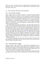
Treatment strategies
Medical treatment
Anti-ischaemic drugs improve symptoms and quality of life, but
have not been shown to reduce mortality or myocardial
infarction. blockers may improve survival in hypertension, in
heart failure, and after myocardial infarction, and so are
considered by many to be first line treatment. Nicorandil has
recently been shown to reduce ischaemic events and need for
hospital admission.
Trials comparing medical treatment with revascularisation
predate the widespread use of antiplatelet and cholesterol
lowering drugs. These drugs reduce risk, both in patients
treated with drugs only and in those undergoing
revascularisation, and so may have altered the risk-benefit ratio
for a particular revascularisation strategy in some patients.
Coronary artery bypass graft surgery
Coronary artery bypass surgery involves the placement of grafts
to bypass stenosed native coronary arteries, while maintaining
cerebral and peripheral circulation by cardiopulmonary bypass.
The grafts are usually saphenous veins or arteries (principally
the left internal mammary artery).
Operative mortality is generally 1-3% but may be much
higher in certain subsets of patients. Scoring systems can
predict operative mortality based on clinical, investigational, and
operative factors. Important developments that have occurred
since trials of bypass surgery versus medical treatment were
conducted include increased use of arterial grafts (which have
much greater longevity than venous grafts), surgery without
extracorporeal circulation (“off-pump” bypass), and minimal
access surgery.
Major factors influencing risks and benefits of coronary
revascularisation
x Advanced age
x Female
x Severe angina
x Smoking
x Diabetes
x Obesity
x Hypertension
x Multiple coronary vessels affected
x Coexisting valve disease
x Impaired left ventricular function
x Impaired renal function
x Cerebrovascular or peripheral vascular disease
x Recent acute coronary syndrome
x Chronic obstructive airwa ys disease
Left internal
mammary
artery with
pedicle
Saphenous
vein graft
Top: Diagrams of saphenous vein and left internal mammary artery grafts
for coronary artery bypass surgery. Bottom: Three completed grafts—(1) left
internal mammary artery (LIMA) to left anterior descending artery (LAD),
and saphenous vein grafts (SVG) to (2) diagonal artery (DG) and (3) obtuse
marginal artery (OM)
Risk score for assessing probable mortality from bypass
surgery in patients with chronic stable angina
Risk factor Weighted score
Age > 60 Score 1 for every
5yearsover
Female sex 1
Chronic obstructive pulmonary disease 1
Extracardiac arteriopathy 2
Neurological dysfunction 2
Previous cardiac surgery 3
Serum creatinine > 200mol/l 2
Reduced left ventricular ejection fraction 1 for 30-50%
3 for < 30%
Myocardial infarction in past 90 days 2
Pulmonary artery systolic pressure > 60 mm Hg 2
Major cardiac procedure as well as bypass surgery 2
Emergency operation 2
x Total score <2 predicts < 1% operative mortality
x Total score of 3-5 predicts 3% operative mor tality
x Total score >6 predicts > 10% operative mortality
A more detailed assessment with logistic analysis is available at www.euroscore.org and
is recommended for assessing high risk patients
12
Percutaneous coronary intervention
The main advantages of percutaneous intervention over bypass
surgery are the avoidance of the risks of general anaesthesia,
uncomfortable sternotomy and saphenous wounds, and
complications of major surgery (infections and pulmonary
emboli). Only an overnight hospital stay is necessary (and many
procedures can be performed as day cases), and the procedure
can be easily repeated. The mortality is low (0.2%), and the most
serious late complication is restenosis.
Pa tient suitability is primarily determined by technical factors.
A focal stenosis on a straight artery without proximal vessel
tortuousness or involv ement of major side branches is ideal for
percutaneous intervention. Long, hea vily calcified stenoses in
tortuous vessels or at bifurca tions and chronic total occlusions are
less suitable. This m ust be borne in mind when interpreting data
from trials of percutaneous intervention and bypass surgery, as
only a minority of patients were suitable for both procedures.
Nowada ys, more and more patients undergo percutaneous
interv ention, and referral rates for bypass surgery are falling.
Comparative studies of
revascularisation strategies
Coronary artery bypass surgery versus medical treatment
In a meta-analysis of seven trials comparing bypass surgery with
medical treatment, surgery conferred a survival advantage in
patients with severe left main stem coronary disease, three
vessel disease, or two vessel disease with severely affected
proximal left anterior descending artery. The survival gain was
more pronounced in patients with left ventricular dysfunction
or a strongly positive exercise test. However, only 10% of trial
patients received an internal mammary artery graft, only 25%
received antiplatelet drugs, and the benefit of lipid lower ing
drugs on long term graft patency was not appreciated when
these studies were carried out. Furthermore, 40% of the
medically treated patients underwent bypass surgery during 10
years of follow up. Thus, these data may underestimate the
benefits of surgery compared with medical treatment alone.
In lower risk patients bypass surgery is indicated only for
symptom relief and to improve quality of life when medical
treatment has failed. Surgery does this effectively, with 95% of
patients gaining immediate relief from angina and 75%
remaining free from angina after five years. Unfortunately,
venous grafts have a median life span of only seven years, and
after 15 years only 15% of patients are free from recurrent
angina or death or myocardial infarction. However, the
increased use of internal mammary artery grafts, which have
excellent long term patency (85% at 10 years), has increased
postoperative survival and reduced long term symptoms.
Subgroup analysis of mortality benefit from coronary artery
bypass surgery compared with medical treatment at 10 years
after randomisation for patients with chronic stable angina
Subgroup Mean (1.96 SE) increased
survival time (months)
P value of
difference
Vessel disease:
1 or 2 vessels 1.8 (3.0) 0.25
3 vessels 5.7 (3.6) 0.001
Left main stem 19.3 (13.7) 0.005
Left ventricular function:
Normal 2.3 (2.4) 0.06
Abnormal 10.6 (6.1) < 0.001
Exercise test:
Normal 3.3 (4.4) 0.14
Abnormal 5.1 (3.3) 0.002
Severity of angina:
CCS class 0, I, II 3.3 (2.7) 0.02
CCS class III, IV 7.3 (4.8) 0.002
CCS=Canadian Cardiovascular Society
Left: Angiogram of a 10 year old diseased venous graft to the obtuse
marginal artery showing proximal aneurysmal dilatation (A) and severe
stenosis in middle segment (B). Right: Removal of this graft after repeat
bypass surgery shows its gross appearance (graft longitudinally opened in
right image), with atherosclerosis in a thin walled aneurysm and a small
residual lumen
Old saphenous vein grafts may contain large amounts of necrotic clotted debris, friable laminated thrombus, and ulcerated atheromatous plaque and are
unattractive for percutaneous intervention because of the high risk of distal embolisation. However, distal embolisation protection devices such as the
FilterWire EX (far right) reduce this risk by trapping any material released. Such a device (far left, B) is positioned in the distal segment of a subtotally
occluded saphenous vein graft of the left anterior descending artery (A) before it is dilated and stented (inner left, C) to restore blood flow (inner right)
Chronic stable angina: treatment options
13
Percutaneous coronary intervention versus medical
treatment
Most percutaneous procedures are undertaken to treat single
vessel or two vessel disease, but few randomised controlled trials
have compared percutaneous intervention with medical
treatment. These showed that patients undergoing the
percutaneous procedure derived greater angina relief and took
less drugs but required more subsequent procedures and had
more complications (including non-fatal myocardial infarction),
with no mortality difference. Patients with few symptoms did
not derive benefit. Therefore, percutaneous intervention is
suitable for low risk patients with one or two vessel disease and
poor symptom control with drugs, at a cost of a slightly higher
risk of non-fatal myocardial infarction. However, the procedure
may not be indicated if symptoms are well controlled.
Percutaneous intervention versus bypass surgery
Single vessel disease
In a meta-analysis by Pocock et al percutaneous intervention in
patients with single vessel disease resulted in mortality similar to
that found with bypass surgery (3.7% v 3.1% respectively) but a
higher rate of non-fatal myocardial infarction (10.1% v 6.1%,
P=0.04). Angina was well treated in both groups, but persistence
of symptoms was slightly higher with percutaneous
intervention. Rates of repeat revascularisation were much
higher with percutaneous intervention than bypass surgery.
Multivessel disease
Since comparative trials could recruit only those patients who
were suitable for either revascularisation strategy, only 3-7% of
screened patients were included. These were predominantly
“low risk” patients with two vessel disease and preserved left
ventricular function
—
patients in whom bypass surgery has not
been shown to improve survival
—
and thus it is unlikely that a
positive effect in favour of percutaneous intervention would
have been detected. The generally benign prognosis of chronic
stable angina means that much larger trials would have been
required to show significant differences in mortality.
A meta-analysis of data available to the end of 2000
revealed similar rates of death and myocardial infarction with
both procedures, but repeat revascularisation rates were higher
with percutaneous intervention. The prevalence of appreciable
angina was greater with percutaneous intervention at one year,
but this difference disappeared at three years.
The nature of percutaneous coronary intervention has
changed considerably over the past 10 years, with important
developments including stenting and improved antiplatelet
drugs. The integrated use of these treatments clearly improves
outcomes, but almost all of the revascularisation trials predate
these developments.
A more recent trial comparing percutaneous intervention
and stenting with bypass surgery in multivessel disease
confirmed similar rates of death, myocardial infarction, and
stroke at one year, with much lower rates of repeat
revascularisation after percutaneous intervention compared
with earlier trials. There was also a cost benefit of nearly $3000
(£1875) per patient associated with percutaneous intervention
at 12 months. The recent introduction of drug eluting (coated)
stents, which seem to reduce substantially the problem of
restenosis, is likely to extend the use of percutaneous
intervention in multivessel disease over the next few years.
Diabetes
Bypass surgery confers a survival advantage in symptomatic
diabetic patients with multivessel disease The BARI trial
Coronary angiogram
showing a severe
focal stenosis (arrow)
in a large oblique
marginal branch of
the left circumflex
artery (LCx), suitable
for percutaneous
coronary
intervention. The left
anterior descending
artery (LAD) has no
important disease
Coronary angiograms of 70 year
old woman with limiting angina.
There were severe stenoses
(arrows) in the proximal and
middle left anterior descending
artery (LAD, top) and in the distal
right coronary artery (RCA, left).
Because of the focal nature of
these lesions, percutaneous
coronary intervention was the
preferred option
Coronary angiograms of a
69 year old man with
limiting angina and
exertional breathlessness.
There was severe proximal
disease (arrows) of the left
anterior descending (LAD)
and left circumflex arteries
(LCx) (top) and occlusion of
the right coronary artery
(RCA, left). The patient was
referred for coronary artery
bypass surgery on prognostic
and symptomatic grounds
ABC of Interventional Cardiology
14
revealed a significant difference in five year mortality (21% with
percutaneous intervention v 6% with bypass surgery). Similar
trends have been found in other large trials. However, the
recent RAVEL and SIRIUS studies, in which the sirolimus
eluting Cypher stent was compared with the same stent
uncoated, showed a remarkable reduction in restenosis rates
within the stented segments in diabetic patients (0% v 42% and
18% v 51% respectively). Ongoing trials will investigate this
issue further.
Other study data
Large registries of outcomes in patients undergoing
revascularisation have the advantage of including all patients
rather than the highly selected groups included in randomised
trials. The registr y data seem to agree with those from
randomised trials: patients with more extensive disease fare
better with bypass surgery, whereas percutaneous intervention
is preferable in focal coronary artery disease.
An unusual observation is that patients screened and
considered suitable for inclusion in a trial fared slightly better if
they refused to participate than did those who enrolled. The
heterogeneous nature of coronary disease means that certain
patient subsets will probably benefit more from one treatment
than another. The better outcome in the patients who were
suitable but not randomised may indicate that cardiolog ists and
surgeons recognise which patients will benefit more from a
particular strategy
—
subtleties that are lost in the randomisation
process of controlled trials.
Refractory coronary artery disease
Increasing numbers of patients with coronary artery disease
have angina that is unresponsive to both maximal drug
treatment and revascularisation techniques. Many will have
already undergone multiple percutaneous interventions or
bypass surgery procedures, or have diffuse and distal coronary
artery disease. In addition to functional limitations, their
prognosis may be poor because of impaired ventricular
function. Emerging treatments may provide alternative
symptomatic improvement for some patients. There is also
renewed interest in the potential anti-ischaemic effects of
angiotensin converting enzyme inhibitors and the plaque
stabilising properties of statins.
The picture showing three completed coronary artery bypass grafts and
the pictures of a 10 year old diseased venous graft to the obtuse marginal
artery were provided by G Singh, consultant cardiothoracic surgeon,
Heath Sciences Centre, Winnipeg, E Pascoe, consultant cardiothoracic
surgeon, St Boniface Hospital, Winnipeg, and J Scatliff, consultant
anaesthetist, St Boniface Hospital. The picture of the FilterWire EX distal
embolisation protection device was provided by Boston Scientific
Corporation, Minneapolis, USA.
Competing interests: None declared.
Names of trials
x BARI
—
Bypass angioplasty revascularisation investigation
x SIRIUS
—
Sirolimus-coated velocity stent in treatment of patients
with de novo coronary artery lesions trial
x RAVEL
—
Randomised study with the sirolimus-eluting velocity
balloon-expandable stent in the treatment of patients with de novo
native coronary artery lesions
Emerging treatment options for refractory angina
x Drugs—Analgesics, statins, angiotensin converting enzyme
inhibitors, antiplatelet drugs
x Neurostimulation—Interruption or modification of afferent
nociceptive signals: transcutaneous electric nerve stimulation
(TENS), spinal cord stimulation (SCS)
x Enhanced external counterpulsation—Non-invasive pneumatic leg
compression, improving coronary perfusion and decreasing left
ventricular afterload
x Laser revascularisation—Small myocardial channels created by laser
beams: transmyocardial laser revascularisation (TMLR),
percutaneous transmyocardial laser revascularisation (PTMLR)
x Therapeutic angiogenesis—Cytokines, vascular endothelial growth
factor, and fibroblast growth factor injected into ischaemic
myocardium, or adenoviral vector for gene transport to promote
neovascularisation
x Percutaneous in situ coronary venous arterialisation (PICVA)—Flow
redirection from diseased coronary artery into adjacent coronary
vein, causing arterialisation of the vein and retroperfusion into
ischaemic myocardium
x Percutaneous in situ coronary artery bypass (PICAB)—Flow redirection
from diseased artery into adjacent coronary vein and then rerouted
back into the artery after the lesion
x Heart transplantation—May be considered when all alternative
treatments have failed
Further reading
x Yusuf S, Zucker D, Peduzzi P, Fisher LD, Takaro T, Kennedy JW, et al.
Effect of coronary artery bypass graft surgery on survival; overview
of 10-year results from randomised trials by the Coronary Artery
Bypass Graft Surgery Trialists Collaboration. Lancet 1994; 344:
563-70
x Pocock SJ, Henderson RA, Rickards AF, Hampton JR, King SB 3rd,
Hamm CW, et al. Meta-analysis of randomised trials comparing
coronary angioplasty with bypass surgery. Lancet 1995;345:1184-9
x Raco DL, Yusuf S. Overview of randomised trials of percutaneous
coronary intervention: comparison with medical and surgical
therapy for chronic coronary artery disease. In: Grech ED,
Ramsdale DR, eds. Practical interventional cardiology. 2nd ed.
London: Martin Dunitz, 2002:263-77
x Serruys PW, Unger F, Sousa JE, Jatene A, Bonnier HJ, Schonberger
JP, et al for the Arterial Revascularisation Therapies Study (ARTS)
Group. Comparison of coronary-artery bypass surgery and stenting
for multivessel disease. N Engl J Med 2001;344:1117-24
x Kim MC, Kini A, Sharma SK. Refractory angina pectoris.
Mechanisms and therapeutic options. J Am Coll Cardiol 2002;39:
923-34
x Morice M-C, Serruys PW, Sousa JE, Fajadet J, Ban Hayashi E, Perin
M, et al. A randomized comparison of a sirolimus-eluting stent with
a standard stent for coronary revascular ization. N Engl J Med
2002;346:1773-80
x Scottish Intercollegiate Guidelines Network. Coronary
revascularisation in the management of stable angina pectoris.
Edinburgh: SIGN, 1998 (SIGN Publication No 32)
Chronic stable angina: treatment options
15
5 Acute coronary syndrome: unstable angina and
non-ST segment elevation myocardial infarction
Ever D Grech, David R Ramsdale
The term acute coronary syndrome refers to a range of acute
myocardial ischaemic states. It encompasses unstable angina,
non-ST segment elevation myocardial infarction (ST segment
elevation generally absent), and ST segment elevation infarction
(persistent ST segment elevation usually present). This article
will focus on the role of percutaneous coronary intervention in
the management of unstable angina and non-ST segment
elevation myocardial infarction; the next article will address the
role of percutaneous intervention in ST segment elevation
infarction.
Although there is no universally accepted definition of
unstable angina, it has been described as a clinical syndrome
between stable angina and acute myocardial infarction. This
broad definition encompasses many patients presenting with
varying histories and reflects the complex pathophysiological
mechanisms operating at different times and with different
outcomes. Three main presentations have been described
—
angina at rest, new onset angina, and increasing angina.
Pathogenesis
The process central to the initiation of an acute coronary
syndrome is disruption of an atheromatous plaque. Fissuring or
rupture of these plaques
—
and consequent exposure of core
constituents such as lipid, smooth muscle, and foam cells
—
leads
to the local generation of thrombin and deposition of fibrin.
This in turn promotes platelet aggregation and adhesion and
the formation of intracoronary thrombus.
Unstable angina and non-ST segment elevation myocardial
infarction are generally associated with white, platelet-rich, and
only partially occlusive thrombus. Microthrombi can detach and
embolise downstream, causing myocardial ischaemia and
infarction. In contrast, ST segment elevation (or Q wave)
myocardial infarction has red, fibrin-rich, and more stable
occlusive thrombus.
Epidemiology
Unstable angina and non-ST segment elevation myocardial
infarction account for about 2.5 million hospital admissions
worldwide and are a major cause of mortality and morbidity in
Western countries. The prognosis is substantially worse than for
chronic stable angina. In-hospital death and re-infarction affect
5-10%. Despite optimal treatment with anti-ischaemic and
antithrombotic drugs, death and recurrent myocardial
infarction occur in another 5-10% of patients in the month after
an acute episode. Several studies indicate that these patients
may have a higher long term risk of death and myocardial
infarction than do patients with ST segment elevation.
Diagnosis
Unstable angina and non-ST segment elevation myocardial
infarction are closely related conditions with clinical
presentations that may be indistinguishable. Their distinction
depends on whether the ischaemia is severe enough to cause
myocardial damage and the release of detectable quantities of
Plaque disruption or erosion
Acute coronary syndromes
Thrombus formation with or without embolisation
Acute cardiac ischaemia
No ST segment elevation
Non-ST segment elevation
myocardial infarction
(Q waves usually absent)
ST segment elevation
myocardial infarction
(Q waves usually present)
Unstable
angina
Elevated markers of
myocardial necrosis
Markers of myocardial
necrosis not elevated
ST segment elevation
Elevated markers of
myocardial necrosis
Spectrum of acute coronary syndromes according to electrocardiographic
and biochemical markers of myocardial necrosis (troponin T, troponin I, and
creatine kinase MB), in patients presenting with acute cardiac chest pain
Three main presentations of unstable angina
x Angina at rest—Also prolonged, usually > 20 minutes
x Angina of new onset—At least CCS class III in severity
x Angina increasing—Previously diagnosed angina that has become
more frequent, longer in duration, or lower in threshold (change in
severity by >1 CCS class to at least CCS class III)
CCS=Canadian Cardiovascular Society
Collagen
Key
Dividing smooth
muscle cell
Oxidised low
density lipoprotein
Lysosomes
Media
Adventitia
Intima
Platelet-rich thrombus
Activated platelets
Lumen
Diagram of an unstable plaque with superimposed luminal thrombus
Distal embolisation of a
platelet-rich thrombus causing
occlusion of intramyocardial
arteriole (arrow). Such an
event may result in
micro-infarction and elevation
of markers of myocardial
necrosis
16
markers of myocyte necrosis. Cardiac troponin I and T are the
preferred markers as they are more specific and reliable than
creatine kinase or its isoenzyme creatine kinase MB.
An electrocardiogram may be normal or show minor
non{specific changes, ST segment depression, T wave inversion,
bundle branch block, or transient ST segment elevation that
resolves spontaneously or after nitrate is given. Physical
examination may exclude important differential diagnoses such
as pleuritis, pericarditis, or pneumothorax, as well as revealing
evidence of ventricular failure and haemodynamic instability.
Management
Management has evolved considerably over the past decade. As
platelet aggregation and thrombus formation play a key role in
acute coronary syndrome, recent advances in treatment (such as
the glycoprotein IIb/IIIa inhibitors, low molecular weight
heparin, and clopidogrel) and the safer and more widespread
use of percutaneous coronary intervention have raised
questions about optimal management.
As patients with unstable angina or non-ST segment
elevation myocardial infarction represent a heterogeneous
group with a wide spectrum of clinical outcomes, tailoring
treatment to match risk not only ensures that patients who will
benefit the most receive appropriate treatment, but also avoids
potentially hazardous treatment in those with a good prognosis.
Therefore, an accurate diagnosis and estimation of the risk of
adverse outcome are prerequisites to selecting the most
appropriate treatment. This should begin in the emergency
department and continue throughout the hospital admission.
Ideally, all patients should be assessed by a cardiologist on the
day of presentation.
Medical treatment
Medical treatment includes bed rest, oxygen, opiate analgesics
to relieve pain, and anti-ischaemic and antithrombotic drugs.
These should be started at once on admission and continued in
those with probable or confirmed unstable angina or non-ST
segment elevation myocardial infarction. Anti-ischaemic drugs
include intravenous, oral, or buccal nitroglycerin, blockers,
and calcium antagonists. Antithrombotic drugs include aspirin,
clopidogrel, intravenous unfractionated heparin or low
molecular weight heparin, and glycoprotein IIb/IIIa inhibitors.
Conservative versus early invasive strategy
“Conservative” treatment involves intensive medical
management, followed by risk stratification by non-invasive
means (usually by stress testing) to identify patients who may
need coronary angiography. This approach is based on the
results of two randomised trials (TIMI IIIB and VANQWISH),
which showed no improvement in outcome when an “early
invasive” strategy was used routinely, compared with a selective
approach.
These findings generated much controversy and have been
superseded by more recent randomised trials (FRISC II,
TACTICS-TIMI 18, and RITA 3), which have taken advantage of
the benefits of glycoprotein IIb/IIIa inhibitors and stents. All
three studies showed that an early invasive strategy
(percutaneous coronary intervention or coronary artery bypass
surgery) produced a better outcome than non-invasive
management. TACTICS-TIMI 18 also showed that the benefit
of early invasive treatment was greatest in higher risk patients
with raised plasma concentrations of troponin T, whereas the
outcomes for lower r isk patients were similar with early invasive
and non-invasive management.
I aVR V1 V4
II aVL V2 V5
III aVF V3 V6
II
Electrocardiogram of a 48 year old woman with unstable angina (top). Note
the acute ischaemic changes in leads V1 to V5 (arrows). Coronary
angiography revealed a severe mid-left anterior descending coronary artery
stenosis (arrow, bottom left), which was successfully stented (bottom right)
Right coronary artery
angiogram in patient with
non-ST segment elevation
myocardial infarction (top
left), showing hazy
appearance of intraluminal
thrombus overlying a severe
stenosis (arrow). Abciximab
was given before direct
stenting (top right), with
good angiographic outcome
(bottom)
Names of trials
x TIMI IIIB
—
Thrombolysis in myocardial infarction IIIB
x VANQWISH
—
Veterans affairs non-Q-wave infarction strategies in
hospital
x GUSTO IV ACS
—
Global use of strategies to open occluded
arteries-IV in acute coronary syndromes
x RITA 3
—
Randomised intervention treatment of angina
x FRISC II
—
Fast revascularisation during instability in coronary
artery disease
x TACTICS-TIMI 18
—
Treat angina with Aggrastat and determine cost
of therapy with an inv asiv e or conservativ e strategy-thrombolysis in
my ocardial infarction
Acute coronary syndrome: unstable angina and non-ST segment elevation myocardial infarction
17
Identifying higher risk patients
Identifying patients a t higher risk of dea th, myocardial infarction,
and recurrent ischaemia allows aggressiv e antithrombotic
treatment and early coronary angiograph y to be targeted to those
who will benefit. The initial diagnosis is made on the basis of a
patient’s history, electrocardiograph y, and the presence o f
elevated plasma concentrations of biochemical markers . The
same information is u sed to assess the risk of an adv erse
outcome. It should b e emphasised that risk assessment is a
continuous process.
The TIMI risk score
Attempts have been made to formulate clinical factors into a
user friendly model. Notably, Antman and colleagues identified
seven independent prognostic risk factors for early death and
myocardial infarction. Assigning a value of 1 for each risk factor
present provides a simple scoring system for estimating risk, the
TIMI risk score. It has the advantage of being easy to calculate
and has broad applicability in the early assessment of patients.
Applying this score to the results in the TACTICS-TIMI 18
study indicated that patients with a TIMI risk score of >3
benefited significantly from an early invasive strategy, whereas
those with a score of <2 did not. Therefore, those with an initial
TIMI score of >3 should be considered for early angiography
(ideally within 24 hours), with a view to revascularisation by
percutaneous intervention or bypass surgery. In addition, any
patient with an elevated plasma concentration of troponin
marker, ST segment changes, or haemodynamic instability
should also undergo early angiography.
Conclusion
The diagnosis of unstable angina or non-ST segment elevation
myocardial infarction demands urgent hospital admission and
coronary monitoring. A clinical history and examination, 12
lead electrocardiography, and measurement of troponin
concentration are the essential diagnostic tools. Bed rest,
aspirin, clopidogrel, heparin, antianginal drugs, and opiate
analgesics are the mainstay of initial treatment.
Early risk stratification will help identify high risk patients,
who may require early treatment with glycoprotein IIb/IIIa
inhibitors, angiography, and coronary revascularisation. Those
deemed suitable for percutaneous intervention should receive a
glycoprotein IIb/IIIa inhibitor and stenting as appropriate.
There seems to be little merit in prolonged stabilisation of
patients before percutaneous intervention, and an early invasive
strategy is generally preferable to a conservative one except for
patients at low risk of further cardiac events. This approach will
shorten hospital stays, improve acute and long term outcomes,
and reduce the need for subsequent intervention.
In the longer term, aggressive modification of risk factors is
warranted. Smoking should be strongly discouraged, and statins
should be used to lower blood lipid levels. Long term treatment
with aspirin, clopidogrel (especially after stenting), blockers,
angiotensin converting enzyme inhibitors, and antihypertensive
drugs should also be considered. Anti-ischaemic drugs may be
stopped when ischaemia provocation tests are negative.
The picture of a microthrombus occluding an intramyocardial arteriole
was provided by K MacDonald, consultant histopathologist, St Boniface
Hospital, Winnipeg.
Competing interests: None declared.
The seven variables for the TIMI risk score
x Age >65 years
x >3 risk factors for coronary artery disease
x >50% coronary stenosis on angiography
x ST segment change > 0.5 mm
x >2 anginal episodes in 24 hours before presentation
x Elevated serum concentration of cardiac markers
x Use of aspirin in 7 days before presentation
No of TIMI risk factors present
Death or myocardial infarction
at 14 days (%)
0 or 1 2
Low risk Higher risk
3 4 5 6 or 7
0
10
15
20
5
Rates of death from all causes and non-fatal myocardial infarction at 14
days, by TIMI risk score. Note sharp rate increase when score >3
Unstable angina or non-ST segment elevation myocardial infarction
TIMI risk assessment on presentation
(aspirin, clopidogrel, heparin, nitrates, β blockers)
Low risk
(TIMI risk score 0-2, negative troponin test)
Conservative management
Higher risk
(TIMI risk score
>
3, positive
troponin test, dynamic ST changes,
or haemodynamically unstable)
Stress test
Negative
Discharge
Positive
Percutaneous coronary
intervention plus
glycoprotein IIb/IIIa inhibitor
Medical
treatment
Coronary
artery bypass
surgery
Possible glycoprotein IIb/IIIa inhibitor
Invasive management
Coronary angiography
Simplified management pathway for patients with unstable angina or
non-ST segment elevation myocardial inf arction
Further reading
x Braunwald E, Antman EM, Beasley JW, Califf RM, Cheitlin MD,
Hochman JS, et al. ACC/AHA 2002 guideline update for the
management of patients with unstable angina and non-ST-segment
elevation myocardial infarction: a report of the American College
of Cardiology/American Heart Association task force on practice
guidelines. J Am Coll Cardiol 2002;40:1366-74
x Bertrand ME, Simoons ML, Fox KA, Wallentin LC, Hamm CW,
McFadden E, et al. Management of acute coronary syndromes:
acute coronary syndromes without persistent ST segment elevation.
Recommendations of the Task Force of the European Society of
Cardiology. EurHeartJ2000;21:1406-32
x Antman EM, Cohen M, Bernink PJ, McCabe CH, Horacek T,
Papuchis G, et al. The TIMI risk score for unstable angina/non-ST
elevation MI: a method for prognostication and therapeutic
decision making. JAMA 2000;284:835-42
x Ramsdale DR, Grech ED. Percutaneous coronary intervention
unstable angina and non-Q-wave myocardial infarction. In: Grech
ED, Ramsdale DR, eds. Practical interventional cardiology.2nded.
London: Martin Dunitz, 2002:165-87
ABC of Interventional Cardiology
18
6 Acute coronary syndrome: ST segment elevation
myocardial infarction
Ever D Grech, David R Ramsdale
Acute ST segment elevation myocardial infarction usually
occurs when thrombus forms on a ruptured atheromatous
plaque and occludes an epicardial coronary artery. Patient
survival depends on several factors, the most important being
restoration of brisk antegrade coronary flow, the time taken to
achieve this, and the sustained patency of the affected artery.
Recanalisation
There are two main methods of re-opening an occluded artery:
administering a thrombolytic agent or primary percutaneous
transluminal coronary angioplasty.
Although thrombolysis is the commonest form of treatment
for acute myocardial infarction, it has important limitations: a
rate of recanalisation (restoring normal flow) in 90 minutes of
only 55% with streptokinase or 60% with accelerated alteplase;
a 5-15% r isk of early or late reocclusion leading to acute
myocardial infarction, worsening ventricular function, or death;
a 1-2% r isk of intracranial haemorrhage, with 40% mortality;
and 15-20% of patients with a contraindication to thrombolysis.
Primary angioplasty (also called direct angioplasty)
mechanically disrupts the occlusive thrombus and compresses
the underlying stenosis, rapidly restoring blood flow. It offers a
superior alternative to thrombolysis in the immediate treatment
of ST segment elevation myocardial infarction. This differs from
sequential angioplasty, when angioplasty is performed after
thrombolysis. After early trials of thrombolytic drugs, there was
much interest in “adjunctive” ang ioplasty (angioplasty used as a
supplement to successful thrombolysis) as this was expected to
reduce recurrent ischaemia and re-infarction. Later studies,
however, not only failed to show any advantage, but found
higher rates of major haemorrhage and emergency bypass
surgery. In contrast, “rescue” (also known as “salvage”)
angioplasty, which is performed if thrombolysis fails to restore
patency after one to two hours, may confer benefit.
Pros and cons of primary angioplasty
Advantages
Large randomised studies have shown that thrombolysis
significantly reduces mortality compared with placebo, and this
effect is maintained long term. Primary angioplasty confers
Histological appearance of a
ruptured atheromatous plaque
(bottom arrow) and occlusive
thrombus (top arrow) resulting
in acute myocardial infarction
Acute ST segment elevation
myocardial infarction
Thrombolytic treatment Primary angioplasty
Infarct artery recanalised,
but significant residual stenosis
Rescue angioplasty
(1-2 hours after failed thrombolysis)
Elective angioplasty (if continued ischaemia)
Adjunctive angioplasty
Deferred angioplasty
(1-7 days after thrombolysis)
Infarct artery not recanalised
Methods of recanalisation for acute myocardial infarction
Incidence (%)
P<0.0001
0
9 studies
(n=58 600)*
* FTT Collaborative Group, Lancet 1994;343:311-22
✝ Keeley et al, Lancet 2003;361:13-20
23 studies
(n=7437)✝
9 studies
(n=58 600)*
23 studies
(n=6271)✝
23 studies
(n=6497)✝
0
10
15
5
P=0.0002
2
3
Mortality
P<0.0001
0
4
6
8
2
Re-infarctionCerebrovascular events
1
P<0.0001
P=0.0004
Controls Thrombolytic PCI
Effects of treatment with placebo, thrombolytic
drugs, or primary percutaneous coronary
intervention (PCI) on mortality, incidence of
cerebrovascular events, and incidence of
non-fatal re-infarction after acute myocardial
infarction in randomised studies. Of the 1%
incidence of cerebrovascular events in patients
undergoing primary percutaneous intervention,
only 0.05% were haemorrhagic. In contrast
patients receiving thrombolytic drugs had a 1%
incidence of haemorrhagic cerebrovascular
events (P<0.0001) and an overall 2% incidence
of cerebrovascular events (P=0.0004)
Comparison of methods of recanalisation
Thrombolysis
Rescue
angioplasty
Primary
angioplasty
Time from admission
to recanalisation
1-3 hours
after start of
thrombolysis
Time to start of
thrombolysis
plus 2 hours
20-60
minutes
Recanalisation with
brisk antegrade flow
55-60% 85% 95%
Systemic fibrinolysis +++ +++ −
Staff and catheter
laboratory “burden”
−++++
Cost of procedure + +++ +++
19
extra benefits in terms of substantial reductions in rates of
death, cerebrovascular events, and re-infarction.
The information provided by immediate coronary
angiography is valuable in determining subsequent
management. Patients with severe three vessel disease, severe
left main coronary artery stenosis, or occluded vessels
unsuitable for angioplasty can be referred for bypass surgery.
Conversely, patients whose arteries are found to have
spontaneously recanalised or who have an insignificant infarct
related artery may be selected for medical treatment, and thus
avoid unnecessary thrombolytic treatment.
Disadvantages
The morbidity and mortality associated with primary
angioplasty is operator dependent, varying with the skill and
experience of the interventionist, and it should be considered
only for patients presenting early ( < 12 hours after acute
myocardial infarction).
Procedural complications are more common than with
elective angioplasty for chronic angina, and, even though it is
usual to deal only with the occluded vessel, procedures may be
prolonged. Ventricular arrhythmias are not unusual on
recanalisation, but these generally occur while the patient is still
in the catheterisation laboratory and can be promptly treated by
intravenous drugs or electrical cardioversion. Right coronary
artery procedures are often associated with sinus arrest,
atrioventricular block, idioventricular rhythm, and severe
hypotension. Up to 5% of patients initially referred for primary
angioplasty require urgent coronary artery bypass surgery, so
surgical backup is essential if risks are to be minimised.
There are logistical hurdles in delivering a full 24 hour
service. Primary angioplasty can be performed only when
adequate facilities and experienced staff are av ailable. The time
from admission to recanalisation should be less than 60 minutes,
which may not be possible if staff are on call from home.
How ever, recent evidence suggests that, even with longer delays,
primary angioplasty may still be superior to thrombolysis.
A catheterisation laboratory requires large initial capital
expenditure and has substantial running costs. However, in an
existing, fully supported laboratory operating at high volume,
primary angioplasty is at least as cost effective as thrombolysis.
Primary angioplasty and coronary
stents
Although early randomised studies of primary angioplasty
showed its clinical effectiveness, outcomes were marred by high
rates of recurrent ischaemia (10-15% of patients) and early
reinfarction of the affected artery (up to 5%). Consequently,
haemodynamic and arrhythmic complications arose, with the
need for repeat catheterisation and revascularisation, prolonged
hospital stay, and increased costs. Furthermore, restenosis rates
in the first six months remained disappointingly high (25-45%),
and a fifth of patients required revascularisation.
Although stenting the lesion seemed an attractive answer, it
was initially thought that deploying a stent in the presence of
thrombus over a ruptured plaque would provoke further
thrombosis. However, improvements in stent deployment and
advances in adjunctive pharmacotherapy have led to greater
technical success. Recent studies comparing primary stenting
with balloon ang ioplasty alone have shown that stented patients
have significantly less recurrent ischaemia, reinfarction, and
subsequent need for further angioplasty. Economic analysis has
shown that, as expected, the initial costs were higher but were
offset by lower follow up costs after a year.
Severe distal left main
stem stenosis (arrow 1)
and partially occluded
mid-left anterior
descending artery due to
thrombus (arrow 2). In
view of the severity of the
lesion salvage angioplasty
was contraindicated. An
intra-aortic balloon
pump was used to
augment blood pressure
and coronary flow before
successful bypass surgery
Pros and cons of primary angioplasty* compared with
thrombolysis
Advantages
x High patency rates ( > 90%) with brisk, antegrade flow
x Lower mortality
x Better residual left ventricular function
x More rapid electrocardiographic normalisation
x Less recurrent ischaemia (angina, reinfarction, exercise induced
ischaemia)
x No systemic fibrinolysis, therefore bleeding problems avoided
x Improved risk stratification by angiography with identification of
patients suitable for coronary artery bypass surgery
Disadvantages
x Higher procedural cost than streptokinase or alteplase (although
long term costs lower)
x Can be performed only when cardiac catheterisation facilities and
experienced staff available
x Recanalisation more rapid than thrombolysis only if 24 hour
on-call team available
x Risks and complications of cardiac catheterisation and
percutaneous intervention
x Reperfusion arrhythmias probably more common because of more
rapid recanalisation
*With or without stenting
Anterior myocardial infarction of 4 hours’ duration and severe
hypotension, caused by a totally occluded proximal left anterior
descending artery (arrow, top left). After treatment with abciximab, a stent
was positioned. Initial inflation showed “waisting” of the balloon (top
right), due to fibrous lesion resistance, which resolved on higher inflation
(bottom left). Successful recanalisation resulted in brisk flow (bottom right),
and the 15 minute procedure completely resolved the patient’s chest pain
ABC of Interventional Cardiology
20