Antiarrhythmic Drugs A practical guide – Part 2 pptx
Bạn đang xem bản rút gọn của tài liệu. Xem và tải ngay bản đầy đủ của tài liệu tại đây (183.91 KB, 19 trang )
12 Chapter 1
occurring simultaneously. For this reason, the ST segmentand the T
wave (the portions of the surface ECG that reflectventricular repo-
larization) give very little directional information,and abnormalities
in the ST segments and T waves are most often (and quite prop-
erly)
interpreted as being nonspecific. The QT interval represents
the time from the beginning of depolarization (the beginning of the
QRS complex) to the end of repolarization (the end of the T wave)
of the ventricular myocardium,and thus reflects the averageaction
potential duration of ventr
icular muscle.
Mechanisms of cardiac tachyarrhythmias
Most rapid cardiac arrhythmias are thought to be duetooneoftwo
general mechanisms: abnormal automaticity or reentry. In recent
years, however, a thirdgeneral mechanism—the “channelopathy”—
has been recognized as the cause of several relatively unusual vari-
eties of car
diac arrhythmias.
Automaticity
As already noted,automaticity isan important feature of the normal
electrical system; the pacemaker function of the heart depends upon
it. Under some circumstances, however, abnormal automaticity can
occur. When an abnormal acceleration of phase 4 a
ctivity occurs
at somelocationwithin the heart, an automatic tachyarrhythmia is
the result. Suchan automatic focus can arise in the atria, the AV
junction, or the ventricles and can lead to automatic atrial tachy-
cardia, automatic junctional tachyc
ardia, or automatic ventricular
tachycardia.
Automatic tachyarrhythmias are not particularly common; they
probably account for less than 10% of all tachyarrhythmias. Fur-
ther, automatic tachyarrhythmias are usually recognizable by their
characteristicsand the clinical settings in which they occur. Con
sid-
eration of some of the features of sinustachycardia, which is the
only normal variety of automatic tachycardia, may be helpful in this
regard.Sinustachycardia usually occurs as a result of appropriately
increased sympathetic tone (e.g., in respon
se to exercise). When si-
nustachycardia develops, the heart rate gradually increases from
the basic (resting)sinus rate;when sinustachycardiasubsides, the
rate likewise decreases gradually.
Similarly, automatic tachyarrhythmias oftendisplay “warm-up”
and “warm-down” in rate when the arrhythmiabeginsa
nd ends.
Mechanismsofcardiac tachyarrhythmias 13
Also, analogoustosinustachycardia, automatic tachyarrhythmias
often have metabolic causes, suchasacute cardiacischemia, hypox-
emia, hypokalemia, hypomagnesemia, acid–base disturbances, high
sympathetic tone, or the use of sympathomimetic agents. Therefore,
automatic arrhyth
mias are frequently seeninacutely ill patients,
usually in the intensive care unit (ICU) setting.
Common examples of automatic tachyarrhythmias are the multi-
focal atrial tachycardias (MATs) that accompanyacute exacerbations
of chronic pulmonary disease, many of the atrial a
nd ventricular
tachyarrhythmias seenduring the induction of and recovery from
general anesthesia(probably a result of surges in sympathetic tone),
and the ventricular arrhythmias seenduring the first minutes to
hours of an acute myocardial infarction.(Enhanced automaticity in
thi
ssituationis thought to be mediated by ischemia.)
Of all tachyarrhythmias, automatic arrhythmias are closest to re-
sembling an“itch” of the heart. The balm of antiarrhythmic drugs is
occasionally helpful, but the primary treatment of these arrhythmias
should always be directed towardide
ntifying and treating the under-
lying metabolic cause. Ingeneral, these “ICU arrhythmias” resolve
once the patient’s acute medical problems have been stabilized.
Reentry
The mechanism of reentry accounts for most clinically significant
tachyarrhythmias. Recognition of thisfactand of the fact that reen-
trant arrhythmias are amenable to study in the laboratory led to
the widespreadproliferation of electrophysiology laboratories in the
1980s.
The mechanism of reen
try, although less intuitive than the mech-
anism of automaticity, can still be reduced to a few simple con-
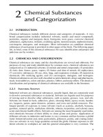
cepts. Reentry cannot occur unless certain underlying conditions
exist (Figure 1.6). First, tworoughly parallel conducting pathways
must be connectedprox
imally and di stally by conducting tissue,
thus forming a potential electrical circuit. Second,one pathway must
have a longer refractory period than the other pathway. Third, the
pathway with the shorter refractory periodmust conduct electrical
impulses more slowly thandoes the opposite p
athway.
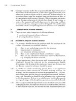
If all these seemingly implausible conditions are met, reentry can
be initiated by introducing an appropriately timedpremature im-
pulse to the circuit(Figure 1.7). The premature impulse must en-
ter the circuit early enough that the pathway with the long refrac-
tory periodi
sstill refractory from the latest depolarization,but late
14 Chapter 1
A
B
Figure 1.6 Prerequisites for reentry. An anatomic circuit must be present in
whichtwo portionsofthecircuit(pathways A and B) have electrophysio-
logic properties that differ from oneanother in a critical way. In this example,
pathway A conducts electrical impulses more slowly thanpath
way B;path-
way B has a longer refractory period thanpathway A.
enough that the pathway with the shorter refractory period has
recovered and is able to conduct the premature impulse. The im-
pulse enters the pathway with the shorter refractory period but is
conducted slowly because that pathway has the electrophysiologic
property of slowconduction. By the time the impulse rea
ches the
long-refractory-periodpathway from below, that pathway has had
timetorecover and is able to conduct the impulse in the retrograde
direction. If the retrograde impulse now reenters the first pathway
and is conducted antegradely (as islikely because of the short re-
fractory period of the first path
way), a continuously circulating im-
pulse is established, which rotates around and around the reentrant
Mechanismsofcardiac tachyarrhythmias 15
A
B
Figure 1.7 Initiation of reentry. If the prerequisites describedinFigure 1.6
are present, an appropriately timed, premature electrical impulse can block
in pathway A (which has a relatively long refractory period) while conduct-
ing down pathway A. Because c
onductiondown pathway A is slow, pathway
B has timetorecover, allowing the impulse to conduct retrogradely up path-
way B. The impulse can then reenter pathway A. A continuously circulating
impulse isthus established.
circuit. All that is necessary for the reentrant impulse to usurp the
rhythm of the heart is for the impulse to exit from the circuitat
some point during eachlap and thereby depolarize the remaining
myocardium outside the circuit.
Because reentry dependsoncritical differences in
the conduction
velocities and refractory periodsamong the various pathways of the
circuit, and because conduction velocities and refractory periods, as
we have seen, are determined by the shape of the actionpotential,
the actionpotentials of the tw
o pathways in any reentrant circuit
16 Chapter 1
must be different from oneanother. Thus, drugs that change the
shape of the actionpotential might be useful in the treatmentof
reentrant arrhythmias.
Reentrant circuits, while always abnormal, occur with some fre-
quency in the human heart. Some reentrant circuits are p
resent
at birth, notably those causing supraventricular tachycardias (e.g.,
reentry associatedwith AV bypass tracts and with dual AV nodal
tracts). However, reentrant circuits that cause ventricular tachycar-
dias are almost never congenital, but come into existenceascardiac
disease develops during life. In the ventricles, reentrant circuits arise
in areas in which normal cardiac tissuebecomes interspersedwith
patches of fibrous(scar) tissue, thus forming potential anatomic cir-
cuits. Thus, ventricular reentrant circuits usu
ally occuronly when
fibrosis develops in the ventricles, such as after a myocardial infarc-
tion or with cardiomyopathic diseases.
Theoretically, if all anatomic and electrophysiologic criteria for
reentry are present, any impulse that enters the circuit at the ap-
propriate instan
t in time induces a ree ntranttachycardia. The time
from the end of the refractory period of the shorter-refractory-period
pathway to the end of the refractory period of the pathway with a
longer refractory time, during which reentry can be induced, is called
the tachycardia zone. Treating reentrant arrhythmias ofteninvolves
try
ing to narrow or abolish the tachycardia zone with antiarrhyth-
mic drugs (by using a drug that, onehopes, might increase the re-
fractory period of the shorter-refractory-periodpathway, or decrease
the refractory period of the longer-refractory-periodpathway).
Because reentrant arrhythmias ca
n be reproducibly induced (and
terminated)byappropriately timed impulses, these arrhythmias are
ideal for study in the electrophysiology laboratory. Inmany instances
(very commonly with supraventricular arrhythmias, butonly occa-
sionally with ventricular arrhythmias), the pathways involvedinthe
reentrant cir
cuit can be precisely mapped, the effectofvarious ther-
apies can be assessed,and critical portions of the circuit can even be
ablated through the electrode catheter.
The channelopathies
In recent years, some varieties of tachyarrhythmias have been at-
tributed to genetic abnormalities in the channels that mediate ionic
fluxes across the cardiaccell membrane. Such “channelopathies”—
abnormally functioning channels duetoinheritable
mutations—can
affectany electrically active cell and are not limited to the heart. For
Mechanismsofcardiac tachyarrhythmias 17
instance, some varieties of migraine, epilepsy, periodic paralysis, and
muscle disorders are apparently duetochannelopathies.
While several distinctive cardiac arrhythmias are now thought
to be caused by channelopathies, the most clinically relevantand
the most co
mmonchannelopathic arrhythmias are those related to
triggered activity.
Triggered activity
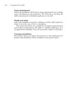
Triggered activity is caused by abnormal fluxes of positive ions into
cardiaccells. These ionic fluxes producean abnormal “bump” in the
actionpotential during late phase 3 or early phase 4 (Figure 1.8).
The bump is called an afterdepolarizat
ion.Inmost if not all cases,
afterdepolarizations are thought to be duetoinherited abnormalities
in the channels that control the movementofcalcium ionsacross
the cell membrane. If the afterdepolarizations are of sufficientam-
plitude, they can
trigger the rapid sodium channels (which, as noted,
are voltage dependent), and thus cause another actionpotential to
be generated.
Digitalis-toxic arrhythmias, torsades de pointes, and someof
the rare ventricular tachycardias that respond to calcium-blocking
age
nts have all been advanced as arrhythmias that are most likely
caused by triggered activity.
Clinical features of the major tachyarrhythmias
Before considering how antiarrhythmic drugs work, it will be help-
fultoreview the salient clinical features of the major cardiac tach-
yarrhythmias.
Supraventricular tachyarrhythmias
Table 1.1 classifies the supraventricular tachyarrhythmias according
to mechanism.
Automatic supraventricular tachyarrhythmias
Automatic supraventricular arrhythmias are seen almost exclusively
in acutely ill patients, most of whom have one of the following condi-
tions:myocardial ischemia, acute exacerbationsofchronic lung dis-
ease, acute alcohol toxicity, or major electrolyte disturbances. Any
of these disorders canproduceectopic automatic foci in the atrial
myocardium.
18 Chapter 1
T-U wave
EAD
(a)
(b)
Figure 1.8 Triggered activity. Both panels show asurface ECG (top)and a
simultaneousventricular actionpotential (bottom). (a) Phase 3 of the action
potential is interrupted by a “bump”—an EAD. The EAD is reflected on the
surface ECG by a prolonged and distorted T wave (T-U wave). (b) The EAD
i
sofsufficientamplitudetoengage the rapid sodium channel and generate
another actionpotential. The resultant premature complex is seen on surface
ECG. Note that just as the premature actionpotential is coincident with the
EAD (since it i
s generated by the EAD), the premature ventricular complex
is also coincident with the T-U wave of the previous complex.
Mechanismsofcardiac tachyarrhythmias 19
Table 1.1 Classification of supraventricular tachyarrhythmias
Automatic arrhythmias
Some atrial tachycardias associated with acute medical conditions
Some multifocal atrial tachycardias
Reentrant arrhythmias
SA nodal reentrant tachycardia
Intra-atrial reentrant tachycardia
Atrial flutter and atrial fibrillation
AV nodal reentrant tachycardia
Macroreentrant (bypass-mediated) reentrant tachycardia
Triggered arrhythmias (probable mechanism)
Digitalis-toxic atrial tachycardia
Some multifocal atrial tachycardias
SA, sinoatrial; AV, atrioventricular.
Clinically, the heart rate with automatic atrial tachycardias is usu-
ally less than200 beats/min.Like all automatic rhythms, the onset
and offset are usually relatively gradual; that is, they oftendisplay
warm-up, in which the heart rate accelerates over several cardiac
cycles. Each QRS complex is preceded
by a discrete P wave, whose
shape generally differs from the normal sinusPwave, depending
on the location of the automatic focus within the atrium.Likewise,
the PR interval is often shorter thanit is during sinus rhythm,since
the ectopic focus may be relatively close to the AV node. Becau
se
automatic atrial tachycardias arise in and are localized to the atrial
myocardium (and thus the arrhythmia itself is not dependenton
the AV node), ifAVblock is produced, atrial arrhythmia itself is
unaffected.
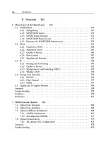
MAT (Figure 1.9) is the most common
form of automatic atrial
tachycardia. It is characterized by multiple (usually at least three)
P-wave morphologies and irregular PR intervals. MAT is thought to
be caused by the presence of several automatic foci within the atria,
firing at different rates. The arrhythmia is usually associatedwith
exac
erbation of chronic lung disease, especially in patients receiving
theophylline.
Pharmacologic therapy is usually not very helpful in treating au-
tomatic atrial tachycardia, though drugs that affect the AV node can
20 Chapter 1
Figure 1.9 MAT isanirregular atrial tachyarrhythmia that superficially re-
sembles atrial fibrillation.However, in MAT (in contrast to atrial fibrillation),
each QRS complex is preceded by a discrete P wave. Further, at least three
distinctP-wave morphologies are present, which reflects the multifocal ori
-
gin of atrial activity in this arrhythmia.
sometimes slow the ventricular rate by creating second-degree block.
The basic strategy for treating automatic atrial arrhythmias istoag-
gressively treat the underlying illness.
Reentrant supraventricular tachyarrhythmias
Ingeneral, patients have reentrantsupraventricular tachyarrhyth-
mias because they are bornwith abnormal electrical pathways that
create potential reentrant circuits. Accordingly (in contrast to pa-
tients with automatic supraventricular arrhythmias), these patients
most often initi
ally experiencesymptoms when they are young and
healthy. Most supraventricular tachyarrhythmias seeninotherwise
healthy patients are caused by the mechanism of reentry.
The five general categories of reentrantsupraventricular arrhyth-
mias are listedinTable 1.1. Many clinicianslump these arrhythmias
tog
ether (except for atrial fibrillation and atrial flutter, which gen-
erally are easily distinguishable) as paroxysmal atrial tachycardia
(PAT). Inmost instances, an astute cliniciancan tell whichspecific
Mechanismsofcardiac tachyarrhythmias 21
category of PAT he or she is dealing with (and therefore caninstitute
appropriate therapy) merely by carefully examining a12-lead ECG
of the arrhythmia.
AV nodal reentrant tachycardia
AV nodal reentranttachycardia is the most common typeofPAT,ac-
counting for nearly 60% of regular supraventricular tachyarrhyth-
mias. In AV nodal reentry, the reentrant circuit can be visualized as
being enclosed entirely within an AV node that isfunc
tionally di-
videdinto twoseparate pathways (Figure 1.10). The dual pathways
form the reentrant circuit responsible for the arrhythmia. Because
αβ
(a)
αβ
(b)
αβ
(c)
Figure 1.10 AV nodal reentranttachycardia. (a) Inpatients with AV nodal
reentry, the AV node isfunctionally dividedinto twoseparate pathways
(alpha (α)and beta (β) pathways). Similar to the example shown in Figures
1.6 and 1.7, the alpha pathway conducts more slowly than the beta pathway,
a
nd the beta pathway has a longer refractory period than the alpha pathway.
Since the beta pathway conducts more rapidly thandoes the alpha pathway,
a normal atrial impulse reaches the ventricles via the beta pathway. (b) A
premature atrial impulse can find the beta pathway still refractory at a time
when the alpha p
athway is not refractory. Because conductiondown the
alpha pathway is slow, the resultantPRinterval is prolonged.(c)Ifconditions
are right, a premature impulse can block in the beta pathway and conduct
down the alpha pathway (as in (b)), then travel retrogra
de up the beta
pathway and reenter the alpha pathway in the antegrade direction.AVnodal
reentranttachycardia results when suchacircuitous impulse is established
within the AV node.
22 Chapter 1
the reentrant circuit is within the AV node, the pharmacologic treat-
mentofAVnodal reentry usually involves giving drugs that act upon
the AV node.
Bypass-tract-mediated macroreentrant tachycardia
Tachycardia mediated by AV bypass tracts (also called accessory
pathways) is the next most common type of reentrantsupraven-
tricular tachycardiaand accounts for approximately 30% of ar-
rhythmias presenting as PAT . Most patients with suchbypass tracts
do not have overt Wolff-Parkinson–Wh
ite syndrome, however.
Instead, they have concealed bypass tracts, that is, bypass tracts
that are incapable of conducting in the antegrade direction (from
the atrium to the ventricles), and therefore never display delta
waves. Concealed bypass tracts are able to conduct electrical im-
pulses only in the retrograde direc
tion (from the ventricles to the
atrium).
The reentrant circuit responsible for these tachycardias is formed
by the bypass tract(whichalmost always constitutes the retrograde
pathway), and the normal AV nodal conducting system (the ante-
grade pathway), conne
cted by the atrial and ventricular myocardium
(Figure 1.11). Because the reentrant circuit is large(involving the
AV node, the His-Purkinje system, the ventricular myocardium, the
bypass tract, and the atrial myocardium), it is termed a macroreen-
trant circuit. Also, because the circuit cons
ists of several types of tis-
sue, it can be attacked onmany levels by many differentkindsof
drugs—drugs that affect the AV node, the bypass tract, the ventric-
ular myocardium, or the atrial myocardium.
Intra-atrial reentry
Intra-atrial reentry accounts for only a small percentage of arrhyth-
mias presenting as PAT. The reentrant circuit in intra-atrial reentry
resides entirely within the atrial myocardium and does not involve
the AV conducting system (Figure 1.12). Intra-atrial reentry resem-
bles automatic atrial tac
hycardiabecause discrete (most often atyp-
ical) P waves precedeeach QRS complex, and AV block can occur
without affecting the arrhythmia itself. Intra-atrial reentry differs
from automatic tachycardiabecause of its sudden onset and termi-
nati
on,and,like all reentrant arrhythmias, it can be induced by
pacing.Intra-atrial reentry is affected only by drugs that affect the
atrial myocardium.
Mechanismsofcardiac tachyarrhythmias 23
(a) (b) (c)
Figure 1.11 Bypass-tract-mediatedmacroreentranttachycardia. (a) Because
abypass tract is present, a normal sinus beat is transmitted to the ventricles
viatwoseparate pathways. Because the ventricle is partially preexcited (i.e.,
someventricular myocardium i
s depolarized early via the bypass tract), the
QRS complex displays a delta wave. A bypass tract usually has a longer refrac-
tory period than the normal conducting system,and the normal conducting
system includes the slow-conducting AV nodeand conducts electrical im-
pu
lses more slowly than the bypass tract. Thus, the substrate for reentry is
present. (b) A premature atrial complex occurs during the refractory period
of the bypass tractand is therefore conducted solely via the normal conduct-
ing system. The resultant QRS complex displays no delta wave. (c)Because
conduction via the normal conducting systemis relatively slow, the bypass
tract may nolonger be refractory by the time the impulse reaches the ventri-
cles. Thus, the bypass tract may be able to conduct the impulse retrogradely
back to the atrium. If so, a reentrant impulse may be established, which trav-
els antegradely down the
normal conducting system and retrogradely up the
bypass tract. The result is a large(macro) reentrant circuit.
Atrial flutter and atrial fibrillation
Atrial flutter and atrial fibrillation are special formsofintra-atrial
reentranttachycardias and are generally distinguishable quite read-
ily from other kinds of atrial tachyarrhythmias (commonly labeled
PAT) by reviewing a12-lead ECG.
In atrial flutter, the atrial activity isregular, in excess of
220 beats/min,and usually displays a typical sawtooth pattern
(Figure 1.13). Atrial flutter isalmost always accompanied by AV
block, most oftenina 2:1 pattern.
24 Chapter 1
RA
LA
AVN
RV
LV
SAN
(a)
RA
LA
AVN
RV
LV
SAN
(b)
RA
LA
AVN
RV
LV
SAN
(c)
Figure 1.12 The components of the reentrant circuit determine whichan-
tiarrhythmic drugs are likely to be effective in treating supraventricular
tachycardia. Both AV nodal reentry (a) and macroreentry (b) include the
AV node within the reentrant circuit. Therefore, drugs that affect the AV
node affec
t the reentrant circuit itself and may be useful in terminating
or preventing the arrhythmia. Incontrast, in intra-atrial reentry (c), the
reentrant circuit does not include the AV node. Drugs that affect the AV
node generally do not affect intra-atrial reentry itself, although they may
be effective in slo
wing the ventricular response during the arrhythmia.
Atrial fibrillation, atrial flutter, and automatic atrial tachycardia are simi-
lar to intra-atrial reentry in that the AV node is not required for initiat-
ing or sustaining these arrhythmias. AVN, atrioventricular node; LA, left
atrium; LV, left ventricle
; RA, right atrium; RV, right ventricle; SAN,sinoatrial
node.
Figure 1.13 Atrial flutter. A surface ECG (top)and anintracardiac electro-
gram that directly records intra-atrial electrical activity (bottom) are shown.
Note the two atrial impulses (seen on the intracardiac electrogram) for every
QRS complex; AV blockoccurs in atypical 2:1 pattern.
Mechanismsofcardiac tachyarrhythmias 25
Figure 1.14 Atrial fibrillation. Note the randomly irregular ventricular re-
sponse and the absenceofdiscrete P waves.
In atrial fibrillation, the atrial activity is continuousand chaotic,
and discrete P waves cannot be distinguished (Figure 1.14). The
ventricular response is completely irregular, reflecting the chaotic
nature of the atrial activity.
Since atrial fibrillatio
n and atrial flutter are intra-atrial arrhyth-
mias, AV block(whichoccurs in almost every case) does not affect
the arrhythmia itself. Drug therapy is usually aimed at converting
the arrhythmiabyuse of drugs that affect the atrial myocardium
or at controlling the ventricular response with drugs that affectAV
co
nduction.
SA nodal reentry
SA nodal reentry is a relatively uncommon arrhythmia in which
the reentrant circuit is thought to be enclosed entirely within the
SA node(i.e., dual SA nodal pathways are thought to exist, simi-
lar to those seeninAV nodal reentry). Discrete P waves identical
to sinusPwaves prece
deeach QRS complex. SA nodal reentry is
distinguishable fromnormal sinustachycardia(which isautomatic
in mechanism)byits sudden onset and offset, and by the fact that
it is inducible with pacing.Itis affected by drugs that affect the SA
and AV nodes.
Triggered supraventricular tachyarrhythmias
The only supraventricular tachycardia commonly attributed to trig-
gered activity is that seenwith digitalis toxicity. Digitalis toxicity
canproduce delayed afterdepolarizations (DADs; see Figure 1.16a)
that can lead to atrial tachycardias. Clinically, since digitali
s toxic-
ity also produces AV block, digitalis-toxic arrhythmias oftenmani-
fest as atrial tachycardia with block. In fact, the presenceofatrial
26 Chapter 1
tachycardia with block should always make one consider the possi-
bility of digitalis toxicity.
Electrocardiographic patterns of supraventricular
tachyarrhythmias
Oftenit is possible to specifically diagnose a patient’s supraventricu-
lar arrhythmiabyexamining a12-lead ECG. Atrial flutter and atrial
fibrillationcanusually be distinguished by simple inspection.In the
supraventricular tachycardias commonly labeled as PAT (i
.e., reg-
ular, narrow-complex tachycardias), both the relationship of the P
waves to the QRS complexes and the morphology of the P waves
during the tachycardia can be very helpful. Figure 1.15 shows the
essential electrocardiographic characteristics of the fourtypes of PAT.
Ventricular tachyarrhythmias
Table 1.2classifies the ventricular tachyarrhythmias according to
mechanism.
Automatic ventricular tachyarrhythmias
Abnormal automaticity accounts for a relatively small proportion of
ventricular tachyarrhythmias. As is the case with automatic atrial
arrhythmias, automatic ventricular arrhythmias are usually associ-
atedwith acute medical conditions, suchasmyocardial ischemi
a,
acid–base disturbances, electrolyte abnormalities, and highadren-
ergic tone. Automatic ventricular arrhythmias are most often seen
in patients with acute myocardial ischemiaorinfarction,orsome
other acute medical illness. Most arrhythmias occurring with
in
the first few hours of an acute myocardial infarction are thought to
be automatic.Once the ischemic tissue dies or stabilizes, however,
the substrate for automaticity is nolonger present.
Ingeneral, the treatmentofautomatic ventricular arrhythmias
consists of treating the
underlying illness. Antiarrhythmic drugs are
occasionally beneficial.
Reentrant ventricular tachyarrhythmias
Most ventricular arrhythmias are reentrant in mechanism.While the
conditions producing automatic ventricular arrhythmias are usually
temporary in nature (e.g., cardiacischemia), the substrate necessary
for producing reentrantventricular arrhythmias, once present, tends
to be per
manent.
Mechanismsofcardiac tachyarrhythmias 27
(a)
(b)
(c)
(d)
Figure 1.15 Typical P-wave relationships in fourkinds of PAT . Surface ECG
lead II is depicted. (a) I n AV nodal reentranttachycardia, the P wave is
usually buriedwithin the QRS complex and is most oftennot discernible
evenwith carefulstudy of all 12-lead ECG. (b) In bypass-tract-
mediated
macroreentranttachycardia, the inferior ECG leads usually show a negative
P wave. (It has a superior axisbecause the atria are activatedinthe retrograde
direction.) Also, the P wave is usually closer to the preceding QRS complex
than to the following QRS complex. (c)I
nintra-atrial reentry, discrete P
waves almost always are seen before each QRS complex. Because the intra-
atrial reentrant circuit can be located anywhere within the atria, the P-wave
morphology can have any configuration. The PR interval is usually normal
or short. (d)In
SA nodal reentry, P waves and the PR interval appear normal.
28 Chapter 1
Table 1.2 Classification of ventricular tachyarrhythmias
Automatic arrhythmias
Some ventricular tachycardias associated with acute medical conditions
Acute myocardial infarction or ischemia
Electrolyte and acid–base disturbances or hypoxia
High sympathetic tone
Reentrant arrhythmias
Ventricular tachycardia and fibrillation associated with some chronic heart
diseases
Previous myocardial infarction
Dilated cardiomyopathy
Hypertrophic cardiomyopathy
Channelopathies
Triggered arrhythmias (probable mechanism)
Pause-dependent torsades de pointes (EADs) associated with drugs that
prolong QT interval
Catechol-dependent torsades de pointes (DADs) associated with digitalis
toxicity or idiopathy
Brugada syndrome and SUNDS
EADs, early afterdepolarizations; DADs, delayed afterdepolarizations; SUNDS, sud-
den unexpected nocturnal death syndrome.
Reentrant circuits within the ventricular myocardium usually
arise after scar tissue develops, a conditionmost commonly seenin
patients who have myocardial infarctionsorcardiomyopathy. Once
the scar tissue gives rise to a reentrant ci
rcuit, the circuit persists, and
the potential for a ventricular arrhythmiaalways exists. Thus, the
“late” suddendeaths that occur after a myocardial infarction (i.e.,
from about12 h to several years after the acute event) are usually a
result of reentrant arrhythmias. Reentrantventricular arrhythmias
are seen only rarely in individuals w
ho have normal ventricles.
Most antiarrhythmic drugs affect the ventricular myocardium
and,accordingly, most are used to treat ventricular tachyarrhyth-
mias.
Channelopathic ventricular tachyarrhythmias
Channelopathies probably account for several distinctive types of
ventricular tachyarrhythmias, at least twoofwhich have now been
Mechanismsofcardiac tachyarrhythmias 29
well characterized. These are the ventricular arrhythmias dueto
triggered activity and Brugadasyndrome.
Triggered activity in the ventricles
Because ventricular tachyarrhythmias duetotriggered activity are
reasonably common,and because the managementoftriggered ven-
tricular arrhythmias
is very different from the managementofmore
typical ventricular arrhythmias, it is importanttorecognize their
characteristics. Twofairly distinct clinical syndromes are caused by
ventricular triggered activity:catechol-dependent arrhythmi
as and
pause-dependent arrhythmias. In eachsyndrome, the resultantven-
tricular arrhythmias are similar. They are the classically polymor-
phic ventricular tachyarrhythmias generally referred to as torsades de
pointes.
Catechol-dependent triggered arrhythmias. Catechol-dependenttrig-
gered arrhythmias are caused by DADs, whichoccur during p
hase 4
of the actionpotential (Figure 1.16a). DADs are seeninsusceptible
patients in the setting of digitalis intoxication and cardiacischemia.
They are also seenincertain patients who have a congenital form of
QT prolongation associatedwith what is thought to be animbalance
in
the sympathetic innervation of the heart, with predominant in-
put coming from the left stellate ganglia—stimulation of which can
reproduce DADs.
The ventricular arrhythmias caused by DADs typically are poly-
morphic,and are seeninconditionsofhighsympathetic tone.
Patients with
catechol-dependenttriggered activity therefore expe-
rience arrhythmias (oftenmanifested by syncopeorcardiac arrest)
in times of severe emotional stress or during exercise. Often they
have normal ECGs at rest but will developQTabnormalities dur-
ing exercise. The onset of the arrhythmia is not associ
atedwith a
pause.
Left stellate sympathectomy has eliminated arrhythmias in some
of these patients. Medical treatment has generally consisted of beta
blockers and calcium-channel blockers (consistent with the fact that
DADs are thought to be mediated by abnormalities in the calcium
chann
els). Many of these patients, however, end up receiving im-
plantable defibrillators.
Pause-dependent triggered arrhythmias. Pause-dependenttriggered
arrhythmias are caused by afterdepolarizations that occur during
30 Chapter 1
Delayed afterdepolarization
Early afterdepolarization
(a)
(b)
Figure 1.16 Early and delayed afterdepolarizations. (a) DADs of the type
thought to be responsible for catechol-dependenttriggered arrhythmias. The
DAD occurs during phase 4 of the actionpotential. (b) EAD of the type
thought to be responsible for pause-dependenttriggered arrhythmias. The
EAD occurs during phase 3 of the act
ionpotential.