Antiarrhythmic Drugs A practical guide – Part 5 pdf
Bạn đang xem bản rút gọn của tài liệu. Xem và tải ngay bản đầy đủ của tài liệu tại đây (154.42 KB, 19 trang )
Class I antiarrhythmic drugs 71
Several drug interactions have been seenwith phenytoin. Pheny-
toin increases plasma levels of theophylline, quinidine, disopyra-
mide, lidocaine, and mexiletine. Phenytoin levels are increased by
cimetidine, isoniazid,sulfonamides, and amiodarone. Plasma levels
of phenytoin can be reduced by theo
phylline.
Like other Class IB drugs, phenytoin rarely causes proarrhythmia.
Class IC
Class IC drugs generated muchexcitement in the early to late 1980s
because they are very effective in suppressing both atrial and ven-
tricular tachyarrhythmias and generally cause only mild end-organ
toxicity. When the proarrhythmic potential of Class IC drugs was
more fully appreciated,however, the drugsq
uickly fell out of favor
and one(encainide) was taken off the market entirely.
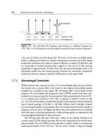
As shown in Figure 3.3, Class IC drugs have a relatively pro-
nounced effecton the rapid sodium channel because of their slow
sodium-channel-binding kinetics. Thus, they significantly slowcon-
duction velocity even at normal heart rates. They have only a m
od-
est effecton repolarization. Class IC drugs have similar effects on
Figure 3.3 Effect of Class IC drugson the cardiac actionpotential. Baseline
actionpotential is displayed as a solid line; the dashed line indicates the effect
of Class IC drugs.
72 Chapter 3
Table 3.5 Clinical pharmacology of Class IC drugs
Flecainide Propafenone Moricizine
GI absorption >90% >90% >90%
Protein binding 40% 90% >90%
Elimination 70% liver
30% kidneys
Liver Liver (metabolized to
>2 dozen compounds)
Half-life 12–24 h 6–7 h Variable; usually 3–12 h
Therapeutic level 0.2–1.0 µg/mL 0.2–1.0 µg/mL —
Dosage range 100–200 mg q12h 150–300 mg q8h 200–300 mg q8h
both atrial and ventricular tissueand are useful for both atrial and
ventricular tachyarrhythmias. The major clinical features of Class IC
antiarrhythmic drugs are summarizedinTable 3.5, and the major
electrophysiologic properties are shown in Table 3.6.
Flecainide
Flecainide was synthesizedin1972 and approved by the FDA in
1984.
Clinical pharmacology
Flecainide is well absorbed from the gastrointestinal tract, and peak
plasma levels are reached2–4 hours after an oral dose. Forty percent
of the drug is protein bound. The drug is mainly metabolized by
the liver (70%), but 30% isexcreted unchanged by the kidneys.
Flecainidehasalong elimination half-li
fe (12–24 h), so a steady
state is not reached for 3–5 days after a change in oral dosage.
Dosage
The usual dosage is 100–400 mg/day orally, in divideddoses. Gen-
erally, the beginning dosage is 100 mg every 12 hours. Dosage can
be increased by 50 mg/dose (at 3- to 5-day intervals) to a maximal
dosageof200 mg every 12 hours.
Class I antiarrhythmic drugs 73
Table 3.6 Electrophysiologic effects of Class IC drugs
Flecainide Propafenone Moricizine
Conduction velocity Decrease +++ Decrease +++ Decrease ++
Refractory periods No change (may
lengthen RP in
atrium)
No change Decrease +
Automaticity – Suppresses Suppresses
Afterdepolarizations – – Suppresses EADs
and DADs
Efficacy
Atrial fibrillation/atrial
flutter
++ ++ +
AVN reentry ++ ++ +
Macroreentry ++ ++ +
PVCs +++ +++ ++
VT/VF ++ ++ ++
AVN, AV node; EADs, early afterdepolarizations; DADs, delayed afterdepolariza-
tions; RP, refractory periods; PVCs, premature ventricular complexes; VT/VF, ven-
tricular tachycardia and ventricular fibrillation.
Electrophysiologic effects
The major electrophysiologic feature of flecainide isasubstantial
slowing in conduction velocity. The prolonged slowing is directly
related to the prolonged binding-unbinding time(i.e., the slow
binding kinetics) of the drug. Although most Class IA agents have
binding times in the rangeof5second
s, and Class IB drugs have
binding times of approximately 0.3 seconds, flecainidehasabinding
timeof30seconds. Thus, flecainide isvirtually continuously bound
to the sodium channel, and therefore produces slowconduction
even at low heart rates (i.e., at rest). Flecainidesubsequently has
a dose-dependent effecto
n the electrocardiogram, manifested by
74 Chapter 3
a progressive prolongation of the PR and QRS intervals (reflecting
its slowing of conduction velocity), with only a minor effecton the
QT interval (reflecting its minimal effecton refractory periods). The
drug depresses conductioninall areas of the heart.
Hemodynamic effects
Flecainide has a pronouncednegative inotropic effectsimilar to that
of disopyramide. The drug shouldnot be given to patients with a
history of congestive heart failure or with significantly depressed
left ventricular ejection fraction.
Therapeutic uses
As one might predict from the universal nature of the drug’s elec-
trophysiologic properties, flecainide has an effecton both atrial and
ventricular tachyarrhythmias. It has been shown to be effective for
terminating and preventing atrial fibrillation and atrial flutter;if the
arrhythmias recur, flecainide c
an slow the ventricular response. Be-
cause it affects accessory pathway function,flecainide is useful in the
treatmentofbypass-tract-mediated tachyarrhythmias. The drug has
a profound suppressive effectonpremature ventricular complexes
and nonsustain
ed ventricular tachycardia. It has been reported to
suppress approximately 20–25% of inducible sustained ventricular
tachycardias in the electrophysiology laboratory.
Flecainide is unsurpassedinsuppressing premature ventricular
complexes and nonsustained ven
tricular tachycardias, but it should
not be used for this indicationinpatients who have underlying heart
disease. Thisfinding was madeapparentbyresults of the Cardiac Ar-
rhythmiaSuppression Trial (CAST [1]), which tested the proposition
that suppression of ventricular ectopy after
myocardial infarction
would reduce mortality. Patients receiving flecainideorencainide in
thistrial had significantly higher mortality rates than did patients
receiving placebo. The significant difference in mortality has been
attributed to the p
roarrhythmic properties of the Class IC drugs.
Adverse effects and interactions
Flecainide is generally better tolerated thanmost antiarrhythmic
agents. Mild-to-moderate visual disturbances are the most common
side effect, usually manifesting as blurred vision.Occasionally, gas-
trointestinal symptomsoccur. However, nosignific
antorgan toxicity
has been reported.
Class I antiarrhythmic drugs 75
By far the most seriousadverse effectofflecainide(and of all
Class IC drugs) is its significant proarrhythmic potential (see the
comparison to other Class I drugs in Table 3.7). Proarrhythmia with
IC agents takes the form of exacerbation of reentrantventricular
tachycardia; torsades de pointes is not seen
.Thus, the risk of proar-
rhythmia with flecainide is mainly limited to patients who have the
potential for developing reentrantventricular arrhythmias, that is,
patients with underlying cardiacdisease. CAST revealed that proar-
rhythmia with Class IC drugs isespecially likely during times of acute
myocardial ischemia. It islikely that ischemia potentiates the effect
of these drugs just as it does with both Class IA and IB drugs. In any
case, flecainideand other Class IC drugsappear to have a tendency
to convert an episodeofanginatoan episodeofsuddendeath. Class
IC drugs shoul
d be avoidedinpatients with known or suspected
coronary artery disease.
Flecainide levels may be increased by amiodarone, cimetidine,
propranolol, and quinidine. Flecainide may modestly increase
digoxin levels.
Encainide
Encainide is a Class IC drug whose electrophysiologic and clinical
properties are very similar to those of flecainide. Encainide was re-
moved from the market after CAST and is nolonger available.
Propafenone
Propafenone was developedinthe late 1960s and released for use
in the United States in 1989.
Clinical pharmacology
Propafenone is well absorbed from the gastrointestinal tractand
achieves peak blood levels 2–3 hours after an oral dose. It issubject
to extensive first-pass hepatic metabolism that results in nonlinear
kinetics—as the dosageofthedrug is increased,hepatic metabolism
becomes sat
urated; thus, a relatively small increase in dosage can
produce a relatively large increase in drug levels. The drug is 90%
protein bound and is metabolized by the liver. The elimination half-
life is 6 or 7 hours after a steady state is reached.Generally, 3 days
at a stable drug dosageachieves steady-state blood levels.
76 Chapter 3
Table 3.7 Common adverse effects of Class I drugs
Proarrhythmia
General toxicity Reentrant VT Torsades de pointes
Quinidine GI (diarrhea), cinchonism,
rashes, hemolytic anemia,
and thrombocytopenia
++ ++
Procainamide Hypotension (IV), lupus, GI
(nausea), and agranulocytosis
++ ++
Disopyramide Cardiac decompensation,
urinary retention, and dry
mouth and eyes
++ ++
Lidocaine CNS (slurred speech,
paresthesias, and seizures)
+ –
Mexiletine GI (nausea) and CNS (tremor
and ataxia)
+ –
Phenytoin GI (nausea), CNS (ataxia and
nystagmus), hypersensitivity
reactions (rashes and
hematologic), osteomalacia,
and megaloblastic anemia
+ –
Flecainide Visual disturbances, GI
(nausea), and cardiac
decompensation
+++ –
Propafenone GI (nausea), CNS (dizziness
and ataxia), and cardiac
decompensation
(uncommon)
+++ –
Moricizine Dizziness, headache, and
nausea
++ –
Dosage
The usual dosageofpropafenone is 150–300 mg every 8 hours. Gen-
erally, the beginning dosage is 150 mg or 225 mg every 8 hours.
Dosage may be increased,but not more often than every thirdday.
Class I antiarrhythmic drugs 77
Electrophysiologic effects
Propafenone produces potent blockade of the sodium channel, sim-
ilar to other Class IC drugs. Unlike other Class IC agents, however,
propafenone also causes a slight increase in the refractory periodsof
all cardiac tissue. I n addition, propafenone has mild beta-blocking
and calcium-blocking properties.
Hemodynamic effects
Propafenone has a negative inotropic effect that is relatively mild,
substantially less than that seenwith disopyramideorflecainide.
The drug also blunts the heart rate during exercise. Both effects may
be a result of its beta-blocking (and perhaps its calcium-blocking)
properties.
Therapeutic uses
Like all Class IC agents, propafenone is effective in treating a wide
variety of atrial and ventricular arrhythmias. Its therapeutic profile
issimilar to that of flecainide.
Adverse effects and interactions
The most common side effects of propafenone are dizziness, light-
headedness, ataxia, nausea, and a metallic aftertaste. Exacerbation
of congestive heart failure can be seen,especially in patients with
histories of heart failure. Propafenone cancausealupuslike fa
cial
rash, and also a conditioncalled exanthematous pustulosis, which
isanasty rash accompanied by fever and ahigh white-blood-cell
count. Generally, propafenonetendstocause more side effects than
other Class IC antiarrhythmic drugs.
As is the case with all Class IC drug
s, proarrhythmia isasignificant
problemwith propafenone, but the problemislimited to patients
with underlying heart disease. Most clinicians believe, and some
clinical trials appear to show, that proarrhythmia with propafenone
issomewhat less frequent thanit is
with flecainide.
Numerous drug interactions have been reportedwith
propafenone. Phenobarbital, phenytoin,and rifampin decrease
levels of propafenone. Quinidineand cimetidine increase levels
of propafenone. Propafenone increases levels of digoxin, propra-
nolol, metoprolol, theop
hylline, cyclosporine, and desipramine. It
increases the effectofwarfarin.
78 Chapter 3
Moricizine
Moricizine, a phenothiazine derivative, has beeninuse in Russia
since the 1970s. It was approved by the FDA in 1990.
Clinical pharmacology
Moricizine is absorbed almost completely when administered orally,
and peak plasma levels occur within 1–2 hours. Moricizine is exten-
sively metabolizedinthe liver to a multitudeofcompounds, someof
which may have electrophysiologic effects. The elimination half-life
of the parent
compound is variable (generally, 3–12 h), but the half-
life of someofits metabolites issubstantially longer. Plasma levels
of moricizine have not reflected the efficacy of the drug.
Dosage
Moricizine is usually initiatedindosages of 200 mg orally every 8
hours and may be increased to 250–300 mg every 8 hours. Generally,
it isrecommended that dosage increases be made no more often
than every thirdday. Dosage should be decreasedinthe presenceof
hepatic insufficiency.
Electrophysiologic effects
Moricizine does not display the same affinity for the sodium channel
displayed by other Class IC drugs. Hence, its effectonconduction
velocity is less pronounced than that for flecainideorpropafenone.
In addition, moricizine decreases the actionpotential duratio
n and
therefore decreases refractory periods, similar to Class IB agents.
Classification of moricizine has thus beencontroversial; some classify
it as a Class IB drug.Itis classified as a Class IC drug in this book
mainly to emphasize its proarrhythmic effects (which are only rarely
seenwith Class IB drugs).
Hemodynamic effects
Moricizine may have a mildnegative inotropic effect, but in general,
exacerbation of congestive heart failure has been uncommonwith
this drug.
Therapeutic uses
Moricizine is moderately effective in the treatment of both atrial
and ventricular arrhythmias. It has beenused successfully in treat-
ing bypass-tract-mediated tachyarrhythmias and may have some ef-
ficacyagainst atrial fibrillation and atrial flutter. Its efficacyagainst
Class I antiarrhythmic drugs 79
ventricular arrhythmias is generally greater than that of Class IB
agents but is clearly less than that for other Class IC drugs. A ten-
dency for higher mortality with moricizine comparedwith that for
placebo was seeninCAST, but the study was terminated before the
tendency reached statistical significance.
Adverse effects and interactions
Ingeneral, moricizine isfairly well tolerated. Most side effects are
related to the gastrointestinal or central nervous systems, similar
to Class IB drugs. Dizziness, headache, and nausea are the most
common side effects.
Proarrhythmia clearly occurs with moricizine more often thanit
does with Class IB drugsbut less often
than that with other Class IC
drugs.
Cimetidine increases moricizine levels and moricizine decreases
theophylline levels.
Reference
1Echt DS, Liebson PR, Mitchell B, et al. Mortality and morbidity in patients
receiving encainide, flecainideorplacebo. N EnglJMed 1991;324:781.
CHAPTER 4
Class II antiarrhythmic
drugs; beta-blocking agents
Beta-blocking drugs exert antiarrhythmic effects by blunting the ar-
rhythmogenic actionsofcatecholamines. Comparedwith other an-
tiarrhythmic drugs, these agents are only mediocre at suppressing
overt cardiac arrhythmias. Nonetheless, beta blockers exert a pow-
erful protective effect in certain clinic
al conditions—they are among
the fewdrugs that have been shown to significantly reduce the inci-
denceofsuddendeath in anysubset of patients (an effect they most
likely achieve by helping to prevent cardiac arrhythmias).
Because of the success of the drugs in treating a myriad of me
dical
problems, more than two dozen beta blockers have been synthesized
and more than a dozen are available for clinical use in the United
States. Incontrast to Class I antiarrhythmic drugs, the antiarrhyth-
mic effects of the various Class II drugstend to be quite similar to
oneanother.
Electrophysiologic effects of beta blockers
For practical purposes, the electrophysiologic effects of beta block-
ers are manifested solely by theirblunting of the actionsofcat-
echolamines. The effect of beta blockers on the cardiac electrical
system, then, reflects the distribution of adrenergic innervation of
the heart. In areas where there isricha
drenergic innervation, beta
blockers can have a pronounced effect. In areas where adrenergic
innervationissparse, the electrophysiologic effect of beta blockers
is relatively minimal.
Since the sympathetic innervation of the heart is greatest in the
sinoatrial (SA) and atrioventricular (AV) nodes, it is in
these struc-
tures that beta blockers have their greatest electrophysiologic effects.
In both the SA and AV nodes, phase 4depolarizationisblunted
by beta-blocking agents, leading to a decrease in automaticity, and
80
Class II antiarrhythmic drugs; beta-blocking agents 81
hence to a slowing in the heart rate. In the AV node, beta blockers
cause a marked slowing in conduction and a prolongationinre-
fractory periods. The drugs have relatively little effecton SA nodal
conductioninnormal individuals but canmarkedly prolong SA
nodal conduction
(leading to sinus nodal exit blockand hence brad-
yarrhythmias) in patients with intrinsic SA nodal disease. Beta block-
ers have very little effectonconduction velocity or refractoriness in
normal atrial or ventricular myocardium.
Beta blockers can have a profound electrophysiologic effect, ho
w-
ever, in ischemic or damagedmyocardium.Byhelping to prevent
ischemia, the drugs can reduce the incidence of arrhythmias. Fur-
ther, beta blockers raise the threshold for ventricular fibrillationinis-
chemic myocardium and have been shown to reduce the risk of ven-
tricular fibrillationduring ische
mia. There is also evidence that beta
blockers can helpprevent the formation of reentrant arrhythmias in
myocardium that has beendamaged by ischemia. In such damaged
myocardium,amaldistribution of autonomic innervationcan arise
and lead to regi
onal differences in adrenergic stimulation.Regional
differences can serve as substrate for reentranttachyarrhythmias by
creating localizeddifferences in refractory periods. By “smoothing
out” localizeddifferences in autonomic stimulation, beta blockers
may hel
p to prevent arrhythmias.
Beta-blocking agents in the treatment
of arrhythmias
Supraventricular arrhythmias
The major electrophysiologic effects of beta blockers are manifested
in the SA and AV nodes;it shouldnot be surprising that the efficacy
of beta blockers in treating supraventricular arrhythmias is mainly
related to the extenttowhich the arrhythmias depend on the SA
and AV nodes. Beta blockers are most effect
ive in treating those
supraventricular arrhythmias in which the SA or AV nodes are in-
cludedwithin the reentrant pathways (namely, SA nodal reentrant
tachycardia, AV nodal reentranttachycardia, and macroreentrant
tachycardias associatedwith bypass tracts). In these cases, beta blo
ck-
ers can have a directsuppressive effecton the pathways of reentry;
thus, they can often terminate the arrhythmias and can helpprevent
theirrecurrence.
For arrhythmias arising within the atrial muscle (automatic or
reentrant atrial tachycardias, atrial fibrillation,and atrial flu
tter),
82 Chapter 4
Table 4.1 Potential effects of beta-blocking drugson supraventricular
tachyarrhythmias
Terminate or prevent
AV nodal reentrant tachycardia
SA nodal reentrant tachycardia
Macroreentrant (bypass-tract-mediated) tachycardia
Slow ventricular response
Atrial tachycardia (automatic or reentrant)
Atrial fibrillation
Atrial flutter
beta blockers have only a minimal directsuppressive effect. In these
atrial arrhythmias, however, beta blockers can still be quite useful
in helping to control the ventricular response by increasing the re-
fractory period of the AV node, and thus allowing fewer impulses to
be transmitted to the ventricles. In rare patien
ts, beta blockers also
help to prevent arrhythmias arising in the atria. In such instances,
the atrial arrhythmias appear to be catechol dependentand patients
often relate the onset of their arrhythmias to exercise. The effects
of beta blockers on supraventricular arrhythmias are summarizedin
Table 4.1.
Ventricular arrhythmias
Ingeneral, beta blockers are not particularly effective in suppressing
ambientventricular ectopyorventricular tachycardias. In some cir-
cumstances, however, generally when arrhythmias are dependent
oncatecholamines or related to myocardial ischemia, beta blockers
c
an be useful. Beta blockers are the drugsofchoice, for instance, for
exercise-induced ventricular arrhythmias. Beta blockers have also
been shown to reduce the number of episodes of ventricular fibril-
lationduring acute myocardial infarction,tosignificantly improve
overall survival, and to reduce the risk of suddendeath a
nd recurrent
infarctioninsurvivors of myocardial infarction.
Beta blockers can also be effective in treating sometypes of con-
genital long QT-interval syndrome. These syndromes are character-
ized by long QT intervals and a propensity for syncopeorsudden
d
eath during exercise or during times of severe emotional stress.
While the arrhythmias associatedwith these conditions are probably
mediated by delayed afterdepolarizations, they are also apparently
associatedwith localizeddifferences in refractory periods caused by a
Class II antiarrhythmic drugs; beta-blocking agents 83
maldistribution of sympathetic fibers in the ventricles. Beta blockers,
which along with left stellate sympathectomy have been effective in
treating many patients with these disorders, can help to smooth out
any resultantsympathetic imbalance, reduce nonuniform refractory
periods, and make arrhyth
mias less likely.
Clinical pharmacology of beta-blocking agents
To a large extent, all the available beta blockers appear to be of
comparable efficacy in the treatment of arrhythmias and ischemia.
Choosing among these agents for the purpose of treating arrhyth-
mias is, then, mainly a matter of selecting a drug with an appropriate
pharmacologic profile for the patientbeing treated.Among the c
on-
siderations in making such a selection are the relative potencies of
the drugsbeing considered and whether they offer receptor selec-
tivity, intrinsic sympathomimetic activity (ISA), vasodilator activity,
and membrane-stabilizing activity. Table 4.2is
not all inclusive, but
itlists the pharmacologic properties of the most commonly used
beta-blocking agents.
Potency of a beta blocker is not a major consideration,but the
recommendeddosages of various beta blockers differ markedly, and
dosages must be a
djusted accordingly for the drug being used.
Receptor selectivity refers to β
1
-receptors (those in the heart)
and β
2
-receptors (those in the peripheral vasculature and bronchi).
Drugs with selectivity, suchasatenolol and metoprolol, produce
minimal blockadeofβ
2
-receptors and thus are potentially safer to
Table 4.2 Clinical pharmacology of beta-blocking drugs
Drug β
1
-Selective ISA Class I Vasodilator Lipid soluble Half-life (h)
Acebutolol +++ 0 Moderate 3–10
Atenolol ++ 0 0 0 Weak 6–9
Carvedilol 0 0 ++ + Moderate 7–10
Esmolol ++ 00 + Weak 9 min
Labetolol 0 + 0 + Weak 3–4
Metoprolol ++ 0 0 0 Moderate 3–4
Pindolol 0 ++ + 0 Moderate 3–4
Propranolol 0 0 ++ 0 High 3–4
Timolol 0 0 0 0 Weak 4–5
ISA, intrinsic sympathomimetic activity.
84 Chapter 4
use in patients with lung disease or with impairedperipheral circu-
lation.
ISA refers to the fact that some beta blockers, suchaspindolol
and acebutolol, produceapartial agonist (stimulating) effecton the
beta receptor sites to which they bind (and block). Thus, in theory,
heart rate depression and de
pression of myocardial function might
not be as potent with beta blockers offering ISA. However, clear-cut
clinical indications for using ISA drugs have not beenidentified.Of
note, drugs offering ISA may not have a protective effect in survivors
of myocardial infarct
ion.
Vasodilator activity is produced by some beta blockers either
throughalpha-receptor blockade(carvedilol), or direct β
2
-receptor
stimulation (dilevalol), or both (labetolol).
Membrane-stabilizing activity refers to the factthatafew beta
blockers exhibit Class I antiarrhythmic activity (slowing of the de-
polarizationphase of the actionpotential) if serum levels are suf-
ficiently high. However, the blood levels that must be achieved to
d
emonstrate such Class I activity are greatly in excess of therapeutic
levels. Thus, whether membrane-stabilizing activity is ever relevant
with the use of beta blockers is very questionable.
The lipid solubility of beta blockers partially determines how the
agents are metabolized (lipid-soluble dr
ugs are generally metabo-
lizedinthe liver and water-soluble drugs are generally excreted by
the kidneys) and whether they cross the blood–brain barrier (drugs
that cross are more pronetocause central nervous system side ef-
fects, suchasfatigue, depression, insomnia, or hallucinations).
In s
ummary, beta blockers as a class generally exhibitsimilar de-
grees of effectiveness in the treatmentofcardiac arrhythmias. The
major considerations in choosing among these drugs are the pre-
dominantroute of elimination (to avoid accumulation of the drug
in a patient
with liver or kidney disease), side effects, and whether
receptor selectivity or vasodilation are desired.Ingeneral, the po-
tential for membrane-stabilizing activity should be ignored and ISA
avoided.
Adverse effects and drug interactions
The most common side effects of beta blockers are a direct con-
sequenceofadrenergic blockade. These include bronchoconstric-
tion, claudication, Raynaud’s phenomenon, intensification of hypo-
glycemic episodes, a
nd fatigue. Notably, while blocking sympathetic
Class II antiarrhythmic drugs; beta-blocking agents 85
stimulation to the heart can lead to some degree of myocardial de-
pression, patients with heart failure only rarely deteriorate signifi-
cantly after the carefuladdition of beta blockers. In fact, beta blockers
improve survival in patients with heart failure. Bradycardia dueto
adrenergic blockade isawell-recognized side effect of beta block-
ers, but patients only rarely develop symptomatic bradyarrhythmias
on these drugs unless they have underlying SA nodal or AV nodal
disease.
The suddenwithdrawal of beta blockers, especially the short-
acting beta blockers like pro
pranolol, can lead to unstable ischemic
heart disease in patients with underlying coronary artery disease.
The withdrawal syndrome issubstantially less likely with the longer-
acting beta blockers.
Other possible but much less common side effects of beta block-
ers include rashes, fever, sexual dysfuncti
on, mental depression,
and gastrointestinal symptoms. Indiabetics, beta blockers canmask
symptomsofhypoglycemiaand cancause hypoglycemiabyreducing
gluconeogenesisorhyperglycemiabyreducing insulin levels.
Some of the side effects related to beta blocka
de itself may be
avoided by appropriate drug selection.Asnoted, drugs with β
2
-
selectivity might helpinavoiding bronchospasm, worsening of hy-
poglycemia, claudication,and Raynaud’s phenomenoninsome in-
dividuals. Using drugs with low lipid solubility might help to prevent
central nervous system side effec
ts.
Hepatic metabolism of lipid-soluble beta blockers can be increased
by cimetidineand decreased by barbiturates. Aluminum hydroxide
candelay absorption of beta blockers. The hepatic metabolism of li-
docaine can be reduced by administration of lipophilic beta blockers,
suchaspro
pranolol.
CHAPTER 5
Class III antiarrhythmic
drugs
Class III antiarrhythmic drugs prolong the duration of the cardiac
actionpotential, usually by blocking the potassium channels that
mediate repolarization,and thus increase the refractory periodsof
cardiac tissue(Figure 5.1).
Despite this defining similarity, n
one of the currently available
Class III drugs behave exactly alike. One reason the drugs are clini-
cally dissimilar is that none are pure Class III agents—all have addi-
tional electrophysiologic effects that contribute to their efficacyand
to their toxicity. Another reason for differences among the Class III
drugs
is that they display varying degrees of reverse use dependence.
The term use dependence,youmay recall, refers to the time-related
effect of Class I drugson the sodium channel; as a result of binding ki-
netics, the degree of sodium-channel blockade increases as the heart
rate increases. As itturnsout, the magnitudeofpotassium-channel
blockad
e manifested by Class III agents also is related to heart rate.
For Class III drugs, however, the strength of blockade decreases as
the heart rate increases; hence, the term reverse use dependence has
beencoined. Reverse use dependence means that at slower heart
rates, the prolongation of the actionpotential is most pronounced;
at faster heart rates, the effect diminishes. Reverse use d
ependence
is related to a drug’s binding characteristics. Drugs that preferentially
bind to closedpotassium channels, for instance, display significant
reverse use dependencebecause phase 4 of the actionpotential is
longer (and thus potassium channels sp
end more time in the closed
state) when the heart rate is slow. Reverse use dependence has two
potential undesirable effects. First, it causes some Class III drugs
to lose potency with rapid heart rates, just when their potency is
neededmost. Second, the fact that actionpotenti
al prolongation by
some Class III drugs is most pronouncedduring bradycardia potenti-
ates the tendency of these drugstocause the pause-dependent early
86
Class III antiarrhythmic drugs 87
Figure 5.1 Effect of Class III drugsoncardiac actionpotential. Baselineaction
potential is displayed as a solid line; the dashed line indicates the effectof
Class III drugs.
afterdepolarizations that produce torsades de pointes. Amiodarone
isaunique Class III agent in several ways, as we will see, butone
way it is different from other Class III drugs is that itbinds preferen-
tially to open potassium channels and therefore displays much less
reverse use dep
endence. Consequently, amiodarone does not lose its
effect when heart rate increases. The lowmagnitude of reverse use
dependence seenwith amiodarone may explain not only its remark-
able efficacyagainst tachyarrhythmias but also its low incid enceof
producing torsades de pointes.
Although the differences among Class III drugs have not yet man-
dated that this class be formally subgrouped as the Class I drugs
have been, it is necessary to keep in mind that these drugs are not
interchangeable. The major clinical features of Class III antiarrhyth-
mic drugs are listedinTable 5.1, and the major electrophysi
ologic
properties are listedinTable 5.2.
Amiodarone
Amiodarone was synthesizedinBelgium in the 1960s as a vasodila-
tor, mainly for the purpose of treating angina. Its antiarrhythmic
88 Chapter 5
Table 5.1 Clinical pharmacology of Class III drugs
Amiodarone Sotalol Ibutilide Dofetilide
GI absorption 30–50% >90% — 100%
Elimination Hepatic* Renal Renal Renal, some
hepatic
Half-life 30–106 days 12 h 2–12 h 8–10 h
Dosage range 800–1600
mg/day for
3–10 days,
then 100–400
mg/day PO
160–320
mg/day PO
10-mg IV
infusion during
10 min, may be
repeated
125–500 µg
twice per day
*Both hepatic and renal elimination are minimal for amiodarone.
GI, gastrointestinal; IV, intravenous; PO, oral.
efficacy was notedinthe early 1970s, and the drug rapidly came into
widespreaduse in manyEuropeancountries as an antiarrhythmic
agent. In the late 1970s, clinical trials with amiodarone were begun
in the United States and the oral form of the drug was a pproved
by the Food and Drug Admini
stration (FDA) in the mid-1980s. The
intravenous formwas approvedin1995.
Electrophysiologic effects
Amiodarone displays activity from all fourantiarrhythmic classes. It
is classified as a Class III antiarrhythmic drug because its major elec-
trophysiologic effect isahomogeneous prolongation of the action
potential, and therefore of refractory perio
ds, due to blockade of the
potassium channels. The drug has this Class III effect in all cardiac tis-
sues. When therapy with amiodarone is first initiated, prolongation
of refractoriness is not seen immediately. Instead, refractory periods
gradually increase during the prolonge
d loading period (see below).
Consequently, amiodarone’s Class III drug effects may not become
maximal for several weeks and notably, are not seen acutely even
with intravenous loading of the drug.
In addition to its potassium-channel effects, amiodarone produces
a mild
-to-moderate blockade of the sodium channel (a Class I effect),
a noncompetitive beta blockade (a Class II effect), and some degree
Table 5.2 Electrophysiologic properties of Class III drugs
Amiodarone Sotalol Ibutilide Dofetilide
Conduction velocity Decrease + 00
0
Refractory periods Increase ++ Increase ++ Increase ++ Increase ++
Automaticity Suppress ++ Suppress + Suppress + Suppress +
Afterdepolarizations May cause EADs May cause EADs May cause EADs May cause EADs
Other effects Class II and
Class IV
Class II None None
Efficacy
Atrial fibrillation/
atrial flutter
++ ++ ++ ++
AVN reentry +++ ++ 00
Macroreentry +++ ++ 00
PVCs +++ ++ 00
VT/VF +++ ++ 0 +
AVN, AV node; EADs, early afterdepolarizations; PVCs, premature ventricular complexes; VT/VF, ventricular tachycardia and ventricular fibrillation.