Antiarrhythmic Drugs A practical guide – Part 9 potx
Bạn đang xem bản rút gọn của tài liệu. Xem và tải ngay bản đầy đủ của tài liệu tại đây (165.89 KB, 19 trang )
Treatmentofsupraventricular tachyarrhythmias 149
Disopyramide, because of its vagolytic effects, may be effective in
treating the relatively uncommon varieties of atrial fibrillation that
are triggered by strong vagal stimulation (suchasswallowing cold
liquids).
Finally, beta blockers may be effective in preventing the recur-
renceofcertain
kinds of atrial fibrillation that seem to be induced
by increased sympathetic tone.
Anticoagulation in atrial fibrillation and atrial flutter
Most often, preventing stroke should be the doctor’s chief goal in
treating patients with atrial fibrillation or atrial flutter. The only
method that has been shown to reliably reduce the risk of stroke
isanticoagulationwith warfarin and, to a lesser extent, with aspirin.
Thus, when seeing a patie
nt who has atrial fibrillation or atrial flut-
ter, the decision as to whether to anticoagulate should always be
actively considered.
In2006, the ACC/AHA/ESC publishedjoint guidelines on the use
of chronic antithrombotic therapy in patients with atrial fibrillation
or atrial flutter [3]. These guidelines are fairly complex and can be
difficult to sort through, but in g eneral they can be summarized as
follows:
Patients with atrial fibrillation or atrial flutter can be categorized
into oneoftwo groups:patients at low risk and patients at h
ighrisk
for thromboembolism. Those in the low-risk categories should be
treatedwith aspirin (81–325 mg/day) unless contraindicated. Those
in the high-risk categories should be treatedwith oral anticoagula-
tioninorder to producean INRof2.0–3.0, unless contraindicate
d.
Determining whether patients fit into a low-orhigh-risk category
dependson two general factors: ageand the presenceofrisk fac-
tors for thromboembolism. The risk factors include heart failure, left
ventricular ejection fraction <0.35, history of hypertensi
on, valvular
heart disease, diabetes, and prior history of thromboembolism.
Patients in the low-risk category include:
Age <75 and norisk factors
Patients in the high-risk category include:
Age75orgreater,
Age <75, but presenceofrisk factors
While pati
ents with paroxysmal atrial fibrillation have long been
thought to have a lower incidenceofembolization than those with
chronic atrial fibrillation, at least two large clinical trials have now
shown similar risks among these patients—and similar benefits from
150 Chapter 11
anticoagulation.Thus, patients with paroxysmal atrial fibrillation
should be treated according to these same guidelines.
Additionally, both the AFFIRM and RACE trials have suggested
that patients treatedwith the goal of restoring and maintaining sinus
rhythm (as opposed to rate control) do not have a s
ubstantially re-
duced risk of thromboembolism.Accordingly, these patients should
also be treated according to these guidelines.
Finally, it isbyno means clear that patients with atrial fibrilla-
tionwho are treated by ablation techniques in order to restore and
maintain sinus rhyth
mwill have a reduced risk of stroke. For now,
chronic anticoagulation should also be strongly consideredinthese
patients.
References
1 Wyse DG, Waldo AL, DiMarco JP, et al. A comparison of rate control
and rhythmcontrol in patients with atrial fibrillation. N EnglJMed
2002;347(23):1825.
2 Van Gelder IC, Hagens VE, Bosker HA, et al. A comparison of rate control
and rhythmcontrol in patients with recurren
t persistent atrial fibrillation.
N EnglJMed2002;347(23):1834.
3Furster V, Ryden LE, Cannom DS, et al. ACC/AHA/ESC guidelines for the
managementofpatients with atrial fibrillation.Areport of the American
College of Cardiology/American Heart Association Task Forc
eon Prac-
ticeGuidelines and the European Society of CardiologyCommittee for
PracticeGuidelines (Writing committee to revise the 2001guidelines for
the managementofpatients with atrial fibrillation). J Am Coll Cardiol
2006;48:e149.
CHAPTER 12
Treatmentofventricular
arrhythmias
Ventricular arrhythmias are responsible for hundreds of thousands
of suddendeaths each year in the United States alone. Therapeuti-
cally, patients at risk for suddendeath usually fall into one of the
two broadcategories. First, there are patients who have already
experienced an episodeofsu
stained ventricular tachycardia (VT)
or ventricular fibrillation (VF). These individuals, having already
demonstrated a propensity for lethal arrhythmias, are at substan-
tial risk for subsequentsuddendeath. The second and much larger
category consists of individuals who are at highrisk but have
not
yet had sustained ventricular arrhythmias. These patients generally
have significant underlying cardiacdisease, whether or not it isac-
companied by complex ventricular ectopy(consisting of frequent
premature ventricular complexes (PVCs), non
sustained VT, or both).
The risk of suddendeath for these patients, although demonstrably
increased over normal levels, is generally not as high as for patients
in the first category.
Treatment of nonsustained ventricular
arrhythmias
The significance of ventricular ectopy
Ventricular ectopy is generally classified as being either simple or com-
plex.Simple ventricular ectopy issaid to be present in patients who
have PVCs, butfewer than 10 PVCs per hour during 24-hour Holter
monitoring and no nonsustained VT. Complex ventricular ectopy
is
generally defined as >10 PVCs per hour during 24-hour monitoring
or the presenceofnonsustained VT. Simple ventricular ectopy car-
ries no prognostic significance. However, in the presenceofunderly-
ing cardiacdisease, complex ventricular ectopy
does have prognos-
tic implications. Indeed, complex ectopy is relatively uncommonin
151
152 Chapter 12
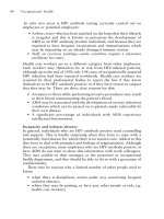
Table 12.1 Relationship of ventricular ectopy to estimated risk of sudden
death
Number of risk factors One-year risk (%)
One
Previous MI 5
LVEF < 0.40
Two
Previous MI + CVE 10
LVEF <0.40 + CVE
Previous MI + LVEF <0.40
Three
Previous MI + LVEF <0.40 + CVE 15
CVE, complex ventricular ectopy; LVEF, left ventricular ejection fraction; MI, my-
ocardial infarction.
patients with normal hearts. The presenceofunexpectedcomplex
ventricular ectopy should thus promptan evaluation for undiag-
nosedcardiacdisease.
It is possible to estimate a patient’s risk of suddendeath by consid-
ering the presence of three simple
clinical factors:previous myocar-
dial infarction, depressed left ventricular ejection fraction (i.e., an
ejection fraction of less than 0.40), and complex ventricular ectopy.
The resultantrisks are shown in Table 12.1. If previous
myocardial
infarction or depressed ventricular function are present (as noted,
the presenceofcomplex ectopy alone carries no prognostic signifi-
cance), the 1-year risk of suddendeath isapproximately 5%. If any
tworisk factors are present, the 1-year risk of suddend
eath isap-
proximately 10%. If all three risk factors are present, the 1-year risk
isapproximately 15%. Thus, patients who have survivedmyocar-
dial infarction or who have depressed ventricular function from any
cause have increased risk of suddendeath. The risk increases with
the presenceofc
omplex ventricular ectopy.
Treating ventricular ectopy
The association betweencomplex ectopyand the risk of sudden
death has been recognized for decades, and for many years, it
was assumed that antiarrhythmic drug therapyaimed at eliminat-
ing complex ectopy would improve that risk. This assumptionwas
provenwrong in the late 1980s c
ourtesy of the Cardiac Arrhythmia
Treatmentofventricular arrhythmias 153
Suppression Trial (CAST), discussedinChapter 9. To review, CAST
randomizedpatients who had survivedmyocardial infarctionsand
who hadcomplex ectopy(and who, therefore had an increased risk
of suddendeath) either to have theirectopysuppressedwith Class
IC drugsortoreceive
placebo. Much to the surprise of many ob-
servers, and in distinct contrast to the predictionsofmost experts,
patients whose ectopyhad been successfully suppressed by the Class
IC agents generally had asignificant increase in mortality as compared
to patients onplacebo. Not o
nly did getting rid of the ectopyfailto
improve outcomes, but also the use of antiarrhythmic drugs itself
(presumably duetoproarrhythmia) increasedmortality. The find-
ings of CAST were reinforced by subsequent meta-analyses, showing
that patients treatedwith Class I antiarrhythmic drugs commonly
have reduc
ed survival as compared to patients onplacebo.
Inconceptualizing the treatmentofcomplex ventricular ectopy,
the bear droppings theory is instructive—ifyou are walking in the
woodsand see bear droppings, your chances of being eaten by a
bear are higher thanif there were no bear d
roppings. However, if
you take outyour gun and shoot the bear droppings, you are not
reducing yourrisk. In fact, you might even induce the bear to come
by to investigate the disturbance. Complex ectopy is best viewed as
an indication of increased risk (like bear droppings), and not as a n
indication for therapy.
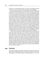
The
prophylactic empiric use of amiodarone has also been ad-
vanced as a way of treating patients with underlying heart dis-
ease who have complex ventricular ectopy, and several random-
ized trials have now examined thisquestion. The results of the trials
are summarizedinTable 12.2.U
nfortunately, these results do not
provide definitive evidence that prophylactic use of amiodarone is
helpful. In the Basel Antiarrhythmic StudyofInfarctSurvival (BA-
SIS) [1], patients treatedwith amiodaronehad improved overall
mortality comparedwith that of control patien
ts. In the Canadian
Amiodarone Myocardial Infarction ArrhythmiaTrial (CAMIAT) [2]
and the EuropeanMyocardial InfarctAmiodaroneTrial (EMIAT)
[3], amiodaroneyielded areductioninarrhythmic death but not in
overall mortality. In the VeteransAdministrat
ion Congestive Heart
Failure Antiarrhythmic Trial (CHF-STAT) [4], no improvement in
mortality with amiodarone was seencomparedwith that of controls.
Overall, these findingssuggest that amiodarone-related toxicity may
largely negate anyreductioninsuddendeath. However, in distinct
con
trast to the Class I drugs, amiodarone is not associatedwith an
154 Chapter 12
Table 12.2 Clinical trials examining the prophylactic use of empiric
amiodarone
Reduction in
Patient arrhythmic or Reduction in
Trial population Randomization cardiac mortality* total mortality*
BASIS MI, CVE amio 200 mg/day
vs. other drugs
or placebo
—Yes
CHF-STAT low EF, CVE amio 200 mg/day
vs. placebo
—No
CAMIAT MI, CVE amio 300 mg/day
vs. placebo
Yes No
EMIAT MI, low EF amio 200 mg/day
vs. placebo
Yes No
*Reduction in indicated mortality with amiodarone versus controls.
BASIS, Basel Antiarrhythmic Study of Infarct Survival; CHF-STAT, Veterans Admin-
istration Congestive Heart Failure Antiarrhythmic Trial; CAMIAT, Canadian Amio-
darone Myocardial Infarction Arrhythmia Trial; EMIAT, European Myocardial Infarct
Amiodarone Trial; amio, amiodarone; CVE, complex ventricular ectopy; EF, left ven-
tricular ejection fraction; MI, myocardial infarction.
increase in mortality whenusedinpatients with complex ectopy
and underlying heart disease.
The bottom line is that treating ventricular ectopy with antiar-
rhythmic drugs has not been associatedwith an improvedclinical
outcome, despite the fact that numerous clinical trials have been
co
nducted to examinethisquestion. Therefore, it is not appropriate
to treat these patients with antiarrhythmic drugs for the purpose of
improving theirsurvival.
However, on occasion, it may be appropriate to treat ventricu-
lar ectopy if the ectopic beats themselves are producing signifi
cant
symptoms. Here, obviously, the goal istoimprove symptoms(and
not necessarily to abolish the ectopy completely). Ingeneral, when
trying to suppress ventricular ectopy for the purpose of relieving
symptoms, the appropriate choiceofan antiarrhythmic drug de-
pendson the p
atient’s clinical condition.
Treatmentofventricular arrhythmias 155
Inpatients with no underlying heart disease, beta blockers should
be the first drugs attempted,since they are well tolerated and have
relatively few side effects. Unfortunately, they are also generally
ineffective in suppressing ventricular ectopy. The use of flecainide
might be a reasonable option,since the drug is reasonably w
ell tol-
erated, isquite effective at suppressing ectopy, and should have little
proarrhythmic potential in patients with structurally normal hearts
and alow risk of developing ischemic heart disease. However, be-
cause of the results of CAST, someexperts are reluctanttorecom-
mend flecainide (or any Class IC dr
ug) for the treatmentofventricu-
lar ectopy in any patients, no matter how healthy he or she appears
to be. Sotalol and dofetilide may be reasonable choices if beta block-
ers are ineffective (despite the fact that their efficacy in suppressing
ventricular ectopy is not well documented), but
precautions must
be takenwith these Class III agents to minimize the risk of torsades
de pointes. Finally, amiodarone can be considered—but its ability to
suppress symptomatic ectopy needstobecarefully weighed against
its propensity to c
ause end-organ toxicities that might well dwarf
the significanceofpalpitations.
Inpatients with underlying heart disease who need to be treated
to reducesymptomatic ventricular ectopy, beta blockers are a clear
first choice, since these drugs need to be used anyway in p
atients
with prior myocardial infarctions or heart failure (because of the
significant improvement in survival they impart to these patients).
If the ventricular ectopyremainsaproblem,amiodarone can be
considered,aswell as sotalol or dofetil
ide.
Treatment of sustained ventricular arrhythmias
Patients who have survived an episodeofsustained VT or VF have an
extraordinarily highrisk of experiencing arecurrent arrhythmia. In
general, 30–50% will have another episodeofsustained ventricular
tachyarrhythmia within 2 years. Therefore, oncesuchan arrhyth-
mia has occurred,aggressive measures must be take
n to reduce the
subsequentrisk of suddendeath.
Treatment of sustained monomorphic VT
Most patients presenting with sustainedmonomorphic VT (i.e., reg-
ular VT with a stable QRS complex, occurring at a rate of more
than 100 beats/min,and persisting for at least 30 s) are survivors of
156 Chapter 12
myocardial infarction.Sustainedmonomorphic VT in any patient is
usually a strong indicator that a fixed reentrant circuitexists within
the ventricular myocardium,and thus, once seen, monomorphic VT
islikely to recur.
Most episod
es of sustainedmonomorphic VT occur after the acute
phase of a myocardial infarction, that is, after the first 48hours, and
usually within the first year, butsometimes as late as several years
after acute myocardial damageoccurs. The prognosisofpatients with
monomorphic VT is relatively
poor, largely because this arrhythmia
tends to be associatedwith poor left ventricular function, heart fail-
ure, and multivessel coronary artery disease. While most episodes
of VF are preceded by at least short episodes of VT, it is not clear that
patients presenting with stable, sustainedmonomorphic VT—at least
those
who survive and are referred to electrophysiologists—have an
extraordinarily highrisk of subsequent VF. The incidenceofsudden
death in patients presenting with well-toleratedmonomorphic VT is
substantially lower than that for patients who have survivedcardiac
arrest, though thei
r overall rate of subsequent mortality (probably
due to the extentofunderlying heart disease) remains elevated.
Acute treatment
Patients presenting with sustainedmonomorphic VT can be treated
acutely with direct-current (DC) cardioversion or with intravenous
antiarrhythmic drugs. Intravenous procainamide is oftenuseful(i.e.,
effective in up to 50% of patie
nts) in terminating hemodynamically
stable VT. Intravenousamiodarone can also be used,and isespecially
useful for controlling sustained VT that isrecurring frequently. In-
travenouslidocaine, for decades the drug of choice, is now
felt to be
only marginally effective in terminating monomorphic VT, unless
the arrhythmia isbeing caused by active myocardial ischemia.
Chronic treatment
Monomorphic VT in the setting of underlying heart disease is al-
most always a reentrant arrhythmia. Unfortunately, it is difficult to
predict the effectofaparticular antiarrhythmic drug on a particular
reentrant circuit. The same drug may have a beneficial effecton one
circuitbutaproarrhyth
mic effecton another. Ideally, some means
should be used to measure the effectofadrug before a patient is com-
mitted to long-term therapy. Two general methodsofguiding drug
therapy have beenusedinpatients with ventricular tachyarrhyth-
mias: Holter monitoring and ele
ctrophysiologic (EP) testing.
Treatmentofventricular arrhythmias 157
Holter monitoring was the only methodology available for guiding
drug therapy until the late 1970s, and it was widely useduntilalmost
1990. The use of this method relied on the suppression of ambient
ventricular ectopy, butaswe have seen,thistechnique was rendered
a death blow
by the CAST study.
The idea behind EP testing to guide drug therapy is essentially
sound, at least in theory. If a reentrant circuit is present that is ca-
pable of generating an arrhythmia, all youneed to do to start the
arrhythmia istointroducean appropriately timed electrical impulse
into the circuit (see Figure 1.7). Thi
s procedure can be accomplished
in the EP laboratory by the techniqueknown as programmed stimu-
lation, in whichatemporary ventricular pacemaker is used to deliver
precisely timed, paced impulses into a presumed reentrant circuit. If
suchacircuitexists and if it has the a ppropriate EP characteristics
(as discussedinChapter 1), VT can be induced.
EP testing, therefore, can help to determine whether a reen-
trant circuit capable of generating aventricular tachyarrhythmia is
present. Among patients p
resenting with sustainedmonomorphic
VT, the presumedclinical arrhythmia can be inducedinapproxi-
mately 90%. Sustained VT can also be inducedin30–60% of patients
whose presenting arrhythmia isVF.In addition to assessing the pres-
ence or absence of a reentrant circuit, EP testing c
an be usedinthe
attempt to assess the effect that an antiarrhythmic drug might have
on the reentrant circuit. The assessment is donebyadministering
one of the antiarrhythmic drugsand then attempting to reinduce
the arrhythmia. If a previously inducible arrhythmia isrendered
noninducible by a drug, it is assumed that the
drug has favorably
changed the characteristics of the reentrant circuit. Chronic therapy
with the drug then seems reasonable.
Thiskind of EP testing was widely used by electrophysiologists
from the early 1980s until the mid-1990s in guiding the therapyof
patients presenting with sustainedmonomorphic
VT. But clinical re-
ports by the mid-1990s began to call into question the ability of such
“EP-guided” therapytoactually improve the outcomes of patients
with this arrhythmiaVT.This growing skepticismwas finally con-
firmed by the Electrophysiologic Testing Versus Electroc
ardiographic
Monitoring (ESVEM)trial [5]. In ESVEM, patients presenting with
sustained VT, who also had both a high degree of ambientventricular
ectopyand inducible VT, were randomized to drug therapy guided
by either EP testing or Holter monitoring. Both groupshad
very sim-
ilar, and very poor, outcomes. The rate of recurrent arrhythmias for
158 Chapter 12
both treatment groups was nearly 40% at 1 year and 66% at 4 years.
Thistrial convincedmost electrophysiologists that EP-guideddrug
testing is no more effective in improving clinical outcomes thanis
Holter-guideddrug testing. Neither methodworks adequately, and
we now know
that neither should be reliedupon to direct therapy
in patients presenting with VT.
Empiric drug therapy
Using antiarrhythmic drugsempirically simply meansadminister-
ing themwithoutan attempttomeasure their efficacy beforehand.
Empiric drug therapyastheprimary treatment for ventricular tach-
yarrhythmias was common before 1980, but was deemedunaccept-
able with the advent of EP testing
. By the time EP testing also fell
out of favor in the late 1990s, the phenomenon of proarrhythmia
with Class I antiarrhythmic drugs was widely recognized,render-
ing the idea of simply going backtoempiric therapy (at least with
most antiarrhythmic drugs), generally unacceptable as the primary
approach to treating patients
with sustained VT.
However, empiric therapy with antiarrhythmic drugs can be use-
fulasasupplementtopatients who have received implantable car-
dioverter defibrillators (ICDs), or in patients who refuse to receive or
are not goodcandidates for one of these devices. Because they have
a relati
vely lowpropensity to exacerbate reentrant VT, the Class III
antiarrhythmic drugstoday are the ones most commonly used for
empiric therapy.
There isevidence fromclinical trials that amiodarone, in particu-
lar, can be effective—certainly more effective than Class I drugs—in
treating patie
nts presenting with sustained VT. The Cardiac Arrest
in Seattle—Conventional VersusAmiodaroneDrug Evaluation
(CASCADE) trial [6], in whichsurvivors of cardiac arrest were
randomized to receive either empiric treatment with amiodarone
or treatment with con
ventional drugs guided by EP testing, Holter
monitoring, or both, showed that amiodarone was significantly
better thanconventional drugs in reducing the incidenceofcardiac
mortality and recurrent arrhythmic events. Implantable defibrilla-
tors were also usedinmany p
atients in the study, so the effectof
amiodarone in reducing mortality couldnot be well evaluatedinthis
trial.
Other Class III agents may also reduce the risk of recurrentar-
rhythmias in patients presenting with sustained VT. Sotalol, in
particular, seemstoprovidesomebenefit in these patients, and
there
Treatmentofventricular arrhythmias 159
is preliminary evidence that dofetilide, as well as the investigational
drug azimilide, may also be helpful. Again,however, whenever pos-
sible, empiric antiarrhythmic drug therapy should be reserved for
patients who have ICDs. Empiric drug therapysimply cannot be re-
liedup
on as the primary treatmentofchoice for patients presenting
with sustained ventricular tachyarrhythmias.
Implantable cardioverter defibrillators
An ICD isapacemakerlike device that automatically detects the on-
set of ventricular tachyarrhythmias and then takes action to termi-
nate them,either by administering a DC shock to the heart (for VF
or very rapid VT) or by delivering bursts of antitachycardia pacing
(for slower sustained VTs). The ICD has beeninclinical use s
ince the
early 1980s, and vast, worldwideexperience with the device has
beengathered. ICDs cannow be implantedwithasurgical mortality
of much less than 1%, and they have proven to be extremely effec-
tive in preventing suddendeath from ventricular tachyarrhythmias.
Survivors of cardiac arrest, whose risk of recurre
ntlife-threatening
arrhythmias is otherwise as highas40% after 2 years, have had the
risk of suddendeath reduced by the ICD to less than2% at 1 year
and less than 6% at 5 years. No other therapy is as effective in elim-
inating the risk of suddendeath in this population.
Whether ICDs produceasignificant decrease in mortality in pa-
tients p
resenting with relatively well-tolerated sustainedmonomor-
phic VT, however, isasomewhat more difficult question.While
many, if not most, episodes of VF are preceded by at least a few
beats of VT, it is unclear how oftenpatients who have recurrent,
prolonged episodes of he
modynamically stable monomorphic VT go
on to develop VF. Nonetheless, there are at least two reasonsto
strongly consider implanting ICDs in these patients.
First, ICDs can often terminate monomorphic VT by means of their
(painless) antitachycardia p
acing algorithms, thus restoring normal
sinus rhythmwithout the need for painful shocks. And second, the
large majority of patients presenting with monomorphic VT will al-
ready have an indication for an ICD. Patients with reduced ejec-
tio
n fractionsand either prior myocardial infarctionsorahistory of
heart failure (i.e., the majority of patients with monomorphic VT)
have now been shown to have significantly improved survival with
ICDs, regardless of whether or not they have hadprior ve
ntricular
arrhythmias. Most patients with monomorphic VT, therefore, will
alreadyfitwidely acceptedcriteria for implantation of an ICD.
160 Chapter 12
Treatment of hemodynamically unstable VT or VF
The chief clinical goal in treating patients who have survived ven-
tricular tachyarrhythmias that produced hemodynamic instability—
that is, patients who have survivedcardiac arrest—istoreduce their
high residual risk of suddendeath. Several randomizedclinical tri
-
als have now shown that in these patients the ICD produces a
significantreductioninmortality, of up to 25%, as compared to
antiarrhythmic drugs, including amiodarone. Guidelines from the
American College of Cardiology/American Heart Association/Heart
Rhythm Society n
ow recommend the ICD for survivors of cardiac
arrest, unless the cardiac arrest was duetotransient or reversible
causes.
If an ICD cannot be used for some reason,empiric therapy with
amiodaroneappears to offer at least some protection from recurrent
cardiac arrest and shoul
d be considered.In addition, beta blockers
have been shown to reduce the risk of suddendeath in both survivors
of myocardial infarction and patients with heart failure, and they
should be given to these patients whether they have had sustained
ventricular arrhythmias or not.
As already noted, Class III antiarrhythmic drugs are frequently
usefulasadjunctive therapy in patients who have ICDs as a means
of reducing the need for shocks. However, since these drugs (espe-
cially amiodarone) have been reported to occasionally increase the
threshold for defibrillation, potentially render
ing the ICD less ef-
fective, and because they (again,especially amiodarone) cancause
significant toxicity, their use as adjunctive therapy in patients with
ICDs shouldnot be taken lightly.
Treatment of less common forms of ventricular
tachyarrhythmias
In Chapter 1, we mentioned several less common formsofven-
tricular tachyarrhythmias, noneofwhich are caused by the typical
intramyocardial reentrant circuits associatedwith coronary artery
disease or cardiomyopathy. Two of these are known to be dueto
channelopathies—the arrhythmi
as caused by triggered activity, and
those related to the Brugadasyndrome—and were coveredinsome
detail in the discussion onchannelopathies in that chapter. The re-
maining uncommon formsofVTwill now be discussedinmore
detail.
Treatmentofventricular arrhythmias 161
VT associated with right ventricular dysplasia
“Arrhythmogenic” right ventricular dysplasia (AVRD) isacondition
of unknown etiology, most commonly seeninyounger individu-
als, characterized by the replacement of the right ventricular my-
ocardium with fibrofatty tissue, and the propensity to d
evelop ven-
tricular tachyarrhythmias. Sustainedmonomorphic VT originating
in the right ventricle (and thus having a left bundle branch block pat-
tern to the QRS complexes) is the most commonpresenting arrhyth-
mia, butsuddendeath (especially during exercise) can be the first
presenting
symptom. The arrhythmias seenwith AVRD are related
to reentrant circuits that arise as a result of the fibrofatty deposits.
Treatment of these VTs generally consists of either drug therapy
with sotalol (which has been reported to suppress arrhythmias asso-
ciatedwith AVRD in over 60% of patients) or amiodarone, or with
an ICD. ICD usage is often supple
mentedwith antiarrhythmic drug
therapy.
Drug therapy alone shouldgenerally be reserved for patients who
have hadwell-tolerated,sustainedmonomorphic VT, since these
patients are thought to have a relatively low risk of suddendeath.
ICDs should be chosen as primary therapy for higher-risk patie
nts,
a category that includes younger patients, those presenting with
syncope, presyncope, or cardiac arrest, or those with a family history
of cardiac arrest or syncope related to this condition.
Bundle branch reentry
Bundle branch reentry is seen occasionally in patients with dilated
cardiomyopathy and intraventricular conductiondelays. While such
patients, duetothenature of their underlying heart disease, have a
high propensity for “typical” reentrantventricular arrhythmias, they
can also develop bundle branch reentry. In bundle branch reentry,
the reentrant circuit is formed by the right and left bundle branches,
the bundle of His, and the intervening ventricular myocardium.An
arrhythmia can be triggeredwhen a premature ventricular impulse
enters both bundle branches in the retro
grade direction, is blocked
in the right bundle branchbut conducts up the left bundle branch
(which has a shorter refractory period)and then turns around at
the bundle of Hisand reenters the right bundle branch in the ante-
grade direction. The resulting VT will therefore have a left bundle
branc
h block configuration.Itis often a very rapid VT that causes
hemodynamic instability.
162 Chapter 12
Radiofrequency ablation of the right bundle branch completely
eliminates this arrhythmia, and is considered the treatmentofchoice
by many electrophysiologists. However, these patients almost always
have a markedly reduced left ventricular ejection fraction and a
h
istory of heart failure, and therefore are indicated for ICDs even
if theirbundle branch reentry is “cured.” Ablation of the bundle
branch reentry circuit, then,ought to be thought of, in most cases,
as an adjunctive therapy, aimed at reducing the need for ICD shocks.
Antiarrhythmic drugs have little or no role in the managemen
tof
bundle branch reentry.
Repetitive monomorphic VT
Repetitive monomorphic VT (RMVT), also known as right ventric-
ular outflow tracttachycardia, presents as bursts of nonsustained,
monomorphic VT with a left bundle branch block pattern and an
inferior axis. These arrhythmias originate, for the most part, in the
o
utflow tract of the right ventricle. They are seen almost exclusively
in young-to-middle-age patients, and they are exacerbated by in-
creased adrenergic tone. Patients with RMVT most oftenpresent
with complaints of palpitationsorlight-headedness associatedwith
exercise or emot
ional stress. In addition, womenwith RMVT will
oftencomplain of the samesymptoms during certain times of the
menstrual cycle. While suddendeath is not unheard of in patients
with RMVT, its incidence is thought to be quite low.
There is now evidenc
e that at least some cases of RMVT may be
related to a form of triggered activity that produces delayed afterde-
polarizations (see Chapter 1). In any case, RMVT tendstorespond to
antiarrhythmic drugs that are generally ineffective in treating more
typical formsofVT,including adenosine, verapamil, and
beta block-
ers. Class I and Class III antiarrhythmic drugs are also effective rea-
sonably often.However, since these arrhythmias are often localiz-
able, they are quite amenable to radiofrequency ablation, which is
reported to be completely effective in over 80% of cases.
There isacondition often referred to as “paroxysmal sustained
VT,”w
hich isvirtually identical to RMVT (including its response to
verapamiland adenosine) except that the episodes of VT persist for
muchlonger than the dozen or so beats usually seenwith RMVT.
Someexperts consider thisadistinctsyndrome, while others con-
sid
er itsimply an exaggerated form of RMVT.
Treatmentofventricular arrhythmias 163
Idiopathic left ventricular tachycardia
Idiopathic left ventricular tachycardia (ILVT) isanother form of VT
associatedwith young patients who have no identifiable underlying
heart disease. These patients present with sustained VT originating
frominferior-apical or mid-septal region of the left ventricle (yield-
ing aright bundle branc
h blockand left superior axis QRS complex).
The arrhythmia is not associatedwith exercise, and symptoms are
usually limited to palpitationsand light-headedness. Suddendeath
in patients with ILVT is thought to be rare. Studies in electrophys-
iology laboratory suggest that ILVT isan unusual form of reentry,
asso
ciatedwith abnormal Purkinjetissue that issensitive to vera-
pamil.
Indeed, the sensitivity of this arrhythmia to verapamil is perhaps
its most distinctive feature, and chronic verapamil therapy is often
very effective in suppressing ILVT. The arrhythmia is also typically
quite am
enable to radiofrequency ablation.
References
1Burkart F, Pfisterer M,and Kiowski W. Effectofantiarrhythmic therapyon
mortality in survivors of MI with asymptomatic complex ventricular ar-
rhythmias. Basel Antiarrhythmic StudyofInfarctSurvival (BASIS). J Am
Coll Cardiol 1990;16:1711.
2 Cairns JA, Connolly SJ, Roberts R, et al. Randomised trial of outcome after
myocar
dial infarctioninpatients with frequentorrepetitive premature
depolarisations: CAMIAT. Lancet 1997;349:675.
3Julian DG, Camm AJ, Frangin G, et al. Randomised trial of effectofamio-
daroneonmortality in patients with left-ventricular dysfunct
ion after re-
cent myocardial infarction: EMIAT. Lancet 1997;349:667.
4 SinghSN, Fletcher RD, Fisher SG, et al. Amiodarone in patients with con-
gestive heart failure and asymptomatic ventricular arrhythmia. Survival
Trial of Antiarrhythmic Therapy in Congest
ive Heart Failure. N EnglJMed
1995;333:77.
5 Mason JW. A comparison of electrophysiologic testing with Holter moni-
toring to predictantiarrhythmic drug efficacy for ventricular tachyarrhyth-
mias. Electrophysiologic Study versus Electrocardiographic Monitoring In
-
vestigators. N EnglJMed 1993;329:445.
6 The CASCADE Investigators. Randomized antiarrhythmic drug therapy in
survivors of cardiac arrest (the CASCADE study). Am J Cardiol 1993;72:
280.
CHAPTER 13
Treatment of arrhythmias
in pregnancy
Pregnancy creates several types of physiologic stress, and as a re-
sult, womenwho are pronetodevelopcardiac arrhythmias are more
likely to experience themwhen they are pregnant. These physiologic
stresses include the hemodynamic stress produced by a “chronic”
high-output state, various hormo
nal shifts, and changes in auto-
nomic tone. Further, womenwith congenital heart disease, evenif
successfully repaired, are especially likely to develop arrhythmias if
they become pregnant.
Womenwho have the electrophysiologic substrate for reentrant
supraventricular arrhyth mias—esp
ecially AV nodal reentranttachy-
cardiaand bypass-tract-mediated tachycardia, that is, arrhythmias in
which the AV node is part of the reentrant circuit—seemparticularly
likely to experience arrhythmias during pregnancy. This is probably
due to the increased adrenergic to
ne that occurs in pregnant women,
most oftenproducing an increase in the resting sinus rate and a de-
crease in the PR interval.
Ventricular arrhythmias are relatively rare during pregnancy un-
less underlying heart disease is present. Indeed, womenwho de-
velop ventricular arrhythmias while pregna
nt should be evaluated
for heart disease (including pregnancy-relatedcardiomyopathy), as
well as accelerated hypertension and thyrotoxicosis.
Using antiarrhythmic drugs in pregnancy
There isarisk to both mother and fetus in using antiarrhythmic drugs
during pregnancy, and these drugs should be avoided altogether un-
less the arrhythmias are intolerable. Furthermore, it should be rec-
ognized that conducting systematic, prospective clinical studies on
the use of ant
iarrhythmic drugs in pregnant women has simply not
been feasible and that, therefore, the quality of informationwe have
164
Treatment of arrhythmias in pregnancy 165
on the safety and efficacy of these drugs during pregnancy isquite
poor and incomplete. The little that isknown about the safe use of
antiarrhythmic drugs during pregnancy will be summarized below.
Class IA antiarrhythmic drugs
Quinidine has beenused for several decades during pregnancy, and
based on thisexperience, it is considered to be relatively safe. In
addition to the usual side effects seenwith quinidine, however, fetal
thrombocytopeniaand premature labor have been reported.
Procainamide has not been
reported to produceany problems
uniquely associatedwith pregnancy, but many of the side effects
of this drug—especially those related to immune reactions—should
preclude its use.
There islittle information on the use of disopyramide during p reg
-
nancy, except that it has beenused to induce labor (by increasing
contractions). This drug, also, should be avoidedif possible.
Quinidineand disopyramide are excretedinto breast milk. The
American AcademyofPediatrics, however, considers these drugsto
be compatible with breast-feeding
.
Class IB antiarrhythmic drugs
Intravenouslidocaineappears to be safe during pregnancy, but blood
levels should be monitored to avoid producing central nervous sys-
tem side effects (which can affect both the mother and the fetus).
Mexiletine has not beenused extensively in pregnant patients.
However, hy
poglycemia in the newborn has been reported after
mothers have taken this drug.Itisexcretedinto breast milk, but
adverse effects to babies being breast-fed have not been reported.
Phenytoin,because of its extensive usage in the treatmentof
seizures, has beenused for decades in pre
gnant women. Ba-
bies whose mothers have takenphenytoin during pregnancy have
roughly twice the risk of developing congenital abnormalities as that
of babies not exposed to this drug. Pregnant women onpheny-
toin should take folic acid each day to helpprevent n
eural tube
defects. Transient blood-clotting defects have been reportedinnew-
borns whose mothers were taking this drug,butvitaminKgiven to
mothers during the last month of pregnancy prevents this problem.
Phenytoin isexcretedinto breast milk in lowconcentrat
ions, but it
is considered safe to breast-feed full-term babies while taking this
drug.
166 Chapter 13
Class IC antiarrhythmic drugs
Flecainide has beenusedinpregnancy withoutadverse effects. The
drug crosses the placenta and has beenuseful for controlling fetal
supraventricular tachycardias. It isexcretedinto breast milk but has
not been reported to cause problems in nursing infants.
Propafenone should be avoide
dduring pregnancybecause par-
ticularly little information exists about its safety. Propafenone also
isexcretedinto breast milk but has not been recognized to cause
problemstonursing babies.
Moricizine, like propafenone, has not been studiedinpregnant
wo
men and should be avoided.Itisexcretedinto breast milk, but
problemstonursing babies have not been seen.
Class II antiarrhythmic drugs
Beta blockers have beenusedduring pregnancy for decades, mainly
to treat nonarrhythmic disorders suchashypertension.How-
ever, reports suggest that beta blockers may be associatedwith
low birth weights, neonatal bradycardiaand hypoglycemia. The
most common antiarrhythmic app
lication of beta blockers, in gen-
eral, istocontrol the heart rate during atrial fibrillation. When
controlling the ventricular response in atrial fibrillationduring
pregnancy, attempts should be made first with digoxin and ve-
rapamil, turning to beta blockers only if these are ineffect
ive.
Most beta blockers are excretedinto breast milk, but it is gener-
ally considered safe to nurse full-terminfants while taking beta
blockers.
Class III antiarrhythmic drugs
Amiodarone is effective in treating most formsoftachyarrhyth-
mias in both the mother and the fetus. However, its impressive
end-organ toxicity and its prolonged half-life mandate that itbe
used only as a last resort during pregnancy. In addition to the array
of “typical” amiodarone-related tox
icities, risks specifically associ-
atedwith pregnancy include premature labor, low birth weight, and
neonatal hypothyroidism and hyperthyroidism.Amiodaroneap-
pears in breast milk, and mothers taking this drug shouldnot breast-
feed.
Sotalol has not beenused widely or studied
adequately during
pregnancyand should be avoided.Itisexcretedinto breast milk,
and its use during breast-feeding is not known to be safe.
Treatment of arrhythmias in pregnancy 167
Class IV antiarrhythmic drugs
Verapamil has beenused fairly commonly during pregnancy to treat
cardiac arrhythmias, and there are noknown adverse effects to the
fetus. The drug does inhibit uterine contractions, which in fact has
led to its use in inhibiting premature labor. Verapa
mil isexcretedinto
breast milk but has noknown adverse effects onnursing babies.
Diltiazem has beenused much less frequently than verapamil dur-
ing pregnancy. Little isknown about its safety, thoughadverse effects
have not been reported.Itisexcretedinto breast milk and, ideally,
shou
ld be avoidedinmothers who are breast-feeding.
Nondrug antiarrhythmic therapy in pregnancy
Implantable defibrillators
The presenceofan implantable cardioverter defibrillator (ICD) dur-
ing pregnancy has not been associatedwith poor outcomes for either
the mother or the fetus. Specifically, complications with ICDs do not
appear to increase with pregnancy, nor do the frequency of ICD
shocks.
Implanting a
n ICD during pregnancy isamuch more difficult issue
because of the necessity to use fluoroscopy. Ingeneral, pregnant
women shouldnot be exposed to radiation for any reason.Ifan ICD
is deemednecessary for the mother’s survival, the procedure
can be
considered—but the mother wouldneed to be fully informed of the
risks, and ifan ICD is chosen, the procedure must be conductedwith
every precaution, including shielding of the mother (to the fullest
extent possible) and the baby, and keeping the use of fluorosco
pyto
an absolute minimum.
Radiofrequency ablation
Radiofrequency ablationgenerally requires the use of largeamounts
of fluoroscopy, far more thanwould be required for implantation
of an ICD, for instance. Therefore, this procedure should virtually
never be performedduring pregnancy—again, with the exception of
al
ife-threatening arrhythmia for which no other viable treatment
option exists.