Cardiovascular Imaging A handbook for clinical practice - Part 1 pptx
Bạn đang xem bản rút gọn của tài liệu. Xem và tải ngay bản đầy đủ của tài liệu tại đây (576.14 KB, 31 trang )
Cardiovascular Imaging
A handbook for clinical practice
BCIPR 6/17/05 9:53 PM Page i
BCIPR 6/17/05 9:53 PM Page ii
THE ESC EDUCATION SERIES
Cardiovascular
Imaging
A handbook for clinical practice
EDITED BY
Jeroen J. Bax
Christopher M. Kramer
Thomas H. Marwick
William Wijns
BCIPR 6/17/05 9:53 PM Page iii
© 2005 European Society of Cardiology
2035 Route des Colles-Les Templiers, 06903 Sophia-Antipolis, France
For further information on the European Society of Cardiology,
Visit our website: www.escardio.org
Published by Blackwell Publishing
Blackwell Publishing, Inc., 350 Main Street, Malden, Massachusetts 02148-5020, USA
Blackwell Publishing Ltd, 9600 Garsington Road, Oxford OX4 2DQ, UK
Blackwell Publishing Asia Pty Ltd, 550 Swanston Street, Carlton, Victoria 3053, Australia
The right of the Authors to be identified as the Authors of this Work has been asserted in
accordance with the Copyright, Designs and Patents Act 1988.
All rights reserved. No part of this publication may be reproduced, stored in a retrieval
system, or transmitted, in any form or by any means, electronic, mechanical, photocopying,
recording or otherwise, except as permitted by the UK Copyright, Designs and Patents Act
1988, without the prior permission of the publisher.
First published 2005
Library of Congress Cataloging-in-Publication Data
Cardiovascular imaging : a handbook for clinical practice / edited by Jeroen J. Bax [et al.].
p. ; cm.
—
(ESC educational series)
Includes index.
ISBN-13: 978-1-4051-3131-5 (alk. paper)
ISBN-10: 1-4051-3131-4 (alk. paper)
1. Heart
—
Imaging
—
Handbooks, manuals, etc. 2. Cardiovascular system
—
Imaging
—
Handbooks, manuals, etc.
[DNLM: 1. Diagnostic Techniques, Cardiovascular. 2. Diagnostic Imaging
—
methods.
3. Risk Assessment. WG 141 C2688 2005] I. Bax, Jeroen J. II. European Society of
Cardiology. III. Series.
RC683.5.I42C378 2005
616.1¢20754
—
dc22
2005007226
ISBN-13: 978-1-4051-3131-5
ISBN-10: 1-4051-3131-4
A catalogue record for this title is available from the British Library
Set in 9.5/12pt Meridien by SNP Best-set Typesetter Ltd., Hong Kong
Printed and bound in India by Replika Press Pvt., Ltd
Commissioning Editor: Gina Almond
Development Editor: Helen Harvey
Production Controller: Kate Charman
For further information on Blackwell Publishing, visit our website:
The publisher’s policy is to use permanent paper from mills that operate a sustainable forestry
policy, and which has been manufactured from pulp processed using acid-free and
elementary chlorine-free practices. Furthermore, the publisher ensures that the text paper
and cover board used have met acceptable environmental accreditation standards.
BCIPR 6/17/05 9:53 PM Page iv
Contents
List of contributors, vii
Preface, xi
Foreword, xiii
Section one: Valve disease
Chapter 1 Mitral stenosis, 3
Kewal Krishnan Talwar, Manojkumar Rohit
Chapter 2 Mitral regurgitation, 13
Frank A. Flachskampf, Fausto Pinto
Chapter 3 Aortic stenosis, 26
Benjamin M. Schaefer, Catherine M. Otto
Chapter 4 Aortic regurgitation, 36
Helmut Baumgartner, Gerald Maurer
Chapter 5 Aortic dissection, 49
Debabrata Mukherjee, Kim A. Eagle
Chapter 6 Evaluation of prosthetic heart valves, 61
Darryl J. Burstow
Chapter 7 Echocardiography in infective endocarditis, 71
Eric Brochet, Agnès Cachier, Alec Vahanian
Section two: Coronary artery disease
Chapter 8 Coronary imaging and screening, 91
Koen Nieman, Pim J. de Feyter
Chapter 9 Diagnosis and prognosis in patients with chest pain, 103
George A. Beller
Chapter 10 Peripheral vascular disease, 118
Serge Kownator
Chapter 11 Risk stratification post-infarction, 129
Frank M. Bengel
Chapter 12 Risk stratification before non-cardiac surgery, 136
Miklos D. Kertai, Don Poldermans
v
BCIPR 6/17/05 9:54 PM Page v
Section three: Heart failure
Chapter 13 Acute dyspnea (diastolic, systolic LV dysfunction,
and pulmonary embolism), 153
Michael V. McConnell, Brett E. Fenster
Chapter 14 Echocardiographic evaluation of patients with chronic
dyspnea, 164
Jong-Won Ha, Jae K. Oh
Chapter 15 Resynchronization therapy, 175
Ole-A. Breithardt
Chapter 16 Hypertrophic cardiomyopathy, 187
Petros Nihoyannopoulos
Chapter 17 Viability in ischemic cardiomyopathy, 203
Gabe B. Bleeker, Jeroen J. Bax, Ernst E. van der Wall
Section four: Uncommon entities
Chapter 18 Cardiac tumors, 221
Joshua Lehrer-Graiwer, Charles B. Higgins
Chapter 19 Evaluation of the transplanted heart, 235
Oberdan Parodi, Maria Frigerio, Benedetta De Chiara
Chapter 20 Unusual cardiomyopathies
—
role of cardiac magnetic resonance
imaging, 251
Sanjay K. Prasad, Ravi G. Assomull, Dudley J. Pennell
Chapter 21 Myocarditis and pericardial disease, 261
Frank E. Rademakers
Chapter 22 Congenital heart disease, 273
Heynric B. Grotenhuis, Lucia J.M. Kroft, Eduard R. Holman, Jaap Ottenkamp,
Albert de Roos
Index, 287
Video clips 1–61 can be found on the accompanying CD in the back of this book.
They are referred to in the text by .
vi Contents
BCIPR 6/17/05 9:54 PM Page vi
List of contributors
Editors
Jeroen J. Bax, MD, PhD, Department of Cardiology, University Hospital Leiden, Albinusdreef
2, 2333A Leiden, The Netherlands
Christopher M. Kramer, MD, Departments of Radiology and Medicine, University of Virginia
Health System, Box 800170, Charlottesville, VA 22908, USA
Thomas H. Marwick, MD, PhD, Professor of Medicine, University of Queensland, Princess
Alexandra Hospital, Brisbane, Q4102, Australia
William Wijns, MD, PhD, Cardiovascular Centre, OLV Hospital, Moorselbaan 164, Aalst,
9300, Belgium
Contributors
Ravi G. Assomull, MRCP, Cardiovascular Magnetic Resonance Unit, Royal Brompton
Hospital, Sydney Street, London SW3 6NP, UK
Helmut Baumgartner, MD, Medical University of Vienna, Department of Cardiology,
Vienna General Hospital, Währinger Gürtel 18–20, A-1090 Vienna, Austria
George A. Beller, MD, Cardiovascular Division, Department of Internal Medicine,
University of Virginia Health System, PO Box 800158, Charlottesville, VA 22908-0158,
USA
Frank M. Bengel, MD, Nuklearmedizinische Klinik der TU München, Klinikum rechts der
Isar, Ismaninger Str. 22, 81675 München, Germany
Gabe B. Bleeker, MD, Department of Cardiology, Leiden University Medical Centre, Leiden,
The Netherlands
Ole-A. Breithardt, MD, I. Medizinische Klinik, Department of Cardiology, Klinikum
Mannheim, University of Heidelberg, Theodor-Kutzer-Ufer 1–3, D-68167 Mannheim,
Germany
Eric Brochet, MD, Department of Cardiology, Hôpital Bichat, 46 rue Henri Huchard, Paris
75018, France
Darryl J. Burstow, MB, BS, FRACP, The Prince Charles Hospital, Rode Road, Chermside,
Brisbane, Queensland, Australia 4032
Agnès Cachier, MD, Department of Cardiology, Hôpital Bichat, 46 rue Henri Huchard, Paris
75018, France
Benedetta De Chiara, MD, CNR Clinical Physiology Institute
—
Milan, Niguarda Ca’ Granda
Hospital, Piazza Ospedale Maggiore, 3-20162 Milan, Italy
Pim J. de Feyter, MD, PhD, Erasmus Medical Center, Department of Cardiology (Thorax
Center), Room BD 410, PO Box 2040, 3000 CA, Rotterdam, The Netherlands
Albert de Roos, MD, PhD, Department of Radiology, Leiden University Medical Center,
C2-S, Albinusdreef 2, 2300 RC Leiden, The Netherlands
Kim A. Eagle, MD, Internal Medicine, North Ingalls Building, 300 North Ingalls, Room NIB
8B02, Ann Arbor, MI 48109-0047, USA
Brett E. Fenster, MD, Department of Medicine, Division of Cardiovascular Medicine,
Stanford University Medical Center, Stanford, CA, USA
Frank A. Flachskampf, MD, FESC, FACC, Med. Klinik II, Universitätsklinikum Erlangen
Ulmenweg 18, 91054 Erlangen, Germany
vii
BCIPR 6/17/05 9:54 PM Page vii
Maria Frigerio, MD, Struttura Complessa Cardiologia II, Dipartimento Cardiologico,
Ospedale Niguarda Ca’ Granda, Piazza Ospedale Maggiore, 3-20162 Milan, Italy
Heynric B. Grotenhuis, MD, Department of Radiology, Leiden University Medical
Center, C2-S, Albinusdreef 2, 2300 RC Leiden, The Netherlands
Jong-Won Ha, MD, PhD, Cardiology Division, Yonsei University College of Medicine, Seoul,
Korea
Charles B. Higgins, MD, University of California, 505 Parnassus Avenue, Suite L308,
Department of Radiology, Box 0628, San Francisco, CA 94143-0628, USA
Eduard R. Holman, MD, PhD, Department of Non-Invasive Cardiology, Leiden University
Medical Center, C2-S, Albinusdreef 2, 2300 RC Leiden, The Netherlands
Miklos D. Kertai, MD, PhD, Departments of Cardiothoracic Surgery, Semmelweis University
Varosmajor Str 68, H-1122 Budapest, Hungary
Serge Kownator, MD, Cardiology Center, 1 Allee Poincare, 57100 Thionville, France
Lucia J.M. Kroft, MD, PhD, Department of Radiology, Leiden University Medical Center,
C2-S, Albinusdreef 2, 2300 RC Leiden, The Netherlands
Joshua Lehrer-Graiwer, MD, University of California, 505 Parnassus Avenue, Suite L308,
Department of Radiology, Box 0628, San Francisco, CA 94143-0628
Gerald Maurer, MD, Medical University of Vienna, Department of Cardiology, Vienna
General Hospital, Währinger Gürtel 18-20, A-1090 Vienna, Austria
Michael V. McConnell, MD, MSEE, Department of Medicine, Division of Cardiovascular
Medicine, Stanford University Medical Center, Stanford, CA, USA
Debabrata Mukherjee, MD, Gill Heart Institute and Division of Cardiovascular Medicine,
University of Kentucky, 900 S Limestone, 326 Wethington Building, Lexington, KY 40536-
0200, USA
Koen Nieman, MD, PhD, Massachusetts General Hospital, CIMIT, 100 Charles River Plaza,
Suite 400, Boston, MA 02114, USA
Petros Nihoyannopoulos, MD, FRCP, FESC, FACC, FAHA, Cardiology Department,
Hammersmith Hospital, National Heart and Lung Institute, Imperial College London,
London W12 0NN, UK
Jae K. Oh, MD, Division of Cardiovascular Diseases and Internal Medicine, Mayo Clinic
College of Medicine, 200 First Street SW, Rochester, MN 55905, USA
Jaap Ottenkamp, MD, PhD, Department of Pediatric Cardiology, Leiden University
Medical Center, C2-S, Albinusdreef 2, 2300 RC Leiden, The Netherlands
Catherine M. Otto, MD, Division of Cardiology, Box 356422, University of Washington,
Seattle, WA 98195, USA
Oberdan Parodi, MD, CNR Clinical Physiology Institute
—
Milan, Niguarda Ca’ Granda
Hospital, Piazza Ospedale Maggiore, 3-20162 Milan, Italy
Dudley J. Pennell, MD, FRCP, FACC, FESC, Cardiovascular Magnetic Resonance Unit, Royal
Brompton Hospital, Sydney Street, London SW3 6NP, UK
Fausto J. Pinto, MD, PhD, FESC, FACC, University Hospital Santa Maria, Lisbon University
Medical School, Division of Cardiology, Avenida Professor Egas Moniz, 1649-035 Lisbon,
Portugal
Don Poldermans, MD, PhD, Department of Vascular Surgery, Room H921, Erasmus Medical
Center, Dr. Molewaterplein 40, 3015 GD Rotterdam, The Netherlands
Sanjay K. Prasad, MD, MRCP, Cardiovascular Magnetic Resonance Unit, Royal Brompton
Hospital, Sydney Street, London SW3 6NP, UK
Frank E. Rademakers, Department of Cardiology, University Hospitals Leuven, Herestraat
49, B-3000 Leuven, Belgium
Manojkumar Rohit, MD, Department of Cardiology, Postgraduate Institute of Medical
Education & Research, Chandigarh, India, 160 012
Benjamin M. Schaefer, MD, Division of Cardiology, Box 356422, University of Washington,
Seattle, WA 98195, USA
Kewal Krishnan Talwar, MD, DM, FAMS, Department of Cardiology, Postgraduate Institute
of Medical Education & Research, Chandigarh, India, 160 012
viii
List of contributors
BCIPR 6/17/05 9:54 PM Page viii
Alec Vahanian, MD, Department of Cardiology, Hôpital Bichat, 46 rue Henri Huchard, Paris
75018, France
Ernst E. van der Wall, MD, Department of Cardiology, Leiden University Medical Center, PO
Box 9600, 2300 RC Leiden, The Netherlands
List of contributors ix
BCIPR 6/17/05 9:54 PM Page ix
BCIPR 6/17/05 9:54 PM Page x
Preface
As part of The European Society of Cardiology Education Series, this book is fo-
cused on the use of non-invasive imaging in clinical cardiology. Currently, the
main non-invasive imaging modalities include echocardiography, nuclear
imaging, cardiac magnetic resonance (CMR), and (multi-slice) computed
tomography (MSCT). Rather than providing another textbook on imaging
techniques, the central theme in this book is how to use these different imaging
modalities to solve clinical problems that physicians encounter on a regular
basis. A variety of clinical syndromes are discussed, including valvular disease,
coronary artery disease, and myocardial and pericardial disease. In these vari-
ous pathologies, the incremental value of echocardiography, nuclear imaging,
CMR and MSCT are highlighted. Timely issues are discussed, for example the
use of all imaging modalities in the assessment of myocardial viability in
ischemic heart failure, the use of tissue Doppler echocardiography in cardiac
resynchronization therapy, non-invasive angiography using MSCT in the
evaluation of coronary artery disease, and the use of CMR in the evaluation of
adult congenital heart disease.
All the chapters are clinically oriented, illustrating the contribution of differ-
ent imaging techniques to the management of these clinical issues. The chapters
reflect the expertise of the authors in managing the clinical problems, and can
serve as a guide to physicians as to how these clinical issues can be addressed.
The majority of the chapters are also illustrated with representative case his-
tories and the moving images are available on the accompanying CD-Rom. The
cases in particular offer excellent examples of how to use the imaging modalities
in clinical cardiology.
The authors were selected based on their knowledge and experience in the
field, and represent a broad panel of expertise both from a scientific and clinical
point-of-view. Contributors are active members of the various Working Groups
and Association of the European Society of Cardiology, including the Working
Groups on CMR and Nuclear Cardiology respectively and the Association of
Echocardiography. Besides contributors from Europe, additional authors from
the United States and Asia have been included to provide a global perspective on
the use of non-invasive imaging in clinical cardiology. Not necessarily all imag-
ing modalities are discussed in each chapter, since different imaging modalities
are more or less useful in the clinical scenarios discussed. The contributors have
provided their own view on how to approach the different clinical problems and
which techniques to use. It is possible that other imaging modalities will emerge
to be as useful in the future; yet we trust that the current state of the art is ade-
quately described.
xi
BCIPR 6/17/05 9:54 PM Page xi
The editors (each representing different imaging modalities) are grateful to
all the authors for their excellent contributions. With this goal in mind, we sin-
cerely hope that this book will be seen by clinicians as a useful handbook and
help them to make the best usage of cardiovascular imaging modalities.
Jeroen J. Bax
Christopher M. Kramer
Thomas Marwick
William Wijns
xii Preface
BCIPR 6/17/05 9:54 PM Page xii
Foreword
Over the last decade, we have witnessed an exponential development in imag-
ing technology. Today, imaging plays a pivotal role in clinical management and
decision making in patients with nearly every disease of the cardiovascular sys-
tem. Accurate information on anatomy, perfusion, function, tissue viability,
and even on molecular mechanisms of the disease process, can be obtained
non-invasively through various techniques, all contributing to refined diagno-
sis and prognosis, and to better understanding of the pathophysiology.
However, the large volume of information can be overwhelming for the clini-
cian who finds it increasingly difficult to select the most appropriate technique
to be used in a specific disease. As a result, patients are often submitted to multi-
ple imaging modalities, which may provide redundant information, contribut-
ing to the rapidly increasing costs of health care.
This new book in the “The ESC Education Series” intends to provide the read-
er with the answer to the most critical question that we ask ourselves every day:
“Which imaging modality should I use for this particular patient with this
specific clinical presentation?”. Thus, it is not another technique-driven text-
book, but rather a practical guide on optimal use of non-invasive cardiovascular
imaging. We trust that this practical, case-based approach, presented by the
leading experts in imaging, will make this book an interesting and useful tool to
most clinical cardiologists.
Michal Tendera, FESC
President, European Society of Cardiology, 2004–2006
xiii
BCIPR 6/17/05 9:54 PM Page xiii
BCIPR 6/17/05 9:54 PM Page xiv
Section one
Valve disease
BCI1 6/15/05 8:26 PM Page 1
BCI1 6/15/05 8:26 PM Page 2
CHAPTER 1
Mitral stenosis
Kewal Krishan Talwar and Manojkumar Rohit
Introduction
Mitral stenosis (MS) is a progressive disease that can result in serious complica-
tions which may be fatal unless an intervention enlarges the mitral valve orifice
enough to permit adequate cardiac output. The predominant cause of MS is
rheumatic heart disease. Approximately 25% of all patients with rheumatic
heart disease have pure MS, and an additional 40% have combined MS and
mitral regurgitation.
1
When MS is symptomatic, the anatomic features consist of thickened mitral
cusps, fusion of the valve commissures, shortening and fusion of the chordae
tendineae, or a combination of these features. Characteristically, mitral valve
cusps fuse at their edge, and fusion of the chordae tendineae results in thicken-
ing and shortening of these structures. Although the major obstruction in pa-
tients with MS is usually caused by fusion of commissures, it may be below the
valve itself, secondary to fusion of the chordae, and this assessment is important
because significant subvalvular involvement leads to suboptimal results with
mitral commissurotomy or balloon dilatation.
Other rare cause of MS include congenital mitral stenosis (e.g. supramitral
ring, cor triatriatum), mitral annular calcification, systemic lupus erythemato-
sus, rheumatoid arthritis, and mucopolysaccharidoses.
Although there are multiple clues to the presence of MS by physical exami-
nation, they are often subtle and likely to be overlooked during a routine physi-
cal examination of an asymptomatic patient. The diagnosis of MS is often made
when the patient presents with a complication (e.g. atrial fibrillation, em-
bolism, acute pulmonary edema, or massive hemoptysis).
The various imaging modalities that are useful in confirming the diagnosis
and assessing the severity of MS are discussed in this chapter.
3
Case Presentation
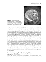
A 25-year-old woman was referred to our Institute with progressive shortness of
breath for 6 months, with chest X-ray as shown in Fig. 1.1. This chest X-ray shows
straightening of left heart border with pulmonary venous hypertension. How
consistent is this with a diagnosis of MS?
BCI1 6/15/05 8:26 PM Page 3
The most frequent roentgenographic findings in MS include left atrial enlarge-
ment, redistribution of blood flow to the upper lobes of the lung, Kerley B lines,
and enlarged pulmonary artery. Although their cardiac silhouette may be
normal in the frontal projection, patients with hemodynamically significant
MS almost invariably have evidence of left atrial enlargement.
Left atrial enlargement is one of the earliest signs of mitral stenosis; however,
its presentation may be subtle and limited to enlargement of the left atrial ap-
pendage, causing a straightening of the left heart border. In more advanced
cases, the left atrium is recognized as a double density and elevation of main
stem bronchus on the postanterior film. Radiologic changes in the lung fields in-
directly reflect the severity of MS. Redistribution of blood flow to the upper
lobes correlates best with the degree of mitral valve obstruction. The presence of
Kerley B lines is an important finding in patients with MS. These are fine
parallel densities in the peripheral lung fields which are perpendicular to a
pleural surface and are most frequently seen in the costophrenic sulci. The
lines are caused by thickened interlobar septa and signify chronic pulmonary
venous hypertension.
This finding is present in 30% of patients with resting pulmonary arterial
wedge pressures less than 20 mmHg and in 70% of patients with pressures
greater than 20 mmHg.
4 Chapter 1
Figure 1.1 Chest X-ray posteroanterior (PA) view showing straightening of left heart
border, cephalization of pulmonary veins, and double atrial shadow.
BCI1 6/15/05 8:27 PM Page 4
Although a decreased E-F slope is almost always present in severe MS, it is not
diagnostic of MS and it is an unreliable indicator of its severity. The specificity of
the diagnosis of MS by M-mode echocardiography is greatly improved by visu-
alizing the initial diastolic movement of the posterior mitral leaflet.
2
In the nor-
mal mitral valve, the posterior leaflet moves away from the anterior leaflet
during early diastole. In MS, the posterior leaflet moves anteriorly during early
diastole.
Mitral stenosis 5
Case Presentation (Continued)
Although the chest X-ray is strongly suggestive of MS, management decisions
need to be informed by details of anatomy and severity, so echocardiography
remains the examination of choice for evaluating MS. Indeed, the chest X-ray
may be unnecessary, and of course should be avoided in a pregnant woman. The
patient’s M-mode showed a decreased E-F slope and the posterior mitral leaflet
moved anteriorly during diastole, indicative of MS (Fig. 1.2). Is the M-mode still
useful?
Figure 1.2 M-mode echo showing paradoxical posterior mitral leaflet (PML).
Case Presentation (Continued)
The patient’s two-dimensional (2D) imaging in the short axis view showed a
typical “fishmouth” appearance of severely stenotic mitral valve with a mitral
valve area of 0.6 cm
2
(Fig. 1.3). Does this confirm the diagnosis of MS on 2D
imaging, and how should we evaluate severity of MS and suitability for
percutaneous mitral commissurotomy?
BCI1 6/15/05 8:27 PM Page 5
The short axis view allows the mitral valve area (MVA) to be measured by
planimetry, although technical factors may compromise the accuracy of this
method
—
not least the difficulty in ensuring that imaging is being performed at
the tips of the leaflets. Heavily calcified leaflets may have indistinct borders
that are difficult to trace, and there may also be dropout of echoes, leaving
gaps in the area to be traced. The hallmark of MS on 2D echocardiography is
thickening and restriction of motion of both mitral valve leaflets, with the pre-
dominant pathologic process being at the tips of the leaflets and proximal chor-
dae. The abnormal motion of the leaflets is apparent in early diastole. Fusion of
the commissures causes restriction in the motion of the tip of the anterior
leaflet. The commissural fusion usually causes the posterior leaflet to move
anteriorly during diastole with the larger anterior leaflet rather than moving
posteriorly.
Doppler echocardiography assesses the severity of the stenotic lesion and
color flow imaging is instrumental in determining associated mitral regurgita-
tion. This is important because moderate mitral regurgitation (more than 2+)
would be a contraindication to perform a closed procedure. MS produces char-
acteristic changes in the mitral flow velocity pattern, involving an increase in
the early diastolic peak velocity of flow and slower than normal rate of fall in
velocity. The transvalvular pressure gradient can be measured continuously
throughout diastole and correlates well with mean pressure gradient measured
by cardiac catheterization. However, the pressure gradient is affected by heart
rate, cardiac output, and valvular regurgitation in addition to orifice area, and
hence it provides only a rough estimate of severity.
The pressure half-time is the time required for the instantaneous gradient
across the valve to fall to half of the peak value (Fig. 1.4). This means of assess-
ing MVA is usually sufficiently accurate for clinical use. The pressure half-time
method is not valid for several days after mitral balloon valvuloplasty, probably
because of a decrease in left atrial pressure without a commensurate improve-
ment in left atrial compliance.
6 Chapter 1
Figure 1.3 Parasternal short axis
view showing typical “fishmouth”
appearance of severely stenotic mitral
valve (mitral valve area [MVA]
0.6 cm
2
).
BCI1 6/15/05 8:27 PM Page 6
An echocardiographic scoring system developed by Wilkins et al.
3
has been used
widely for assessment of suitability for percutaneous mitral commissurotomy.
Leaflet rigidity, thickening, valvular calcification, and subvalvular involvement
are each scored from 0 to 4. A score of 8 or less is usually associated with ex-
cellent immediate and long-term results, whereas scores exceeding 8 are
associated with less impressive results. In our experience, slight commissural
calcium is not a contraindication for percutaneous mitral commissurotomy, but
when the calcium score is more than 2+, the incidence of restenosis is higher
and thus surgical repair of the mitral valve is preferable. Significant subvalvular
pathology is a more important determinant of suboptimal results following per-
cutaneous mitral commissurotomy.
In addition to determining the presence and severity of MS, it is important to
evaluate the heart for secondary effects of MS. These include left atrial enlarge-
ment, stasis, thrombus formation, and secondary pulmonary hypertension.
The aortic, tricuspid, and pulmonic valves can likewise be directly evaluated for
evidence of rheumatic involvement.
Mitral stenosis 7
Figure 1.4 Continuous wave Doppler showing gradient across severely stenotic mitral
valve.
Case Presentation (Continued)
Pressure half-time measurement in this case showed a mean gradient of
16 mmHg at the heart rate of 66 b/min (Fig. 1.4). Color Doppler performed on our
index patient confirmed the stenotic mitral valve and fortunately showed no
mitral regurgitation. Is the patient suitable for percutaneous intervention?
BCI1 6/15/05 8:27 PM Page 7
Transesophageal echocardiography
Transesophageal echocardiography (TEE) provides excellent images of the mi-
tral valve leaflets, the left atrium, and the left atrial appendage (Fig. 1.5).
Transthoracic imaging is usually diagnostic of MS and can accurately assess the
severity of the stenosis. However, in some cases the transthoracic accoustic win-
dow may be inadequate. Multiplane TEE visualizes most of this anatomically
complex structure and thrombus in the appendage can be accurately diagnosed,
although experience is needed to avoid mistaking the pectinate muscle for
thrombus.
TEE is well established as the gold standard for detecting thrombi in the left
atrium (LA) and LA appendage, with a sensitivity and specificity of 100% and
99%, respectively.
4
The semi-invasive nature and safety of the test make it
ideal for serial follow-up of thrombi in the LA body and appendage. TEE is
also indicated if there is doubt regarding the presence or severity of mitral re-
gurgitation and assessment of subvalvular pathology, if this is unclear on
transthoracic echocardiography (Fig. 1.6). In our practice, all patients under-
going balloon valvuloplasty with atrial fibrillation and suspicion of clot on
transthoracic echocardiography undergo TEE before the procedure. Finally,
TEE may be used to guide the atrial trans-septal puncture during the balloon
valvuloplasty procedure,
5
although we do not use TEE during the procedure at
our institution.
8 Chapter 1
Case Presentation (Continued)
According to the Wilkins scoring system, our patient’s mitral valve score was 6.
There was no calcium on the mitral valve. This is an ideal candidate for
percutaneous transmitral commissurotomy (PTMC) and a good result could be
anticipated. What other steps are required before PTMC?
Figure 1.5 Transesophageal
echocardiography (TEE) showing
large left atrium with dense
spontaneous contrast and amputated
left atrial appendage.
BCI1 6/15/05 8:27 PM Page 8
Recently, there has been much interest in cardiac magnetic resonance imaging
(MRI) and three-dimensional echocardiography in the assessment of valve
lesions. We do not believe that MRI gives any additional information over
echocardiography for MS, and MRI is expensive, time-consuming, and may be
compromised by atrial fibrillation, which is not uncommon in MS. Three-
dimensional echocardiography is a newer imaging modality and, in selected
cases, will be useful, especially to assess the subvalvular apparatus.
Three-dimensional echocardiography
Real-time three-dimensional (R3D) echocardiography is a novel technique
that permits visualization of mitral valvular anatomy in any desired plane of ori-
entation. The use of R3D echocardiography in evaluation of mitral stenosis has
been studied by Zamorano et al.
6
In 76 patients with significant MS, these
Mitral stenosis 9
Figure 1.6 Parasternal long axis
showing stenotic mitral valve and
significant subvalvular thickening.
Case Presentation (Continued)
In this case, TEE was performed before PTMC. TEE showed dense spontaneous
contrast but there was no clot in the left atrium (Fig. 1.5).
At cardiac catheterization before PTMC, left heart pressure was measured by
retrograde catheterization of the left ventricle. Left atrial pressure was initially
measured by pulmonary artery (which is accurate) and later by direct entry into
the left atrium through trans-septal puncture. The mean gradient across the
mitral valve was 15 mmHg before the PTMC (Fig. 1.7). The mitral valve was
successfully dilated with a 26-mm Inoue balloon and the mean gradient across
the mitral valve after the procedure was 4 mmHg with mitral valve area of
1.7 cm
2
and trivial mitral regurgitation.
BCI1 6/15/05 8:27 PM Page 9
authors demonstrated that R3D echocardiography is a feasible, accurate, and
highly reproducible technique for assessing MVA. MVA calculation with R3D
echocardiography has the best agreement with invasive methods (average dif-
ference between both methods: 0.08cm
2
). R3D echocardiography may im-
prove the assessment of MS severity in patients with discordant results between
different methods and in clinical scenarios where these methods have limita-
tions, particularly after balloon valvoplasty.
7
Magnetic resonance imaging
Magnetic resonance imaging can be used to identify the presence of valvular
stenosis. The high-velocity flow across the narrow valvular orifice may be rec-
ognized as a signal void on cine MRI. Imaging may be performed with steady-
state free precession (SSFP) imaging for semi-quantitative assessment of
valvular dysfunction or with a standard breath-hold segmented gradient–
recalled echoplanar imaging sequence (GE-EPI) (Fig. 1.8). A study by
10 Chapter 1
Figure 1.7 Left heart catheterization of left ventricle (LV) and left atrium (LA) tracing
showing gradient across mitral valve before and after percutaneous transmitral
commissurotomy (PTMC).
BCI1 6/15/05 8:27 PM Page 10