Cardiovascular Imaging A handbook for clinical practice - Part 9 pptx
Bạn đang xem bản rút gọn của tài liệu. Xem và tải ngay bản đầy đủ của tài liệu tại đây (335.42 KB, 31 trang )
Mader MT, Poulton TB, White RD. Malignant tumors of the heart and great vessels: MR
imaging appearance. Radiographics 1997;17:145.
Meng Q, Lai H, Lima J, et al. Echocardiographic and pathologic characteristics of primary
cardiac tumors: a study of 149 cases. Int J Cardiol 2002;84:69.
Schvartzman PR, White RD. Imaging of cardiac and paracardiac masses. J Thorac Imaging
2000;15:265.
Siripornpitak S, Higgins CB. MRI of primary malignant cardiovascular tumors. J Comput
Assist Tomogr 1997;21:462.
234 Chapter 18
BCI18 6/15/05 8:41 PM Page 234
CHAPTER 19
Evaluation of the
transplanted heart
Oberdan Parodi, Maria Frigerio, and Benedetta De Chiara
Introduction
Cardiac transplantation is an established treatment for advanced heart failure.
Clinical experience and progress in immunosuppression have increased recipi-
ent survival to more than 80% at 1 year; 10-year survival is more than 50%
at many centers.
1
Common complications after heart transplantation (HTx)
include acute rejection, infections, cardiac allograft vasculopathy (CAV), and
lymphoproliferative disorders and other malignancies, as well as other condi-
tions mainly related to side-effects of immunosuppressive drugs (Fig. 19.1).
This chapter summarizes briefly the role of cardiac imaging techniques for the
diagnosis of acute rejection, and will provide a more in-depth review of current
techniques for invasive and non-invasive evaluation of CAV.
235
Diagnosis of acute cardiac allograft rejection
Acute rejection is an important cause of death in HTx recipients, accounting for
20% of the deaths occurring in the first post-transplant year, and up to 15%
thereafter.
1
Nevertheless, the majority of acute rejections can be safely man-
aged, providing diagnosis precedes the occurrence of graft dysfunction. Minor
clinical signs of acute cardiac allograft rejection may be absent and are non-
specific (Table 19.1). Thus, surveillance for preclinical diagnosis is of utmost
importance.
Case Presentation
A 55-year-old man with idiopathic dilated cardiomyopathy underwent heart
transplantation in 1999, from a donor of the same age and gender. The patient
had cytomegalovirus infection and one treated acute rejection episode in the
early postoperative months. One year after heart transplantation, the resting
ECG showed an incomplete right bundle branch block, and further evaluation
was sought for surveillance for rejection and allograft vasculopathy.
BCI19 6/17/05 9:49 PM Page 235
Endomyocardial biopsy (EMB) is the most widely used and reliable tool for
the diagnosis of acute rejection. Acute cardiac allograft rejection may show a
wide spectrum of lesions with standard staining (hematoxylin and eosin), rang-
ing from sparse perivascular or interstitial infiltrates of small lymphocytes, to
more widespread and aggressive infiltration of large, activated lymphocytes, as-
236 Chapter 19
First month
Post-surgical complications
Acute graft failure
Acute rejection
Infections (bacterial, fungal)
Side-effects of immunosuppressive drugs
2nd month-1 year
Acute rejection
Infections
(typically CMV and other opportunistic infections)
Side-effects of immunosuppressive drugs
> 12 months
CAV
Post-transplant lymphoproliferative disorders
Other malignancies
Late acute rejection
Side-effects of immunosuppressive drugs
Diabetes
Dyslipidemia
Arterial hypertension
Renal insufficiency
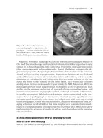
Figure 19.1 Common complications after cardiac transplantation. Side-effects of
immunosuppressive drugs occur early after heart transplantation and contribute to
endothelial dysfunction and progression of cardiac transplant vasculopathy. CAV,
coronary allograft vasculopathy; CMV, cytomegalovirus.
Table 19.1 Clinical symptoms and signs of acute cardiac allograft rejection.
None
Malaise, fatigue
Fever
Tachycardia
Supraventricular arrhythmias, conduction disturbances
Reduced QRS amplitude (peripheral leads)
Reduced systolic blood pressure
Reduced pulse pressure
Dyspnea
Congestive heart failure
Sudden death
BCI19 6/17/05 9:49 PM Page 236
sociated or not with granulocytes, with various degrees of myocyte disruption
and necrosis, up to severe, extensive necrosis, associated with edema, polymor-
phonuclear infiltrates and hemorrhages. A less common but ominous condi-
tion is the so-called “humoral” or “vascular” rejection, which can be briefly
defined as an immunomediated, acute graft dysfunction despite a normal
(“negative”) EMB. The main limitations of EMB are patient discomfort, risk of
complications (less than 1% when performed by skilled personnel: right ventri-
cle perforation, tricuspid leaflet disruption, bleeding, pneumothorax), costs,
and sampling error. Despite the debates about rejection classification, the dis-
agreement observed between pathologists in EMB interpretation, and the un-
certainties regarding therapeutic implications of the EMB results (except in case
of very low- or high-grade rejections), EMB remains the cornerstone for rejec-
tion surveillance at most HTx centers. Nevertheless, various non-invasive alter-
native techniques have been evaluated, with the aim of reducing the need for
repeated EMBs, to optimize their timing, and to add information relevant for
clinical management.
Echocardiography is the most extensively used diagnostic technique for non-
invasive monitoring of HTx recipients. During the first postoperative weeks, the
echocardiogram reflects the mutual adaptation of the donor’s heart and of the
recipient’s hemodynamic profile to the new condition, and postoperative se-
quelae: right ventricular dilatation, tricuspid regurgitation, paradoxic septal
motion, and pericardial effusion are common. After 1 month, the echocardio-
graphic pattern remains quite stable in the absence of significant rejection in
most patients, generally up to 1 year. In the long term, the effects of hyperten-
sion and CAV may interfere with echocardiographic findings. The main features
of acute rejection are alterations of indexes of left ventricular (LV) diastolic
function, commonly detected by pulsed wave Doppler (reduction of pressure
half-time and of isovolumic relaxation time), increased wall thickness, in-
creased myocardial echogenicity, dilated right ventricle, and increased peri-
cardial effusion. LV systolic dysfunction is less frequent, is prognostically
unfavorable, and is more often associated with high-grade rejection or with
biopsy-negative (humoral/vascular) rejection. The accuracy of echocardio-
graphic findings may vary according to the operators’ experience and patient
body structure; obesity, not uncommon after HTx, may reduce the quality of
echocardiography. Recently, tissue Doppler imaging (TDI) has been proposed
for early detection of rejection-related diastolic dysfunction in HTx recipients;
high sensitivity (93%) and high negative predictive value (96%) have been re-
ported, with a favorable impact on the number of EMBs as well as prognostic
implications.
2
Magnetic resonance imaging (MRI) has been more recently explored, both in
experimental and human research. Labeling of macrophages with dextran-
coated, ultrasmall, superparamagnetic iron oxide (USPIO) particles can be used
to detect the accumulation of macrophages in rejecting tissue.
3
Furthermore,
USPIO can be used alone (i.e. not in macrophages) because blood pool contrast
agents leak into the interstitial space in areas of inflammation associated with
Evaluation of the transplanted heart 237
BCI19 6/17/05 9:49 PM Page 237
rejection, where the vessels display increased permeability. Moreover, the
myocardial T2 relaxation time, determined using a black-blood MRI sequence,
has been demonstrated to predict acute heart transplant rejection in humans.
An intriguing radioisotope technique is represented by myocardial scintigra-
phy with radiolabeled (indium-111 pentetreotide) somatostatin receptor ana-
logue.
4
The pathophysiologic hypothesis is that somatostatin receptors are
expressed on activated lymphocytes and up-regulated during cardiac allograft
rejection. It is noteworthy that somatostatin receptor imaging seems to predict
impending rejection at least 1 week before the EMB becomes positive, because
of the interval between lymphocyte activation and relevant myocardial infiltra-
tion or damage. The possibility of anticipating the occurrence of EMB-proven
rejection by means of analysis of gene expression in the peripheral blood is cur-
rently under evaluation.
Another interesting approach, which implies the possibility of telemonitor-
ing of HTx recipients, is represented by continuous recording of high-
resolution, intramyocardial electrocardiogram (ECG) by means of special
electrodes implanted at the time of transplantation.
However, these innovative approaches have not yet gained widespread clini-
cal use. At present, echocardiography is used at most HTx centers as an adjunct
to clinical, laboratory, ECG, and EMB data: it is helpful in deciding if and how to
change the immunosuppressive regimen and for planning patient follow-up,
but it is not a substitute for EMB except in small pediatric patients.
238 Chapter 19
Case Presentation (Continued)
Echocardiographic evaluation demonstrated normal regional and global left
ventricular function (ejection fraction 0.59) and diastolic parameters. A routine
biopsy was normal. However, further evaluation was sought for allograft
vasculopathy.
Coronary allograft vasculopathy
CAV is the main factor limiting long-term survival after transplantation, and ac-
counts for more than 20% of later mortality.
1
Pretransplant conditions, donor
characteristics, and events occurring during the first post-transplant year and
thereafter are implicated in the pathogenesis of CAV (Table 19.2).
5
The initial
endothelial dysfunction and injury are followed by intimal hyperplasia and vas-
cular smooth muscle cell proliferation. The process has been angiographically
documented in 40–50% of patients surviving 5 years after transplantation.
1
The
histologic hallmark of CAV (intimal proliferation in graft coronary arteries) can
be observed in all surviving recipients as soon as 1 year after HTx; its in vivo
equivalent can be appreciated by means of intracoronary ultrasound (ICUS).
Unfortunately, warning anginal symptoms are often absent, as a result of car-
BCI19 6/17/05 9:49 PM Page 238
diac denervation; clinical manifestations of CAV are frequently severe, and in-
clude congestive heart failure, myocardial infarction, life-threatening ventricu-
lar arrhythmias, and sudden death.
Treatment of CAV remains a difficult challenge. The solution for severe, dif-
fuse disease is retransplantation, although this option is limited by donor avail-
ability. Focal stenoses can be approached by percutaneous angioplasty and
stenting, with satisfactory angiographic short-term results, but little is known
regarding long-term success and the prognostic relevance of these procedures.
Recently, the use of proliferation inhibitors (sirolimus and everolimus) appears
promising for preventing, stopping, and perhaps reversing intimal prolifera-
tion. This section describes the advantages and limitations of non-invasive ap-
proaches, the place of invasive techniques in the detection of CAV and in
prognostic stratification of HTx recipients, and the potential of new imaging
modalities.
Non-invasive testing
The availability of accurate non-invasive tests for diagnosis of the presence (and
of the functional and prognostic relevance) of CAV is highly desirable for clini-
cal, organizational, and economic reasons. Moreover, non-invasive tests may
provide information regarding microvascular circulation, which can be im-
paired after HTx. Unfortunately, the sensitivity and specificity of non-invasive
tests for diagnosis of CAV is difficult to establish in relation to angiography, be-
cause anatomic narrowing does not always induce ischemia and, conversely, is-
chemia may occur in patients with small vessel coronary artery vasculopathy
undetected by angiography. The diffuse nature of CAV may result in balanced is-
chemia that is difficult to recognize by imaging modalities, such as perfusion
scintigraphy, which are based on intrapatient comparison of different myocar-
dial areas. Moreover, new events (e.g. late acute rejection) may occur at any
time after HTx, and may accelerate the progression of CAV, thus limiting the
Evaluation of the transplanted heart 239
Table 19.2 Factors involved in the pathogenesis of coronary allograft vasculopathy.
Non-immune
Immune mechanisms mechanisms
Recurrent/persistent acute rejection Older donor age
Vascular/humoral rejection Hyperlipidemia
HLA mismatch Hypertension
Diabetes
Ischemia reperfusion injury*
CMV infection*
* These factors are in an intermediate position, because they may be considered partially
“immune” inasmuch as they imply exposure of endothelial antigens and/or activation of
immune reaction.
CMV, cytomegalovirus.
BCI19 6/17/05 9:49 PM Page 239
predictive value of any test, irrespective of its accuracy. However, for the indi-
vidual patient, a positive non-invasive test indicating inducible myocardial is-
chemia may have powerful prognostic value.
Stress electrocardiography
Most transplant patients have resting ECG abnormalities (mostly incomplete
right bundle branch block and T-wave inversion) that make the interpretation
of stress ECG less sensitive and specific than in general population. Further-
more, sensitivity is reduced because angina is rarely present, and the target
heart rate during exercise usually is not achieved because of heart denervation
and, in some patients, inadequate physical conditioning. Reported sensitivity
and specificity of exercise ECG for the detection of CAV are in the ranges 0–38%
and 77–100%, respectively. Ambulatory ECG monitoring is similarly insensi-
tive. Arterial hypotension during exercise is quite specific for significant CAV.
Additional information may be provided by ECG data when combined with im-
aging techniques. In our experience, the appearance of complete right bundle
branch block increases the specificity of myocardial perfusion defects for pre-
dicting CAV; a blunted heart rate response during dipyridamole-induced va-
sodilatation predicts stress perfusion defects, and higher probability of cardiac
events during follow-up.
Echocardiography
Baseline ejection fraction (EF) and regional wall motion are generally normal in
HTx recipients, even in the presence of CAV. Thus, regional wall motion abnor-
malities and/or a reduced ejection fraction have a low sensitivity for diagnosis of
CAV (50–60%), although their prevalence is higher in HTx patients with CAV.
Spes et al.
6
reported that resting wall motion abnormalities in any left ventricu-
lar territory had a 90% positive predictive value and an 88% specificity, but only
a 57% sensitivity in detecting CAV. Furthermore, normal resting echocardiog-
raphy had a 90% negative predictive value for cardiac events. In a previous
study,
7
we showed that normal resting wall motion at echocardiography cou-
pled to normal stress myocardial perfusion scintigraphy ruled out the presence
of significant CAV. Conversely, resting wall motion abnormalities and perfusion
defects strongly predicted cardiac events. Resting echocardiography alone de-
tected significant CAV only in 50% of cases, but it was an independent prognos-
tic determinant of cardiac events. The addition of a pharmacologic stress (e.g.
dipyridamole or dobutamine) may improve the limited sensitivity of resting
echocardiography for detection of CAV (Table 19.3). Ciliberto et al.
8
first report-
ed that high-dose dipyridamole stress echocardiography is useful for identifying
patients with significant CAV, and more recently found that dipyridamole-
induced wall motion abnormalities were associated with adverse prognosis.
9
Dobutamine stress echocardiography (DSE) provides accurate diagnosis as well
as useful prognostic information in cardiac transplant recipients.
6
Dobutamine
increases contractility, heart rate, and wall stress in a dose-dependent fashion,
an attractive approach to evaluate both microvasculature and epicardial coro-
240 Chapter 19
BCI19 6/17/05 9:49 PM Page 240
nary vessels in HTx patients. DSE is more sensitive than exercise echocardiogra-
phy because the transplanted heart is more responsive to catecholamine stimu-
lation than a normal heart, while the exercise-induced increase in heart rate is
limited. Spes et al.
6
suggested that serial routine coronary angiography could be
deferred in HTx recipients with normal DSE, because the prognostic value of
this test is comparable to that of ICUS. Therefore, resting echocardiography plus
DSE appear a reliable method for routine surveillance of patients after HTx. As
usual, echocardiography may be limited by poor image quality in obese pa-
tients, and its accuracy relies upon the operator’s experience.
Myocardial perfusion scintigraphy
Stress myocardial scintigraphy with thallium-201 or technetium-99m labeled
perfusion tracers and single photon emission computed tomography (SPECT)
has a well-established role in detection of atherosclerotic coronary lesions in pa-
tients with known or suspected ischemic heart disease. In HTx recipients, stress
myocardial scintigraphy provides a low-to-moderate sensitivity and a good
specificity for the detection of CAV, with exercise testing performing better than
a dipyridamole test (Table 19.4). However, the limitations of exercise testing in
HTx patients have been already described. Moreover, most published studies
utilized a qualitative (visual) assessment of perfusion defects, or a semi-quanti-
tative evaluation of myocardial tracer distribution, without any reference to
maps of normal regional perfusion pattern. The limited sensitivity of the tech-
nique reported by these studies might be related to the lack of quantitative as-
Evaluation of the transplanted heart 241
Table 19.3 Accuracy of stress echocardiography in the detection of coronary allograft
vasculopathy.
Patients Sensitivity Specificity
Study Journal Year (n) Stress (%) (%)
Derumeaux et al. J Am Coll 1995 37 Dobutamine 86 91
Cardiol
Herregods et al. J Heart Lung 1994 28 Dobutamine 0 100
Transplant
Akosah et al. J Heart Lung 1994 41 Dobutamine 95 55
Transplant
Ciliberto et al. Eur Heart J 1993 80 Dipyridamole 32 100
Spes et al. Am J Cardiol 1996 46 Dobutamine 79 83
Collings et al. J Heart Lung 1994 51 Exercise 25 86
Transplant
Cohn et al. Am J Cardiol 1996 51 Exercise 15 85
Spes et al. Circulation 1999 109 Dobutamine 94 57
BCI19 6/17/05 9:49 PM Page 241
sessment of the regional tracer uptake. More recently, we evaluated the accura-
cy of high-dose dipyridamole sestamibi SPECT in the detection of CAV and in
prognostic stratification utilizing a semi-quantitative technique corrected for
bull’s eye maps of perfusion normalcy rates in 78 HTx recipients.
7
Our findings
indicate that this approach is sensitive in the detection of significant CAV (sensi-
tivity 92%), and that its combination with resting echocardiography can be a
safe and reasonable non-invasive approach for prediction of long-term progno-
sis after HTx. In this study, concordant negative tests occurred in over two-
thirds of cases, in whom non-invasive testing had an optimal accuracy in ruling
out significant CAV (specificity 82%, negative predicting value 100%). These
patients also had a high event-free survival. Conversely, a significant CAV was
present in 100% of the five patients with abnormalities in both non-invasive
tests. Patients with abnormal resting echocardiography had a 10-fold relative
risk of cardiac events at follow-up, while a positive dipyridamole SPECT con-
ferred a 4 : 1 relative risk of cardiac events. Thus, the association of these tests
may be useful to rule out the need for coronary angiography when both are
negative, and to recommend it when at least one is positive. In our hands, the
242 Chapter 19
Table 19.4 Accuracy of stress myocardial scintigraphy in the detection of coronary
allograft vasculopathy.
Patients Sensitivity Specificity
Study Journal Year (n) Stress (%) (%)
Smart et al. Am J Cardiol 1991 57 Dipyridamole 21 88
Redonnet et al. Transplant 1995 43 Dipyridamole 58 64
Proc
Ciliberto et al. Eur Heart J 1993 50 Exercise 67 100
Rodney et al. J Heart Lung 1994 25 Exercise 77 100
Transplant
Smart et al. Transplant 1991 35 Exercise 21 81
Proc
Valantine et al. Circulation 1988 20 Exercise 36 78
McKillop et al. Clin Radiol 1981 7 Exercise 100 0
Mairesse et al. J Heart Lung 1995 37 Exercise NA 84–92
Transplant
Ambrosi et al. Eur Heart J 1994 34 Exercise NA 97
Carlsen et al. J Heart Lung 2000 67 Dip /Exercise 80 92
Transplant
Ciliberto et al. Eur Heart J 2001 78 Dipyridamole 92 86
NA, not assessed.
BCI19 6/17/05 9:49 PM Page 242
sensitivity of this imaging approach for significant CAV favorably compares
with previous studies that used either dipyridamole or exercise thallium-201
scans
10
or visual interpretation of exercise myocardial perfusion imaging by
technetium-99m labeled compounds.
11
High-dose dipyridamole may augment
the differences in regional tracer distribution among areas with different coro-
nary vasodilating capability, improving the detection of minor coronary lesions.
It is not yet clearly established which stressor is preferable in the evaluation of
blunted coronary flow reserve in heart transplant recipients. In our experience,
the dipyridamole test is safe, reproducible, and feasible (in up to 95% of trans-
planted patients), and it provides good sensitivity in CAV detection and relevant
prognostic information when associated to quantitative evaluation of myocar-
dial tracer uptake.
To refine the capability of detecting perfusion defects by quantitative bull’s
eye imaging in this specific patient population, an ongoing study is being carried
out at our institution, where correction for normal perfusion has been per-
formed utilizing a map obtained from a database of transplanted patients with
normal cardiac function, no rejection, and normal coronary angiography, who
underwent dipyridamole SPECT imaging 1 year after HTx. This approach may
optimize the rest and stress regional cut-off values used for definition of nor-
malcy versus perfusion defects.
Regional perfusion and coronary flow reserve may be accurately measured
in HTx recipients by positron emission tomography in conjunction with flow
tracers (
13
N-ammonia,
15
O-water). The diffuse nature of CAV is a challenge for
all cardiac imaging techniques. Absolute measurements of myocardial blood
flow and coronary flow reserve may circumvent the limitations of other imag-
ing techniques, which explore regional myocardial differences by means
of relative tracer distribution. However, the accuracy of positron emission
tomography in the detection of CAV has not yet been tested, probably also be-
cause this technique is expensive and is not always clinically feasible, and not
widely available.
Invasive techniques
Coronary angiography
After recognition of the occurrence and clinical relevance of CAV, annual coro-
nary angiography has been utilized to monitor its development and progres-
sion. The classification of angiographic abnormalities of the transplanted heart
was proposed by Gao et al. in 1988.
12
Three types of coronary lesions were
defined:
1 Type A, with discrete, tubular or multiple stenoses, which resembles coro-
nary artery disease of the native heart
2 Type B, characteristic of HTx recipients, which is subclassified as B
1
(sharp
onset of distal diffuse concentric narrowing and obliteration, with apparently
normal or nearly normal proximal segments) or B
2
(progressive, concentric
tapering from proximal to distal segments, with some residual flow in the
periphery)
Evaluation of the transplanted heart 243
BCI19 6/17/05 9:49 PM Page 243
3 Type C, with relative proximal dilatation and irregular narrowing of distal
branches, which may show non-tapered, abrupt interruption.
Several studies have demonstrated the prognostic relevance of CAV as
detected by coronary angiography in large patient cohorts. After 3–4 years, the
relative risk of any cardiac event is more than tripled in patients with angio-
graphic evidence of obstructive disease compared with those without evidence
of disease. However, coronary angiography has several major limitations: it is
invasive, requires hospitalization, and the nephrotoxicity of contrast agents
may be harmful in HTx recipients, who may suffer from drug-induced renal
insufficiency. Moreover, the organizational and economic burden of annual
coronary angiography of an increasing population of long-term HTx recipients
is not negligible. The main limitation of coronary angiography is represented
by its “lumenographic” approach, which is insensitive in detecting a diffuse,
concentric disease. Thus, angiography may underestimate the severity of CAV.
Intracoronary ultrasound
The ICUS technique provides images of both the lumen and the vessel wall, and
is at the present time the best tool for early diagnosis and quantification of CAV.
According to the experience of the Stanford HTx program, an intimal thicken-
ing of more than 0.3 mm is associated with a significantly reducedprobability of
survival and an increased risk for cardiac events during follow-up, regardless
of angiographic findings.
13
Furthermore, functional impairment, such as de-
creased coronary blood flow reserve, can be evaluated by ICUS and is more fre-
quently associated to intimal thickening. Increased intimal thickening is
currently proposed as the best surrogate end-point for therapies aimed at limit-
ing or reversing the evolution of CAV.
14
Unfortunately, ICUS also has some limitations: it implies more risks with re-
spect to standard angiography (vessel spasm, dissection, and complications re-
lated to guidewire manipulation); and it is expensive and time-consuming.
Standardization of the procedure facilitates comparison of serial examinations,
and interobserver agreement is good among trained personnel. The character-
istics of the probe do not allow the examination of peripheral segments. How-
ever, it is likely that given the diffuse nature of CAV, measurements of wall
thickness from one artery should reflect the overall extent of the disease, or at
least its prognostic relevance.
ICUS represents the contemporary gold standard for the diagnosis and serial
evaluation of CAV, particularly when therapies targeted at CAV (e.g. pro-
liferation inhibitors such as sirolimus or everolimus, extensive use of statins
irrespective of cholesterol values) become available or are under evaluation.
Unfortunately, its invasive nature and costs continue to limit its use.
244 Chapter 19
BCI19 6/17/05 9:49 PM Page 244
New imaging modalities
Magnetic resonance imaging
Magnetic resonance perfusion imaging (MRPI) using gadolinium-based con-
trast agents has recently been validated as a clinical tool to quantify myocardial
perfusion. Using MRPI, it is possible to quantify myocardial perfusion reserve,
a parameter that mirrors coronary flow reserve as a measure of the functional
significance of epicardial lesions in the pertinent perfusion territories. Because
resting endomyocardial : epimyocardial perfusion ratio decreases with im-
paired coronary circulation, calculation of this index by MRPI may represent a
simple measurement of myocardial perfusion at rest that could be sufficient to
detect early CAV. Perfusion imaging during rest and adenosine- or dipyri-
damole-induced hyperemia can be performed in the same session. Adenosine
or dipyridamole are intravenously infused according to protocols commonly
used in echocardiography and nuclear medicine laboratories. Perfusion is usu-
ally determined in three LV short-axis slices. The first slice is located close to the
base of the heart just below the aortic outflow tract, the second in the middle of
the LV, and the third close to the apex just below the base of the papillary mus-
cles. A single-shot gradient-echo sequence with saturation recovery magneti-
zation preparation for T1 weighting and linear k-spacing is used for imaging.
Temporal resolution allows acquisition of one image in each of the three select-
ed slices within one heart beat up to a heart rate of 110 b/min. Sixty images per
Evaluation of the transplanted heart 245
Case Presentation (Continued)
The patient also underwent dipyridamole technetium-99m sestamibi SPECT. At
the time of the examination he was treated with a triple-drug
immunosuppression protocol (cyclosporine, prednisone, and azathioprine).
Dipyridamole myocardial perfusion scintigraphy showed a fixed perfusion
defect in inferior wall and a slight reversible antero-apical defect; quantitative
analysis better clarified the reduction of tracer uptake (under the physiologic
threshold) in the antero-apical segments (Fig. 19.2). Coronary angiography did
not show any coronary artery lesion; ICUS was employed to assess vascular
composition of the left descending coronary artery. An obvious intimal
proliferation of this vessel was found, confirming the scintigraphic findings (Fig.
19.3); conversely, no abnormalities were detected in the right coronary artery,
suggesting that the fixed perfusion defect in the inferior wall was likely a result
of previous acute rejections. The patient underwent aggressive pharmacologic
treatment with statins, alpha- and beta-blockers. At 4-year follow-up,
myocardial stress scintigraphy did not show any progression of CAV, and no
cardiac event occurred.
This case illustrates how careful observation of transplanted patients by
imaging techniques in the first 1–2 years after transplantation may help select
patients for treatments that appear to slow the progression of established CAV.
BCI19 6/17/05 9:49 PM Page 245
slice location are usually acquired with a spatial resolution of 2–3 mm. Patients
are asked to hold their breath at end expiration for the first 15–20 s of each per-
fusion scan, such that the tracking of the first pass of the bolus at the three
chosen slice locations is not affected by respiratory motion. Immediately after
initiation of the sequence, a compact bolus of 0.03 mmol/kg bodyweight
gadolinium-DTPA is injected over an antecubital vein at a rate of 7 mL/s using a
power injector. Analysis of MRPI curves for calculation of myocardial perfusion
reserve has been described previously in details, and its application for the de-
tection of CAV provided interesting findings.
15
A significant correlation was
found between invasive measurement of coronary flow reserve and the non-
246 Chapter 19
STRESS REST
Short axis
Long axis vert.
Long axis horiz.
Bull’s eye
Figure 19.2 Myocardial single photon emission computed tomography (SPECT)
imaging in the short- and long-axis views and bull’s eye analysis of the case report. A
fixed perfusion defect in the inferior wall and a slight reversible antero-apical defect is
detectable at visual inspection; quantitative analysis better clarifies the reversible
reduction of tracer uptake (under the physiologic threshold) in the antero-apical
segments.
BCI19 6/17/05 9:49 PM Page 246
invasive evaluation of resting endomyocardial : epimyocardial perfusion ratio.
HTx patients with CAV showed a reduced myocardial perfusion reserve and
resting endomyocardial : epimyocardial perfusion ratio. When patients with
left ventricular hypertrophy and/or prior rejection were excluded, a normal
resting endomyocardial : epimyocardial perfusion ratio was able to exclude
CAV (negative predictive value 100% with a cut-off value of 1.3), suggesting
that further stress tests or invasive examinations are not required. A myocardial
perfusion reserve of more than 2.3 as assessed with adenosine infusion exclud-
ed CAV (negative predictive value 100%) in the overall population, suggesting
that this test is useful in HTx recipients with left ventricular hypertrophy and/or
history of acute rejection(s).
These data were obtained in a small sample of HTx patients; if confirmed in
larger cohorts, MRPI might be a good method for routine non-invasive surveil-
lance for CAV after HTx. The potential of dobutamine stress MRI has not yet
been evaluated in HTx recipients.
Evaluation of the transplanted heart 247
Figure 19.3 Angiograms of the left
coronary artery (upper panel) and
intracoronary ultrasound (lower
panel) in the same patient. Note that
the angiogram does not reveal any
significant abnormalities, whereas
intracoronary ultrasound
demonstrates significant intimal
thickening (arrows). No abnormalities
were detected in the right coronary
artery by both coronary angiography
and intracoronary ultrasound, despite
obvious stable perfusion defects in the
inferior wall at myocardial
scintigraphy.
BCI19 6/17/05 9:49 PM Page 247
Electron beam computed tomography
Recently, electron beam computed tomography (EBCT) has been proposed
in the setting of HTx recipients. Contrary to the notion that coronary artery
calcification is an atypical or late feature of CAV, quantification of coronary
calcification by EBCT has recently been reported to correlate closely with
the occurrence of coronary artery lesions in HTx recipients. Calcium scores
revealed close correlation with the ICUS degree of intimal proliferation and
showed high sensitivity (94%) and rather good specificity (79%) for detecting
CAV;
16
in addition, EBCT appeared to be associated with coronary events after
HTx. Because of its relatively low cost, EBCT could be useful to detect the pres-
ence of CAV non-invasively, and to select high-risk patients for invasive proce-
dures; moreover, it could serve for follow-up purposes. However, types B
2
and
C distal coronary artery lesions might be underestimated by this approach, and
the functional significance of these calcifications cannot be ascertained. Very re-
cently, another study warned about the lack of usefulness of EBCT in detecting
documented CAV,
17
confirming the concerns reported above. Finally, the radia-
tion exposure of CT should be considered before recommending a widespread
indication for this technique.
Practical implication of cardiovascular imaging in
heart transplantation
Follow-up of HTx recipients is a complex task for both physicians and the pa-
tient, and non-invasive tests that could diagnose or anticipate significant car-
diac events, such as acute rejection or clinically relevant CAV, are certainly
warranted. At present, the diagnosis of acute rejection still relies upon routine
or extemporary EMBs. Echocardiography at rest is useful for increasing the
clinical suspicion of rejection, for monitoring its impact on cardiac function, and
for optimizing the timing of follow-up biopsies. In the long-term, resting and
stress (with dobutamine) echocardiography may raise the suspicion for CAV,
and provides prognostic information. It is of utmost importance to compare
each echocardiogram with previous examinations, and the specific experience
of the reviewer in follow-up of HTx patients is relevant to the reliability of
echocardiographic findings. DSE is important in prognostication after HTx,
6
and provides a good accuracy for detecting significant CAV.
10
However, early
vasculopathy not associated with obvious impairment of coronary flow reserve
may be missed. ICUS appears the best tool for early recognition of these prob-
lems. Nevertheless, non-invasive assessment of (initially) blunted coronary
flow reserve by myocardial perfusion scintigraphy at maximal coronary artery
vasodilatation is feasible and validated in ischemic heart disease. Myocardial
perfusion scintigraphy with high-dose dipyridamole has a role in detecting CAV,
with the advantage of being less patient- and operator-dependent than
echocardiography. The use of a reference map for normalcy derived from
“healthy” HTx recipients (i.e. without either CAV or history of acute rejections)
instead of from healthy non-transplant individuals could increase its accuracy.
248 Chapter 19
BCI19 6/17/05 9:49 PM Page 248
However, the diffuse nature of CAV may result in balanced ischemia that is dif-
ficult to recognize even by quantitative perfusion imaging. Among recently in-
troduced imaging techniques, MRI appears promising for diagnosis of both
acute rejection and CAV, but more experience and data are needed.
In general, the evaluation of any non-invasive technique for the diagnosis of
CAV meets some problems. First of all, a reference “gold standard” must be de-
fined: at present, ICUS is the best technique for characterizing the specific mor-
phology of CAV, but in practice it is not widely employed. The use of coronary
angiography could result in an inappropriate classification of some cases as
false-positive by other imaging modalities, because of the low sensitivity of an-
giography in detecting diffuse disease. In theory, this problem could be circum-
vented by testing the imaging technique with respect to the occurrence of
relevant clinical events (e.g. death, heart failure, myocardial infarction, reduc-
tion in EF) during follow-up. This approach is attractive, but it must be kept in
mind that the same events can occur as a consequence of other causes (e.g.
acute rejection, complication of long-standing hypertension and/or renal
failure). Acute rejection, especially if occurring late after HTx, may promote
acceleration of the immune process of CAV, thus frustrating the ability of non-
invasive tests to predict patient course beyond characterizing his or her status at
the time of the test.
In practice, it must be kept in mind that sensitivity should be selected, rather
than specificity, when choosing a technique for non-invasive screening of HTx
recipients during long-term follow-up. In fact, despite the limitations of thera-
peutic options, the diagnosis of CAV is relevant for the following reasons:
1 HTx recipients form a small, heterogeneous, and very challenging and costly
patient cohort. The more we learn about the mechanisms of disease, the greater
the probability of finding a strategy for prevention and treatment, in terms of
general and individualized protocols.
2 Patients desire prognostic information, even if there are no direct therapeutic
consequences.
3 Perhaps most important, new immunosuppressive therapies are emerging
(proliferation inhibitors: sirolimus and everolimus) that could improve our ca-
pability to prevent and treat CAV, when utilized either in de novo patients or as
“rescue” after CAV has been diagnosed. The use of reliable non-invasive tests for
monitoring of CAV and prediction of its consequences on patient outcome will
facilitate the evaluation of their efficacy in clinical practice.
References
1Taylor DO, Edwards LB, Boucek MM, et al. The registry of the International Society
for Heart and Lung Transplantation: twenty-first official adult heart transplant re-
port, 2004. J Heart Lung Transplant 2004;23:796–803.
2 Dandel M, Hummel M, Muller J, et al. Reliability of tissue Doppler wall motion mon-
itoring after heart transplantation for replacement of invasive routine screenings by
optimally timed cardiac biopsies and catheterizations. Circulation 2001;104:1184–91.
Evaluation of the transplanted heart 249
BCI19 6/17/05 9:49 PM Page 249
3 Kanno S, Wu YJ, Lee PC, et al. Macrophage accumulation associated with rat cardiac
allograft rejection detected by magnetic resonance imaging with ultrasmall super-
paramagnetic iron oxide particles. Circulation 2001;104:934–8.
4 Aparici CM, Narula J, Puig M, et al. Somatostatin receptor scintigraphy predicts im-
pending cardiac allograft rejection before endomyocardial biopsy. Eur J Nucl Med
2000;27:1754–9.
5Vassalli G, Gallino A, Weis M, et al. Alloimmunity and non-immunologic risk factors
in cardiac allograft vasculopathy. Eur Heart J 2003;24:1180–8.
6 Spes HC, Klauss V, Mudra H, et al. Diagnostic and prognostic value of serial dobuta-
mine stress echocardiography for non-invasive assessment of cardiac allograft vascu-
lopathy: comparison with coronary angiography and intravascular ultrasound.
Circulation 1999;100:509–15.
7 Ciliberto GR, Ruffini L, Mangiavacchi M, et al. Resting echocardiography and quanti-
tative dipyridamole technetium-99m sestamibi tomography in the identification of
cardiac allograft vasculopathy and the prediction of long-term prognosis after heart
transplantation. Eur Heart J 2001;22:964–71.
8 Ciliberto GR, Massa D, Mangiavacchi M, et al. High-dose dipyridamole echocardiog-
raphy test in coronary artery disease after heart transplantation. Eur Heart J 1993;
14:48–52.
9 Ciliberto GR, Parodi O, Cataldo G, et al. Prognostic value of contractile response dur-
ing high-dose dipyridamole echocardiography test in heart transplant recipients. J
Heart Lung Transplant 2003;22:526–32.
10 Fang JC, Rocco T, Jarcho J, et al. Non-invasive assessment of transplant-associated ar-
teriosclerosis. Am Heart J 1998;134:980–7.
11 Mairesse GH, Marwick TH, Melin JA, et al. Use of exercise electrocardiography, tech-
netium-99m-MIBI perfusion tomography, and two-dimensional echocardiography
for coronary disease surveillance in a low-prevalence population of heart transplant
recipients. J Heart Transplant 1995;14:222–9.
12 Gao SZ, Alderman EL, Schroeder JS, et al. Accelerated coronary vascular disease
in heart transplant patients: coronary arteriographic findings. J Am Coll Cardiol
1988;12:334–40.
13 Rickenbacher PR, Pinto FJ, Lewis NP, et al. Prognostic importance of intimal thickness
as measured by intracoronary ultrasound after cardiac transplantation. Circulation
1995;92:3445–52.
14 Kobashigawa JA, Katznelson S, Laks H, et al. Effect of pravastatin on outcomes after
cardiac transplantation. N Engl J Med 1995;333:621–7.
15 Muehling OM, Wilke NM, Panse P, et al. Reduced myocardial perfusion reserve and
transmural perfusion gradient in heart transplant arteriopathy assessed by magnetic
resonance imaging. J Am Coll Cardiol 2003;42:1054–60.
16 Knollmann FD, Bocksch W, Spiegelsberger S, et al. Electron-beam computed tomog-
raphy in the assessment of coronary artery disease after heart transplantation. Circu-
lation 2000;101:2078–82.
17 Ratliff NB III, Jorgensen CR, Gobel FL, et al. Lack of usefulness of electron beam com-
puted tomography for detecting coronary allograft vasculopathy. Am J Cardiol 2004;
93:202–6.
250 Chapter 19
BCI19 6/17/05 9:49 PM Page 250
CHAPTER 20
Unusual cardiomyopathies
—
role of cardiac magnetic
resonance imaging
Sanjay K. Prasad, Ravi G. Assomull, and Dudley J. Pennell
Introduction
In the WHO/ISFC classification, cardiomyopathies are classified on the basis
of their predominant pathophysiologic features. The best characterized and
clinically most prevalent cardiomyopathies are hypertrophic cardiomyopathy
(HCM) and dilated cardiomyopathy (DCM). However, there are other less com-
mon but clinically important diseases that affect the myocardium. These are ei-
ther associated with a particular cardiac disorder or are part of a generalized
systemic disorder. The latter are termed specific cardiomyopathies. Within the
framework of this classification, there are a range of uncommon cardiomy-
opathies that may present in diverse ways and where diagnosis can be both
challenging and frustrating. Examples include predominant restrictive car-
diomyopathies such as amyloidosis and Fabry disease; specific cardiomy-
opathies resulting from sarcoidosis; toxic reactions caused by iron overload;
arrhythmogenic right ventricular dysplasia (ARVD); and unclassified condi-
tions including myocardial non-compaction. Accurate diagnosis is important in
directing correct treatment strategies. Traditionally, invasive procedures in-
cluding endomyocardial biopsy have played a key part in diagnosis but carry
the problems of associated morbidity and mortality and, importantly, are very
prone to sampling errors. More recently, cardiac magnetic resonance imaging
(CMR) has had an important role in defining the severity of systolic and dias-
tolic function and in tissue characterization. In many cases, a near definitive di-
agnosis can be obtained non-invasively with the benefit of serial monitoring
and the opportunity to assess therapeutic responsiveness. The non-invasive na-
ture of CMR coupled with the lack of ionizing radiation make it an ideal modal-
ity for initial assessment and serial monitoring of many of these conditions.
Amyloidosis
Amyloidosis is a multisystem disease characterized by the deposition of amyloid
fibrils in the extracellular compartments of various organs, including the heart,
251
BCI20 6/17/05 10:05 PM Page 251
liver, kidneys, and neurologic system. These deposits result in widespread organ
dysfunction and death. Classification of amyloidosis is based on the immuno-
chemical analysis of the protein fibrils involved. Primary amyloidosis is charac-
terized by monoclonal immunoglobulin light-chain amyloid protein deposition
and is associated with multiple myeloma. Secondary amyloidosis (AA amyloid)
is associated with disorders of chronic inflammation (e.g. rheumatoid arthritis
and tuberculosis) and involves the deposition of protein A. The rarest form, fa-
milial amyloidosis, is characterized by a mutation in the plasma protein pre-
albumin (transthyretin). Up to 50% of patients with amyloidosis have signifi-
cant deposition of amyloid in the heart. Although the syndrome of congestive
heart failure (CHF) is only seen in approximately 25% of amyloidosis patients,
its development is associated with a survival of less than 6 months. In addition,
more than half of all amyloidosis-related deaths are attributed to cardiac
infiltration.
The hallmark of this disorder is a restrictive cardiomyopathy with the “stiff
heart syndrome,” characterized by early impairment of diastolic function and
relatively preserved systolic function until late in the disease (Fig. 20.1). Typi-
cally, there is concentric hypertrophy of the ventricle and interatrial septal
thickening. Histologically, there is accumulation of amyloid fibrils mainly in the
myocardial interstitium, resulting in interstitial expansion with amyloid pro-
252 Chapter 20
Figure 20.1 Horizontal long-axis view of the heart in a patient with cardiac amyloid.
Panel 1 (left) shows images following gadolinium enhancement. There is widespread
late enhancement in the left and right ventricles. The degree of late enhancement is al-
most transmural with some sparing of the lateral epicardial wall (see pointer). Panel 2
(right) shows gradient echo cine image demonstrating concentric left ventricular hyper-
trophy, biatrial dilatation with hypertophy of the intra-atrial septum (see pointer).
BCI20 6/17/05 10:05 PM Page 252
tein and associated endomyocardial fibrosis. The restrictive physiology results
not only from the physical presence of amyloid infiltrates in the myocardium,
but also from direct depression of diastolic function by circulating immunoglob-
ulin light chains. Unlike HCM, left ventricular (LV) systolic and diastolic func-
tion is impaired, with a reduced ejection fraction.
1
In contrast to the accelerated
early LV diastolic filling found in constrictive pericarditis, cardiac amyloidosis is
characterized by an impaired rate of early diastolic filling, which can be detect-
ed by flow mapping. Following administration of Gd-DTPA, CMR shows a char-
acteristic pattern of global subendocardial late enhancement coupled with
abnormal myocardial and blood-pool gadolinium kinetics.
2
The findings accord
with the transmural histologic distribution of amyloid protein and the cardiac
amyloid load, and may potentially enable earlier diagnosis and follow-up. The
mitral and tricuspid valve leaflets may also be thickened. See Video clips 24–26
.
Fabry disease
This condition, also known as angiokeratoma corporis diffusum universale, is
an X-linked recessive disorder of glycosphingolipid metabolism resulting from a
deficiency of the lysosomal enzyme alpha-galactosidase A. The disease is char-
acterized by an intracellular accumulation of glycosphingolipids with promi-
nent involvement of the skin and kidneys as well as the myocardium in the
classic form. Histologic examination often reveals widespread involvement of
the myocardium, vascular endothelium, conducting tissues, and valves, par-
ticularly the mitral valve. The major clinical manifestations result from the
accumulation of the glycolipid substrate in endothelial cells, with eventual
occlusion of small arterioles. The CMR usually reveals increased LV wall thick-
ness producing diastolic dysfunction which is usually mild. Generally, LV sys-
tolic function is preserved, and there is mild mitral regurgitation. Delayed
enhancement is useful in differentiating Fabry from other hypertrophic
processes, as there is a characteristic pattern of mid-wall late enhancement of
the basal lateral wall.
3
See Video clips 27–29 .
Sarcoidosis
Sarcoidosis is a multisystem, granulomatous disease of unknown etiology. It
may affect almost any organ and is characterized typically by the presence of
non-caseating granulomas (Table 20.1). These are composed mainly of an ag-
gregate of epithelioid cells and Langhans or foreign body-type giant cells in the
center, surrounded by lymphocytes, plasma cells, and mast cells. It is thought
that granuloma formation results from an exaggerated cellular immune
response to a variety of antigens or self-antigens which cause CD4 (helper-
inducer) T-cell accumulation, activation, and release of inflammatory cy-
tokines. Underlying infectious, environmental, and genetic factors have been
implicated, but no clear relationship has been established.
Unusual cardiomyopathies 253
BCI20 6/17/05 10:05 PM Page 253
The occurrence of cardiac involvement depends on the population and oc-
curs in 10–27% of sarcoid patients in Europe and the USA but may be as high as
40% in Japan. In the University of Southern California postmortem series,
granulomatous lesions in the heart were found in 24 of 123 patients (19.5%)
with sarcoidosis who had autopsies performed.
From a cardiac perspective, most sarcoid patients are asymptomatic; clinical
evidence of cardiac involvement is present in less than 5% of patients and gen-
erally occurs in patients with multisystem disease. Isolated cardiac involvement
has been described in a few case reports but is extremely rare and usually pre-
cedes future systemic sarcoidosis. The most common presentation is with con-
duction abnormalities. However, patients may also demonstrate ventricular
arrhythmias, mitral regurgitation, CHF, ventricular aneurysms, pericardial ef-
fusion, and pericarditis. The two most common causes of mortality are ventric-
ular arrhythmias and heart failure.
Accurate diagnosis by CMR relies on an awareness of which regions of the
heart are affected and identifying the extent of the myocardium involved. In
1977, Roberts et al. reported the result of 113 necropsy patients with cardiac sar-
coidosis.
4
They found that the LV free wall is the most common location for
granulomas and scars, followed by the intraventricular septum, right ventricu-
lar (RV) free wall, and then right and left atria. Skold et al. reported the presence
of both systolic and diastolic dysfunction in patients with cardiac sarcoidosis
documented by CMR.
5
Patients with CHF syndrome may show clinical features
of restrictive and/or DCM. Where there is extensive pulmonary parenchymal
254 Chapter 20
Table 20.1 Guidelines for diagnosing cardiac sarcoidosis, the Japanese Ministry of
Health and Welfare, 1993.
• Histological diagnosis
Cardiac sarcoidosis is confirmed when histological analysis of operative or endomyocardial
biopsy demonstrates non-caseating granuloma
Or
• Clinical diagnosis
In patients with a histological diagnosis of extracardiac sarcoidosis, cardiac sarcoidosis is
suspected when item 1 and one or more of items 2–5 are present
1. Complete RBBB, left-axis deviation, atrioventricular block, VT, premature ventricular
contractions (>grade 2 of Lown’s classification of PVC), or abnormal Q or ST-T wave
abnormalities on the ECG
2. Abnormal wall motion, regional wall thinning, or dilatation of the left ventricle in
echocardiographic studies
3. Perfusion defects by 201 Tl-myocardial scintigraphy or abnormal accumulation by 67 Ga-
citrate or 99 m TC-myocardial scintigraphy
4. Abnormal intra-cardiac pressure, low cardiac output, or abnormal wall motion or depressed
LV ejection fraction in cardiac catheterization
5. Non-specific interstitial fibrosis or cellular infiltration in myocardial biopsy
BCI20 6/17/05 10:05 PM Page 254
fibrotic involvement, secondary pulmonary hypertension may develop leading
to RV hypertrophy and eventually to RV failure.
In active disease, granulomatous infiltrates may show patchy regions of in-
creased signal on T2-weighted images, reflecting myocardial edema or inflam-
mation (Fig. 20.2). Following administration of gadolinium, regional patterns
of delayed enhancement can be seen that reflect postinflammatory scarring.
6
The most common pattern is mid-wall enhancement but papillary muscle and
subendocardial involvement can also be seen. A late feature of cardiac sarcoid
involvement is ventricular dilatation associated with wall thinning and im-
paired function. CMR can be useful to distinguish the etiology from an idio-
pathic DCM. Valvular dysfunction resulting from papillary muscle involvement
is more common than direct destruction of the valvular leaflets by sarcoidosis
and has been observed in up to 68% of patients with cardiac sarcoidosis. Pa-
tients may present either with the acute onset of mitral regurgitation and he-
modynamic decompensation caused by acute papillary muscle dysfunction or
rupture, or with more insidious and chronic state characterized by LV enlarge-
ment and compensatory eccentric hypertrophy. CMR can be used to assess
accurately the severity of the mitral regurgitation through a combination of
flow-mapping and measurement of right and left ventricular stroke volumes.
Use of delayed enhancement can also identify papillary muscle involvement.
Unusual cardiomyopathies 255
Figure 20.2 Short-axis (SA) view of the heart in a patient with cardiac sarcoid. Panel 1
(left) shows a T2-weighted STIR (short tau inversion recovery) sequence. There is
increased signal in the intraventricular septum indicating an area of myocardial edema
(see pointer). Panel 2 (right) shows a gadolinium-enhanced sequence of the same SA
slice. There is delayed enhancement seen in the same area of the intraventricular
septum (see pointer). This area represents fibrosis with an element of myocardial
inflammation.
BCI20 6/17/05 10:05 PM Page 255
On a wider front, extracardiac involvement can be imaged. Much less com-
monly, rare cases of cardiac sarcoidosis mimicking RV dysplasia or hypertrophic
cardiomyopathy have been described.
In the serial use of CMR as a method of evaluating and monitoring cardiac
sarcoidosis, Vignaux et al.
7
studied 12 patients. CMR abnormalities, consisting
of cardiac signal intensity and thickness, were grouped in the following
three patterns: nodular; focal increase in signal on Gd-DTPA T1-weighted im-
ages; and focal increased signal on T2-weighted images without Gd-DTPA
uptake. CMR scans were obtained initially and after a 12-month follow-up
interval. In six patients who had received corticosteroid therapy, the CMR
improved either partially or completely, whereas the images from the patients
who had received no corticosteroid therapy either worsened or remained un-
changed. While these findings are encouraging, larger multicentre trials are re-
quired that incorporate correlation of myocardial histology with CMR features
to ascertain the true sensitivity and specificity of CMR in detection and moni-
toring of cardiac sarcoid involvement. Currently, there is much interest in the
use of CMR to direct biopsies, guide initiation of steroids, and monitor thera-
peutic response. See Video clips 30 and 31 .
Arrhythmogenic right ventricular cardiomyopathy
Arrhythmogenic right ventricular cardiomyopathy (ARVC) is a genetically de-
termined heart muscle disease that is a recognized cause of sudden death in
young people. Replacement of the RV myocardium by adipose and fibrous tis-
sue is characteristic. The natural history of the disease includes four main phas-
es. In the early, “concealed” phase, minor ventricular arrhythmia may occur
while morphologic changes remain subtle. Subsequently, overt electrical disor-
ders and RV fibrofatty changes are more apparent. Fibrofatty replacement com-
mences at the epicardium and gradually extends through the myocardium
towards the subendocardium. The right ventricular outflow tract (RVOT),
apex, and subtricuspid region are most frequently affected. Later, progression
of myocardial disease may lead to isolated RV failure. Advanced cases may
demonstrate LV involvement with biventricular pump dysfunction.
Possible underlying mechanisms at a cellular level include mutations in
desmoplakin and plakoglobin. Both are cell adhesion proteins involved in
maintaining the structural integrity of tissues. The RV may be more susceptible
to mechanical stress than the LV because of its relatively thin walls. An interna-
tional task force has proposed guidelines to facilitate the clinical diagnosis
of ARVC. Structural, histologic, electrocardiographic, arrhythmic, and genetic
features of the disease are incorporated into major and minor criteria. The pres-
ence of two major, one major plus two minor, or four minor criteria is consid-
ered confirmatory. Morphologic and functional abnormalities that qualify as
major criteria include severe dilatation and reduction of RV ejection fraction
with minimal or no LV impairment, localized RV aneurysms (dyskinetic areas
with diastolic bulging), and severe segmental dilatation of the RV. Minor crite-
256 Chapter 20
BCI20 6/17/05 10:05 PM Page 256
ria include mild global RV dilatation and/or reduced ejection fraction with nor-
mal LV, mild segmental dilatation of the RV and regional RV hypokinesia.
CMR is well placed to detect the structural and functional abnormalities asso-
ciated with ARVC, as it enables clear visualization of both the RV and its outflow
tract, unlike 2D echocardiography.
8,9
Cine MRI in the short axis and transverse
planes offers good depiction of RV dilatation, regional wall motion abnormality,
aneurysms, trabecular disarray, and wall thinning. RV volumes can be meas-
ured accurately, which is a very useful quantitative indication of early disease.
T1-weighted spin-echo images can show fatty replacement. This may be patchy,
and care is needed to prevent over-reading because of the normal presence of
epicardial fat. Furthermore, intramyocardial fat may be a normal finding in eld-
erly subjects, particularly at the apex of the RV. Overall, CMR is an accurate and
reliable means of defining anatomic abnormalities in experienced hands. In the
concealed phase of the disease, where structural changes are more subtle, cor-
relation with other clinical information is mandatory. Early data suggest that an
abnormal CMR study is of prognostic importance in ARVC. See Video clips
32–34 .
Iron overload cardiomyopathy
Iron overload cardiomyopathy is an important global cause of mortality. Beta-
thalassemia major is the main predisposing condition, and 60,000 homozygote
children are born with this annually. More than 70% of these patients die of
heart failure resulting from the tissue iron overload caused by the requirement
for repeat blood transfusions. Myocardial iron cannot be predicted from other
surrogate measures such as serum ferritin or liver iron, and direct assessment of
myocardial iron by biopsy is complicated because of safety issues, and patchy
myocardial iron distribution. Recently, iron measurement using myocardial
T2* CMR has been shown to be useful
10
(Fig. 20.3). T2* is a measure of magnet-
ic relaxation, and this falls as iron levels rise. The lower limit of normal of my-
ocardial T2* is 20 ms. Classic ventricular remodeling of heart failure occurs in
patients with low T2*, with increased volumes and mass but reduced ejection
fraction. The important advantage of myocardial T2* as an indicator of myo-
cardial iron overload is that it can be easily recognized early because of the
well-defined limit of normality, whereas ventricular function parameters have
a much wider variation and early recognition of abnormality can be proble-
matic. T2* CMR has been used to evaluate the myocardial effects of different
chelation therapies, and can be completed rapidly in a single breath-hold with
excellent reproducibility.
11
Iron overload can also be assessed in primary
hemochromatosis.
Left ventricular non-compaction
Isolated non-compaction of the ventricular myocardium is a rare, unclassified
cardiomyopathy characterized by an excessively prominent trabecular
Unusual cardiomyopathies 257
BCI20 6/17/05 10:05 PM Page 257
meshwork and deep intertrabecular recesses.
12
There is altered structure of the
myocardial wall as a result of intrauterine arrest of compaction of the endocar-
dium and myocardium in the absence of any coexisting congenital lesion. There
is continuity between the LV cavity and the intratrabecular recesses without
evidence of communication to the epicardial coronary artery system. It may
have an autosomal dominant pattern of inheritance. Clinical presentation is
usually because of heart failure, ventricular dysrhythmias, or embolic events.
CMR demonstrates these patterns of abnormal LV trabeculation well and in
particular because of differences in signal intensity, readily distinguishes the
compacted from the non-compacted layer (Fig. 20.4). The most common sites
affected are the LV apical and inferior walls followed by the mid-ventricular lat-
eral wall. LV function is typically impaired with both systolic and diastolic dys-
function. A restrictive filling pattern may be seen. A quantitative approach to
diagnosis has been widely used by determining the ratio of maximal thickness
of the non-compacted to compacted layers (measured at end-systole in a short-
axis view), with a ratio > 2 diagnostic of isolated myocardial non-compaction.
This technique allows for differentiation of the trabeculations of non-
compaction from that observed with dilated or hypertensive cardiomyopathy.
258 Chapter 20
2ms 4ms 6ms 8ms
10ms 12ms 14ms 16ms
Figure 20.3 Short-axis (SA) views of the heart in a patient with myocardial iron
overload. The eight panels show the same SA slice with images taken at increased echo
times using a T2* sequence. The images are all acquired in a single breath-hold and
demonstrate significant iron loading at an echo time below 20 ms. This corresponds to
clinically severe myocardial iron loading.
BCI20 6/17/05 10:05 PM Page 258