Báo cáo y học: " State of the Art: Why do the lungs of patients with cystic fibrosis become infected and why can''''t they clear the infection?" pps
Bạn đang xem bản rút gọn của tài liệu. Xem và tải ngay bản đầy đủ của tài liệu tại đây (402.87 KB, 12 trang )
Respiratory Research
BioMed Central
Open Access
Review
State of the Art: Why do the lungs of patients with cystic fibrosis
become infected and why can't they clear the infection?
James F Chmiel and Pamela B Davis*
Address: Department of Pediatrics, Case Western Reserve University School of Medicine at Rainbow Babies and Children's Hospital, Cleveland,
OH U.S.A
Email: James F Chmiel - ; Pamela B Davis* -
* Corresponding author
Published: 27 August 2003
Respiratory Research 2003, 4:8
Received: 27 May 2003
Accepted: 27 August 2003
This article is available from: />© 2003 Chmiel and Davis; licensee BioMed Central Ltd. This is an Open Access article: verbatim copying and redistribution of this article are permitted in
all media for any purpose, provided this notice is preserved along with the article's original URL.
cystic fibrosiscystic fibrosis transmembrane conductance regulatorinflammationlungPseudomonas aeruginosa
Abstract
Cystic Fibrosis (CF) lung disease, which is characterized by airway obstruction, chronic bacterial
infection, and an excessive inflammatory response, is responsible for most of the morbidity and
mortality. Early in life, CF patients become infected with a limited spectrum of bacteria, especially
P. aeruginosa. New data now indicate that decreased depth of periciliary fluid and abnormal
hydration of mucus, which impede mucociliary clearance, contribute to initial infection. Diminished
production of the antibacterial molecule nitric oxide, increased bacterial binding sites (e.g., asialo
GM-1) on CF airway epithelial cells, and adaptations made by the bacteria to the airway
microenvironment, including the production of virulence factors and the ability to organize into a
biofilm, contribute to susceptibility to initial bacterial infection. Once the patient is infected, an
overzealous inflammatory response in the CF lung likely contributes to the host's inability to
eradicate infection. In response to increased IL-8 and leukotriene B4 production, neutrophils
infiltrate the lung where they release mediators, such as elastase, that further inhibit host defenses,
cripple opsonophagocytosis, impair mucociliary clearance, and damage airway wall architecture.
The combination of these events favors the persistence of bacteria in the airway. Until a cure is
discovered, further investigations into therapies that relieve obstruction, control infection, and
attenuate inflammation offer the best hope of limiting damage to host tissues and prolonging
survival.
Introduction
Cystic fibrosis (CF) is an autosomal recessive disease
caused by lack of function of a cAMP-regulated chloride
channel, called CFTR (for the cystic fibrosis transmembrane conductance regulator), which normally resides at
the apical surface of many epithelial cell types. Epithelial
cells in the sweat glands, salivary glands, airways, nasal
epithelium, vas deferens in males, bile ducts, pancreas,
intestinal epithelium, as well as many other sites normally
express CFTR. The function of CFTR is important in many
of these organs, for its absence causes disease. However,
the most important site of disease, which accounts for
much of the morbidity and mortality in CF, is the lung.
Early in life, patients become infected with bacteria, and
eventually Pseudomonas aeruginosa becomes the predominant organism. Chronic infection leads to bronchiectasis,
respiratory failure, and death [1]. The mechanism by
which a defect in chloride transport leads to suppurative
Page 1 of 12
(page number not for citation purposes)
Respiratory Research 2003, 4
disease in the lung, but not elsewhere, is only now being
elucidated.
Vulnerability to infection in CF occurs only in the airways,
and not at other sites such as skin or urinary tract, so there
is no systemic immune defect in CF. However, excess
inflammation occurs at other sites: the prevalence of
inflammatory bowel disease and pancreatitis is markedly
increased [2,3]. Nevertheless, there is unquestionably
something special about the lung, which is intended to be
sterile, yet is continuously challenged by inhaled pathogens. Bacteria, when inhaled in small quantities, are ordinarily
cleared
without
provoking
significant
inflammation. The lungs of patients with CF do not deal
with this challenge appropriately. In this review, we ask
two questions: Why do the lungs of patients with CF
become infected? And why do they not clear these
infections?
Why do CF patients become infected?
Mechanical factors
In the lung, the CFTR channel is found in surface airway
epithelial cells and the cells of the submucosal glands [4].
Recent functional data indicate that there may be CFTR
expression in the alveolar epithelium [5], and some of the
migratory cells in the lung as well, including lymphocytes
[6]. However, the most obvious defects in the lungs of CF
patients appear to arise from defective salt transport across
the airway epithelium and failure to properly hydrate airway secretions. CFTR is a cAMP-regulated chloride channel, so in CF, chloride secretion through CFTR (and any
chloride channel whose activity depends on active CFTR,
such as the outwardly rectifying chloride channel) is
reduced, as is the amount of water which follows the salt.
Although the calcium regulated chloride channel is upregulated in CF, this channel, at least in the murine airway,
appears not to contribute to surface fluid depth. In the
basal state, the depth of airway surface fluid in CF mice is
reduced compared to normal mice [7]. Since calcium-regulated chloride channels induce secretion when stimulated in both normal and CF murine airways, reduced
basal state fluid depth in CF patients indicates the lack of
participation of such channels in the maintenance of
basal state fluid balance. In addition, CFTR lives up to its
name as a "conductance regulator" and affects the function of many other channels in the epithelium [8]. Notable among them is the amiloride-sensitive epithelial
sodium channel (ENaC), which accounts for the bulk of
salt and water transport in the airways [9]. ENaC is
expressed in airway and alveolar epithelium, and is
responsible for the reabsorption of sodium (with water
following) from airway surface liquid. Such resorption is
necessary to maintain the relatively constant depth of airway surface fluid in spite of marked reduction of cross sectional area of the airway surface from the alveoli to the
/>
trachea. ENaC is downregulated by functional CFTR: in
the absence of CFTR function, ENaC activity increases
[10–13]. This increase in activity increases salt and water
reabsorption across the epithelium. The combination of
increased resorption and decreased secretion results in too
little fluid in the airways of patients with CF, although the
ionic composition of the fluid remains normal [14–18].
Although it is difficult to measure salt and water content
of airway surface directly in uninfected lungs of patients
with CF, it is possible to make such measurements in mice
engineered with defects in CFTR. The first such mouse to
be developed was the S489X mouse, a "knockout" mouse
in which a stop codon has been inserted at position 489.
Since this first mouse was engineered, several other
knockout mice and mice with the ∆F508 mutation and
other amino acid substitutions have been produced.
These mice, for the most part, lack function of the CFTR
chloride channel. However, their clinical manifestations
differ from those of humans. The CF mice reliably have
intestinal obstruction, which is usually the dominant and
fatal manifestation. On the other hand, these mice do not
spontaneously develop lung infection, though they are
more vulnerable to direct inoculation with various CF
pathogens. This feature allows pristine, uninfected lungs
with the CF ion transport defect to be studied. Direct
measurements of sodium, chloride, potassium, and calcium concentrations, as well as osmolarity, in the airway
surface liquid in the trachea of living CF and non-CF mice,
and measurements in well-differentiated cultured airway
epithelial cells grown at the air-liquid interface support
this "isotonic, low-volume" hypothesis for the result of
the ion transport abnormalities in the airways of patients
with CF [7,14–18]. Measured ion composition and osmolarity of the airway surface liquid is comparable in CF and
non-CF mice. However, the depth of the airway surface
fluid is less in CF mice, and fluid volume is reduced atop
well-differentiated CF airway epithelial cell cultures compared to non-CF. The "low volume" hypothesis predicts
that reduced airway surface liquid volume interferes with
proper ciliary function, reducing mucociliary clearance. In
CF mice, mucociliary clearance also is reduced. However,
reductions in mucociliary clearance have been difficult to
demonstrate unequivocally in CF patients. The measurements themselves are quite variable, which may be part of
the difficulty, although the same techniques demonstrate
changes in mucociliary clearance with drug interventions,
as well as the markedly reduced mucociliary clearance that
occurs in patients with primary ciliary dyskinesia. Interpretation of results in CF patients is complicated by the
secondary effects of disease, which are unevenly distributed throughout the lung. Nevertheless, since failure of
mucociliary clearance is an important link in the logical
chain connecting CFTR dysfunction with infection in the
Page 2 of 12
(page number not for citation purposes)
Respiratory Research 2003, 4
"low volume" model, it seems important to evaluate this
mechanism further in patients.
Besides abnormal periciliary fluid depth, the CF defect
probably leads to abnormal mucus hydration as well.
Mucus is packaged into granules before secretion and
unfolds during the secretory process. The water content of
CF mucus is reduced, even at sites that are not infected
(such as the uterine cervix) [19]. This reduced water content may contribute to abnormal properties that make
mucus difficult to clear in CF. However, in experimental
model systems, clearance is affected only in minor ways
over a wide range of viscosity and hydration of mucus
[20,21]. Therefore, it is likely that other factors combine
with the properties of the mucus itself to produce the
putative reduction in mucociliary clearance in CF.
During the later stages of disease, impaction of mucus and
failure of mucociliary clearance are unequivocally present.
In patients with bronchiectasis, stagnant pools of secretions collect in the saccular dilations of the bronchi. Histological evaluation of CF airways from patients who have
died, or undergone lung transplantation or resection,
show that dense plaques of mucus become adherent to
the epithelial surface, at least during the later stages of disease in these patients. Bacteria in this mucous layer cannot
be cleared normally. It is not certain that such dense
mucus plaques plastered to the airway wall occur in the
earlier stages of disease. However, as the disease
progresses, stagnation of secretions and failure to clear
bacteria clearly contributes to the maintenance of pulmonary infection.
Failure to kill bacteria properly
Prior to birth, the lungs are bathed in amniotic fluid, and
appear to be normal in CF. At the time of birth, however,
marked changes in fluid flux across the airway occur, the
air-liquid interface at the epithelial surface is established,
and the pattern of ion transport is altered. On histopathologic examination of the lungs of uninfected neonates
with CF who died of meconium ileus, there was little histopathology and no inflammation. However, there is widening of the orifices of the submucosal glands, as if
already, in the prenatal period, plugging of the ducts has
occurred [22]. However, airway tissue retrieved from CF
fetuses and implanted into the backs of immunosuppressed mice shows submucosal collection of neutrophils,
which do not reach the lumen unless some stimulus is
applied [23]. These xenografts also appear to produce an
excess of interleukin (IL)-8, and so may be primed to
respond vigorously to inflammatory stimuli [24]. After
birth, the infant airway is challenged repeatedly with
small doses of bacteria. Nonspecific airway defenses such
as defensins, lysozyme, and nitric oxide (NO) ordinarily
dispatch these small inocula. However, in the airways of
/>
patients with CF, the non-specific defenses may be compromised. Although there has been considerable attention paid to the possibility of defective function of
defensins in the airways of patients with CF, it now
appears that these molecules are intact and function normally. However, the major isoform of nitric oxide synthase (NOS) in airway epithelial cells, NOS-2, is reduced
in CF airway epithelium [25]. Reduction in local NO production probably compromises the host's ability to handle small bacterial inocula.
The lack of NOS-2 expression in CF airway epithelial cells
appears to be related directly to CFTR function. In CF
mice, NOS-2 expression in epithelia is markedly reduced
[25]. In CF mice in which the basic defect is partially corrected by transgenic expression of human CFTR driven by
the FABP promoter in the gut only, NOS-2 expression is
restored only in the epithelium of the gut, not in the airway. In mice that have received human CFTR by gene
transfer; the cells that show the immunohistochemical
signal of hCFTR also have NOS-2 activity restored. Similar
results are obtained in CF models in cell culture. Cells
with a CF phenotype have markedly reduced NOS-2
expression at the mRNA and protein level compared to
their matched non-CF controls. These results are also confirmed by immunohistochemical staining for NOS-2 in
human postmortem tracheal samples. At first glance,
reduction in NOS-2 expression in the airways of patients
with CF is puzzling because NOS-2 is transcribed in
response to nuclear factor-kappaB (NF-κB) activation,
which is known to be increased in the CF epithelium.
However, NOS-2 transcription also requires phosphorylated signal transducer and activator of transcription
(Stat)-1. Further investigation demonstrated that Stat-1 is
present in abundance in the CF epithelial cells and is normally phosphorylated. However, the protein inhibitor of
activated Stat 1 is also markedly upregulated [26]. This
protein binds to phosphorylated Stat-1 and prevents its
transcriptional activity. This mechanism predicts that
other Stat-1 regulated proteins might also be downregulated in CF. This prediction is confirmed for interferon
regulatory factor-1 and regulated on activation normal T
cell expressed and secreted. Thus, dysregulation of Stat-1
activity may well have broad functional consequences in
CF for the defense of the airway.
One hypothesis for the specific defense of the airway
against inhaled P. aeruginosa is that CFTR itself constitutes
a specific receptor for this organism [27]. If CFTR is lacking at the airway epithelial surface, then this receptor is
absent. Once bound to CFTR, P. aeruginosa are internalized, the epithelial cell undergoes apoptosis, and is
sloughed, thereby clearing the internalized organisms.
However, this hypothesis does not explain how patients
with activation or channel mutants of CFTR, that reach the
Page 3 of 12
(page number not for citation purposes)
Respiratory Research 2003, 4
cell surface (e.g., G551D), are just as vulnerable to P. aeruginosa infection as are patients that lack CFTR at the cell
surface.
If the nonspecific defenses of the airway are compromised, this, in combination with reduced clearance of
bacteria, may supply a stimulus sufficient to recruit an
inflammatory response in the CF airway, whereas a comparable bacterial challenge to a normal infant would simply result in killing by nonspecific host defense
mechanisms and efficient clearance. Once the bacteria initiate an inflammatory response, many other systems are
called into play, some of which are deleterious to the
airway.
Abnormal retention of specific bacteria in the airway
The post-translational modification of cell surface and
secreted molecules may be altered to facilitate binding of
P. aeruginosa and other infecting agents. Several mechanisms for retention of bacteria in the airways of patients
with CF have been proposed. Asialo-GM1, a ligand for P.
aeruginosa, S. aureus, and H. influenzae, is increased on CF
airway epithelial cells [28], sufficient to explain a two-fold
increase in bacterial binding [29], a modest change, but
one which could become important over time. Some
investigators have questioned the importance of adherence, since histopathologic examination reveals relatively
few bacteria in apposition to the epithelial surface, suggesting that the dense mucus layer may prevent access
[30,31]. However, these studies were performed on samples taken at autopsy or transplantation, and thus represent end-stage lung disease. At this point, bacteria have
adapted to their environment by a downregulation of the
genes for pilin and flagelin, two important epithelial
adhesins. Even at this late stage in the disease process,
however, other bacteria, notably Burkholderia cepacia, can
penetrate the mucus layer and even the epithelial barrier
[32], suggesting that the dense mucus is not an absolute
barrier to epithelial access. Earlier in the disease process,
however, when pilin and flagelin are expressed by P. aeruginosa, the classical infectious disease paradigm of adherence followed by infection may still be valid. In addition,
even if surface binding of bacteria is not quantitatively
important in the establishment of infection, it may be
important for the initiation of an exuberant inflammatory
response [33]. The binding of pilin to asialo-GM1 activates the pro-inflammatory transcription factor NF-κB
and induces production of the neutrophil chemoattractant IL-8 [34,35]. Flagelin, like pilin, extends from the bacterial cell surface and promotes P. aeruginosa retention in
the airways of patients with CF by binding to mucin oligosaccharides via flagellar cap protein FliD [36,37] and to
epithelial cell oligosaccharides where it stimulates the
production of pro-inflammatory cytokines [38]. Specific
attachment of bacteria, combined with the putative com-
/>
promise of mucociliary clearance as a direct result of the
salt transport defect, promotes retention of bacteria in the
mucus of the airways of patients with CF and allows them
to multiply there. The relationship between mutant CFTR
and these pathophysiologic processes is shown in Figure
1.
Adaptation of bacteria to live in the CF airway
It is impossible to consider the host's response to infection without considering the character of the infecting
organisms. In CF, chronic endobronchial bacterial infections display a limited spectrum of organisms including
H. influenzae, S. aureus, P. aeruginosa, B. cepacia, Stenotrophomonas maltophilia, and Alcaligenes xylosoxidans [39,40].
While H. influenzae and S. aureus may predominate early
in life, over 30% of patients three years of age and 80% of
young adults are chronically infected with P. aeruginosa
[39–41], a ubiquitous and highly adaptable, aerobic,
Gram-negative bacillus that is motile by means of a single
polar flagellum. It is non-pathogenic in normal hosts, but
becomes a pathogen in individuals with weakened
defenses [42]. P. aeruginosa, like a few other CF pathogens,
has the ability to develop resistance to multiple antibiotics. The mechanism of resistance is in large part due to the
presence of efflux pumps, which, in addition to antibiotics, export detergents, dyes, and homoserine lactones [43].
Also, the CF airway confers special selective advantages to
P. aeruginosa. Some strains of P. aeruginosa have the ability
to mutate rapidly in the lungs of patients with CF. These
hypermutable strains demonstrate an increased ability to
resist antibiotics [44]. In explanted lung tissue obtained
from end-stage CF patients, some investigators demonstrated that P. aeruginosa resides primarily within the
intraluminal mucus [45], thus suggesting that the mucociliary escalator is an important host defense mechanism
for those bacteria trapped in mucus (Fig. 2). In the airways
of patients with CF, P. aeruginosa initially continues its
usual non-mucoid phenotype, but ultimately produces
mucoid exopolysaccharide (MEP) or alginate, which gives
its colonies their typical appearance. A steep oxygen concentration gradient exists between the airway lumen and
the interior of the mucus [45]. P. aeruginosa responds to
the hypoxic environment of mucus by producing even
more MEP [45,46], which contributes to the persistence of
P. aeruginosa in the CF airway by interfering with host
defenses and delivery of antibiotics to the bacterial cell.
Although MEP is highly antigenic, antibodies directed
against it are not effective opsonins, but they can participate in formation of immune complexes that intensify
local tissue damage [47–49]. P. aeruginosa also produces
an alginate lyase enzyme that cleaves MEP and may allow
spread of the organism to contiguous sites [50]. Although
the conversion to mucoidy typically is associated with
decreased virulence [51], the decline in lung function that
occurs in CF is actually accelerated after P. aeruginosa
Page 4 of 12
(page number not for citation purposes)
Respiratory Research 2003, 4
/>
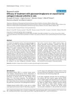
Figureof mutant cystic fibrosis transmembrane conductance regulator (CFTR) on cellular physiology
Impact 1
Impact of mutant cystic fibrosis transmembrane conductance regulator (CFTR) on cellular physiology. Mutant CFTR promotes
initial bacterial infection by upregulating epithelial cell adhesion molecules for bacteria such as asialo-GM1 and by decreasing
production of innate host defense molecules such as nitric oxide (NO). Defects in CFTR also lead to increased sodium absorption through the epithelial sodium channel (ENaC) and decreased chloride secretion. Water follows its concentration gradient
and results in decreased depth of airway surface liquid. Bacterial persistence is promoted by alterations in airway wall architecture, impaired host defense mechanisms, an excessive inflammatory response, and adaptations made by the bacteria to the
microenvironment of the cystic fibrosis airway.
assumes the mucoid phenotype [52]. This implies that the
progressive deterioration in lung function experienced by
a CF patient is due more to the long-term deleterious
effects of host inflammatory responses rather than direct
damage from the bacteria itself. Residence in the CF lung
also seems to alter the properties of P. aeruginosa lipopolysaccharide (LPS): LPS isolated from a large percentage of
CF patients has little or no O-side chain, conferring a
rough appearance to colonies when grown on agar plates.
Changes at this site in the molecule may have clinical
implications because complement fixation occurs on the
O-side chain. Furthermore, P. aeruginosa may synthesize
specific structures in the lipid-A moiety of its endotoxin,
which provoke increased host inflammatory responses
and resistance to antimicrobial peptides [53]. In addition
to the advantages conferred upon it by the appropriate
environmental conditions, P. aeruginosa itself possesses
special characteristics that allow it to persist in the lungs
of patients with CF, including the production of virulence
factors and the ability to organize into a biofilm.
Page 5 of 12
(page number not for citation purposes)
Respiratory Research 2003, 4
/>
Figure 2
Schematic Representation of the mucociliary escalator in the non-cystic fibrosis and cystic fibrosis (CF) airways
Schematic Representation of the mucociliary escalator in the non-cystic fibrosis and cystic fibrosis (CF) airways. In the non-CF
airway (Fig. 2A), where the depth of the periciliary fluid is normal, islands of mucus float on top and are propelled upward
toward the mouth by the coordinated beating of cilia. In the CF airway (Fig 2B), the mucus is poorly hydrated and hypoxic.
Because of the decreased depth of the periciliary fluid, the abnormal mucus is plastered down upon the cilia, thus inhibiting
normal ciliary beating. Eventually the bacteria present in the airway become trapped in the mucus and adapt to the local environment. In the case of P. aeruginosa, this includes production of mucoid exopolysaccharide (MEP) and organization into a
biofilm.
Numerous P. aeruginosa virulence factors contribute to its
pathogenicity in CF by altering the host's defenses. Pseudomonas elastase and alkaline protease are proteolytic
enzymes that may damage host tissues, disrupt tight junctions, and impair opsonophagocytosis [54]. Pseudomonas
elastase
degrades
immunoglobulins,
coagulation factors, complement components, cytokines,
and alpha proteinase inhibitor [55] and stimulates mucin
release from goblet cells [56], likely enhancing the already
increased production of mucin that occurs in the CF airway. Pseudomonas elastase is more potent than neu-
trophil elastase, on a per mg basis, with respect to elastin
degradation, and thus may contribute to CF lung pathology, even though the predominant elastase in CF sputum
is from neutrophils [57]. Exotoxin A promotes tissue
necrosis by inhibiting protein synthesis in eukaryotic cells
by a similar mechanism to that described for diphtheria
toxin. Exotoxin A catalyzes the transfer of the ADP-ribosyl
moiety of nicotinamide adenine dinucleotide onto elongation factor 2, which then is inactive in protein synthesis.
Exotoxin A also attracts neutrophils into the lungs of mice
[58]. Exoenzyme S is an ADP-ribosyltransferase that
Page 6 of 12
(page number not for citation purposes)
Respiratory Research 2003, 4
disrupts eukaryotic cell signal transduction, stimulates
actin reorganization, inhibits tissue regeneration, serves as
a potent T lymphocyte mitogen, maintains the site of
infection by promoting P. aeruginosa adhesion, and is
cytotoxic, especially to epithelial cells, [59–64]. Phospholipase C hydrolyzes lecithin, decreases the neutrophil's respiratory burst, and stimulates IL-8 release by
monocytes in vitro. It also induces local production of
tumor necrosis factor-alpha (TNF-α), IL-1β, interferongamma, macrophage inflammatory protein-1α, and macrophage inflammatory protein-2 in addition to stimulating neutrophil infiltration, thereby likely contributing to
the vigorous inflammatory response seen in the CF airway
[58]. Pigments such as pyocyanin bind iron, inhibit the
growth of other bacteria, and inhibit ciliary beat frequency [65,66]. Since P. aeruginosa virulence factors
increase with acute pulmonary exacerbations and
decrease after the administration of systemic antibiotics
[67,68], virulence factors may contribute, at least in part,
to acute deteriorations in lung function.
Recently, the ability of P. aeruginosa to organize into a biofilm has garnered much attention. Donlan and Costerton
define a biofilm as "a microbially derived sessile community characterized by cells that are irreversibly attached to
a substratum or interface or to each other, are embedded
in a matrix of extracellular polymeric substances that they
have produced, and exhibit an altered phenotype with
respect to growth rate and gene transcription" [69]. Biofilm formation protects bacteria from changes in environmental conditions, antibiotics, and host defenses, and
thus may consolidate the ability of the bacterium to persist in the airways of patients with CF. Bacteria within a
biofilm communicate with one other via a mechanism
known as quorum sensing, which also downregulates virulence factors, allowing the bacteria to live in symbiosis
with the host [70]. The altered phenotype of bacteria in a
biofilm may have clinical importance. Growth characteristics differ significantly for bacteria in a biofilm than for
those in the free-living, planktonic state. Antibiotic sensitivity testing performed on bacteria in the planktonic
state, as occurs in the clinical microbiology laboratory,
may not accurately reflect the true sensitivities of bacteria
in a biofilm [71]. This difference may account for the clinical efficacy of macrolide antibiotics that has been
described in CF [72,73]. Quorum sensing signals provide
a promising potential therapeutic target in CF.
No article on bacterial infections in CF would be complete
without at least mentioning B. cepacia. What was once
thought to be a single organism, "B cepacia" actually
includes several related organisms now known as B. cepacia complex [74,75]. Although infrequent pathogens in
CF, organisms of the B. cepacia complex often have major
clinical impact. The clinical course after acquisition of B.
/>
cepacia complex organisms spans the spectrum of no discernable clinical change to severe and rapidly progressive
respiratory failure, often associated with bacteremia and
death ("cepacia syndrome") [75]. Organisms of the B.
cepacia complex, especially the organisms implicated in
cepacia syndrome, have been proposed to provoke a more
robust host inflammatory response than P. aeruginosa
with respect to production of TNF-α by monocyte cell
lines in vitro, neutrophil recruitment, and priming of the
neutrophil respiratory burst [76,77]. However, this
increased inflammatory response has not been documented in CF patients [78].
Chronic endobronchial bacterial infection with one or
more typical organisms is the hallmark of CF lung disease.
The host inflammatory response in CF to the bacterial
infection dictates the clinical manifestations of the lung
disease. In general, CF patients experience a progressive
decline in pulmonary function that is punctuated by intermittent exacerbations, which are characterized by
increased cough, sputum production, anorexia, and
malaise. Antimicrobial therapy for CF bacterial infections,
especially for P. aeruginosa, frequently requires the administration of a combination of two or more antibiotics due
to the bacteria's ability to become resistant to a single
agent. Moreover, CF patients typically require greater than
normal antibiotic doses to penetrate the large endobronchial mucus sink and to counter the altered pharmacokinetics that occur in CF patients due to increased volume
of distribution from malnutrition and, for some drugs,
increased renal clearance [42]. Most CF patients return to
pre-exacerbation pulmonary function values after completing a course of parenteral antibiotics, but this is not
always the case. It is possible that the cumulative effects of
multiple pulmonary exacerbations contribute to the
decline in lung function and that those patients with more
frequent and/or severe exacerbations have shorter life
spans.
Why do patients with CF fail to clear bacterial
infection?
Excess inflammation provides an environment favorable to
bacterial growth
CF infants develop bacterial infection early, and respond
to it with a vigorous inflammatory response. Epithelial
cells respond to bacteria and their products by increasing
production of cytokines such as IL-6, IL-8, granulocyte
macrophage colony stimulating factor (GM-CSF), expression of intercellular adhesion molecule-1, and production
of mucins. IL-8, a potent chemokine, attracts neutrophils
to the inflammatory site, where their transepithelial passage is facilitated by intercellular adhesion molecule-1
and their survival prolonged by GM-CSF. When lung macrophages encounter bacteria, they respond not only by
producing their own IL-8, but also by producing TNF-α
Page 7 of 12
(page number not for citation purposes)
Respiratory Research 2003, 4
and IL-1β, which in turn can drive epithelial cell production of pro-inflammatory molecules by a signaling
pathway different from that accessed by the bacterial
products. Quickly, the airway recruits large numbers of
neutrophils, which early in the course of the infection, are
often able to contain the bacteria. Initial infections are frequently cleared, and colonization that is only intermittent
is common in the first few years of life.
The inflammatory process appears to go awry in the lungs
of patients with CF, even in infancy. Clinical studies indicate that, for a given lung bacterial burden, the neutrophil
and IL-8 responses of CF infants, measured in bronchoalveolar lavage fluid, are excessive compared to those of
normal infants. This is true whether all organisms recovered from the lung are considered, or whether analysis is
restricted only to infants whose cultures reveal only H.
influenzae [79,80]. Inflammation is in excess in CF even if
the neutrophil and IL-8 responses are adjusted for the
amount of endotoxin in the bronchoalveolar lavage. Initially, this response seems to contain the bacteria. Indeed,
in other, cross-sectional studies of inflammatory
responses in CF bronchoalveolar lavage fluid, many CF
infants have no detectable bacteria, but even some of
these infants have a modest inflammatory response,
which exceeds that observed in other, uninfected, non-CF
infants undergoing bronchoalveolar lavage [81]. However, other studies show that at least some CF infants, particularly those who have never had lung infection, have
no detectable inflammatory response [82]. The picture
emerges of a lung which, although initially pristine and
uninflamed, mounts an excessive inflammatory response
to bacterial stimulation, which continues to reverberate
even after the infection is controlled. Eventually, all the
factors, which serve to retain bacteria in the CF lung, overwhelm the defenses of the lung, even the phagocytic
defenses, and the bacterial signals for inflammation persist. At this point, the excessive inflammatory response
becomes deleterious and even promotes continuing
infection.
One striking feature of CF airways disease is the progressive accumulation of neutrophils over a period of years.
This "acute inflammation" never converts to a more
"chronic" pattern. Since neutrophils do not survive long
after exiting the circulation, there must be a persistent
stimulus to attract these neutrophils. There is certainly an
excess of chemoattractants such as IL-8 and leukotriene B4
recovered in bronchoalveolar lavage fluid [83,84]. Bacteria provide additional chemoattractants. The neutrophils
may survive longer in the airways of patients with CF
because of the production of excess GM-CSF and the relative lack of IL-10 [83,85,86], which, when present, promotes neutrophil apoptosis. When present in excess,
neutrophils and their products actually impair the host's
/>
ability to clear bacterial infection. Neutrophil elastase, in
particular, interacts with airway epithelial cells to promote
the transcription of IL-8 and macromolecular secretion,
further fueling airway inflammation and obstruction [87–
91]. Elastase cleaves IgG at the hinge region [92,93]. Since
macrophages use antibodies to ingest P. aeruginosa,
opsonophagocytosis is reduced in the presence of excess
elastase. On the other hand, neutrophils employ complement for opsonophagocytosis of P. aeruginosa. This system
consists of two receptors, CR-1 and CR-3 and two complement opsonins, C3b (ligand for CR-1) and C3bi (ligand
for CR-3). Elastase cleaves the CR1 receptor and the C3bi
ligand, so that neither of the opsonin-receptor pairs is left
intact [94,95]. Thus, all the usual mechanisms of ingestion of P. aeruginosa are crippled in the presence of free
elastase activity (Fig. 3). In one study of CF patients, all
patients above the age of one year, and many of those less
than one year of age, had excess neutrophil elastase activity in their bronchoalveolar lavage fluid [96]. Most
patients over 1 year of age have concentrations in excess of
1 µM. Since the opsonins and receptors are cleaved at concentrations of free elastase of 10-8 M, 1 µM is more than
sufficient to turn the vicious cycle of inflammation and
infection, and to destroy the fabric of the lung.
Structural damage to the lung allows for mechanical
retention of secretions and retention of bacteria
We have argued here that inflammation in the CF lung
occurs in excess compared to the response mounted by
non-CF individuals, and that it is ultimately ineffective
against the bacteria. The result of all this is that CF infants
develop bacterial infections very early in life. In the beginning, colonization may be intermittent, but eventually, it
becomes chronic. The special binding properties of P. aeruginosa, combined with its ubiquitous presence in our
environment and therefore regular exposure, and its particular ability to adjust quickly and perfectly to conditions
in the CF lung likely account for its predilection for the CF
lung. Eventually most patients with CF acquire this organism, develop a vigorous and persistent neutrophilic
inflammatory response, and settle into a vicious cycle of
airway obstruction, infection, and excess inflammation
that results in lung destruction, further damage to the
clearance processes, and additional vulnerability to infection or phenotypic transformation of the P. aeruginosa
into a biofilm, which is impossible to eradicate despite
the most vigorous antibiotic therapy.
The persistent bacterial stimulation of an overzealous
inflammatory response results in excess neutrophils and
neutrophil products in the airways. Many of the proteases
secreted by the neutrophil are capable of digesting the
structural proteins of the CF lung, including collagen and
elastin. Small breaks in the epithelial barrier expose these
structural proteins, and the normal antiprotease defenses
Page 8 of 12
(page number not for citation purposes)
Respiratory Research 2003, 4
/>
Figure
Adverse3effects of elastase on host defense mechanisms and inflammation
Adverse effects of elastase on host defense mechanisms and inflammation. In the cystic fibrosis airway, the concentration of
elastase exceeds the concentration of inhibitors of elastase by several hundred to several thousand fold. While the vast majority of elastase is produced by the neutrophil, a small but significant amount is derived from bacteria. In addition to causing
structural damage directly, elastase stimulates the production of pro-inflammatory mediators such as IL-8, which further
induces neutrophil influx. Elastase also impairs mucociliary clearance by direct effects on ciliary function and by stimulating
increased mucus production. Elastase inhibits opsonophagocytosis by cleaving the Fc portion of immunoglobulin G and complement receptors on both the neutrophil (CR1) and P. aeruginosa (C3bi), resulting in an opsonin-receptor mismatch.
are overwhelmed by the massive quantities of enzymes
released by the enormous neutrophil infiltration. Reactive
oxygen species are also potent agents of tissue damage.
The antioxidant defenses of the lung are markedly
reduced in CF, although the reasons for this are not
entirely clear. It has been speculated that CFTR transports
glutathione as well as chloride ion, and in the absence of
functional CFTR, less glutathione reaches the airway to
defend against oxidant damage. In any event, reduced oxidant defenses can be demonstrated even in the uninfected
airways of CF mice [97]. Another class of proteases is also
elevated in the bronchoalveolar lavage fluid of patients
with CF. Matrix metalloproteinases, implicated in remodeling of inflamed areas of the lung, are found in excess in
the lungs of patients with CF. These proteinases can be
produced by epithelial cells, and their transcription is activated by NF-κB. All of these damaging proteases and oxidants combine to destroy the supporting structures of the
airway and ultimately lead to bronchiectasis. Once the
fabric of the airway wall is compromised, outpouching of
the airway wall (saccular bronchiectasis) occurs. In these
damaged areas, pooling of secretions and failure of clear-
Page 9 of 12
(page number not for citation purposes)
Respiratory Research 2003, 4
ance is inevitable. It is rare that infection can be cleared
once such structural damage has occurred. In the later
stages of the disease, all of the complications of bronchiectasis of any cause emerge in patients with CF –
engorgement of the bronchial blood vessels with risk for
massive hemoptysis, persistent secretions and cough, and
persistent bacterial infection that is impossible to clear.
However, all of the features noted above that allow bacteria to be retained in the CF lung in the first place are still
present, and all of the abnormalities in signaling that
make for increased inflammatory responses are also in
play. Therefore, the progression of bronchiectasis in the
lungs of patients with CF tends to be more rapid than it is
in patients with bronchiectasis of other causes, such as
post-infectious bronchiectasis or bronchiectasis associated with primary ciliary dyskinesia. Many patients with
bronchiectasis of non-CF etiology survive well into adulthood or even old age, whereas such survival is rare in
patients with CF.
/>
Abbreviations
CF cystic fibrosis
CFTR cystic
regulator
fibrosis
transmembrane
conductance
ENaC epithelial sodium channel
GM-CSF granulocyte macrophage colony stimulating
factor
IL interleukin
LPS lipopolysaccharide
MEP mucoid exopolysaccharide
NF-κB nuclear factor-kappaB
NO nitric oxide
Summary and conclusions
The lungs of patients with CF are vulnerable to bacterial
infection, and once the infection becomes established, it
is not eradicated despite prolonged and vigorous antibiotic and airway clearance therapy. This aspect of the disease has long provided an inviting therapeutic target,
though it is essentially a rear guard action which delays
but does not prevent the progression of the lung disease.
Successful strategies to prevent the initial colonization,
assist in the clearance of initial infections, prevent the
adaptation of P. aeruginosa to the CF lung environment, or
even to limit the excess inflammatory response (although
not the response required to kill the bacteria), would have
great therapeutic benefit to patients with CF. Indeed, a
number of strategies have been proposed to interfere at
each of these steps: aerosolized dextrans to prevent pseudomonas adherence, intravenous IgG to assist in clearance, and many proposed anti-inflammatory treatments
to limit the excessive inflammation already have reached
clinical trial. Once infection has been established, lung
damage might be slowed by inhibiting the excess of oxidants in the CF airway or by inhibiting the proteolytic
damage to the structural proteins of the airways with antiproteases (or, at a more fundamental level, limiting the
access of the neutrophils to the airway). Drugs aimed at
these steps are also in development. Strategies directed at
the basic defect, if applied sufficiently early in the course
of the disease, might abort the entire process and provide
the best therapeutic result of all. Once structural damage
has occurred, however, bronchiectasis may take on a life
of its own, and even complete correction of the underlying genetic defect may not completely halt disease progression. For these patients, further development of the
means to control the inflammatory response and its consequences likely will be necessary.
NOS nitric oxide synthase
Stat signal transducer and activator of transcription
TNF-α tumor necrosis factor-alpha
References
1.
2.
3.
4.
5.
6.
7.
8.
9.
10.
11.
12.
Davis PB, Drumm ML and Konstan MW: State of the Art: Cystic
Fibrosis. Am J Resp Crit Care Med 1996, 154:1229-1256.
Lloyd-Still JD: Crohn's disease and cystic fibrosis. Dig Dis Sci
1994, 39:880-885.
Taylor CJ and Aswani N: The pancreas in cystic fibrosis. Paediatr
Respir Rev 2002, 3:77-81.
Pilewski JM and Frizzell RA: Role of CFTR in airway disease. Physiol Rev 1999, 79(1 Suppl):S215-S255.
Fang X, Fukuda N, Barbry P, Sartori C, Verkman AS and Matthay MA:
Novel role for CFTR in fluid absorption from the distal airspaces of the lung. J Gen Physiol 2002, 119:199-207.
Bubien JK: CFTR may play a role in regulated secretion by
lymphocytes: a new hypothesis for the pathophysiology of
cystic fibrosis. Pflugers Arch 2001, 443(Suppl 1):S36-S39.
Tarran R, Loewen ME, Paradiso AM, Olsen JC, Gray MA, Argent BE,
Boucher RC and Gabriel SE: Regulation of Murine Airway Surface Liquid Volume by CFTR and Ca(2+)-activated Cl(-)
Conductances. J Gen Physiol 2002, 120:407-418.
Greger R, Mall M, Bleich M, Ecke D, Warth R, Riedemann N and
Kunzelmann K: Regulation of epithelial ion channels by the
cystic fibrosis transmembrane conductance regulator. J Mol
Med 1996, 74:527-534.
Hummler E, Barker P, Gatzy J, Beermann F, Verdumo C, Schmidt A,
Boucher R and Rossier BC: Early death due to defective neonatal lung liquid clearance in alpha-ENaC-deficient mice. Nat
Genet 1996, 12:325-328.
Kunzelmann K, Schreiber R, Nitschke R and Mall M: Control of epithelial Na+ conductance by the cystic fibrosis transmembrane conductance regulator. Pflugers Arch 2000, 440:193-201.
Jiang Q, Li J, Dubroff R, Ahn YJ, Foskett JK, Engelhardt J and Kleyman
TR: Epithelial sodium channels regulate cystic fibrosis transmembrane conductance regulator chloride channels in
Xenopus oocytes. J Biol Chem 2000, 275:13266-13274.
Briel M, Greger R and Kunzelmann K: Cl-transport by cystic fibrosis transmembrane conductance regulator (CFTR) contributes to the inhibition of epithelial Na+ channels (ENaCs) in
Xenopus oocytes co-expressing CFTR and ENaC. J Physiol
1998, 508(Pt 3):825-836.
Page 10 of 12
(page number not for citation purposes)
Respiratory Research 2003, 4
13.
14.
15.
16.
17.
18.
19.
20.
21.
22.
23.
24.
25.
26.
27.
28.
29.
30.
31.
32.
33.
Stutts MJ, Rossier BC and Boucher RC: Cystic fibrosis transmembrane conductance regulator inverts protein kinase A-mediated regulation of epithelial sodium channel single channel
kinetics. J Biol Chem 1997, 272:14037-14040.
Zahm JM, Baconnais S, Davidson DJ, Webb S, Dorin J, Bonnet N, Balossier G and Puchelle E: X-ray microanalysis of airway surface
liquid collected in cystic fibrosis mice. Am J Physiol Lung Cell Mol
Physiol 2001, 281:L309-L313.
Jayaraman S, Joo NS, Reitz B, Wine JJ and Verkman AS: Submucosal
gland secretions in airways from cystic fibrosis patients have
normal [Na(+)] and pH but elevated viscosity. Proc Natl Acad
Sci U S A 2001, 98:8119-8123.
Jayaraman S, Song Y, Vetrivel L, Shankar L and Verkman AS: Noninvasive in vivo fluorescence measurement of airway-surface
liquid depth, salt concentration, and pH. J Clin Invest 2001,
107:317-324.
Matsui H, Davis CW, Tarran R and Boucher RC: Osmotic water
permeabilities of cultured, well-differentiated normal and
cystic fibrosis airway epithelia. J Clin Invest 2000, 105:1419-1427.
Matsui H, Grubb BR, Tarran R, Randell SH, Gatzy JT, Davis CW and
Boucher RC: Evidence for periciliary liquid layer depletion,
not abnormal ion composition, in the pathogenesis of cystic
fibrosis airways disease. Cell 1998, 95:1005-1015.
Kopito LE, Kosasky HJ and Shwachman H: Water and electrolytes
in cervical mucus from patients with cystic fibrosis. Fertil Steril
1973, 24:512-516.
King M: Experimental models for studying mucociliary
clearance. Eur Respir J 1998, 11:222-228.
Atsuta S and Majima Y: Nasal mucociliary clearance of chronic
sinusitis in relation to rheological properties of nasal mucus.
Ann Otol Rhinol Laryngol 1998, 107:47-51.
Sturgess J and Imrie J: Quantitative evaluation of the development of tracheal submucosal glands in infants with cystic
fibrosis and control infants. Am J Pathol 1982, 106:303-311.
Tirouvanziam R, de Bentzmann S, Hubeau C, Hinnrasky J, Jacquot J,
Peault B and Puchelle E: Inflammation and infection in naive
human cystic fibrosis airway grafts. Am J Respir Cell Mol Biol 2000,
23:121-127.
Tabary O, Zahm JM, Hinnrasky J, Couetil JP, Cornillet P, Guenounou
M, Gaillard D, Puchelle E and Jacquot J: Selective up-regulation of
chemokine IL-8 expression in cystic fibrosis bronchial gland
cells in vivo and in vitro. Am J Pathol 1998, 153:921-930.
Kelley TJ and Drumm ML: Inducible nitric oxide synthase
expression is reduced in cystic fibrosis murine and human
airway epithelial cells. J Clin Invest 1998, 102:1200-1207.
Kelley TJ and Elmer HL: In vivo alterations of IFN regulatory factor-1 and PIAS1 protein levels in cystic fibrosis epithelium. J
Clin Invest 2000, 106:403-410.
Pier GB, Grout M, Zaidi TS, Olsen JC, Johnson LG, Yankaskas JR and
Goldberg JB: Role of mutant CFTR in hypersusceptibility of
cystic fibrosis patients to lung infections. Science 1996,
271:64-67.
Saiman L and Prince A: Pseudomonas aeruginosa pili bind to
asialoGM1, which is increased on the surface of cystic fibrosis
epithelial cells. J Clin Invest 1993, 92:1875-1880.
Saiman L, Cacalano G, Gruenert D and Prince A: Comparison of
adherence of Pseudomonas aeruginosa to respiratory epithelial cells from cystic fibrosis patients and healthy subjects.
Infect Immun 1992, 60:2808-2814.
Baltimore RS, Christie CD and Smith GJ: Immunohistopathologic
localization of Pseudomonas aeruginosa in lungs from patients
with cystic fibrosis. Implications for the pathogenesis of progressive lung deterioration. Am Rev Respir Dis 1989,
140:1650-1661.
Ulrich M, Herbert S, Berger J, Bellon G, Louis D, Munker G and Doring G: Localization of Staphylococcus aureus in infected airways of patients with cystic fibrosis and in a cell culture
model of S. aureus adherence. Am J Respir Cell Mol Biol 1998,
19:83-91.
Sajjan U, Corey M, Humar A, Tullis E, Cutz E, Ackerley C and Forstner J: Immunolocalisation of Burkholderia cepacia in the lungs
of cystic fibrosis patients. J Med Microbiol 2001, 50:535-546.
Scheid P, Kempster L, Griesenbach U, Davies JC, Dewar A, Weber
PP, Colledge WH, Evans MJ, Geddes DM and Alton EW: Inflammation in cystic fibrosis airways: relationship to increased bacterial adherence. Eur Respir J 2001, 17:27-35.
/>
34.
35.
36.
37.
38.
39.
40.
41.
42.
43.
44.
45.
46.
47.
48.
49.
50.
51.
52.
53.
54.
55.
56.
DiMango E, Ratner AJ, Bryan R, Tabibi S and Prince A: Activation of
NF-kappaB by adherent Pseudomonas aeruginosa in normal
and cystic fibrosis respiratory epithelial cells. J Clin Invest 1998,
101:2598-2605.
DiMango E, Zar HJ, Bryan R and Prince A: Diverse Pseudomonas
aeruginosa gene products stimulate respiratory epithelial
cells to produce interleukin-8. J Clin Invest 1995, 96:2204-2210.
Arora SK, Ritchings BW, Almira EC, Lory S and Ramphal R: The
Pseudomonas aeruginosa flagellar cap protein, FliD, is responsible for mucin adhesion. Infect Immun 1998, 66:1000-1007.
Ramphal R, Arora SK and Ritchings BW: Recognition of mucin by
the adhesin-flagellar system of Pseudomonas aeruginosa. Am J
Respir Crit Care Med 1996, 154(4 Pt 2):S170-S174.
Feldman M, Bryan R, Rajan S, Scheffler L, Brunnert S, Tang H and
Prince A: Role of flagella in pathogenesis of Pseudomonas aeruginosa pulmonary infection. Infect Immun 1998, 66:43-51.
Bauernfeind A, Bertele RM, Harms K, Horl G, Jungwirth R, Petermuller C, Przyklenk B and Weisslein-Pfister C: Qualitative and
quantitative microbiological analysis of sputa of 102 patients
with cystic fibrosis. Infection 1987, 15:270-277.
Gilligan PH: Microbiology of airway disease in patients with
cystic fibrosis. Clin Microbiol Rev 1991, 4:35-51.
Rosenfeld M, Emerson J, Accurso F, Armstrong D, Castile R, Grimwood K, Hiatt P, McCoy K, McNamara S, Ramsey B and Wagener J:
Diagnostic accuracy of oropharyngeal cultures in infants and
young children with cystic fibrosis. Pediatr Pulmonol 1999,
28(2):321-328.
Pitt TL: Biology of Pseudomonas aeruginosa in relation to pulmonary infection in cystic fibrosis. J R Soc Med 1986, 79(Suppl
12):13-18.
Poole K: Multidrug efflux pumps and antimicrobial resistance
in Pseudomonas aeruginosa and related organisms. J Mol Microbiol Biotechnol 2001, 3:255-264.
Oliver A, Canton R, Campo P, Baquero F and Blazquez J: High Frequency of hypermutable Pseudomonas aeruginosa in cystic
fibrosis lung infection. Science 2000, 288:1251-1254.
Worlitzsch D, Tarran R, Ulrich M, Schwab U, Cekici A, Meyer KC,
Birrer P, Bellon G, Berger J, Weiss T, Botzenhart K, Yankaskas JR,
Randell S, Boucher RC and Doring G: Effects of reduced mucus
oxygen concentration in airway Pseudomonas infections of
cystic fibrosis patients. J Clin Invest 2002, 109:317-325.
Vasil ML: Pseudomonas aeruginosa : biology, mechanisms of
virulence, epidemiology. J Pediatr 1986, 108(5 Pt 2):800-805.
Pier GB and Elcock ME: Nonspecific immunoglobulin synthesis
and elevated IgG levels in rabbits immunized with mucoid
exopolysaccharide from cystic fibrosis isolates of Pseudomonas aeruginosa. J Immunol 1984, 133:734-739.
Pier GB, Takeda S, Grout M and Markham RB: Immune complexes
from immunized mice and infected cystic fibrosis patients
mediate murine and human T cell killing of hybridomas producing protective, opsonic antibody to Pseudomonas
aeruginosa. J Clin Invest 1993, 91:1079-1087.
Marshall BC and Carroll KC: Interaction between Pseudomonas
aeruginosa and host defenses in cystic fibrosis. Semin Respir
Infect 1991, 6:11-18.
Boyd A and Chakrabarty AM: Role of alginate lyase in cell
detachment of Pseudomonas aeruginosa. Appl Environ Microbiol
1994, 60:2355-2359.
Burke V, Robinson JO, Richardson CJ and Bundell CS: Longitudinal
studies of virulence factors of Pseudomonas aeruginosa in
cystic fibrosis. Pathology 1991, 23:145-148.
Demko CA, Byard PJ and Davis PB: Gender differences in cystic
fibrosis: Pseudomonas aeruginosa infection. J Clin Epidemiol 1995,
48:1041-1049.
Ernst RK, Yi EC, Guo L, Lim KB, Burns JL, Hackett M and Miller SI:
Specific lipopolysaccharide found in cystic fibrosis airway
Pseudomonas aeruginosa. Science 1999, 286:1561-1565.
Bainbridge T and Fick RB Jr: Functional importance of cystic
fibrosis immunoglobulin G fragments generated by Pseudomonas aeruginosa elastase. J Lab Clin Med 1989, 114:728-733.
Wretlind B and Pavlovskis OR: Pseudomonas aeruginosa elastase
and its role in pseudomonas infections. Rev Infect Dis 1983,
5(Suppl):S998-S1004.
Klinger JD, Tandler B, Liedtke CM and Boat TF: Proteinases of
Pseudomonas aeruginosa evoke mucin release by tracheal
epithelium. J Clin Invest 1984, 74:1669-1678.
Page 11 of 12
(page number not for citation purposes)
Respiratory Research 2003, 4
57.
58.
59.
60.
61.
62.
63.
64.
65.
66.
67.
68.
69.
70.
71.
72.
73.
74.
75.
76.
77.
78.
Williams JC, Lucas BJ, Knee C, Renzetti M and Donahue J: Acute
lung injury induced by Pseudomonas aeruginosa elastase in
hamsters. Exp Lung Res 1992, 18:155-171.
Wieland CW, Siegmund B, Senaldi G, Vasil ML, Dinarello CA and Fantuzzi G: Pulmonary inflammation induced by Pseudomonas
aeruginosa lipopolysaccharide, phospholipase C, and exotoxin A: role of interferon regulatory factor 1. Infect Immun
2002, 70:1352-1358.
Frank DW: The exoenzyme S regulon of Pseudomonas
aeruginosa. Mol Microbiol 1997, 26:621-629.
Bruno TF, Buser DE, Syme RM, Woods DE and Mody CH: Pseudomonas aeruginosa exoenzyme S is a mitogen but not a
superantigen for human T lymphocytes. Infect Immun 1998,
66:3072-3079.
Baker NR, Minor V, Deal C, Shahrabadi MS, Simpson DA and Woods
DE: Pseudomonas aeruginosa exoenzyme S is an adhesion.
Infect Immun 1991, 59:2859-2863.
Barbieri JT: Pseudomonas aeruginosa exoenzyme S, a bifunctional type-III secreted cytotoxin. Int J Med Microbiol 2000,
290:381-387.
Herard AL, Pierrot D, Hinnrasky J, Kaplan H, Sheppard D, Puchelle E
and Zahm JM: Fibronectin and its alpha 5 beta 1-integrin
receptor are involved in the wound-repair process of airway
epithelium. Am J Physiol 1996, 271:L726-733.
Krall R, Sun J, Pederson KJ and Barbieri JT: In vivo rho GTPaseactivating protein activity of Pseudomonas aeruginosa cytotoxin ExoS. Infect Immun 2002, 70:360-367.
Cox CD: Role of pyocyanin in the acquisition of iron from
transferrin. Infect Immun 1986, 52:263-270.
Wilson R, Pitt T, Taylor G, Watson D, MacDermot J, Sykes D, Roberts D and Cole P: Pyocyanin and 1-hydroxyphenazine produced by Pseudomonas aeruginosa inhibit the beating of
human respiratory cilia in vitro. J Clin Invest 1987, 79:221-229.
Grimwood K, Semple RA, Rabin HR, Sokol PA and Woods DE: Elevated exoenzyme expression by P. aeruginosa is correlated
with exacerbations of lung disease in cystic fibrosis. Pediatr
Pulmonol 1993, 15:135-139.
Jaffar-Bandjee MC, Lazdunski A, Bally M, Carrere J, Chazalette JP and
Galabert C: Production of elastase, exotoxin A and alkaline
protease in sputa during pulmonary exacerbation of cystic
fibrosis in patients chronically infected by Pseudomonas
aeruginosa. J Clin Microbiol 1995, 33:924-929.
Donlan RM and Costerton JW: Biofilms: survival mechanisms of
clinically relevant microorganisms. Clin Microbiol Rev 2002,
15:167-193.
Singh PK, Schaefer AL, Parsek MR, Moninger TO, Welsh MJ and
Greenberg EP: Quorum-sensing signals indicate that cystic
fibrosis lungs are infected with bacterial biofilms. Nature 2000,
407:762-764.
Drenkard E and Ausubel FM: Pseudomonas biofilm formation
and antibiotic resistance are linked to phenotypic variation.
Nature 2002, 416:740-743.
Equi A, Balfour-Lynn I, Bush A and Rosenthal M: Long term azithromycin in children with cystic fibrosis: a randomised, placebo-controlled crossover trial. Lancet 2002, 360:978.
Wolter J, Seeney S, Bell S, Bowler S, Masel P and McCormack J: Effect
of long term treatment with azithromycin on disease parameters in cystic fibrosis: a randomised trial. Thorax 2002,
57:212-216.
Coenye T, Vandamme P, Govan JR and LiPuma JJ: Taxonomy and
identification of the Burkholderia cepacia complex. J Clin
Microbiol 2001, 39:3427-3436.
LiPuma JJ: Burkholderia cepacia. Management issues and new
insights. Clin Chest Med 1998, 19:473-486.
Zughaier SM, Ryley HC and Jackson SK: Lipopolysaccharide (LPS)
from Burkholderia cepacia is more active than LPS from
Pseudomonas aeruginosa and Stenotrophomonas maltophilia in
stimulating tumor necrosis factor alpha from human
monocytes. Infect Immun 1999, 67:1505-1507.
Hughes JE, Stewart J, Barclay GR and Govan JR: Priming of neutrophil respiratory burst activity by lipopolysaccharide from
Burkholderia cepacia. Infect Immun 1997, 65:4281-4287.
Hendry J, Elborn JS, Nixon L, Shale DJ and Webb AK: Cystic fibrosis: Inflammatory response to infection with Burkholderia
cepacia and Pseudomonas aeruginosa. Eur Respir J 1999,
14:435-438.
/>
79.
80.
81.
82.
83.
84.
85.
86.
87.
88.
89.
90.
91.
92.
93.
94.
95.
96.
97.
Noah TL, Black HR, Cheng PW, Wood RE and Leigh MW: Nasal and
bronchoalveolar lavage fluid cytokines in early cystic fibrosis.
J Infect Dis 1997, 175:638-647.
Muhlebach MS, Stewart PW, Leigh MW and Noah TL: Quantitation
of inflammatory response to bacteria in young cystic fibrosis
and control patients. Am J Respir Crit Care Med 1999, 160:186-191.
Khan TZ, Wagener JS, Bost T, Martinez J, Accurso FJ and Riches
DWH: Early pulmonary inflammation in infants with cystic
fibrosis. Am J Respir Crit Care Med 1995, 151:1075-1082.
Armstrong DS, Grimwood K, Carlin JB, Carzino R, Gutierrez JP, Hull
J, Olinsky A, Phelan EM, Robertson CF and Phelan PD: Lower airway inflammation in infants and young children with cystic
fibrosis. Am J Respir Crit Care Med 1997, 156:1197-1204.
Bonfield TL, Panuska JR, Konstan MW, Hillard KA, Hillard JB, Ghnaim
H and Berger M: Inflammatory cytokines in cystic fibrosis
lungs. Am J Respir Crit Care Med 1995, 152:2111-2118.
Konstan MW, Walenga RW, Hilliard KA and Hilliard JB: Leukotriene B4 is markedly elevated in the epithelial lining fluid of
patients with cystic fibrosis. Am Rev Respir Dis 1993, 148:896-901.
Bonfield TL, Konstan MW, Burfeind P, Panuska JR, Hillard JB and
Berger M: Normal bronchial epithelial cells constitutively produce the anti-inflammatory cytokine interleukin-10, which is
downregulated in cystic fibrosis. Am J Respir Cell Mol Biol 1995,
13:257-261.
Bonfield TL, Konstan MW and Berger M: Altered respiratory epithelial cell cytokine production in cystic fibrosis. J Allergy Clin
Immunol 1999, 104:72-78.
McElvaney NG, Nakamura H, Birrer P, Hebert CA, Wong WL,
Alphonso M, Baker JB, Catalano MA and Crystal RG: Modulation of
airway inflammation in cystic fibrosis: In vivo suppression of
interleukin-8 levels on the respiratory epithelial surface by
aerosolization of recombinant secretory leukoprotease
inhibitor. J Clin Invest 1992, 90:1296-1301.
Nakamura H, Yoshimura K, McElvaney NG and Crystal RG: Neutrophil elastase in respiratory epithelial lining fluid of
individuals with cystic fibrosis induces interleukin-8 gene
expression in human bronchial epithelial cell line. J Clin Invest
1992, 89:1478-1484.
Sommerhoff CP, Nadel JA, Basbaum CB and Caughey GH: Neutrophil elastase and cathepsin G stimulate secretion from
cultured bovine airway gland serous cells. J Clin Invest 1990,
85:682-689.
Schuster A, Ueki I and Nadel JA: Neutrophil elastase stimulates
tracheal submucosa gland secretion that is inhibited by ICI
200,355. Am J Physiol 1992, 262:L86-L91.
Tegner H, Ohlsson K, Toremalm NG and von Mecklenbeurg C:
Effect of human leukocyte enzymes on tracheal mucosa and
mucociliary activity. Rhinology 1979, 17:199-206.
Tosi MF and Berger M: Functional differences between the 40
kDa and 50 to 70 kDa IgG Fc receptors on human neutrophils revealed by elastase treatment and antireceptor
antibodies. J Immunol 1988, 141:2097-2103.
Tosi MF and Zakem H: Surface expression of Fc gamma receptor III (CD 16) on chemoattractant-stimulated neutrophils is
determined by both surface shedding and translocation from
intracellular storage compartments. J Clin Invest 1992,
90:462-470.
Tosi MF, Zakem H and Berger M: Neutrophil elastase cleaves
C3bi on opsonized pseudomonas as well as CR1 on neutrophils to create a functionally important opsonin receptor
mismatch. J Clin Invest 1990, 86:300-308.
Berger M, Sorensen RU, Tosi MF, Dearborn DG and Doring G:
Complement receptor expression on neutrophils at an
inflammatory site, the pseudomonas-infected lung in cystic
fibrosis. J Clin Invest 1989, 84:1302-1313.
Birrer P, McElvaney NG, Rudeberg A, Sommer CW, Liechti-Gallati S,
Kraemer R, Hubbard R and Crystal RG: Protease-antiprotease
imbalance in the lungs of children with cystic fibrosis. Am J
Respir Crit Care Med 1994, 150:207-213.
Velsor LW, van Heeckeren A and Day BJ: Antioxidant imbalance
in the lungs of cystic fibrosis transmembrane conductance
regulator protein mutant mice. Am J Physiol Lung Cell Mol Physiol
2001, 281:L31-L38.
Page 12 of 12
(page number not for citation purposes)