Báo cáo y học: " Early steps of retrovirus replicative cycle" potx
Bạn đang xem bản rút gọn của tài liệu. Xem và tải ngay bản đầy đủ của tài liệu tại đây (1.17 MB, 20 trang )
BioMed Central
Page 1 of 20
(page number not for citation purposes)
Retrovirology
Open Access
Review
Early steps of retrovirus replicative cycle
Sébastien Nisole
2
and Ali Saïb*
1
Address:
1
CNRS UPR9051, Hôpital Saint-Louis, 1 Avenue Claude Vellefaux, 75475 Paris cedex 10, France and
2
Division of Virology, National
Institute for Medical Research, The Ridgeway, Mill Hill, London NW7 1AA, United Kingdom
Email: Sébastien Nisole - ; Ali Saïb* -
* Corresponding author
Abstract
During the last two decades, the profusion of HIV research due to the urge to identify new
therapeutic targets has led to a wealth of information on the retroviral replication cycle. However,
while the late stages of the retrovirus life cycle, consisting of virus replication and egress, have been
partly unraveled, the early steps remain largely enigmatic. These early steps consist of a long and
perilous journey from the cell surface to the nucleus where the proviral DNA integrates into the
host genome. Retroviral particles must bind specifically to their target cells, cross the plasma
membrane, reverse-transcribe their RNA genome, while uncoating the cores, find their way to the
nuclear membrane and penetrate into the nucleus to finally dock and integrate into the cellular
genome. Along this journey, retroviruses hijack the cellular machinery, while at the same time
counteracting cellular defenses. Elucidating these mechanisms and identifying which cellular factors
are exploited by the retroviruses and which hinder their life cycle, will certainly lead to the
discovery of new ways to inhibit viral replication and to improve retroviral vectors for gene
transfer. Finally, as proven by many examples in the past, progresses in retrovirology will
undoubtedly also provide some priceless insights into cell biology.
Introduction
The life cycle of retroviruses is arbitrarily divided into two
distinct phases: the early phase refers to the steps of infec-
tion from cell binding to the integration of the viral cDNA
into the cell genome, whereas the late phase begins with
the expression of viral genes and continues through to the
release and maturation of progeny virions (see Figure 1 for
a schematic view of the retroviral life cycle). During the
long journey from the cell surface to the nucleus, retrovi-
ruses will face multiple obstacles, since in addition to
finding a path through the cytoplasm to the nucleus they
have to cross two main barriers, the plasma and nuclear
membranes, whilst at the same time avoiding or counter-
acting cellular defences that can interfere with many of
these steps. The surge in Human Immunodeficiency Virus
(HIV) research in order to identify new therapeutic targets
has led to a better understanding of the retroviral life
cycle. However, in comparison with the later events of ret-
rovirus infection (for a review, see [1,2]), early steps are
still poorly understood (for reviews, see [3,4]).
In the case of HIV entry, for example, while the mecha-
nisms of receptor binding, conformational changes and
fusion appear to be relatively well defined, the involve-
ment of attachment molecules and the importance of
lipid rafts in fusion or in recruitment of coreceptors
remain uncertain. Similarly, though the molecular proc-
ess of reverse transcription is well described, very little is
known about the concurrent uncoating process. One of
the most poorly understood steps is the trafficking of pre-
integration complexes (PICs) from the cell surface to the
vicinity of the nucleus, despite a growing body of
Published: 14 May 2004
Retrovirology 2004, 1:9
Received: 06 March 2004
Accepted: 14 May 2004
This article is available from: />© 2004 Nisole and Saïb; licensee BioMed Central Ltd. This is an Open Access article: verbatim copying and redistribution of this article are permitted in all
media for any purpose, provided this notice is preserved along with the article's original URL.
Retrovirology 2004, 1 />Page 2 of 20
(page number not for citation purposes)
knowledge arising from the study of other viral models
such as adenoviruses (Ad) [5] or Herpes simplex viruses
(HSV) [6]. Much has been learned regarding nuclear
entry, but the cellular proteins involved are still unknown
and the exact role of each viral component remains con-
troversial [7]. Finally, the molecular mechanisms of inte-
gration, the last event of the early phase of retroviral life
cycle, are now well understood, but the choice of target
site remains mysterious. Thus, while certain of these steps
have been characterized, we are still far from obtaining a
complete picture of these processes.
Fully elucidating the early steps of retrovirus replication is
therefore crucial not only for identifying new antiretrovi-
ral drugs, but also for improving the design of retroviral
vectors for gene therapy. Cellular inhibitors that interfere
with these steps can represent useful tools for better char-
acterizing the molecular processes involved and, in this
respect, the recent discovery of cellular factors that block
the lentiviral cycle at an early stage in primates provides
novel directions for AIDS research [8].
In this review, we will summarise our current understand-
ing of the early steps of the retroviral cycle, focussing par-
ticularly on the most recent and controversial findings in
the field.
Binding
The initial step of the retroviral replicative cycle is the
adsorption of viral particles to the surface of their target
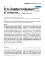
cells (see morphology of different retroviral particles on
Figure 2). It remains unclear whether this binding occurs
through specific interactions, but it is thought that such
attachment usually involves molecules which are distinct
The retroviral life cycleFigure 1
The retroviral life cycle. A schematic view of early and late stages of the retroviral replication cycle is represented. Exam-
ples of cellular factors interfering with early steps are indicated: Lv1/Ref1; CEM15, also known as APOBEC3G (apolipoprotein
B mRNA-editing enzyme-catalytic polypeptide-like-3G) ; Fv2; Fv1. The question marks indicates the exact step affected by the
restriction factors has not precisely been determined. Lv1 and Ref1 block incoming particles before reverse-transcription
whereas Fv1 and Fv2 act at a stage between reverse-transcription and integration. See text for detailed discussion. Abbrevia-
tions: RTC, reverse transcription complex; PIC, pre-integration complex.
Retrovirology 2004, 1 />Page 3 of 20
(page number not for citation purposes)
from the viral receptor responsible for the entry process
[9]. For example, the initial binding of Murine Leukemia
Virus (MLV) does not involve a specific interaction
between the envelope glycoprotein (Env) and the receptor
that is required for viral entry [10]. Furthermore, whereas
HIV entry into target cells involves CD4 and a coreceptor
(see below), the early attachment of virions to the cell sur-
face has been attributed to a variety of cell-surface mole-
cules (for a review, see [11]), including heparan sulfate
proteoglycan [12], LFA-1 [13] and nucleolin [14]. As the
affinity of HIV envelope glycoproteins for CD4 is rela-
tively low, especially in the case of primary virus isolates
[15], the existence of other attachment factors may serve
to concentrate the virus on the target cell surface prior to
specific receptor engagement. Indeed, the attachment of
virions to the cell surface appears to be the rate-limiting
step of HIV-1 entry [16]. Heparan sulfates (HS) are highly
sulfated polysaccharides, widely expressed on the surface
of cells and which have been shown to be utilized as cell
surface attachment factors by numerous viruses, bacteria
and parasites (for a review, see [17]). Among retroviruses,
they are believed to be implicated in the attachment of
Human T Cell Leukemia Virus (HTLV) [18], MLV [19] and
HIV-1 [12] to their target cells. However, although the
involvement of HS in HIV-1 attachment has been widely
documented, its exact role remains somewhat controver-
sial (reviewed in [20]). It is interesting to note that even
retrovirus-like particles lacking envelope proteins are able
to bind cells via interactions with HS [21], confirming that
the initial attachment of retroviruses to cells is, at least to
a certain extent, Env-independent. However, it is known
that Env-independent and/or receptor-independent bind-
ing of HIV leads to the endocytosis of particles, which is a
dead end with respect to cell infection [22,23].
HIV-1, HIV-2 and Simian Immunodeficiency Virus (SIV)
are known to bind the surface of dendritic cells through
interaction of their envelope glycoproteins with the C-
type mannose binding lectins DC-SIGN (Dendritic cell-
specific intercellular adhesion molecule 3-grabbing non-
integrin) and DC-SIGNR (DC-SIGN related) [24,25].
These molecules cannot be considered as receptors since
they do not promote viral entry leading to productive
infection. Instead, they allow DC to bind and capture viral
particles and should therefore be considered as efficient
binding factors. In the case of HIV-1, it seems that high
mannose structures on gp120 are recognized by DC-SIGN
[26-28], but there may also be a direct interaction
between the two proteins [29]. This interaction allows
HIV particles to use DC as a Trojan horse. Indeed, DCs are
Morphology of budding and mature particles from various retrovirusesFigure 2
Morphology of budding and mature particles from various retroviruses. Electron micrographs of retroviral particles
budding from infected cells (top panel) and of particles after the protease-mediated maturation (bottom panel). Abbreviations:
MLV, murine leukemia virus; HTLV, human T cell leukemia virus ; HIV, human immunodeficiency virus; FV, foamy virus. Note
that FV capsid assembly occurs in the cytoplasm similar to B/D typre retroviruses.
Retrovirology 2004, 1 />Page 4 of 20
(page number not for citation purposes)
thought to capture virions at peripheral sites of infection
and carry them to the lymph nodes, so promoting
efficient infection in trans of target cells expressing appro-
priate entry receptors [24,25]. But the involvement of den-
dritic cells in lentivirus pathogenesis may be more
complex, since various DC subsets express distinct arrays
of receptors capable of binding HIV gp120 [30].
Interestingly, this strategy seems to be shared by many
other viruses (for a recent review, see [31]) and even by
non-viral pathogens such as Mycobacterium tuberculosis
[32].
Entry
Following the initial step of binding, retroviral particles
use cell-surface proteins as specific receptors to enter their
target cells through interactions with the viral envelope
glycoproteins. As illustrated by the growing list of recep-
tors identified, retroviruses are able to utilize a variety of
cellular proteins to initiate infection, such as the amino-
acid transporter CAT-1 for ecotropic MLV [33,34], the T-
cell surface marker CD4 for HIV [35], the glucose trans-
porter GLUT-1 for HTLV [36] or the phosphate transport-
ers PIT-1 and PIT-2 used by Gibbon ape Leukemia Virus
(GaLV) [37] and amphotropic MLV [38,39], respectively.
In the case of Foamy viruses (FVs), although the receptor
is still unknown, it appears to be ubiquitous since these
retroviruses can infect a very wide range of cell lines,
although CD4+ and CD8+ lymphocytes appear to be the
main in vivo reservoirs [40-42].
Retroviral entry is a complex multi-step mechanism that
has been particularly well studied for HIV. Firstly, the
envelope glycoprotein gp120, present on the surface of
viral particles as gp41/gp120 trimers, recognises the pri-
mary receptor CD4. This interaction leads to conforma-
tional changes in both CD4 and gp120 and to the
recruitment of coreceptors belonging to the chemokine
receptor family, mainly CXCR4 and CCR5 (for a review,
see [43]). A second interaction then takes place between
gp120 and one of these coreceptors, which triggers new
conformational shifts in the envelope glycoproteins [44].
These sequential conformational changes finally lead to
the dissociation of gp120 from gp41, and to the transition
of gp41 to its fusogenic conformation. Entry of virions
into the cell is achieved by insertion of the gp41 fusion
peptide into the target membrane, resulting in the fusion
of viral and cellular membranes and the release of the
viral core in the cytoplasm (for recent reviews, see
[45,46]).
Although it has been suspected for some time that galac-
tosyl ceramide (GalCer) may be used by HIV-1 as an alter-
native receptor to infect neural cells [47], until recently
little else was known about the role of lipids in retroviral
entry. The discovery that lipids are distributed heterogene-
ously within cell membranes has led to the proposal that
sphingolipids and cholesterol tend to segregate into
microdomains called lipid rafts [48]. Several observations
support the hypothesis that lipid rafts may be involved in
the HIV entry process. Firstly, binding of HIV-1 to CD4
has been reported to result in a direct interaction between
gp120 and certain glycosphingolipids in membrane
microdomains [49]. Furthermore, disruption of target cell
membrane rafts by cholesterol depletion prevents HIV-1
infection [50], as does targeting CD4 to non-raft mem-
brane domains [51]. Finally, binding of virus to permis-
sive cells induces the clustering of CD4, CXCR4 and CCR5
within lipid-rafts [50,52,53]. Despite these lines of evi-
dence, the contribution of lipid rafts to HIV entry remains
controversial, as some studies have shown that the locali-
zation of CD4 and CCR5 to non-raft membrane domains
may not prevent HIV entry [54,55]. Interestingly, mem-
brane microdomains also seem to be involved in late
events of the retroviral cycle, since HIV-1 particles have
been found to bud preferentially through raft microdo-
mains of the plasma membrane [56]. This explains the
unusually high cholesterol and sphingomyelin content of
HIV membranes [57], a composition that is thought to be
important for fusion, since cholesterol-depleted virions
fail to enter cells [58].
Most retroviruses, including HIV, enter target cells by
direct fusion with the plasma membrane, as indicated by
their resistance to drugs blocking the acidification of
endosomes [59]. Interestingly, although HIV entry is
strictly pH independent, the majority of viral particles that
bind to the cell surface enters by endocytosis [22]. It seems
that a balance exists between these two entry pathways of
HIV-1 into T-lymphocytes, since the inhibition of one
route increases entry of particles by the alternative mech-
anism [23]. However, particles entering by endocytosis do
not support productive infection as they are degraded by
the proteasome [60], a conclusion supported by the
observation that inhibition of endosomal/lysosomal deg-
radation increases the infectivity of HIV-1 [61]. The only
known exceptions in the retrovirus family are ecotropic
and amphotropic MLV [62], and FVs [63], which seem to
enter target cells by endocytosis, although in the case of
FVs, the possibility of entry by direct fusion cannot be
excluded. However, the route of penetration into the cyto-
plasm can depend of the type of cell being infected.
Indeed, whereas the ecotropic MLV enters mouse NIH 3T3
cells by endocytosis, its entry into rat XC cells occurs by
fusion at the cell surface [64]. It is interesting to note that
the involvement of pH in retroviral entry has been recon-
sidered, since the distinction between pH-dependence
and independence has been shown to be more relative
than initially thought. Indeed, while the entry mechanism
of avian leukosis viruses (ALV) has originally been classi-
fied as pH-independent in comparison to influenza virus
Retrovirology 2004, 1 />Page 5 of 20
(page number not for citation purposes)
(for a review, see [65]), it has been shown to involve a low
pH step [66]. In contrast to influenza virus, it is the
interaction of ALV with its receptor that converts the enve-
lope glycoprotein to a pH-sensitive form, capable of pro-
moting fusion at low pH [66].
Finally, in the case of lentiviruses, there are some exam-
ples of direct infection from cell to cell. This is the case of
dendritic cells which can transmit HIV particles to T-cells
by direct contact without themselves being infected
[25,67,68]. The fact that most of the infectious HIV pro-
duced by primary macrophages is assembled on late
endocytic membranes rather than at the plasma mem-
brane suggests that a direct transmission of virions from
infected macrophages to T-cells during antigen presenta-
tion could also occur [69].
Uncoating and reverse transcription
The fusion of viral and cellular membranes delivers the
viral core into the cytoplasm, where the viral RNA is
reverse transcribed by the virion-packaged reverse tran-
scriptase (RT), generating a linear double-stranded DNA
molecule (for a review, see [70]). Although there is evi-
dence for limited DNA synthesis in virions prior to infec-
tion [71-73], reverse transcription usually occurs after the
release of the viral core into the cytoplasm of the target
cell. The only exceptions are FVs, which also reverse tran-
scribe their RNA during a late stage of their life cycle [74-
76]. Although unique among retroviruses, this feature is
shared with Hepadnaviruses, a viral family that has many
other similarities with FVs (for a review see [77]). The trig-
ger for the initiation of reverse transcription is not clearly
understood, but exposure of the incoming viral ribonucle-
oprotein complex to a significant concentration of deox-
yribonucleotides in the cytoplasm is thought to play an
important role (for a review, see [4]).
Immediately after its release into the cytoplasm, the viral
core undergoes a partial and progressive disassembly,
known as uncoating, that leads to the generation of subvi-
ral particles called reverse-transcription complexes (RTCs)
and pre-integration complexes (PICs). It seems that initi-
ation of reverse transcription is coupled to the onset
uncoating of the viral core [78]. It should be noted that
the distinction between RTCs and PICs is somewhat arbi-
trary, since uncoating is believed to occur progressively,
but PICs are usually defined as the integration-competent
complexes, whereas reverse-transcription is incomplete in
RTCs [79]. Attempts to define the composition of RTCs
and/or PICs have not yielded a clear answer, since the
nature of the viral and cellular components found to be
associated with the viral genome depends on the tech-
nique used for purifying the complexes, which are very
sensitive to detergents. Furthermore, it is known that the
vast majority of viruses entering a cell will not lead to a
productive infection, meaning that purified complexes
may not necessarily represent those particles able to per-
form reverse-transcription, nuclear import or integration.
Indeed, in the case of HIV-1, it has been reported that the
infectivity to particle ratio is as low as 1 in 60,000 [80,81],
even if some mathematical analyses tend to prove that
more than 10% of particles in a viral stock is theoretically
able to infect cells [82].
As a result of these practical restraints, it is still unclear
which proteins remain associated with the viral genome
in the RTCs/PICs. For HIV, RTCs have been shown to asso-
ciate rapidly with the host cytoskeleton after infection,
possibly through a direct interaction between the matrix
protein and the actin network [83]. They appear as large
nucleoprotein structures by electron microscopy and have
a sedimentation velocity of approximately 350 S and a
density of 1.34 g/ml in equilibrium gradients [84,85].
While most studies show that HIV PICs contain protease
(PR), reverse-transcriptase (RT), integrase (IN) and Vpr,
the presence of the structural proteins is more controver-
sial. The capsid proteins (CA) are thought to be released
soon after infection and only trace amounts are found in
PICs. Whereas nucleocapsid (NC) and matrix (MA) were
initially thought to be associated with PICs [86,87], more
recent studies revealed that the majority of these proteins
are lost during the uncoating process [85]. Interestingly, as
some viral structural components are released, certain cel-
lular proteins associate with the PICs during their journey
to the nucleus, such as the high mobility group protein
HMG I(Y), which has been proposed to be important for
integration [88].
It seems that the MLV core persists longer than that of HIV
since NC, MA and CA can all be detected in structures at
the vicinity of the nuclear membrane by electron micros-
copy [89]. However, whereas NC and IN can be detected
in the nucleus, MA and CA were found only in the cyto-
plasm [89,90]. Similarly, in the case of FVs, electron
microscopy studies revealed that incoming capsids seem
to retain an intact structure during their journey from the
cell surface to the microtubule-organizing centre (MTOC)
[91]. Interestingly, FV capsids were never detected either
within the nucleus, or close to nuclear pores, even later
during the replication cycle, whereas unassembled Gag
proteins and the viral genome are detected in the nucleus
early after infection [92]. Therefore, in contrast to viruses
such as Adenovirus type 2 (Ad2) or Herpes Simplex Virus
type 1 (HSV-1), whose capsids dock to the nuclear pore
triggering nuclear translocation of the viral genome [93-
95], nuclear import of FV Gag and genome must be
accompanied by disassembly or significant deformation
of the core particle at the MTOC.
Retrovirology 2004, 1 />Page 6 of 20
(page number not for citation purposes)
Some viral and cellular proteins appear to influence the
uncoating and/or the reverse-transcription of retroviruses.
This has been exemplified by HIV-1 Nef and Vif and the
cellular protein cyclophilin A. These three proteins,
present in incoming virions by virtue of their association
with the viral core, have been shown to modulate early
events of the replicative cycle of HIV, but their mode of
action is still unclear. Indeed, viral particles lacking one of
these proteins are less infectious than wild-type and this
defect seems to occur early in the viral cycle. Nef-defective
viruses for example display a strong decrease in infectivity
[96-98]. Since it does not appear to alter virion binding or
entry but does enhance viral DNA synthesis, Nef has been
proposed to act either at the level of viral uncoating or
reverse transcription [99,100]. Nef appears likely to mod-
ulate viral entry only when it occurs by fusion at the
plasma membrane [101], as HIV-1 virions pseudotyped
with the amphotropic MLV envelope [100,102], but not
with the envelope glycoprotein from the vesicular stoma-
titis virus (VSV-G) [100] display Nef-mediated enhance-
ment of infectivity membrane. This mechanism,
dependent on the route used by the virus to enter its target
cell, may be related to the high content of cholesterol
present in the viral particle membrane [57]. Indeed, it has
been proposed that Nef may enhance viral infectivity by
increasing the synthesis and incorporation of cholesterol
into progeny virions [103].
Vif, another HIV-1 accessory protein known to be incor-
porated into virions, also seems to play a role in an early
step of the HIV replicative cycle, as ∆-Vif viruses are unable
to complete viral DNA synthesis [104] and their RTCs are
less stable than wild-type viruses [105]. These observa-
tions may now be explained by recent studies. Indeed, Vif
has been shown to counteract the antiviral activity of
CEM15/APOBEC3G by preventing its incorporation into
progeny virions [106-110]. The fact that this cellular pro-
tein inhibits HIV replication at the step of reverse-tran-
scription is consistent with the observed phenotype of ∆-
Vif viruses. This latter will be discussed in more detail
below.
Finally, the cellular protein cyclophilin A (CypA), which
is incorporated into virions through its interaction with
viral capsid [111-113], has been shown to play a critical
role in the correct disassembly of the HIV-1 cores early
after infection [114], since particles lacking CypA display
a defect between entry and reverse-transcription. How-
ever, these observations are probably due to the failure of
CA to bind CypA rather than the absence of the cellular
protein in the virions. Indeed, some data suggest that
CypA incorporation into virions is dispensable, since
CypA can associate with the CA of incoming particles
within the target cells [115]. CypA is believed to protect
the viral capsid from the human restriction factor Ref1,
leading to an increase in HIV-1 infectivity [115]. The
mechanism of Ref1 restriction will be discussed below.
Additionally, it should be noted that early expression of
viral genes from unintegrated viral cDNA has also been
described [116-120]. Although the role of this early
expression is not clear, it is enhanced in the presence of
Vpr [121].
Trafficking of incoming viruses through the
cytoplasm
After penetration into the host cell, pathogens have to
reach their sites of replication, the nucleus in the case of
retroviruses. The cytoplasm, containing a high protein
concentration in addition to organelles and the cytoskele-
ton, constitutes a medium in which incoming particles
cannot rely on simple passive diffusion to move. Conse-
quently, viruses have evolved numerous and specific
mechanisms to hijack cellular machinery, and in particu-
lar the cytoskeleton, to facilitate their spread within the
infected cells, [122]. For example, microtubules (MT) are
essential for HSV-1 [6] and Ad [5] to reach the nucleus of
the infected cells, while vaccinia virus exploits first the
microtubule network for its intracellular movement
[123], and then the actin cytoskeleton to enhance its cell-
to-cell spread [124].
Initial studies have revealed that the use of specific drugs
altering the integrity of the cytoskeleton can interfere with
the retroviral cycle, either by directly affecting the intracel-
lular trafficking of incoming viruses or by interfering with
other steps of the early phase of infection such as reverse
transcription. Indeed, it has been shown that an intact
actin cytoskeleton is essential for efficient reverse tran-
scription of HIV-1 [83]. Additional reports have described
specific interactions between retroviral proteins and
cytoskeleton components. For example, HIV-1 IN and NC
have been shown to interact with yeast microtubule-asso-
ciated proteins [125], and actin [126-128], respectively,
but the precise role of such interactions in intracellular
trafficking of incoming viruses remains to be elucidated.
In contrast, several reports have described the effect of ret-
roviral proteins on the cytoskeleton, which might assist
viral replication. This is exemplified by the effect of the
HIV-1 Rev and Vpr proteins on the polymerisation of the
microtubule network [129] or on the nuclear membrane
(see below), respectively, or the ability of Vif to alter the
structure of vimentin network [130]. But once again, a
direct link between these observations and intracellular
trafficking remains to be clarified. Interestingly, the micro-
tubule network has been reported to be implicated in the
intracellular trafficking of incoming retroviruses. Such
movement has been demonstrated for incoming FVs
which target the microtubule organizing centre (MTOC)
prior to nuclear translocation. Centrosomal targeting of
Retrovirology 2004, 1 />Page 7 of 20
(page number not for citation purposes)
incoming viral proteins and subsequent viral replication
were inhibited by a treatment with nocodazole, demon-
strating the involvement of the MT network in
intracellular trafficking [92]. Remarkably, the Gag protein
by itself can target the MTOC in transfected cells through
interaction with the cytoplasmic light chain 8 (LC8) of the
minus-end directed MT motor dynein [91]. A similar role
for LC8 has been described for ASFV (African Swine Fever
Virus) and rabies virus, two other viruses which use the
MT network to move within infected cells [131-134].
Interestingly, this evolutionarily conserved molecule has
been shown to interact with numerous cellular complexes
such as nitric oxide synthase, or myosin V, an actin-based
motor mainly located at the plasma membrane which
shuttles between the cell periphery and the MTOC along
the MT network (for a review, see [135]). Therefore, inter-
action between incoming retroviral capsids and the multi-
functional LC8 could provide a bridge to shuttle between
an actin-based motor beneath the plasma membrane and
the MT network within the cytoplasm. Remarkably,
McDonald and al. have observed the migration of HIV-1
particles along MT toward the centrosome by following
GFP-tagged viral particles in the cytoplasm of infected
cells. [79]. A MT-dependent movement of retroviral Gag
proteins from the MTOC has also been described during
late stages of the life cycle for HTLV-I [136], the Mason
Pfizer Monkey virus [137,138] and also intracisternal type
A particles [139,140]. Although the viral and cellular pro-
tagonists involved in this transport were not determined,
these observations suggest that distinct classes of retroele-
ments may use the dynein-dynactin complex motor on
the MT network to make their way to or from the nucleus,
through the cytoplasm.
Nuclear entry
The retroviral life cycle requires the integration of the viral
DNA into the host cell genome to form the so-called pro-
virus. To achieve this, the reverse-transcribed DNA associ-
ated with viral proteins to form PICs, must enter the
nucleus (for a review, see [7]). PICs from most retrovi-
ruses are unable to enter intact nuclei and must therefore
"wait" for the breakdown of the nuclear membrane occur-
ring during mitosis [141,142]. Consequently, these retro-
viruses, such as MLV, are dependent on the cell cycle and
cannot replicate in non-dividing cells. In contrast, lentivi-
ruses such as HIV-1 are able to productively infect non-
dividing cells [143], such as macrophages or quiescent T
lymphocytes, indicating that PICs are able to actively cross
the nuclear membrane [144]. Some other retroviruses
seem to have an intermediate capacity to enter the
nucleus, since the PICs of Rous sarcoma virus [145] and
FVs [92,146] are able to penetrate intact nuclei with a low
efficiency, but their replication is dramatically increased
in dividing cells. HIV PICs, composed of the double-
stranded linear DNA associated with the viral proteins
MA, RT, IN and Vpr, have a estimated Stokes diameter of
56 nm [86]. Since the central channel of the nuclear pore
has a maximum diameter of 25 nm and the pore is known
to be able to transport macromolecules up to 39 nm
[147], HIV has developed a strategy to achieve the chal-
lenge of passing through these structures.
Nuclear pore complexes (NPCs) are large supramolecular
protein structures that span the nuclear membrane and
protrude into both cytoplasm and nucleoplasm (for a
recent review, see [148]). Signal-mediated nuclear import
involves the interaction of nuclear localization signals
(NLS) in proteins with nucleocytoplasmic shuttling recep-
tors, belonging to the karyopherin β family, also known as
importins. NLSs are typically short stretches of amino
acids, the best studied of which are basic amino acid-rich
sequences that interact with the receptor importin β,
either directly or through the adapter importin α [148].
Importin β interacts with other classes of NLS using differ-
ent adapters, including snurportin, RIP (for Rev interact-
ing protein), and importin 7. This latter has recently been
proposed to play a key role in nuclear import of HIV-1
PICs in primary macrophages [84]. Four different viral
components have been identified to contribute to the
nuclear import of HIV-1. Among the constituents that are
believed to form the PIC, IN, MA, Vpr and the viral DNA
are suspected to play a significant role in this complex
process, either directly or indirectly, although the exact
function of each remains to be fully understood (for
reviews, see [7,149]).
Integrase has been considered to be the main mediator of
HIV-1 nuclear translocation for some time, but its exact
implication is now being re-evaluated. This viral protein,
which harbours a non-classical NLS, has been shown to
be both necessary and sufficient to promote the nuclear
accumulation of viral PICs [150,151]. The nature of the
pathway used by this NLS is not known, but interestingly,
the nuclear import function of IN was found to be essen-
tial for productive infection of both non-dividing and
dividing cells [151]. This unexpected result suggests that
nuclear entry of HIV-1 PICs during mitosis may not be a
passive process. Supporting this finding, it has been
reported that nuclear import of HIV-1 PICs might be
mitosis-independent in cycling cells [152]. However, new
questions have been raised concerning the karyophilic
properties of IN and the role of its NLS. Indeed, IN has
been found to enter the nucleus even when the NLS has
been mutated [153,154], and some data suggest that
nuclear accumulation of IN does not involve members of
the karyopherinfamily [155]. Furthermore, it has been
proposed that the observed nuclear localization of IN may
result from its ability to bind DNA, in combination to its
degradation in the cytoplasm [156]. Hence, more studies
Retrovirology 2004, 1 />Page 8 of 20
(page number not for citation purposes)
are required in order to elucidate the exact role of IN in
PIC nuclear import.
Two other HIV-1 proteins have been proposed to possess
karyophilic properties. The first of these is the MA, which
has been found to contain a classical basic NLS in its N-
terminal region (GKKKYK), responsible for targeting the
PIC into the nucleus [157,158]. The mutation of this
signal has been found to block HIV replication in non-
dividing cells [157], whereas it does not interfere with
virus growth in replicating cells [158]. However, the role
of this NLS was later disputed, with several reports dem-
onstrating its dispensability for infection in non-dividing
cells [159-161]. A second NLS has been identified in the
C-terminal region of MA [162], re-igniting the controversy
surrounding the exact role of MA in nuclear import.
The third protein that has been proposed to be involved
in nuclear import of HIV-1 PICs, Vpr [163,164], is proba-
bly the most controversial. This small viral protein (11.7
kD) has been shown to be a component of PICs and,
despite not containing a canonical NLS, various sequences
have been reported to target fusion proteins to NPCs
[165]. Vpr has been found to interact directly with compo-
nents of the NPC, such as importin α [163,166] and
nucleoporin hCG1 [167,168]. These interactions are
believed to enhance nuclear import efficiency [166]. Inter-
estingly, Vpr expression has been shown to induce tran-
sient bulges in the nuclear envelope, which sometimes
burst, creating a channel between the nucleus and the
cytoplasm [169]. However, the precise role of these
nuclear envelope disruptions in PIC nuclear import
remains uncertain, since Vpr-deficient viruses can infect
non-dividing cells efficiently [151,159]. In contrast, the
Vpx protein encoded by HIV-2 and SIV has been shown to
be both necessary and sufficient for the nuclear import of
PICs [170].
Lastly, another component of the HIV-1 PIC that has been
described to be important for nuclear entry is not a pro-
tein but rather an unusual DNA structure present in the
viral DNA of lentiviruses resulting from the reverse tran-
scription mechanism [171]. During this process, the plus
strand DNA is synthesised discontinuously as two halves,
the synthesis of one half being initiated from the central
copy of the polypurine tract sequence (cPPT), whereas the
other starts from the 3' PPT. Consequently, the final prod-
uct is a linear DNA molecule bearing in its centre a stable
99 nucleotide-long plus strand overlap [172], referred to
as the central DNA flap, which has been proposed to act
as a cis-determinant of HIV-1 DNA nuclear import
[171](Figure 3). Zennou et al. have shown that viruses car-
rying a mutated flap are able to complete reverse-tran-
scription but the linear cDNA then accumulates at the
nuclear periphery, instead of entering the nucleus. In con-
trast, the insertion of a central DNA flap into HIV-based
vectors lacking a cPPT dramatically enhances the ability of
these vectors to enter the nucleus of growth-arrested cells
[171,173]. The mechanism by which this triple-stranded
DNA structure acts as an import signal remains unclear.
One possibility could be that the DNA flap induces the
viral DNA to adopt a conformation that permits, or at
least facilitates, its translocation through the nuclear
pores. Alternatively, the DNA flap may be involved in
interactions with cellular proteins such as import cargos
or NPC components. However, other studies showed that
cPPT mutant viruses were still able to replicate efficiently
in both dividing and non-dividing cells [153,174], casting
doubt on the importance of the central DNA flap in HIV-
1 nuclear import. A last report however confirmed the
importance of the DNA FLAP [175] by showing it is nec-
essary and sufficient for efficient HIV-1 single-cycle repli-
cation in both dividing and non-dividing cells [175]. It is
also interesting to note that this structure was implicated
in the integration step of HIV-1 cDNA [173].
In addition to lentiviruses, other retro-elements possess a
cPPT, such as FVs [176,177], the yeast Ty1 retrotranspo-
son [178] and the fish retroviruses Walleye dermal sar-
coma virus (WDSV) [179] and Walleye epidermal
hyperplasia virus (WEHV) [180]. Consequently, the
reverse transcription process in these viruses generates a
cDNA containing a single-stranded gap (Figure 3). How-
ever, the possible implications of this particular structure
in nuclear import of the corresponding PIC have not yet
been investigated. Another issue, which is still debated,
concerns the role of the circular viral DNA forms arising
during the replication cycle of many retroviruses. Firstly,
the so-called 1- or 2-LTR circles, which were initially
thought to be markers of a recent infection and dead-end
complexes, may be in fact stable structures [181]. Further-
more, whereas these circular DNA molecules have been
used as a marker for PIC nuclear translocation and inte-
gration, 2-LTR circles can be detected in the cytoplasm of
MLV infected cells as soon as 2 hours post-viral entry, in
dividing or non-dividing cells [182]. Thus, these different
observations indicate that the exact nature and function of
circular viral DNA must be reconsidered.
Therefore, although several factors were shown to regulate
nuclear import of retroviral genomes in particular in non-
dividing cells, one can bet that future works will precise
the role of each of them and will certainly implicate other
proteins, as recently suggested in the case of HIV-1 CA
[183], in this stage of the replication cycle.
Integration
Although the process of proviral integration has been
intensively studied in in vitro assays in the presence of
recombinant integrase, the molecular basis of in vivo inte-
Retrovirology 2004, 1 />Page 9 of 20
(page number not for citation purposes)
gration of animal retroviruses remains poorly understood.
This unique property of retroviruses maintains the genetic
information life-long in the cell genome and constitutes a
major advantage for retroviral vectors when gene correc-
tion must be continuous. Initially, integration events fol-
lowing the use of retroviral vectors into the host genome
were accepted to be random and the chance of acciden-
tally disruption or deregulated expression of a host gene
was considered to be extremely low. MLV-derived vectors
were used in the first definitive cure of a genetic disease by
gene therapy [184]. Children with SCID-X1 syndrome
recovered a functional immune system following admin-
istration of their own haematopoietic stems cells trans-
duced ex vivo with an MLV vector carrying the γc chain
cytokine receptor gene. Unfortunately, two of the ten chil-
dren developed a leukaemia-like disorder due to the inte-
gration of the retroviral vector near the lmo2 oncogene,
leading to clonal expansion of the corresponding trans-
duced T cells [185,186]. This represents the first descrip-
tion of insertional mutagenesis following a clinical trial of
a murine retroviral vector in humans, raising the old ques-
tion of the potential danger of such viruses, which are
known to cause somatic and germline mutations that lead
to cancers and inherited disorders in their natural hosts.
Indeed, this property of murine leukemia viruses is also
successfully used for the identification of essential cellular
genes involved in tumour development, a technique
called provirus tagging (for a review, see [187]).
Initial studies on retrovirus integration have demon-
strated that proviral insertion generally occurs in a non
sequence-specific fashion but may be influenced by the
A schematic representation of the reverse-transcription process of retroviral RNAFigure 3
A schematic representation of the reverse-transcription process of retroviral RNA. The generation of the central
DNA FLAP in HIV-1 cDNA and the corresponding gap in the FV cDNA is represented. Abbreviations: PBS, primer-binding site;
cPPT, central polypurine tract; 3'PPT, 3' polypurine tract; FVs, foamy viruses.
Retrovirology 2004, 1 />Page 10 of 20
(page number not for citation purposes)
structure of the neighbouring chromatin [188]. In this
respect, MLV integration was shown to occur within
DNaseI-hypersensitive chromatin regions, suggesting that
actively transcribed genes are preferred targets for provirus
insertion [189], while HIV-1 integration was never
observed in centromeric alphoid repeats [190]. Con-
versely, transcriptionally active regions are not favoured as
sites of integration for ALV [191]. Gaining a global picture
of the integration pattern of a given retrovirus has now
become possible, thanks to the complete sequencing of
the human genome. Schröder et al. have mapped over 500
integration events of HIV-1 and of derived retroviral vec-
tors following infection of a human T cell line, revealing
that integration preferentially occurs in genes highly tran-
scribed by the RNA PolII [192]. This specificity may there-
fore favour efficient HIV-1 gene expression, maximizing
virus propagation whilst being deleterious to host sur-
vival. Similarly, Wu et al. have mapped 903 different inte-
gration sites of MLV, revealing preferential integration
into highly transcribed genes [193]. MLV integration
events distribute evenly upstream and downstream of the
transcriptional start site of actively transcribed genes, +/- 1
kb from the CpG islands, whereas HIV-1 proviruses are
found on the entire length of the transcriptional unit.
Such regional preferences along the host genome, in the
absence of sequence specificity, suggest that integration
may be influenced by specific interaction occurring
between host proteins and viral components or by specific
chromatin architecture in these regions.
Several studies have suggested that the integrase is a key
factor in determining the site of integration and, in this
respect, it is interesting to note that this protein can dock
to mitotic chromosomes in the absence of other viral pro-
teins or viral genome [194-196]. IN, which is a member of
the D, D(35)E transposase/IN superfamily of proteins,
mediates integration of the viral DNA into the host
genome [197]. We know for example that the integrase of
FIV, HIV and Visna virus display distinct preference of
integration sites when given an identical DNA target in
vitro [198-200]. In the case of HIV-1, several cellular DNA
binding proteins have been described to interact with the
integrase and may therefore constitute good candidates
for directing the PIC to its target site. The integrase inter-
actor 1 (Ini1, also called hSNF5), a subunit of the SWI/
SNF chromatin-remodeling complex, was initially iso-
lated by a yeast two hybrid screen for human proteins
interacting with the IN [201] and was proposed to stimu-
late the in vitro DNA-joining activity of the IN and to target
the viral genome to active genes in an as yet undetermined
manner. Equally, HMG-I(Y) [88], a non-histone chromo-
somal protein important for transcriptional control and
chromosome architecture, and the barrier-to-autointegra-
tion factor (BAF) [202], a cellular protein involved in the
reorganization of post-mitotic nuclei, have been identi-
fied as partners of the HIV-1 IN. Both proteins appear to
be required for efficient integration in vitro, but their
respective role in directing the PIC to precise sites of the
host genome was not evaluated.
Two other IN-binding partners were isolated which seem
to be critical for directing the PIC to the host chromatin.
This is the case for the EED protein which is encoded by
the human homologue of the mouse embryonic ectoderm
development (eed) gene product and of the Drosophila esc
gene, and which interacts also with the matrix protein of
HIV-1 [203-205]. These genes belong to the family of
widely conserved Polycomb group of genes, involved in the
maintenance of the silent state of chromatin and reduc-
tion of DNA accessibility. An interaction occurring
between EED and the viral proteins MA and IN might not
only direct the PIC to the host chromatin but also trigger
transcriptional activation [203]. Finally, the lens epithe-
lium-derived growth factor (LEDGF/p75), a protein
implicated in the regulation of gene expression and in the
cellular stress response was found to interact with the
HIV-1 IN [195]. Interestingly, this interaction is not essen-
tial for nuclear accumulation of the HIV-1 IN, but seems
to be absolutely required to dock the PIC to the host chro-
matin ([194] and S. Emiliani, personal communication).
Although the molecular basis of site specificity is unclear
for retroviruses, much more is known about other retrovi-
rus-like elements known to preserve the integrity of the
host genome during their replication. Retrotransposons
contain a similar arrangement of their genes to mamma-
lian retroviruses, and also are flanked by direct repeats
(LTRs), use similar mechanisms to replicate and share
strong reverse transcriptase homologies. However, they
harbour at least two major differences. First, an extracellu-
lar phase of the life cycle is not generally observed in the
case of retrotransposons since most of them do not
encode an envelope glycoprotein. More importantly,
some retrotransposons are non-randomly distributed
along the genome they colonize. This has been evidenced,
for example, by the clustering of retrotransposons in inter-
genic regions of maize [206] or the association of some
retroelements with heterochromatin and telomeres in
Drosophila [207]. The pressure on target site selection is
even more extreme in the case of yeast retrotransposons,
as these elements must integrate their DNA into a gene-
rich, densely packed and timely haploid genome without
disruption of essential host genes. This is the case for Ty1,
a yeast copia-like element, which integrates within a tight
window of 1 to 4 nucleotides upstream of RNA pol III
dependent promoter start sites without deleterious effects
on host survival. Similarly, Ty5, another yeast retrotrans-
posons, specifically inserts into regions of silent chroma-
tin. Such site selection is driven by specific interactions
between the viral integration machinery, especially the
Retrovirology 2004, 1 />Page 11 of 20
(page number not for citation purposes)
integrase, and host proteins, allowing a balance between
the fitness of the host and the ability of the
retrotransposon to propagate and survive in the host
genome (for reviews, see [208-210]). A similar mecha-
nism may also account for site selection of animal retrovi-
ruses [211,212].
Understanding the stepwise molecular interactions occur-
ring between cell components and the PIC proteins
responsible for guiding the viral genome to its integration
site will be essential to fully understand the risk factors
and benefits of different retroviral gene-therapy systems.
Moreover, this knowledge is clearly indispensable for the
development of new generations of engineered safer retro-
viral vectors harbouring chosen site specificity. For that
purpose, comparing the specificity of different retroviral
integrases, and other components of the PIC which may
influence chromatin docking, and defining the protein
domains involved in determining site selection will allow
the possibility to engineer this enzyme without loss of in
vivo function. The yeast model has unexpectedly, but sig-
nificantly, improved our understanding of the integration
process regarding animal retroviruses [211,212]. This pro-
vides an excellent system in which to study both the
mechanics of retrotransposon integration and the influ-
ence of host genes, which can affect distinct steps of the
retrotransposon life cycle [213,214]. Indeed, functional
genomics screens for host factors that influence Ty retro-
transposition reveal that several gene products, identified
as host defence factors which are able to limit Ty activity,
were conserved in other organisms [214]. This model will
be useful to provide a starting point for identifying host
factors implicated in retroviral restriction of pathogenic
viruses [215].
Cellular factors interfering with early events of
retroviral life cycle
While providing all the molecules, proteins and machin-
ery required by viruses to achieve their replicative cycle,
mammalian cells have developed specific defences to pro-
tect themselves against viral infection. Among the array of
antiretroviral genes, some act by interfering with early
steps of the retroviral cycle. However, at the same time,
retroviruses have found strategies to avoid or counteract
many of these host defence mechanisms. For example, the
human apolipoprotein B mRNA-editing enzyme-catalytic
polypeptide-like-3G (APOBEC3G), also known as
CEM15, has recently been reported to be an endogenous
inhibitor of HIV-1 replication [107,110,216]. This cellular
protein is a DNA deaminase that is incorporated into vir-
ions during egress and subsequently exerts antiviral activ-
ity during reverse transcription by triggering G-to-A
hypermutation in the nascent retroviral DNA. It has been
shown that APOBEC3G can inhibit a broad range of retro-
viruses, including HIV, SIV, and MLV, as well as the Hep-
atitis B Virus (HBV), a pararetrovirus whose life cycle also
involves a reverse-transcription step [217]. HIV-1 Vif was
demonstrated to counteract this antiviral protein by pre-
venting the encapsidation of APOBEC3G into virions,
either through inhibition of its expression and packaging
[218,219] or by promoting its degradation by the protea-
some [108,109,220]. The hypermutation of reverse tran-
scripts catalyzed by APOBEC3G may be directly lethal or
may result in instability of the RTCs, consistent with the
described phenotypes of ∆-Vif viruses [104,105].
The search for host genes affecting the susceptibility of
mice to infection by MLV has been particularly extensive,
starting in the early 1970s with the description of a series
of genes controlling responses to Friend virus infection,
known as Fv1-Fv6 (for Friend Virus susceptibility genes 1 to
6). Since then, many other murine genes have been
described affecting the sensitivity of mice to other strains
of MLV. While many of these genes influence the immu-
nological response, others act directly on virus replication
(for a review, see [221]). Most of these latter genes inter-
fere with viral entry by one of two distinct mechanisms.
The first group of genes encodes variant forms of the
receptor used by viruses, such as Slc7a1, an allelic variant
of the ecotropic CAT1 receptor [222] or Svx, a polymor-
phism of the polytropic/xenotropic receptor [223,224].
The second group of resistance factors block MLV entry
through an interference mechanism. The best-character-
ized of these genes, Fv4, expresses high levels of an enve-
lope glycoprotein closely related to that of the ecotropic
MLVs, interfering with receptor binding of exogenous eco-
tropic viruses [225-227]. Another gene, called Rmcf, has
been shown to act by a similar mode of action, and inter-
feres with the binding of polytropic mink cell-focus form-
ing (MCF) MLVs [228,229].
The Fv1 gene is unique among murine antiviral genes
since it interferes with the replication of MLV at a stage
between reverse-transcription and integration (for
reviews, see [230,231]). There are two major alleles of Fv1
among inbred mice, Fv1
n
and Fv1
b
, each displaying a spe-
cific inhibitory activity. MLV strains have been classified
into two groups of tropism, depending which allele of Fv1
blocks their infection: the Fv1
n
allele, found in NIH Swiss
mice, allows replication of N-tropic but not B-tropic
strains of MLV, whereas Fv1
b
inhibits MLV N but has no
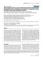
effect on B viruses (Figure 4). Virus resistance mediated by
Fv1 is semi-dominant in genetic crosses, so that Fv1
n/b
het-
erozygous animals are resistant to both N- and B-tropic
viruses. The cloning of the Fv1 gene [232] revealed that it
displays a strong sequence similarity (60% of identity
over 1.3 kb) to families of human and murine endog-
enous retroviruses called HERV or MuERV-L, respectively
[233]. Based on its position within the gag gene, Fv1
apparently encodes for a CA-like protein.
Retrovirology 2004, 1 />Page 12 of 20
(page number not for citation purposes)
While the MLV capsid protein was rapidly suspected to
represent the viral target of Fv1 restriction [234,235], the
restriction specificity has been shown to be mainly deter-
mined by a single amino-acid at position 110 in CA
[236,237]. This latter finding and the fact that Fv1 seems
to be a CA-like protein is consistent with a mechanism in
which Fv1 would interfere with an early event of the MLV
cycle by competing with the capsid of incoming virions.
This is supported by the observation that Fv1 can be satu-
rated by an excess of restricted virus or by the pre-treat-
ment of cells with inactive virion particles, a mechanism
referred to as abrogation [238]. However, the fact that (i)
Fv1 was found to be expressed at extremely low levels, (ii)
is completely unrelated to MLV CA and, (iii) that Gag pro-
teins have never previously been implicated in viral inter-
ference, has led to the suggestion that Fv1 may act via a
more subtle mechanism. So far, this mechanism is still
unknown, but it is believed to involve a direct interaction
between Fv1 and CA [231,239].
Interestingly, similar restriction activities have recently
been described in non-murine cells, and have been shown
to be due to an Fv1-like factor present in these cells (for a
review, see [215]). The first factor, called Ref1 (for Restric-
tion factor 1), interferes with N-MLV and Equine
Infectious Anemia Virus (EIAV) infection in human and
other primate and non-primate species [240-242]. This
factor shows many similarities with its murine counter-
part, and in particular with Fv1
b
, since it can be abrogated
by an excess of MLV N but not by MLV B [243]. Surpris-
ingly, the same residue 110 in MLV CA that confers the
specificity of inhibition to Fv1 is also responsible for Ref1
specificity [240]. However, Ref1 has been found to act at
a stage between entry and reverse-transcription, whereas
the Fv1 block is subsequent to reverse-transcription [242].
Interestingly, cyclophilin A which is known to be associ-
ated to HIV-1 Gag in virions [112,244] and to facilitate an
early step of infection [114], has been shown to modulate
the sensitivity of HIV-1 to restriction factors [115]. In
human cells, its association with the viral CA prevents it
from being the target of the Ref1 restriction factor,
whereas in certain non-human primates, this association
may be responsible for the restriction of HIV-1 cells by
Ref1 [115]. Surprisingly, the incorporation of CypA into
virions is not a prerequisite for the protection of HIV-1
against Ref1 antiviral activity, as the relevant CA-CypA
interaction takes place in the target cells [115].
The second Fv1-like factor is expressed in certain non-
human primates and, depending on the species, can
inhibit the replication of various lentiviruses, including
N-MLV, HIV-1, HIV-2, SIV and EIAV [245-247]. Because it
shares many characteristics with Fv1, this factor was called
Lv1, for Lentivirus susceptibility 1. Like Fv1 and Ref1, the
viral determinant of Lv1 restriction maps to the viral cap-
sid and, like Ref1, it blocks infection before reverse tran-
scription occurs. The relationship between Fv1, Ref1 and
Lv1 remains to be investigated.
The Fv1 specific inhibition of MLV infectionFigure 4
The Fv1 specific inhibition of MLV infection. There are two major alleles of the gene Fv1 among inbred mice, Fv1
n
and
Fv1
b
. Mice that carry the Fv1
n
allele are protected from B-tropic MLV infection (MLV B), whereas those carrying the Fv1
n
allele
cannot be infected by N-tropic strains (MLV N). See text for detailed discussion. Abbreviations: MLV, murine leukemia virus.
Retrovirology 2004, 1 />Page 13 of 20
(page number not for citation purposes)
It is likely that the list of genes influencing restriction of
lentiviruses in mammals will grow, as did the series of
genes controlling the sensitivity of mice to Friend virus
infection in the 1970s [248] (Table 1). A restriction factor
interfering with the replication of HIV-2 in certain human
cells has been described [248,249]. This new factor, called
Lv2, which interferes with a step of the HIV-2 life cycle
between the reverse transcription and nuclear entry, is
believed to be unrelated to Fv1, Ref1 and Lv1 [248]. Both
Env and Gag have been found to be viral determinants of
Lv2 activity, the residue 207 in CA being responsible for
the Gag restriction whereas the Env-mediated block is due
to a single amino-acid in gp120 [248]. Recently,
Sodroski's group has identified a protein in rhesus mon-
key that restricts HIV-1 replication [250]. This protein,
named TRIM5 belongs to the tripartite motif (TRIM)
family harbouring a RING domain, one or two B boxes
and a coiled-coil region. Although rhesus TRIM5α dis-
plays Lv1 activity, whether other TRIM proteins also play
a role in the restriction mechanism remains to be
answered. Interestingly, PML, another member of this
protein family, also known as RBCC (for RING domain, B
box, Coiled-Coil), has been shown to limit replication of
certain RNA and DNA viruses (for a review, see [251]).
Finally, among the many questions that remain to be
addressed, it would be interesting to investigate if some
TRIM proteins are involved in Fv1 and Ref1 restriction.
All these results illustrate the striking ability of retrovi-
ruses to counteract the antiviral mechanisms developed
by their hosts, either by direct use of a viral protein, or by
hijacking a cellular factor, thus allowing early steps of the
replicative cycle to proceed.
It is interesting to note that the FV accessory protein Bet
seems to display a similar activity to the cellular restriction
factors described above. This protein is translated from a
multispliced mRNA transcribed from an internal pro-
moter (IP) located between the env gene and the 3' LTR
[252], which also encodes for Tas, the transactivator of
gene expression from both the 5' LTR and the IP. Bet is
highly expressed in infected cells, where it localizes to
both the cytoplasm and the nucleus [253]. Interestingly,
Bet has also been shown to be secreted by infected cells,
and to be internalised by surrounding naive cells [254]
where it targets the nucleus through its C-terminal bipar-
tite NLS [253]. Finally, this protein is believed to be impli-
cated in the establishment and/or maintenance of viral
persistence in vivo. Indeed, a Tas-defective genome
(∆HFV) has been described to behave like a defective
interfering virus and to interfere with the replication of
wild-type viruses by the production of Bet [255]. Further-
more, expression of Bet has been shown to interfere with
an early stage of FV replication, between virus entry and
integration [256]. The capacity of Bet to prevent up-regu-
lation of basal IP activity might also be a factor in its abil-
ity to block superinfection of cells [257]. Although its role
and mechanism of action are still unclear, these observa-
tions suggest that Bet may help FVs to control their own
spread in order to persist in their host. This protein there-
fore represents an atypical inhibitor of early steps of the
retrovirus replicative cycle.
Perspectives
The stepwise events allowing retroviruses to enter the tar-
get cell, to move within the cytoplasm, to penetrate into
the nucleus and to integrate its genome into host chromo-
somes, are beginning to be unravelled, but many issues
are still unanswered. This is particularly evident concern-
ing the uncoating of incoming viruses, a complex process
involving cellular and viral proteins and which takes place
all along this early journey. It is interesting to note that
among the PIC proteins the viral protease, which is critical
for the late phase of infection, could also be involved in
the uncoating process, as already described for certain ret-
roviruses [258-260] and other viral families [261].
Table 1: Examples of cellular factors inhibiting early steps of retroviral cycle.
Restricted
Retroviruses
a
Step being
affected
b
Viral determinant
c
Distribution Cloned
CEM15 HIV-1 RT Vif human yes
Fv1 N-MLV or B-MLV between RT and
integration
CA Mouse yes
Ref1 N-MLV, EIAV between entry and RT CA various mammals yes*
Lv1 N-MLV, HIV-1, HIV-2,
SIVmac, EIAV
between entry and RT CA various mammals yes
Lv2 HIV-2 between RT and
nuclear entry
CA / Env human no
Abbreviations:
a
HIV-1, human immunodeficiency virus type 1 ; HIV-2, human immunodeficiency virus type 2 ; SIVmac, simian immunodeficiency
virus macaques ; N-MLV, N-tropic murine leukemia virus ; B-MLV, B-tropic murine leukemia virus ; EIAV, equine infectious anemia virus.
b
RT,
reverse-transcription.
c
CA, capsid protein ; Env, envelope glycoproteins. *Personal communications and unpublished data.
Retrovirology 2004, 1 />Page 14 of 20
(page number not for citation purposes)
Similarly, post-translational modifications of viral com-
ponents such as phosphorylation, ubiquitination and/or
sumoylation, could also influence and regulate these early
steps. The way in which retroviruses activate signalling
cascades [262] which might also regulate the behaviour of
the incoming viral components, is still unknown. How-
ever, it has already been demonstrated that HIV-1 virions
hijack many cellular proteins harbouring signalling prop-
erties such as CypA or mitogen-activated protein kinase,
two pivotal proteins known to be implicated in signalling
pathways (for a review, see [263]). An apparent block in
HIV-1 replication was described in resting CD4
+
T cells
prior to the integration of the viral genome into host cell
chromosomes in a state called preintegration latency,
awaiting stimulation and a transition to productive infec-
tion. Several studies have demonstrated that resting CD4
+
T cells isolated from the blood of HIV-1-infected individ-
uals contain completely reverse transcribed unintegrated
viral DNA, likely constituting a latent reservoir (for
reviews, see [264,265]), since these forms of DNA were
shown to be transcriptionally active [120]. Therefore, it
will also be important to precisely define the intracellular
compartments in which these unintegrated viral structures
localize and how they are maintained. The fact that 2-LTR
junctions can be detected in the cytoplasm of MLV
infected cells soon after viral entry, whereas these struc-
tures were believed to appear in the nucleus only if inte-
gration had occurred [182], may provide a clue to unravel
this unknown mechanism.
Interestingly, the discovery of host gene products that can
interfere with early steps of retroviral infection has
strengthen the idea that the incoming virus is not simply
an inert cage protecting the viral genome but rather inter-
acts widely with cellular components. The identification
of restriction factors and the characterization of their
mode of action may lead to new approaches for blocking
retroviral replication.
Understanding the precise interactions between cellular
and viral partners occurring during the early steps of infec-
tion will certainly open new fields of research leading to
the discovery of new antiretroviral drugs. Towards this
goal, the study of retroelements from distinct organisms,
such as retrotransposons in the yeast model, will help us
to define conserved and non-conserved cellular mecha-
nisms involved in the early steps of infection. This will
also allow the development of safer therapeutic long-term
expression vectors, targeting the transgene to specific
regions of the host genome without deleterious effects
[210,211]. Finally, one can also assume that the study of
early steps of infection will certainly contribute to a better
understanding of principal cell functions.
Acknowledgements
We thank Jonathan Stoye and Laura Burleigh for critical review of the man-
uscript. S.N. is supported by the European Molecular Biology Organization
(EMBO Long-term Fellowship ALTF 343-2001). A.S. is supported by
Ensemble contre le SIDA/SIDACTION and ANRS.
References
1. Amara A, Littman DR: After Hrs with HIV. J Cell Biol 2003,
162:371-375.
2. Perez OD, Nolan GP: Resistance is futile: assimilation of cellu-
lar machinery by HIV-1. Immunity 2001, 15:687-690.
3. Dvorin JD, Malim MH: Intracellular trafficking of HIV-1 cores:
journey to the center of the cell. Curr Top Microbiol Immunol 2003,
281:179-208.
4. Goff SP: Intracellular trafficking of retroviral genomes during
the early phase of infection: viral exploitation of cellular
pathways. J Gene Med 2001, 3:517-528.
5. Suomalainen M, Nakano MY, Keller S, Boucke K, Stidwill RP, Greber
UF: Microtubule-dependent plus- and minus end-directed
motilities are competing processes for nuclear targeting of
adenovirus. J Cell Biol 1999, 144:657-672.
6. Sodeik B, Ebersold MW, Helenius A: Microtubule-mediated
transport of incoming herpes simplex virus 1 capsids to the
nucleus. J Cell Biol 1997, 136:1007-1021.
7. Sherman MP, Greene WC: Slipping through the door: HIV entry
into the nucleus. Microbes Infect 2002, 4:67-73.
8. Towers GJ, Goff SP: Post-entry restriction of retroviral
infections. AIDS Rev 2003, 5:156-164.
9. Sharma S, Miyanohara A, Friedmann T: Separable mechanisms of
attachment and cell uptake during retrovirus infection. J Virol
2000, 74:10790-10795.
10. Pizzato M, Marlow SA, Blair ED, Takeuchi Y: Initial binding of
murine leukemia virus particles to cells does not require spe-
cific Env-receptor interaction. J Virol 1999, 73:8599-8611.
11. Ugolini S, Mondor I, Sattentau QJ: HIV-1 attachment: another
look. Trends Microbiol 1999, 7:144-149.
12. Mondor I, Ugolini S, Sattentau QJ: Human immunodeficiency
virus type 1 attachment to HeLa CD4 cells is CD4 independ-
ent and gp120 dependent and requires cell surface heparans.
J Virol 1998, 72:3623-3634.
13. Fortin JF, Cantin R, Tremblay MJ: T cells expressing activated
LFA-1 are more susceptible to infection with human immu-
nodeficiency virus type 1 particles bearing host-encoded
ICAM-1. J Virol 1998, 72:2105-2112.
14. Nisole S, Krust B, Callebaut C, Guichard G, Muller S, Briand JP, Hov-
anessian AG: The anti-HIV pseudopeptide HB-19 forms a
complex with the cell-surface-expressed nucleolin independ-
ent of heparan sulfate proteoglycans. J Biol Chem 1999,
274:27875-27884.
15. Moore JP, McKeating JA, Huang YX, Ashkenazi A, Ho DD: Virions
of primary human immunodeficiency virus type 1 isolates
resistant to soluble CD4 (sCD4) neutralization differ in sCD4
binding and glycoprotein gp120 retention from sCD4-sensi-
tive isolates. J Virol 1992, 66:235-243.
16. O'Doherty U, Swiggard WJ, Malim MH: Human immunodefi-
ciency virus type 1 spinoculation enhances infection through
virus binding. J Virol 2000, 74:10074-10080.
17. Spillmann D: Heparan sulfate: anchor for viral intruders? Bio-
chimie 2001, 83:811-817.
18. Pinon JD, Klasse PJ, Jassal SR, Welson S, Weber J, Brighty DW, Sat-
tentau QJ: Human T-cell leukemia virus type 1 envelope glyc-
oprotein gp46 interacts with cell surface heparan sulfate
proteoglycans. J Virol 2003, 77:9922-9930.
19. Walker SJ, Pizzato M, Takeuchi Y, Devereux S: Heparin binds to
murine leukemia virus and inhibits Env-independent attach-
ment and infection. J Virol 2002, 76:6909-6918.
20. Zhang YJ, Hatziioannou T, Zang T, Braaten D, Luban J, Goff SP,
Bieniasz PD: Envelope-dependent, cyclophilin-independent
effects of glycosaminoglycans on human immunodeficiency
virus type 1 attachment and infection. J Virol 2002,
76:6332-6343.
21. Guibinga GH, Miyanohara A, Esko JD, Friedmann T: Cell surface
heparan sulfate is a receptor for attachment of envelope
protein-free retrovirus-like particles and VSV-G pseudo-
Retrovirology 2004, 1 />Page 15 of 20
(page number not for citation purposes)
typed MLV-derived retrovirus vectors to target cells. Mol Ther
2002, 5:538-546.
22. Marechal V, Clavel F, Heard JM, Schwartz O: Cytosolic Gag p24 as
an index of productive entry of human immunodeficiency
virus type 1. J Virol 1998, 72:2208-2212.
23. Schaeffer E, Soros VB, Greene WC: Compensatory link between
fusion and endocytosis of human immunodeficiency virus
type 1 in human CD4 T lymphocytes. J Virol 2004, 78:1375-1383.
24. Pohlmann S, Soilleux EJ, Baribaud F, Leslie GJ, Morris LS, Trowsdale J,
Lee B, Coleman N, Doms RW: DC-SIGNR, a DC-SIGN homo-
logue expressed in endothelial cells, binds to human and sim-
ian immunodeficiency viruses and activates infection in
trans. Proc Natl Acad Sci U S A 2001, 98:2670-2675.
25. Geijtenbeek TB, Kwon DS, Torensma R, van Vliet SJ, van Duijnhoven
GC, Middel J, Cornelissen IL, Nottet HS, KewalRamani VN, Littman
DR, Figdor CG, van Kooyk Y: DC-SIGN, a dendritic cell-specific
HIV-1-binding protein that enhances trans-infection of T
cells. Cell 2000, 100:587-597.
26. Hong PW, Flummerfelt KB, de Parseval A, Gurney K, Elder JH, Lee B:
Human immunodeficiency virus envelope (gp120) binding to
DC-SIGN and primary dendritic cells is carbohydrate
dependent but does not involve 2G12 or cyanovirin binding
sites: implications for structural analyses of gp120-DC-SIGN
binding. J Virol 2002, 76:12855-12865.
27. Lin CL, Sewell AK, Gao GF, Whelan KT, Phillips RE, Austyn JM: Mac-
rophage-tropic HIV induces and exploits dendritic cell
chemotaxis. J Exp Med 2000, 192:587-594.
28. Lue J, Hsu M, Yang D, Marx P, Chen Z, Cheng-Mayer C: Addition of
a single gp120 glycan confers increased binding to dendritic
cell-specific ICAM-3-grabbing nonintegrin and neutraliza-
tion escape to human immunodeficiency virus type 1. J Virol
2002, 76:10299-10306.
29. Geijtenbeek TB, van Duijnhoven GC, van Vliet SJ, Krieger E, Vriend
G, Figdor CG, van Kooyk Y: Identification of different binding
sites in the dendritic cell-specific receptor DC-SIGN for
intercellular adhesion molecule 3 and HIV-1. J Biol Chem 2002,
277:11314-11320.
30. Turville SG, Cameron PU, Handley A, Lin G, Pohlmann S, Doms RW,
Cunningham AL: Diversity of receptors binding HIV on den-
dritic cell subsets. Nat Immunol 2002, 3:975-983.
31. van Kooyk Y, Geijtenbeek TB: DC-SIGN: escape mechanism for
pathogens. Nat Rev Immunol 2003, 3:697-709.
32. Tailleux L, Schwartz O, Herrmann JL, Pivert E, Jackson M, Amara A,
Legres L, Dreher D, Nicod LP, Gluckman JC, Lagrange PH, Gicquel B,
Neyrolles O: DC-SIGN is the major Mycobacterium tubercu-
losis receptor on human dendritic cells. J Exp Med 2003,
197:121-127.
33. Kim JW, Closs EI, Albritton LM, Cunningham JM: Transport of cat-
ionic amino acids by the mouse ecotropic retrovirus
receptor. Nature 1991, 352:725-728.
34. Wang H, Kavanaugh MP, North RA, Kabat D: Cell-surface recep-
tor for ecotropic murine retroviruses is a basic amino-acid
transporter. Nature 1991, 352:729-731.
35. Maddon PJ, Dalgleish AG, McDougal JS, Clapham PR, Weiss RA, Axel
R: The T4 gene encodes the AIDS virus receptor and is
expressed in the immune system and the brain. Cell 1986,
47:333-348.
36. Manel N, Kim FJ, Kinet S, Taylor N, Sitbon M, Battini JL: The ubiqui-
tous glucose transporter GLUT-1 is a receptor for HTLV. Cell
2003, 115:449-459.
37. O'Hara B, Johann SV, Klinger HP, Blair DG, Rubinson H, Dunn KJ,
Sass P, Vitek SM, Robins T: Characterization of a human gene
conferring sensitivity to infection by gibbon ape leukemia
virus. Cell Growth Differ 1990, 1:119-127.
38. van Zeijl M, Johann SV, Closs E, Cunningham J, Eddy R, Shows TB,
O'Hara B: A human amphotropic retrovirus receptor is a sec-
ond member of the gibbon ape leukemia virus receptor
family. Proc Natl Acad Sci U S A 1994, 91:1168-1172.
39. Miller DG, Miller AD: A family of retroviruses that utilize
related phosphate transporters for cell entry. J Virol 1994,
68:8270-8276.
40. Hill CL, Bieniasz PD, McClure MO: Properties of human foamy
virus relevant to its development as a vector for gene
therapy. J Gen Virol 1999, 80(Pt 8):2003-2009.
41. Mergia A, Heinkelein M: Foamy virus vectors. Curr Top Microbiol
Immunol 2003, 277:131-159.
42. von Laer D, Neumann-Haefelin D, Heeney JL, Schweizer M: Lym-
phocytes are the major reservoir for foamy viruses in periph-
eral blood. Virology 1996, 221:240-244.
43. Berger EA, Murphy PM, Farber JM: Chemokine receptors as HIV-
1 coreceptors: roles in viral entry, tropism, and disease. Annu
Rev Immunol 1999, 17:657-700.
44. Kwong PD, Wyatt R, Robinson J, Sweet RW, Sodroski J, Hendrickson
WA: Structure of an HIV gp120 envelope glycoprotein in
complex with the CD4 receptor and a neutralizing human
antibody. Nature 1998, 393:648-659.
45. Gallo SA, Finnegan CM, Viard M, Raviv Y, Dimitrov A, Rawat SS, Puri
A, Durell S, Blumenthal R: The HIV Env-mediated fusion
reaction. Biochim Biophys Acta 2003, 1614:36-50.
46. Moore JP, Doms RW: The entry of entry inhibitors: a fusion of
science and medicine. Proc Natl Acad Sci U S A 2003,
100:10598-10602.
47. Harouse JM, Bhat S, Spitalnik SL, Laughlin M, Stefano K, Silberberg
DH, Gonzalez-Scarano F: Inhibition of entry of HIV-1 in neural
cell lines by antibodies against galactosyl ceramide. Science
1991, 253:320-323.
48. Simons K, Ikonen E: Functional rafts in cell membranes. Nature
1997, 387:569-572.
49. Hammache D, Yahi N, Maresca M, Pieroni G, Fantini J: Human
erythrocyte glycosphingolipids as alternative cofactors for
human immunodeficiency virus type 1 (HIV-1) entry: evi-
dence for CD4-induced interactions between HIV-1 gp120
and reconstituted membrane microdomains of glycosphin-
golipids (Gb3 and GM3). J Virol 1999, 73:5244-5248.
50. Manes S, del Real G, Lacalle RA, Lucas P, Gomez-Mouton C, Sanchez-
Palomino S, Delgado R, Alcami J, Mira E, Martinez AC: Membrane
raft microdomains mediate lateral assemblies required for
HIV-1 infection. EMBO Rep 2000, 1:190-196.
51. Del Real G, Jimenez-Baranda S, Lacalle RA, Mira E, Lucas P, Gomez-
Mouton C, Carrera AC, Martinez AC, Manes S: Blocking of HIV-1
infection by targeting CD4 to nonraft membrane domains. J
Exp Med 2002, 196:293-301.
52. Popik W, Alce TM, Au WC: Human immunodeficiency virus
type 1 uses lipid raft-colocalized CD4 and chemokine recep-
tors for productive entry into CD4(+) T cells. J Virol 2002,
76:4709-4722.
53. Nisole S, Krust B, Hovanessian AG: Anchorage of HIV on permis-
sive cells leads to coaggregation of viral particles with sur-
face nucleolin at membrane raft microdomains. Exp Cell Res
2002, 276:155-173.
54. Percherancier Y, Lagane B, Planchenault T, Staropoli I, Altmeyer R,
Virelizier JL, Arenzana-Seisdedos F, Hoessli DC, Bachelerie F: HIV-1
entry into T-cells is not dependent on CD4 and CCR5 locali-
zation to sphingolipid-enriched, detergent-resistant, raft
membrane domains. J Biol Chem 2003, 278:3153-3161.
55. Popik W, Alce TM: CD4 receptor localized to non-raft mem-
brane microdomains supports HIV-1 entry. Identification of
a novel raft localization marker in CD4. J Biol Chem 2004,
279:704-712.
56. Nguyen DH, Hildreth JE: Evidence for budding of human immu-
nodeficiency virus type 1 selectively from glycolipid-enriched
membrane lipid rafts. J Virol 2000, 74:3264-3272.
57. Aloia RC, Tian H, Jensen FC: Lipid composition and fluidity of
the human immunodeficiency virus envelope and host cell
plasma membranes. Proc Natl Acad Sci U S A 1993, 90:5181-5185.
58. Guyader M, Kiyokawa E, Abrami L, Turelli P, Trono D: Role for
human immunodeficiency virus type 1 membrane choles-
terol in viral internalization. J Virol 2002, 76:10356-10364.
59. McClure MO, Marsh M, Weiss RA: Human immunodeficiency
virus infection of CD4-bearing cells occurs by a pH-inde-
pendent mechanism. Embo J 1988, 7:513-518.
60. Schwartz O, Marechal V, Friguet B, Arenzana-Seisdedos F, Heard JM:
Antiviral activity of the proteasome on incoming human
immunodeficiency virus type 1. J Virol 1998, 72:3845-3850.
61. Fredericksen BL, Wei BL, Yao J, Luo T, Garcia JV: Inhibition of
endosomal/lysosomal degradation increases the infectivity
of human immunodeficiency virus. J Virol 2002, 76:11440-11446.
62. Katen LJ, Januszeski MM, Anderson WF, Hasenkrug KJ, Evans LH:
Infectious entry by amphotropic as well as ecotropic murine
leukemia viruses occurs through an endocytic pathway. J Virol
2001, 75:5018-5026.
Retrovirology 2004, 1 />Page 16 of 20
(page number not for citation purposes)
63. Picard-Maureau M, Jarmy G, Berg A, Rethwilm A, Lindemann D:
Foamy virus envelope glycoprotein-mediated entry involves
a pH-dependent fusion process. J Virol 2003, 77:4722-4730.
64. Kizhatil K, Albritton LM: Requirements for different compo-
nents of the host cell cytoskeleton distinguish ecotropic
murine leukemia virus entry via endocytosis from entry via
surface fusion. J Virol 1997, 71:7145-7156.
65. Skehel JJ, Wiley DC: Receptor binding and membrane fusion in
virus entry: the influenza hemagglutinin. Annu Rev Biochem
2000, 69:531-569.
66. Mothes W, Boerger AL, Narayan S, Cunningham JM, Young JA: Ret-
roviral entry mediated by receptor priming and low pH trig-
gering of an envelope glycoprotein. Cell 2000, 103:679-689.
67. Sanders RW, de Jong EC, Baldwin CE, Schuitemaker JH, Kapsenberg
ML, Berkhout B: Differential transmission of human immuno-
deficiency virus type 1 by distinct subsets of effector den-
dritic cells. J Virol 2002, 76:7812-7821.
68. McDonald D, Wu L, Bohks SM, KewalRamani VN, Unutmaz D, Hope
TJ: Recruitment of HIV and its receptors to dendritic cell-T
cell junctions. Science 2003, 300:1295-1297.
69. Pelchen-Matthews A, Kramer B, Marsh M: Infectious HIV-1
assembles in late endosomes in primary macrophages. J Cell
Biol 2003, 162:443-455.
70. Telesnitsky A, Goff SP: Reverse transcriptase and the genera-
tion of retroviral DNA. In Retroviruses Edited by: Coffin JM, Hughes
SH, Varmus HE. Cold Spring Harbor Laboratory Press; 1997:121-160.
71. Lori F, di Marzo Veronese F, de Vico AL, Lusso P, Reitz MS Jr, Gallo
RC: Viral DNA carried by human immunodeficiency virus
type 1 virions. J Virol 1992, 66:5067-5074.
72. Trono D: Partial reverse transcripts in virions from human
immunodeficiency and murine leukemia viruses. J Virol 1992,
66:4893-4900.
73. Zhu J, Cunningham JM: Minus-strand DNA is present within
murine type C ecotropic retroviruses prior to infection. J Virol
1993, 67:2385-2388.
74. Yu SF, Sullivan MD, Linial ML: Evidence that the human foamy
virus genome is DNA. J Virol 1999, 73:1565-1572.
75. Moebes A, Enssle J, Bieniasz PD, Heinkelein M, Lindemann D, Bock M,
McClure MO, Rethwilm A: Human foamy virus reverse tran-
scription that occurs late in the viral replication cycle. J Virol
1997, 71:7305-7311.
76. Delelis O, Saib A, Sonigo P: Biphasic DNA synthesis in
spumaviruses. J Virol 2003, 77:8141-8146.
77. Lecellier CH, Saib A: Foamy viruses: between retroviruses and
pararetroviruses. Virology 2000, 271:1-8.
78. Zhang H, Dornadula G, Orenstein J, Pomerantz RJ: Morphologic
changes in human immunodeficiency virus type 1 virions sec-
ondary to intravirion reverse transcription: evidence indicat-
ing that reverse transcription may not take place within the
intact viral core. J Hum Virol 2000, 3:165-172.
79. McDonald D, Vodicka MA, Lucero G, Svitkina TM, Borisy GG, Emer-
man M, Hope TJ: Visualization of the intracellular behavior of
HIV in living cells. J Cell Biol 2002, 159:441-452.
80. Kimpton J, Emerman M: Detection of replication-competent
and pseudotyped human immunodeficiency virus with a sen-
sitive cell line on the basis of activation of an integrated beta-
galactosidase gene. J Virol 1992, 66:2232-2239.
81. Piatak M Jr, Saag MS, Yang LC, Clark SJ, Kappes JC, Luk KC, Hahn BH,
Shaw GM, Lifson JD: High levels of HIV-1 in plasma during all
stages of infection determined by competitive PCR. Science
1993, 259:1749-1754.
82. Andreadis S, Lavery T, Davis HE, Le Doux JM, Yarmush ML, Morgan
JR: Toward a more accurate quantitation of the activity of
recombinant retroviruses: alternatives to titer and multi-
plicity of infection. J Virol 2000, 74:3431-3439.
83. Bukrinskaya A, Brichacek B, Mann A, Stevenson M: Establishment
of a functional human immunodeficiency virus type 1 (HIV-
1) reverse transcription complex involves the cytoskeleton.
J Exp Med 1998, 188:2113-2125.
84. Fassati A, Gorlich D, Harrison I, Zaytseva L, Mingot JM: Nuclear
import of HIV-1 intracellular reverse transcription com-
plexes is mediated by importin 7. Embo J 2003, 22:3675-3685.
85. Fassati A, Goff SP: Characterization of intracellular reverse
transcription complexes of human immunodeficiency virus
type 1. J Virol 2001, 75:3626-3635.
86. Miller MD, Farnet CM, Bushman FD: Human immunodeficiency
virus type 1 preintegration complexes: studies of organiza-
tion and composition. J Virol 1997, 71:5382-5390.
87. Bukrinsky MI, Sharova N, McDonald TL, Pushkarskaya T, Tarpley
WG, Stevenson M: Association of integrase, matrix, and
reverse transcriptase antigens of human immunodeficiency
virus type 1 with viral nucleic acids following acute infection.
Proc Natl Acad Sci U S A 1993, 90:6125-6129.
88. Farnet CM, Bushman FD: HIV-1 cDNA integration: require-
ment of HMG I(Y) protein for function of preintegration
complexes in vitro. Cell 1997, 88:483-492.
89. Risco C, Menendez-Arias L, Copeland TD, Pinto da Silva P, Oroszlan
S: Intracellular transport of the murine leukemia virus during
acute infection of NIH 3T3 cells: nuclear import of nucleo-
capsid protein and integrase. J Cell Sci 1995, 108(Pt
9):3039-3050.
90. Fassati A, Goff SP: Characterization of intracellular reverse
transcription complexes of Moloney murine leukemia virus.
J Virol 1999, 73:8919-8925.
91. Petit C, Giron ML, Tobaly-Tapiero J, Bittoun P, Real E, Jacob Y, Tordo
N, De The H, Saib A: Targeting of incoming retroviral Gag to
the centrosome involves a direct interaction with the dynein
light chain 8. J Cell Sci 2003, 116:3433-3442.
92. Saib A, Puvion-Dutilleul F, Schmid M, Peries J, de The H: Nuclear
targeting of incoming human foamy virus Gag proteins
involves a centriolar step. J Virol 1997, 71:1155-1161.
93. Ojala PM, Sodeik B, Ebersold MW, Kutay U, Helenius A: Herpes
simplex virus type 1 entry into host cells: reconstitution of
capsid binding and uncoating at the nuclear pore complex in
vitro. Mol Cell Biol 2000, 20:4922-4931.
94. Greber UF, Suomalainen M, Stidwill RP, Boucke K, Ebersold MW,
Helenius A: The role of the nuclear pore complex in adenovi-
rus DNA entry. Embo J 1997, 16:5998-6007.
95. Trotman LC, Mosberger N, Fornerod M, Stidwill RP, Greber UF:
Import of adenovirus DNA involves the nuclear pore com-
plex receptor CAN/Nup214 and histone H1. Nat Cell Biol 2001,
3:1092-1100.
96. Jamieson BD, Aldrovandi GM, Planelles V, Jowett JB, Gao L, Bloch LM,
Chen IS, Zack JA: Requirement of human immunodeficiency
virus type 1 nef for in vivo replication and pathogenicity. J Virol
1994, 68:3478-3485.
97. Chowers MY, Spina CA, Kwoh TJ, Fitch NJ, Richman DD, Guatelli JC:
Optimal infectivity in vitro of human immunodeficiency
virus type 1 requires an intact nef gene. J Virol 1994,
68:2906-2914.
98. Miller MD, Feinberg MB, Greene WC: The HIV-1 nef gene acts as
a positive viral infectivity factor. Trends Microbiol 1994,
2:294-298.
99. Aiken C, Trono D: Nef stimulates human immunodeficiency
virus type 1 proviral DNA synthesis. J Virol 1995, 69:5048-5056.
100. Aiken C: Pseudotyping human immunodeficiency virus type 1
(HIV-1) by the glycoprotein of vesicular stomatitis virus tar-
gets HIV-1 entry to an endocytic pathway and suppresses
both the requirement for Nef and the sensitivity to
cyclosporin A. J Virol 1997, 71:5871-5877.
101. Schaeffer E, Geleziunas R, Greene WC: Human immunodefi-
ciency virus type 1 Nef functions at the level of virus entry by
enhancing cytoplasmic delivery of virions. J Virol 2001,
75:2993-3000.
102. Luo T, Douglas JL, Livingston RL, Garcia JV: Infectivity enhance-
ment by HIV-1 Nef is dependent on the pathway of virus
entry: implications for HIV-based gene transfer systems.
Virology 1998, 241:224-233.
103. Zheng YH, Plemenitas A, Fielding CJ, Peterlin BM: Nef increases the
synthesis of and transports cholesterol to lipid rafts and HIV-
1 progeny virions. Proc Natl Acad Sci U S A 2003, 100:8460-8465.
104. von Schwedler U, Song J, Aiken C, Trono D: Vif is crucial for
human immunodeficiency virus type 1 proviral DNA synthe-
sis in infected cells. J Virol 1993, 67:4945-4955.
105. Ohagen A, Gabuzda D: Role of Vif in stability of the human
immunodeficiency virus type 1 core. J Virol 2000,
74:11055-11066.
106. Lecossier D, Bouchonnet F, Clavel F, Hance AJ: Hypermutation of
HIV-1 DNA in the absence of the Vif protein. Science 2003,
300:1112.
Retrovirology 2004, 1 />Page 17 of 20
(page number not for citation purposes)
107. Mangeat B, Turelli P, Caron G, Friedli M, Perrin L, Trono D: Broad
antiretroviral defence by human APOBEC3G through lethal
editing of nascent reverse transcripts. Nature 2003, 424:99-103.
108. Marin M, Rose KM, Kozak SL, Kabat D: HIV-1 Vif protein binds
the editing enzyme APOBEC3G and induces its degradation.
Nat Med 2003, 9:1398-1403.
109. Sheehy AM, Gaddis NC, Malim MH: The antiretroviral enzyme
APOBEC3G is degraded by the proteasome in response to
HIV-1 Vif. Nat Med 2003, 9:1404-1407.
110. Zhang H, Yang B, Pomerantz RJ, Zhang C, Arunachalam SC, Gao L:
The cytidine deaminase CEM15 induces hypermutation in
newly synthesized HIV-1 DNA. Nature 2003, 424:94-98.
111. Luban J, Bossolt KL, Franke EK, Kalpana GV, Goff SP: Human
immunodeficiency virus type 1 Gag protein binds to cyclo-
philins A and B. Cell 1993, 73:1067-1078.
112. Thali M, Bukovsky A, Kondo E, Rosenwirth B, Walsh CT, Sodroski J,
Gottlinger HG: Functional association of cyclophilin A with
HIV-1 virions. Nature 1994, 372:363-365.
113. Gamble TR, Vajdos FF, Yoo S, Worthylake DK, Houseweart M, Sun-
dquist WI, Hill CP: Crystal structure of human cyclophilin A
bound to the amino-terminal domain of HIV-1 capsid. Cell
1996, 87:1285-1294.
114. Braaten D, Franke EK, Luban J: Cyclophilin A is required for an
early step in the life cycle of human immunodeficiency virus
type 1 before the initiation of reverse transcription. J Virol
1996, 70:3551-3560.
115. Towers GJ, Hatziioannou T, Cowan S, Goff SP, Luban J, Bieniasz PD:
Cyclophilin A modulates the sensitivity of HIV-1 to host
restriction factors. Nat Med 2003, 9:1138-1143.
116. Butera ST, Perez VL, Besansky NJ, Chan WC, Wu BY, Nabel GJ, Folks
TM: Extrachromosomal human immunodeficiency virus
type-1 DNA can initiate a spreading infection of HL-60 cells.
J Cell Biochem 1991, 45:366-373.
117. Stevenson M, Haggerty S, Lamonica CA, Meier CM, Welch SK,
Wasiak AJ: Integration is not necessary for expression of
human immunodeficiency virus type 1 protein products. J
Virol 1990, 64:2421-2425.
118. Teo I, Veryard C, Barnes H, An SF, Jones M, Lantos PL, Luthert P,
Shaunak S: Circular forms of unintegrated human immunode-
ficiency virus type 1 DNA and high levels of viral protein
expression: association with dementia and multinucleated
giant cells in the brains of patients with AIDS. J Virol 1997,
71:2928-2933.
119. Cara A, Cereseto A, Lori F, Reitz MS Jr: HIV-1 protein expression
from synthetic circles of DNA mimicking the extrachromo-
somal forms of viral DNA. J Biol Chem 1996, 271:5393-5397.
120. Wu Y, Marsh JW: Early transcription from nonintegrated DNA
in human immunodeficiency virus infection. J Virol 2003,
77:10376-10382.
121. Poon B, Chen IS: Human immunodeficiency virus type 1 (HIV-
1) Vpr enhances expression from unintegrated HIV-1 DNA.
J Virol 2003, 77:3962-3972.
122. Ploubidou A, Way M: Viral transport and the cytoskeleton. Curr
Opin Cell Biol 2001, 13:97-105.
123. Ploubidou A, Moreau V, Ashman K, Reckmann I, Gonzalez C, Way M:
Vaccinia virus infection disrupts microtubule organization
and centrosome function. Embo J 2000, 19:3932-3944.
124. Rietdorf J, Ploubidou A, Reckmann I, Holmstrom A, Frischknecht F,
Zettl M, Zimmermann T, Way M: Kinesin-dependent movement
on microtubules precedes actin-based motility of vaccinia
virus. Nat Cell Biol 2001, 3:992-1000.
125. de Soultrait VR, Caumont A, Durrens P, Calmels C, Parissi V, Recor-
don P, Bon E, Desjobert C, Tarrago-Litvak L, Fournier M: HIV-1
integrase interacts with yeast microtubule-associated
proteins. Biochim Biophys Acta 2002, 1575:40-48.
126. Ibarrondo FJ, Choi R, Geng YZ, Canon J, Rey O, Baldwin GC, Krogs-
tad P: HIV type 1 Gag and nucleocapsid proteins: cytoskeletal
localization and effects on cell motility. AIDS Res Hum
Retroviruses 2001, 17:1489-1500.
127. Liu B, Dai R, Tian CJ, Dawson L, Gorelick R, Yu XF: Interaction of
the human immunodeficiency virus type 1 nucleocapsid with
actin. J Virol 1999, 73:2901-2908.
128. Wilk T, Gowen B, Fuller SD: Actin associates with the nucleo-
capsid domain of the human immunodeficiency virus Gag
polyprotein. J Virol 1999, 73:1931-1940.
129. Watts NR, Sackett DL, Ward RD, Miller MW, Wingfield PT, Stahl SS,
Steven AC: HIV-1 rev depolymerizes microtubules to form
stable bilayered rings. J Cell Biol 2000, 150:349-360.
130. Strebel K, Bour S: Molecular interactions of HIV with host
factors. Aids 1999, 13(Suppl A):S13-24.
131. Alonso C, Miskin J, Hernaez B, Fernandez-Zapatero P, Soto L, Canto
C, Rodriguez-Crespo I, Dixon L, Escribano JM: African swine fever
virus protein p54 interacts with the microtubular motor
complex through direct binding to light-chain dynein. J Virol
2001, 75:9819-9827.
132. Raux H, Flamand A, Blondel D: Interaction of the rabies virus P
protein with the LC8 dynein light chain. J Virol 2000,
74:10212-10216.
133. Poisson N, Real E, Gaudin Y, Vaney MC, King S, Jacob Y, Tordo N,
Blondel D: Molecular basis for the interaction between rabies
virus phosphoprotein P and the dynein light chain LC8: dis-
sociation of dynein-binding properties and transcriptional
functionality of P. J Gen Virol 2001, 82:2691-2696.
134. Jacob Y, Badrane H, Ceccaldi PE, Tordo N: Cytoplasmic dynein
LC8 interacts with lyssavirus phosphoprotein. J Virol 2000,
74:10217-10222.
135. Harrison A, King SM: The molecular anatomy of dynein. Essays
Biochem 2000, 35:75-87.
136. Igakura T, Stinchcombe JC, Goon PK, Taylor GP, Weber JN, Griffiths
GM, Tanaka Y, Osame M, Bangham CR: Spread of HTLV-I
between lymphocytes by virus-induced polarization of the
cytoskeleton. Science 2003, 299:1713-1716.
137. Sfakianos JN, Hunter E: M-PMV capsid transport is mediated by
Env/Gag interactions at the pericentriolar recycling
endosome. Traffic 2003, 4:671-680.
138. Sfakianos JN, LaCasse RA, Hunter E: The M-PMV cytoplasmic tar-
geting-retention signal directs nascent Gag polypeptides to
a pericentriolar region of the cell. Traffic 2003, 4:660-670.
139. Calafat J, Hilkens JG: Ultrastructural study of virus-like particles
in Chinese hamster lung cells. J Gen Virol 1978, 41:417-420.
140. Heine UI, Kramarsky B, Wendel E, Suskind RG: Enhanced prolifer-
ation of endogenous virus in Chinese hamster cells associ-
ated with microtubules and the mitotic apparatus of the host
cell. J Gen Virol 1979, 44:45-55.
141. Roe T, Reynolds TC, Yu G, Brown PO: Integration of murine
leukemia virus DNA depends on mitosis. Embo J 1993,
12:2099-2108.
142. Lewis PF, Emerman M: Passage through mitosis is required for
oncoretroviruses but not for the human immunodeficiency
virus. J Virol 1994, 68:510-516.
143. Weinberg JB, Matthews TJ, Cullen BR, Malim MH: Productive
human immunodeficiency virus type 1 (HIV-1) infection of
nonproliferating human monocytes. J Exp Med 1991,
174:1477-1482.
144. Bukrinsky MI, Sharova N, Dempsey MP, Stanwick TL, Bukrinskaya
AG, Haggerty S, Stevenson M: Active nuclear import of human
immunodeficiency virus type 1 preintegration complexes.
Proc Natl Acad Sci U S A 1992, 89:6580-6584.
145. Hatziioannou T, Goff SP: Infection of nondividing cells by Rous
sarcoma virus. J Virol 2001, 75:9526-9531.
146. Bieniasz PD, Weiss RA, McClure MO: Cell cycle dependence of
foamy retrovirus infection. J Virol 1995, 69:7295-7299.
147. Pante N, Kann M: Nuclear pore complex is able to transport
macromolecules with diameters of about 39 nm. Mol Biol Cell
2002, 13:425-434.
148. Fried H, Kutay U: Nucleocytoplasmic transport: taking an
inventory. Cell Mol Life Sci 2003, 60:1659-1688.
149. Cullen BR: Journey to the center of the cell. Cell 2001,
105:697-700.
150. Gallay P, Hope T, Chin D, Trono D: HIV-1 infection of nondivid-
ing cells through the recognition of integrase by the impor-
tin/karyopherin pathway. Proc Natl Acad Sci U S A 1997,
94:9825-9830.
151. Bouyac-Bertoia M, Dvorin JD, Fouchier RA, Jenkins Y, Meyer BE, Wu
LI, Emerman M, Malim MH: HIV-1 infection requires a functional
integrase NLS. Mol Cell 2001, 7:1025-1035.
152. Katz RA, Greger JG, Boimel P, Skalka AM: Human immunodefi-
ciency virus type 1 DNA nuclear import and integration are
mitosis independent in cycling cells. J Virol 2003,
77:13412-13417.
Retrovirology 2004, 1 />Page 18 of 20
(page number not for citation purposes)
153. Dvorin JD, Bell P, Maul GG, Yamashita M, Emerman M, Malim MH:
Reassessment of the roles of integrase and the central DNA
flap in human immunodeficiency virus type 1 nuclear import.
J Virol 2002, 76:12087-12096.
154. Limon A, Devroe E, Lu R, Ghory HZ, Silver PA, Engelman A: Nuclear
localization of human immunodeficiency virus type 1 pre-
integration complexes (PICs): V165A and R166A are pleio-
tropic integrase mutants primarily defective for integration,
not PIC nuclear import. J Virol 2002, 76:10598-10607.
155. Depienne C, Mousnier A, Leh H, Le Rouzic E, Dormont D, Benichou
S, Dargemont C: Characterization of the nuclear import path-
way for HIV-1 integrase. J Biol Chem 2001, 276:18102-18107.
156. Devroe E, Engelman A, Silver PA: Intracellular transport of
human immunodeficiency virus type 1 integrase. J Cell Sci
2003, 116:4401-4408.
157. Bukrinsky MI, Haggerty S, Dempsey MP, Sharova N, Adzhubel A, Spitz
L, Lewis P, Goldfarb D, Emerman M, Stevenson M: A nuclear local-
ization signal within HIV-1 matrix protein that governs infec-
tion of non-dividing cells. Nature 1993, 365:666-669.
158. von Schwedler U, Kornbluth RS, Trono D: The nuclear localiza-
tion signal of the matrix protein of human immunodefi-
ciency virus type 1 allows the establishment of infection in
macrophages and quiescent T lymphocytes. Proc Natl Acad Sci
U S A 1994, 91:6992-6996.
159. Reil H, Bukovsky AA, Gelderblom HR, Gottlinger HG: Efficient
HIV-1 replication can occur in the absence of the viral matrix
protein. Embo J 1998, 17:2699-2708.
160. Fouchier RA, Meyer BE, Simon JH, Fischer U, Malim MH: HIV-1
infection of non-dividing cells: evidence that the amino-ter-
minal basic region of the viral matrix protein is important
for Gag processing but not for post-entry nuclear import.
Embo J 1997, 16:4531-4539.
161. Freed EO, Englund G, Martin MA: Role of the basic domain of
human immunodeficiency virus type 1 matrix in macro-
phage infection. J Virol 1995, 69:3949-3954.
162. Haffar OK, Popov S, Dubrovsky L, Agostini I, Tang H, Pushkarsky T,
Nadler SG, Bukrinsky M: Two nuclear localization signals in the
HIV-1 matrix protein regulate nuclear import of the HIV-1
pre-integration complex. J Mol Biol 2000, 299:359-368.
163. Vodicka MA, Koepp DM, Silver PA, Emerman M: HIV-1 Vpr inter-
acts with the nuclear transport pathway to promote macro-
phage infection. Genes Dev 1998, 12:175-185.
164. Heinzinger NK, Bukinsky MI, Haggerty SA, Ragland AM, Kewalramani
V, Lee MA, Gendelman HE, Ratner L, Stevenson M, Emerman M: The
Vpr protein of human immunodeficiency virus type 1 influ-
ences nuclear localization of viral nucleic acids in nondividing
host cells. Proc Natl Acad Sci U S A 1994, 91:7311-7315.
165. Popov S, Rexach M, Ratner L, Blobel G, Bukrinsky M: Viral protein
R regulates docking of the HIV-1 preintegration complex to
the nuclear pore complex. J Biol Chem 1998, 273:13347-13352.
166. Popov S, Rexach M, Zybarth G, Reiling N, Lee MA, Ratner L, Lane
CM, Moore MS, Blobel G, Bukrinsky M: Viral protein R regulates
nuclear import of the HIV-1 pre-integration complex. Embo J
1998, 17:909-917.
167. Le Rouzic E, Mousnier A, Rustum C, Stutz F, Hallberg E, Dargemont
C, Benichou S: Docking of HIV-1 Vpr to the nuclear envelope
is mediated by the interaction with the nucleoporin hCG1. J
Biol Chem 2002, 277:45091-45098.
168. Fouchier RA, Meyer BE, Simon JH, Fischer U, Albright AV, Gonzalez-
Scarano F, Malim MH: Interaction of the human immunodefi-
ciency virus type 1 Vpr protein with the nuclear pore
complex. J Virol 1998, 72:6004-6013.
169. de Noronha CM, Sherman MP, Lin HW, Cavrois MV, Moir RD, Gold-
man RD, Greene WC: Dynamic disruptions in nuclear envelope
architecture and integrity induced by HIV-1 Vpr. Science 2001,
294:1105-1108.
170. Fletcher TM 3rd, Brichacek B, Sharova N, Newman MA, Stivahtis G,
Sharp PM, Emerman M, Hahn BH, Stevenson M: Nuclear import
and cell cycle arrest functions of the HIV-1 Vpr protein are
encoded by two separate genes in HIV-2/SIV(SM). Embo J
1996, 15:6155-6165.
171. Zennou V, Petit C, Guetard D, Nerhbass U, Montagnier L, Charneau
P: HIV-1 genome nuclear import is mediated by a central
DNA flap. Cell 2000, 101:173-185.
172. Charneau P, Mirambeau G, Roux P, Paulous S, Buc H, Clavel F: HIV-
1 reverse transcription. A termination step at the center of
the genome. J Mol Biol 1994, 241:651-662.
173. Van Maele B, De Rijck J, De Clercq E, Debyser Z: Impact of the
central polypurine tract on the kinetics of human immuno-
deficiency virus type 1 vector transduction. J Virol 2003,
77:4685-4694.
174. Limon A, Nakajima N, Lu R, Ghory HZ, Engelman A: Wild-type lev-
els of nuclear localization and human immunodeficiency
virus type 1 replication in the absence of the central DNA
flap. J Virol 2002, 76:12078-12086.
175. Ao Z, Yao X, Cohen EA: Assessment of the role of the central
DNA flap in human immunodeficiency virus type 1 replica-
tion by using a single-cycle replication system. J Virol 2004,
78:3170-3177.
176. Tobaly-Tapiero J, Kupiec JJ, Santillana-Hayat M, Canivet M, Peries J,
Emanoil-Ravier R: Further characterization of the gapped
DNA intermediates of human spumavirus: evidence for a
dual initiation of plus-strand DNA synthesis. J Gen Virol 1991,
72(Pt 3):605-608.
177. Kupiec JJ, Tobaly-Tapiero J, Canivet M, Santillana-Hayat M, Flugel RM,
Peries J, Emanoil-Ravier R: Evidence for a gapped linear duplex
DNA intermediate in the replicative cycle of human and sim-
ian spumaviruses. Nucleic Acids Res 1988, 16:9557-9565.
178. Heyman T, Wilhelm M, Wilhelm FX: The central PPT of the yeast
retrotransposon Ty1 is not essential for transposition. J Mol
Biol 2003, 331:315-320.
179. Holzschu DL, Martineau D, Fodor SK, Vogt VM, Bowser PR, Casey
JW: Nucleotide sequence and protein analysis of a complex
piscine retrovirus, walleye dermal sarcoma virus. J Virol 1995,
69:5320-5331.
180. LaPierre LA, Holzschu DL, Bowser PR, Casey JW: Sequence and
transcriptional analyses of the fish retroviruses walleye epi-
dermal hyperplasia virus types 1 and 2: evidence for a gene
duplication. J Virol 1999, 73:9393-9403.
181. Pierson TC, Kieffer TL, Ruff CT, Buck C, Gange SJ, Siliciano RF:
Intrinsic stability of episomal circles formed during human
immunodeficiency virus type 1 replication. J Virol 2002,
76:4138-4144.
182. Serhan F, Penaud M, Petit C, Leste-Lasserre T, Trajcevski S, Klatz-
mann D, Duisit G, Sonigo P, Moullier P: Early detection of a 2-LTR
junction molecule in the cytoplasm of recombinant murine
leukemia virus-infected cells. J Virol 2004 in press.
183. Yamashita M, Emerman M: Capsid is a dominant determinant of
retrovirus infectivity in nondividing cells. J Virol 2004,
78:5670-5678.
184. Cavazzana-Calvo M, Hacein-Bey S, de Saint Basile G, Gross F, Yvon E,
Nusbaum P, Selz F, Hue C, Certain S, Casanova JL, Bousso P, Deist
FL, Fischer A: Gene therapy of human severe combined immu-
nodeficiency (SCID)-X1 disease. Science 2000, 288:669-672.
185. Hacein-Bey-Abina S, Von Kalle C, Schmidt M, McCormack MP, Wulf-
fraat N, Leboulch P, Lim A, Osborne CS, Pawliuk R, Morillon E,
Sorensen R, Forster A, Fraser P, Cohen JI, de Saint Basile G, Alexan-
der I, Wintergerst U, Frebourg T, Aurias A, Stoppa-Lyonnet D,
Romana S, Radford-Weiss I, Gross F, Valensi F, Delabesse E, Macin-
tyre E, Sigaux F, Soulier J, Leiva LE, Wissler M, Prinz C, Rabbitts TH,
Le Deist F, Fischer A, Cavazzana-Calvo M: LMO2-associated
clonal T cell proliferation in two patients after gene therapy
for SCID-X1. Science 2003, 302:415-419.
186. Hacein-Bey-Abina S, von Kalle C, Schmidt M, Le Deist F, Wulffraat N,
McIntyre E, Radford I, Villeval JL, Fraser CC, Cavazzana-Calvo M,
Fischer A: A serious adverse event after successful gene ther-
apy for X-linked severe combined immunodeficiency. N Engl
J Med 2003, 348:255-256.
187. Suzuki T, Shen H, Akagi K, Morse HC, Malley JD, Naiman DQ, Jenkins
NA, Copeland NG: New genes involved in cancer identified by
retroviral tagging. Nat Genet 2002, 32:166-174.
188. Pryciak PM, Sil A, Varmus HE: Retroviral integration into mini-
chromosomes in vitro. Embo J 1992, 11:291-303.
189. Craigie R: Hotspots and warm spots: integration specificity of
retroelements. Trends Genet 1992, 8:187-190.
190. Carteau S, Hoffmann C, Bushman F: Chromosome structure and
human immunodeficiency virus type 1 cDNA integration:
centromeric alphoid repeats are a disfavored target. J Virol
1998, 72:4005-4014.
Retrovirology 2004, 1 />Page 19 of 20
(page number not for citation purposes)
191. Weidhaas JB, Angelichio EL, Fenner S, Coffin JM: Relationship
between retroviral DNA integration and gene expression. J
Virol 2000, 74:8382-8389.
192. Schroder AR, Shinn P, Chen H, Berry C, Ecker JR, Bushman F: HIV-
1 integration in the human genome favors active genes and
local hotspots. Cell 2002, 110:521-529.
193. Wu X, Li Y, Crise B, Burgess SM: Transcription start regions in
the human genome are favored targets for MLV integration.
Science 2003, 300:1749-1751.
194. Maertens G, Cherepanov P, Pluymers W, Busschots K, De Clercq E,
Debyser Z, Engelborghs Y: LEDGF/p75 is essential for nuclear
and chromosomal targeting of HIV-1 integrase in human
cells. J Biol Chem 2003, 278:33528-33539.
195. Cherepanov P, Maertens G, Proost P, Devreese B, Van Beeumen J,
Engelborghs Y, De Clercq E, Debyser Z: HIV-1 integrase forms
stable tetramers and associates with LEDGF/p75 protein in
human cells. J Biol Chem 2003, 278:372-381.
196. Cherepanov P, Pluymers W, Claeys A, Proost P, De Clercq E,
Debyser Z: High-level expression of active HIV-1 integrase
from a synthetic gene in human cells. Faseb J 2000,
14:1389-1399.
197. Asante-Appiah E, Skalka AM: Molecular mechanisms in retrovi-
rus DNA integration. Antiviral Res 1997, 36:139-156.
198. Shibagaki Y, Chow SA: Central core domain of retroviral inte-
grase is responsible for target site selection. J Biol Chem 1997,
272:8361-8369.
199. Shibagaki Y, Holmes ML, Appa RS, Chow SA: Characterization of
feline immunodeficiency virus integrase and analysis of func-
tional domains. Virology 1997, 230:1-10.
200. Katzman M, Sudol M: Mapping domains of retroviral integrase
responsible for viral DNA specificity and target site selection
by analysis of chimeras between human immunodeficiency
virus type 1 and visna virus integrases. J Virol 1995,
69:5687-5696.
201. Kalpana GV, Marmon S, Wang W, Crabtree GR, Goff SP: Binding
and stimulation of HIV-1 integrase by a human homolog of
yeast transcription factor SNF5. Science 1994, 266:2002-2006.
202. Mansharamani M, Graham DR, Monie D, Lee KK, Hildreth JE, Siliciano
RF, Wilson KL: Barrier-to-autointegration factor BAF binds
p55 Gag and matrix and is a host component of human
immunodeficiency virus type 1 virions. J Virol 2003,
77:13084-13092.
203. Violot S, Hong SS, Rakotobe D, Petit C, Gay B, Moreau K, Billaud G,
Priet S, Sire J, Schwartz O, Mouscadet JF, Boulanger P: The human
polycomb group EED protein interacts with the integrase of
human immunodeficiency virus type 1. J Virol 2003,
77:12507-12522.
204. Denisenko ON, Bomsztyk K: The product of the murine
homolog of the Drosophila extra sex combs gene displays
transcriptional repressor activity. Mol Cell Biol 1997,
17:4707-4717.
205. Peytavi R, Hong SS, Gay B, d'Angeac AD, Selig L, Benichou S, Bena-
rous R, Boulanger P: HEED, the product of the human homolog
of the murine eed gene, binds to the matrix protein of HIV-
1. J Biol Chem 1999, 274:1635-1645.
206. SanMiguel P, Tikhonov A, Jin YK, Motchoulskaia N, Zakharov D,
Melake-Berhan A, Springer PS, Edwards KJ, Lee M, Avramova Z, Ben-
netzen JL: Nested retrotransposons in the intergenic regions
of the maize genome. Science 1996, 274:765-768.
207. Pardue ML, DeBaryshe PG: Retrotransposons provide an evolu-
tionarily robust non-telomerase mechanism to maintain
telomeres. Annu Rev Genet 2003, 37:485-511.
208. Zhu Y, Dai J, Fuerst PG, Voytas DF: Controlling integration spe-
cificity of a yeast retrotransposon. Proc Natl Acad Sci U S A 2003,
100:5891-5895.
209. Sandmeyer S: Targeting transposition: at home in the genome.
Genome Res 1998, 8:416-418.
210. Sandmeyer S: Integration by design. Proc Natl Acad Sci U S A 2003,
100:5586-5588.
211. Bushman FD: Targeting survival: integration site selection by
retroviruses and LTR-retrotransposons. Cell 2003,
115:135-138.
212. Bushman F: Targeting retroviral integration? Mol Ther 2002,
6:570-571.
213. Curcio MJ, Garfinkel DJ: New lines of host defense: inhibition of
Ty1 retrotransposition by Fus3p and NER/TFIIH. Trends Genet
1999, 15:43-45.
214. Griffith JL, Coleman LE, Raymond AS, Goodson SG, Pittard WS, Tsui
C, Devine SE: Functional genomics reveals relationships
between the retrovirus-like Ty1 element and its host Sac-
charomyces cerevisiae. Genetics 2003, 164:867-879.
215. Bieniasz PD: Restriction factors: a defense against retroviral
infection. Trends Microbiol 2003, 11:286-291.
216. Harris RS, Bishop KN, Sheehy AM, Craig HM, Petersen-Mahrt SK,
Watt IN, Neuberger MS, Malim MH: DNA deamination mediates
innate immunity to retroviral infection. Cell 2003, 113:803-809.
217. Turelli P, Mangeat B, Jost S, Vianin S, Trono D: Inhibition of hepa-
titis B virus replication by APOBEC3G. Science 2004, 303:1829.
218. Stopak K, de Noronha C, Yonemoto W, Greene WC: HIV-1 Vif
blocks the antiviral activity of APOBEC3G by impairing both
its translation and intracellular stability. Mol Cell 2003,
12:591-601.
219. Kao S, Khan MA, Miyagi E, Plishka R, Buckler-White A, Strebel K: The
human immunodeficiency virus type 1 Vif protein reduces
intracellular expression and inhibits packaging of
APOBEC3G (CEM15), a cellular inhibitor of virus infectivity.
J Virol 2003, 77:11398-11407.
220. Yu X, Yu Y, Liu B, Luo K, Kong W, Mao P, Yu XF: Induction of
APOBEC3G ubiquitination and degradation by an HIV-1 Vif-
Cul5-SCF complex. Science 2003, 302:1056-1060.
221. Chesebro B, Miyazawa M, Britt WJ: Host genetic control of spon-
taneous and induced immunity to Friend murine retrovirus
infection. Annu Rev Immunol 1990, 8:477-499.
222. Eiden MV, Farrell K, Warsowe J, Mahan LC, Wilson CA: Character-
ization of a naturally occurring ecotropic receptor that does
not facilitate entry of all ecotropic murine retroviruses. J Virol
1993, 67:4056-4061.
223. Kozak CA: Susceptibility of wild mouse cells to exogenous
infection with xenotropic leukemia viruses: control by a sin-
gle dominant locus on chromosome 1. J Virol 1985, 55:690-695.
224. Marin M, Tailor CS, Nouri A, Kozak SL, Kabat D: Polymorphisms
of the cell surface receptor control mouse susceptibilities to
xenotropic and polytropic leukemia viruses. J Virol 1999,
73:9362-9368.
225. Ikeda H, Laigret F, Martin MA, Repaske R: Characterization of a
molecularly cloned retroviral sequence associated with Fv-4
resistance. J Virol 1985, 55:768-777.
226. Taylor GM, Gao Y, Sanders DA: Fv-4: identification of the defect
in Env and the mechanism of resistance to ecotropic murine
leukemia virus. J Virol 2001, 75:11244-11248.
227. Suzuki S: FV-4: a new gene affecting the splenomegaly induc-
tion by Friend leukemia virus. Jpn J Exp Med 1975, 45:473-478.
228. Ruscetti S, Davis L, Feild J, Oliff A: Friend murine leukemia virus-
induced leukemia is associated with the formation of mink
cell focus-inducing viruses and is blocked in mice expressing
endogenous mink cell focus-inducing xenotropic viral enve-
lope genes. J Exp Med 1981, 154:907-920.
229. Hartley JW, Yetter RA, Morse HC 3rd: A mouse gene on chro-
mosome 5 that restricts infectivity of mink cell focus-form-
ing recombinant murine leukemia viruses. J Exp Med 1983,
158:16-24.
230. Jolicoeur P: The Fv-1 gene of the mouse and its control of
murine leukemia virus replication. Curr Top Microbiol Immunol
1979, 86:67-122.
231. Stoye JP: Fv1, the mouse retrovirus resistance gene. Rev Sci
Tech 1998, 17:269-277.
232. Best S, Le Tissier P, Towers G, Stoye JP: Positional cloning of the
mouse retrovirus restriction gene Fv1. Nature 1996,
382:826-829.
233. Benit L, De Parseval N, Casella JF, Callebaut I, Cordonnier A, Hei-
dmann T: Cloning of a new murine endogenous retrovirus,
MuERV-L, with strong similarity to the human HERV-L ele-
ment and with a gag coding sequence closely related to the
Fv1 restriction gene. J Virol 1997, 71:5652-5657.
234. Rommelaere J, Donis-Keller H, Hopkins N: RNA sequencing pro-
vides evidence for allelism of determinants of the N-, B- or
NB-tropism of murine leukemia viruses. Cell 1979, 16:43-50.
235. Hopkins N, Schindler J, Hynes R: Six-NB-tropic murine leukemia
viruses derived from a B-tropic virus of BALB/c have altered
p30. J Virol 1977, 21:309-318.
Publish with BioMed Central and every
scientist can read your work free of charge
"BioMed Central will be the most significant development for
disseminating the results of biomedical research in our lifetime."
Sir Paul Nurse, Cancer Research UK
Your research papers will be:
available free of charge to the entire biomedical community
peer reviewed and published immediately upon acceptance
cited in PubMed and archived on PubMed Central
yours — you keep the copyright
Submit your manuscript here:
/>BioMedcentral
Retrovirology 2004, 1 />Page 20 of 20
(page number not for citation purposes)
236. DesGroseillers L, Jolicoeur P: Physical mapping of the Fv-1 tro-
pism host range determinant of BALB/c murine leukemia
viruses. J Virol 1983, 48:685-696.
237. Kozak CA, Chakraborti A: Single amino acid changes in the
murine leukemia virus capsid protein gene define the target
of Fv1 resistance. Virology 1996, 225:300-305.
238. Boone LR, Innes CL, Heitman CK: Abrogation of Fv-1 restriction
by genome-deficient virions produced by a retrovirus pack-
aging cell line. J Virol 1990, 64:3376-3381.
239. Goff SP: Operating under a Gag order: a block against incom-
ing virus by the Fv1 gene. Cell 1996, 86:691-693.
240. Towers G, Bock M, Martin S, Takeuchi Y, Stoye JP, Danos O: A con-
served mechanism of retrovirus restriction in mammals. Proc
Natl Acad Sci U S A 2000, 97:12295-12299.
241. Hatziioannou T, Cowan S, Goff SP, Bieniasz PD, Towers GJ: Restric-
tion of multiple divergent retroviruses by Lv1 and Ref1. Embo
J 2003, 22:385-394.
242. Besnier C, Ylinen L, Strange B, Lister A, Takeuchi Y, Goff SP, Towers
GJ: Characterization of murine leukemia virus restriction in
mammals. J Virol 2003, 77:13403-13406.
243. Towers G, Collins M, Takeuchi Y: Abrogation of Ref1 retrovirus
restriction in human cells. J Virol 2002, 76:2548-2550.
244. Franke EK, Yuan HE, Luban J: Specific incorporation of cyclophi-
lin A into HIV-1 virions. Nature 1994, 372:359-362.
245. Cowan S, Hatziioannou T, Cunningham T, Muesing MA, Gottlinger
HG, Bieniasz PD: Cellular inhibitors with Fv1-like activity
restrict human and simian immunodeficiency virus tropism.
Proc Natl Acad Sci U S A 2002, 99:11914-11919.
246. Besnier C, Takeuchi Y, Towers G: Restriction of lentivirus in
monkeys. Proc Natl Acad Sci U S A 2002, 99:11920-11925.
247. Munk C, Brandt SM, Lucero G, Landau NR: A dominant block to
HIV-1 replication at reverse transcription in simian cells. Proc
Natl Acad Sci U S A 2002, 99:13843-13848.
248. Schmitz C, Marchant D, Neil SJD, Aubin K, Reuter S, Dittmar MT,
McKnight A: Lv2, a novel postentry restriction, is mediated by
both capsid and envelope. J Virol 2004, 78:2006-2026.
249. McKnight A, Griffiths DJ, Dittmar M, Clapham P, Thomas E: Charac-
terization of a late entry event in the replication cycle of
human immunodeficiency virus type 2. J Virol 2001,
75:6914-6922.
250. Stremlau M, Owens CM, Perron MJ, Kiessling M, Autissier P, Sodroski
J: The cytoplasmic body component TRIM5a restricts HIV-1
infection in Old World monkeys. Nature 2004, 427:848-853.
251. Regad T, Chelbi-Alix MK: Role and fate of PML nuclear bodies
in response to interferon and viral infections. Oncogene 2001,
20:7274-7286.
252. Muranyi W, Flugel RM: Analysis of splicing patterns of human
spumaretrovirus by polymerase chain reaction reveals com-
plex RNA structures. J Virol 1991, 65:727-735.
253. Lecellier CH, Vermeulen W, Bachelerie F, Giron ML, Saib A: Intra-
and intercellular trafficking of the foamy virus auxiliary bet
protein. J Virol 2002, 76:3388-3394.
254. Giron ML, de The H, Saib A: An evolutionarily conserved splice
generates a secreted env-Bet fusion protein during human
foamy virus infection. J Virol 1998, 72:4906-4910.
255. Saib A, Koken MH, van der Spek P, Peries J, de The H: Involvement
of a spliced and defective human foamy virus in the establish-
ment of chronic infection. J Virol 1995, 69:5261-5268.
256. Bock M, Heinkelein M, Lindemann D, Rethwilm A: Cells expressing
the human foamy virus (HFV) accessory Bet protein are
resistant to productive HFV superinfection. Virology 1998,
250:194-204.
257. Meiering CD, Linial ML: Reactivation of a complex retrovirus is
controlled by a molecular switch and is inhibited by a viral
protein. Proc Natl Acad Sci U S A 2002, 99:15130-15135.
258. Nagy K, Young M, Baboonian C, Merson J, Whittle P, Oroszlan S:
Antiviral activity of human immunodeficiency virus type 1
protease inhibitors in a single cycle of infection: evidence for
a role of protease in the early phase. J Virol 1994, 68:757-765.
259. Tozser J, Shulenin S, Kadas J, Boross P, Bagossi P, Copeland TD, Nair
BC, Sarngadharan MG, Oroszlan S: Human immunodeficiency
virus type 1 capsid protein is a substrate of the retroviral
proteinase while integrase is resistant toward proteolysis.
Virology 2003, 310:16-23.
260. Rumlova M, Ruml T, Pohl J, Pichova I: Specific in vitro cleavage of
Mason-Pfizer monkey virus capsid protein: evidence for a
potential role of retroviral protease in early stages of
infection. Virology 2003, 310:310-318.
261. Greber UF, Webster P, Weber J, Helenius A: The role of the ade-
novirus protease on virus entry into cells. Embo J 1996,
15:1766-1777.
262. Kinet S, Bernard F, Mongellaz C, Perreau M, Goldman FD, Taylor N:
gp120-mediated induction of the MAPK cascade is depend-
ent on the activation state of CD4(+) lymphocytes. Blood 2002,
100:2546-2553.
263. Greber UF: Signalling in viral entry. Cell Mol Life Sci 2002,
59:608-626.
264. Persaud D, Zhou Y, Siliciano JM, Siliciano RF: Latency in human
immunodeficiency virus type 1 infection: no easy answers. J
Virol 2003, 77:1659-1665.
265. Persaud D, Pierson T, Ruff C, Finzi D, Chadwick KR, Margolick JB,
Ruff A, Hutton N, Ray S, Siliciano RF: A stable latent reservoir for
HIV-1 in resting CD4(+) T lymphocytes in infected children.
J Clin Invest 2000, 105:995-1003.