Báo cáo y học: "Liver sinusoidal endothelial cells represents an important blood clearance system in pigs" doc
Bạn đang xem bản rút gọn của tài liệu. Xem và tải ngay bản đầy đủ của tài liệu tại đây (718.74 KB, 14 trang )
BioMed Central
Page 1 of 14
(page number not for citation purposes)
Comparative Hepatology
Open Access
Research
Liver sinusoidal endothelial cells represents an important blood
clearance system in pigs
Geir I Nedredal*
1
, Kjetil H Elvevold
2
, Lars M Ytrebø
1
, Randi Olsen
3
,
Arthur Revhaug
1
and Bård Smedsrød
2
Address:
1
Department of Digestive Surgery, University Hospital of Tromsø, 9038 Tromsø, Norway,
2
Department of Experimental Pathology,
Institute of Medical Biology, University of Tromsø, 9037 Tromsø, Norway and
3
Department of Electron Microscopy, Institute of Medical Biology,
University of Tromsø, 9037 Tromsø, Norway
Email: Geir I Nedredal* - ; Kjetil H Elvevold - ;
Lars M Ytrebø - ; Randi Olsen - ; Arthur Revhaug - ;
Bård Smedsrød -
* Corresponding author
Abstract
Background: Numerous studies in rats and a few other mammalian species, including man, have
shown that the sinusoidal cells constitute an important part of liver function. In the pig, however,
which is frequently used in studies on liver transplantation and liver failure models, our knowledge
about the function of hepatic sinusoidal cells is scarce. We have explored the scavenger function
of pig liver sinusoidal endothelial cells (LSEC), a cell type that in other mammals performs vital
elimination of an array of waste macromolecules from the circulation.
Results:
125
I-macromolecules known to be cleared in the rat via the scavenger and mannose
receptors were rapidly removed from the pig circulation, 50% of the injected dose being removed
within the first 2–5 min following injection. Fluorescently labeled microbeads (2 µm in diameter)
used to probe phagocytosis accumulated in Kupffer cells only, whereas fluorescently labeled soluble
macromolecular ligands for the mannose and scavenger receptors were sequestered only by LSEC.
Desmin-positive stellate cells accumulated no probes. Isolation of liver cells using collagenase
perfusion through the portal vein, followed by various centrifugation protocols to separate the
different liver cell populations yielded 280 × 10
7
(range 50–890 × 10
7
) sinusoidal cells per liver
(weight of liver 237.1 g (sd 43.6)). Use of specific anti-Kupffer cell- and anti-desmin antibodies,
combined with endocytosis of fluorescently labeled macromolecular soluble ligands indicated that
the LSEC fraction contained 62 × 10
7
(sd 12 × 10
7
) purified LSEC. Cultured LSEC avidly
endocytosed ligands for the mannose and scavenger receptors.
Conclusions: We show here for the first time that pig LSEC, similar to what has been found
earlier in rat LSEC, represent an effective scavenger system for removal of macromolecular waste
products from the circulation.
Background
Pig liver is frequently used to study liver transplantation
and failure, and also serves as a source of cells for bioarti-
ficial livers [1]. On this background it is surprising that the
knowledge about a central liver function, namely blood
clearance, in the pig, has been insufficiently dealt with in
Published: 3 January 2003
Comparative Hepatology 2003, 2:1
Received: 24 October 2002
Accepted: 3 January 2003
This article is available from: />© 2003 Nedredal et al; licensee BioMed Central Ltd. This is an Open Access article: verbatim copying and redistribution of this article are permitted in all
media for any purpose, provided this notice is preserved along with the article's original URL.
Comparative Hepatology 2003, 2 />Page 2 of 14
(page number not for citation purposes)
the literature. The concept of the reticuloendothelial sys-
tem (RES) was launched by Aschoff in 1924 [2]. A fact that
is often forgotten nowadays is that Aschoff included both
Kupffer cells (KC) and sinusoidal endothelial cells (LSEC)
as equally important members of hepatic RES. However,
with time, the liver RES came to be synonymous with the
liver macrophage. In fact, all major text books of patholo-
gy used today describe the RES as consisting only of mac-
rophages. Nevertheless, very recent studies on the biology
of LSEC have shown that these cells in rodents, and the
few other mammals that have been studied, represent the
most important site of elimination of nearly all tested sol-
uble waste macromolecules, spanning from the unphysi-
ological colloidal vital stains used by Aschoff and his
predecessors to a number of physiological macromolecu-
lar waste products such as major matrix components [3],
serum components [4], lysosomal enzymes [5], and
pathophysiological substances such as oxidized low den-
sity lipoprotein (LDL) [6] and advanced glycation end
products [7]. Studies carried out to compare the scavenger
function of KC and LSEC have shown that these two cell
types contribute to the hepatic RES function in different
yet complementary ways: KC eliminate large, insoluble
waste fragments by phagocytosis, whereas LSEC are
geared to non-phagocytic endocytosis of soluble macro-
molecules [3]. In line with this notion is the curious fact
that most of the colloidal vital stain that Aschoff and his
predecessors used to demonstrate the existence of a RES,
was recently shown to be taken up exclusively by LSEC [8].
Thus, blood clearance of soluble waste macromolecules, a
major liver function, resides largely in LSEC. It should be
noted that these findings have been obtained using rats
and some other rodents. Furthermore, it has been shown
that most vertebrates carry their so-called scavenger en-
dothelial cells (endothelial cells endowed with the same
RES-function as rat LSEC) in organs other than liver [9].
These findings justify a careful study to determine whether
the liver of pig is equipped with the same type of scaven-
ger LSEC that is present in rat liver.
With the motivation to determine if pig liver contains
LSEC that resemble rat LSEC, we set out to study the scav-
enger function of pig LSEC. Although some laboratories
have reported on isolation of pig liver sinusoidal cells,
those methods either yield very low purity or a very low
cell number [10,11]. For this reason, we established a pro-
tocol consisting of collagenase perfusion, differential and
density centrifugation, and centrifugal elutriation. This
method yields both high purity and functionally intact pig
liver sinusoidal cells that can be cultivated in monolayer
cultures. Notably, the yield of sinusoidal cells was four or-
ders of magnitude higher with the presently described
method compared to a recently reported protocol [10].
With this method we show, for the first time, that pig
LSEC are as endocytically active as their rat liver counter-
parts.
Results
Rate of elimination and organ distribution of circulating
formaldehyde-treated serum albumin (FSA) and
α
-man-
nosidase
The circulatory survival of FSA and α-mannosidase was
determined after intravenous administration of trace
amounts of
125
I-tyramine cellobiose-FSA (
125
I-TC-FSA)
and
125
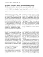
I-α-mannosidase. Decay plots indicated efficient
clearance of either probe, with 50% of injected dose being
eliminated from the blood during 2–5 min (Fig. 1). The
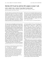
liver was the main site of uptake (Fig. 2), while a surpris-
ing finding was uptake in the lungs. Blood radioactivity af-
ter 15–20 min was 15–20% of injected dose. This equals
the amount of unbound
125
I after gel filtration through a
PD-10 column of a sample of the intravenously adminis-
tered ligands.
In vivo liver cell identification
Intravenuosly administered TRITC-monodisperse poly-
mer particles (MDPP) for identification of phagocytosing
KC accumulated mainly periportally in liver acini (Figs.
3A, 3B, 3C). Immunoelectron microscopy of liver sections
that had been reacted with anti-TRITC-antibodies and
protein A-gold revealed the presence of gold particles
along the periphery of the surface of the particles, allow-
ing a reliable identification and intracellular location of
TRITC-MDPP (Figs. 4A, 4B). In contrast to these particles,
FITC-FSA was taken up exclusively in LSEC-like cells lin-
ing the liver sinusoids (Fig. 3B). To distinguish LSEC from
stellate cells, double immunolabeling was performed to
visualize FITC-FSA and desmin in transmission electron
microscopy. FITC-FSA and desmin were observed in dis-
tinct cell types along the sinusoidal lining (Fig. 5). FITC-
FSA was associated with organelles judged as lysosomes of
LSEC.
Cell separation
The number of non-parenchymal cells (NPC) obtained
per liver following collagenase dispersion and isopycnic
density separation in iodixanol was 280 × 10
7
(range 50–
890 × 10
7
) (weight of liver 237.1 g (43.6)) with a viability
of 95.4% (2.5) as judged by trypan blue exclusion (Table
1). The corresponding figures for hepatocytes were 1880 ×
10
7
(1110 × 10
7
) and 94.1% (2.2). The cells obtained af-
ter iodixanol separation were subjected to centrifugal elu-
triation and collected in 4 fractions. The corresponding
recoveries expressed as number of NPC and percentages of
total are displayed in Table 2.
Identification of cultured cells
Cells, seeded on fibronectin-coated substrate, obtained
from the elutriation fractions yielded LSEC cultures of var-
Comparative Hepatology 2003, 2 />Page 3 of 14
(page number not for citation purposes)
ying purity (Table 3). We used in vivo (Fig. 6A) or in vitro
administered FITC-FSA as a specific LSEC marker, positive
reaction with anti-desmin antibodies as a specific marker
of stellate cells (Fig. 7A), and a specific anti-pig macro-
phage antibody (Fig. 7B) or phagocytosis of TRITC-MDPP
(Fig. 6B) as KC specific markers. Using these criteria, cul-
tures resulting from elutriation fraction 1 were shown to
contain 63.9% stellate cells; cultures established from
fraction 2 contained 80.4% LSEC, and fractions 3 and 4
contained 66.2% and 61.0% LSEC. Cells that reacted with
anti-pig-macrophage antibodies or phagocytosed TRITC-
MDPP contained no FITC-FSA. Stellate cells were distin-
guished by immunolabeling with anti-desmin antibodies
or by their content of characteristic autofluorescence from
vitamin A droplets when irradiated with light of 328 nm
of wavelength ([12]) (Fig. 6C).
Specificity of endocytosis in cultured LSECs and hepato-
cytes
The specificity of endocytosis of
125
I-FSA and
125
I-asialo-
orusomucoid protein (ASOR) in cultured LSEC and hepa-
tocytes was studied by attempting to inhibit the uptake of
trace amounts of radiolabeled ligands using excess
amounts of unlabeled ligands. Incubation of LSEC cul-
tures with
125
I-FSA in the presence of excess amounts of
unlabeled FSA (100 mg·mL
-1
) resulted in a 90% inhibi-
tion of uptake (Fig. 8). The presence of galactose (50
mmol·L
-1
) did not inhibit endocytosis of
125
I-FSA by
LSEC. Incubation of hepatocytes with
125
I-ASOR in the
presence of excess amounts of galactose (50 mmol·L
-1
)
inhibited uptake by 85%. Unlabeled FSA did not inhibit
endocytosis of
125
I-ASOR by hepatocytes (Fig. 8).
Discussion
Although it is assumed that pig LSEC perform the same
physiological scavenger function as it has been observed
in rat LSEC [3], it has actually never been shown. Since en-
dothelial cells of the liver of most vertebrate species are as-
sociated with clearance activity [9], we wanted to study
whether pig liver clearance function resides in the scaven-
ger activity of LSEC in the same way as it has been shown
in the rat. To this end, endocytosis of both foreign and
physiological waste macromolecules in pig LSEC was
studied in vivo and in vitro. For the in vitro studies we also
developed a method for mass isolation and culture of pig
LSEC.
Rate of elimination and organ distribution of FSA and
α
-
mannosidase
First we studied the circulatory survival and anatomical
distribution of FSA, a frequently used test ligand for the
LSEC scavenger receptor in rat [13], and α-mannosidase,
a physiological ligand for the mannose receptor of rat
LSEC [14]. Studies in the rat and other vertebrates have
shown that
125
I-FSA is degraded very rapidly after uptake,
resulting in rapid escape of radiotracer from the site of up-
take. For this reason, FSA was labeled with
125
I-TC, which
is trapped in the lysosomes at the cellular site of uptake,
Figure 1
Clearance kinetics. Approximately 100 × 10
6
cpm of
125
I-
tyramine cellobiose-formaldehyde-treated serum albumin
(TC-FSA) and
125
I-α-mannosidase were injected intrave-
nously. Radioactivity in the blood sample collected immedi-
ately after injection was taken as 100%. Blood samples were
collected every minute during the first 10 minutes, then
every 5 minutes for one hour. (Open boxes:
125
I-TC-FSA; n
= 2, closed boxes:
125
I-α-mannosidase; n = 1).
0
20
40
60
80
100
0 102030405060
min
Radioactivity (% remaining)
Figure 2
Anatomical distribution. The animals used in the blood
clearance studies (Fig. 1) were analyzed for anatomical distri-
bution of radioactivity 1 h after injection. More than 90% of
the injected doses were recovered in the organs listed.
Results are expressed as percent total radioactivity recov-
ered. (Grey bars:
125
I-TC-FSA; n = 2, white bars:
125
I-α-man-
nosidase; n = 1).
organ
liver
lung
blood
spleen
kidney
heart
urine
duodenum
stomach
thymus
thyroidea
muscle
lymphnode
aorta
Radioactivity (% of recovered)
0
10
20
30
40
50
60
70
Comparative Hepatology 2003, 2 />Page 4 of 14
(page number not for citation purposes)
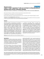
Figure 3
Fluorescence micrographs of liver section. Following intravenous administration of fluorescently labeled substances, sec-
tions were prepared as described in the Methods section. A heterogeneous distribution of yellow fluorescence from TRITC-
labeled monodisperse polymer particles (MDPP) phagocytosed by Kupffer cells was located mainly in the periportal region of
the liver acinus (arrows) (A). Green fluorescence along the lining of the liver sinusoids identifies endocytosed FITC-formalde-
hyde-treated serum albumin (FSA) by liver sinusoidal endothelial cells (LSEC), while the localization of phagocytosed MDPP is
shown by arrows (B). Uptake of FITC-FSA (arrowheads) and MDPP (arrow) is shown more clearly at higher magnification in C.
(Scale bars; A: 80 µm, B: 20 µm, C: 8 µm).
Comparative Hepatology 2003, 2 />Page 5 of 14
(page number not for citation purposes)
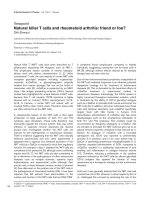
Figure 4
Uptake of monodisperse polymer particles (MDPP) in Kupffer cells (KC). Following intravenous administration of
fluorescently labeled substances, sections were prepared as described in the Methods section for transmission electron micro-
scopy. MDPP are located intracellularly in Kupffer cells, as judged by their characteristic phagocytosis of the particles (A).
Hepatocytes (Hep) contain numerous mitochondria. The cells that contain fat vacuoles (FV) may represent stellate cells (SC).
To distinguish between vacuoles containing fat and phagocytosed MDPP, sections were immunolabeled with monoclonal anti-
mouse TRITC-conjugate. Gold particles are located in the periphery of MDPP where the TRITC-molecules are attached (B).
(Scale bars; A: 2 µm, B: 500 nm).
Comparative Hepatology 2003, 2 />Page 6 of 14
(page number not for citation purposes)
Figure 5
Stellate cells (SC) and liver sinusoidal endothelial cells (LSEC). Following intravenous administration of fluorescently
labeled substances, sections were prepared as described in the Methods section for transmission electron microscopy.
Ultrathin sections were immunodouble labeled to visualize both FITC-labeled formaldehyde-treated serum albumin (FSA) in
LSEC and desmin in SC. Figures B and C are higher magnification of segments of figure A. Cells lining the sinusoids (A) are
LSEC as judged by the localization of small gold particles (5 nm, small arrow) in organelles taken as lysosomes (B). The cell con-
taining large fatty vacuoles (FV) and large gold particles (10 nm, large arrow), was judged as a stellate cell (SC) (C). (Scale bars;
A: 1 µm, B: 200 nm, C: 500 nm).
Comparative Hepatology 2003, 2 />Page 7 of 14
(page number not for citation purposes)
thus preventing
125
I escape from the uptake site [15]. Pre-
vious studies in the rat and other vertebrates showed that
α-mannosidase, after its rapid uptake by the mannose re-
ceptor, accumulates within lysosomes and is reused for
several hours before being degraded [5]. Therefore, α-
mannosidase was labeled with
125
I in a direct, conven-
tional manner. Both
125
I-TC-FSA and
125
I-α-mannosidase
were rapidly eliminated from the circulation, with 50% of
the ligands being removed during the first 2–5 min after
intravenous administration. This rapid removal suggested
a very efficient uptake mechanism. Monitoring of radioac-
tivity in the organs showed that the liver contained 53%
(FSA) and 62% (α-mannosidase) of injected dose, sug-
gesting that a cell type(s) in liver was responsible for clear-
ance via the scavenger and mannose receptors.
Surprisingly, as much as 26% FSA and 18% α-mannosi-
dase were recovered in lungs. This is clearly different than
in the rat, where uptake in the lungs of these and other
soluble macromolecular waste products have not been
observed [3]. A recent report [16] showed that ligands for
studies of reticuloendothelial function were taken up in
both lung and liver of pig, similarly to what we found us-
ing α-mannosidase and FSA. It was concluded from that
study that
198
Au colloidal particles and iron oxide parti-
cles were taken up in pulmonary intravascular macro-
phages. The possibility that these ligands might have been
taken up by scavenger endothelial cells was not men-
tioned in that paper.
In vivo liver cell identification
To determine the role of different sinusoidal cells in the
clearance function of pig liver, the cellular site of uptake
of FITC-FSA was compared with that of TRITC-MDPP (a
functional marker of phagocytosing KC), and immunore-
active desmin (a marker of stellate cells). Since light mi-
croscopy does not allow a clear distinction between
particles that are truly internalized and those that are as-
sociated with the cell surface, liver tissue was prepared for
electron microscopy. To enable a distinction between vi-
tamin A-containing lipid droplets in stellate cells and in-
ternalized MDPP in KC, sections were first incubated with
anti-TRITC-antibodies, then with protein A-gold. Obser-
vations of these sections revealed gold staining along the
surface of the MDPP particles, corresponding to the sur-
face localization of TRITC. Double immunolabeling
showed that FITC-FSA (5 nm gold) was always associated
with endothelial like lining cells that neither took up
MDPP nor contained desmin (10 nm gold), indicating
that the hepatic uptake in vivo of FSA was exclusively in
LSEC, similar to what has been found in the rat [13].
Separation, cultivation, and characterization of cells in vit-
ro
To allow a more detailed study of the tentative scavenger
function of pig LSEC, we developed a protocol for isola-
tion of sinusoidal cells. The protocol was modified as
compared to rat [17] and mouse liver. According to the lit-
erature, rat and mouse liver sinusoidal cells can be isolat-
ed in high yield and purity using isopycnic separation. We
found that this method was insufficient to isolate such
cells from pig due to the high number of desmin-positive
cells; therefore, we included centrifugal elutriation to sep-
arate the cells according to size. Using collagenase per-
fusion through the portal vein, followed by differential
centrifugation, isopycnic centrifugation, and centrifugal
elutriation we obtained 4 fractions, of which fraction 2,
Table 1: Parameters of liver perfusions, recovery of non-parenchymal cells (NPC), and viability (n = 10).
Body wt (kg) Liver wt (g) Collagenase perfusion
(min)
Portal-flow (mL·min
-1
) Total NPC (×10
7
) Viability NPC (%)
7.6 (0.6)* 237.1 (43.6)* 16.5 (3.2)* 304.9 (47.4)* 280 (50–890)
#
95.4 (2.5)*
*The values are expressed as: mean (standard deviation).
#
The value is expressed as: mean (range).
Table 2: Yield of non-parenchymal cells (NPC) from elutriation fractions (n = 4).
Fraction Flow rate (mL·min
-1
) Number of NPC (×10
7
) % of total NPC
1 18.5 (1.0) 190 (68) 69.2
2 32.0 (0.0) 62 (12) 23.0
3 37.0 (0.0) 13 (8) 5.0
4 45.0 (0.0) 7 (2) 2.7
The values are expressed as: mean (standard deviation).
Comparative Hepatology 2003, 2 />Page 8 of 14
(page number not for citation purposes)
Figure 6
Fluorescence micrographs of cultured liver sinusoidal endothelial cells (LSEC). Cultures were prepared as
described in the Methods section. The cultures were fixed in 4% paraformaldehyde, after 6 h of incubation. FITC-labeled for-
maldehyde-treated serum albumin (FSA) and TRITC-labeled monodisperse polymer particles (MDPP) were administered intra-
venously prior to isolation of liver cells. Fluorescent microscopy reveals a homogeneous LSEC culture contaminated by a few
cells with TRITC-MDPP and lipid containing vacuoles. The green fluorescence from endocytosed FITC-FSA demonstrates that
most cells are LSEC, and that the probe is localized in cytoplasmic vacuoles (A), whereas the yellow fluorescence from phago-
cytosed TRITC-MDPP identifies Kupffer cells (arrows) (B). Autofluorescence from vitamin A identifies stellate cells (arrows)
(C). (Scale bars; 20 µm).
Comparative Hepatology 2003, 2 />Page 9 of 14
(page number not for citation purposes)
Figure 7
Fluorescent micrographs of cultured stellate cells and Kupffer cells. Cultures were prepared as described in Meth-
ods. The cultures were fixed in 4% paraformaldehyde, after 1 h of incubation. Micrographs of cultured stellate cells stained with
monoclonal anti-desmin antibody (A) and cultured Kupffer cells stained with monoclonal anti-pig macrophage antibody (B).
(Scale bars; 20 µm).
Comparative Hepatology 2003, 2 />Page 10 of 14
(page number not for citation purposes)
Table 3: Identification of cells after cultivation of elutriation fractions.
Fraction FITC – FSA Desmin KC Hepatocytes
1 32.1 (12.6) 63.9 (15.4) 4.0 (6.9) 0.0 (0.0)
2 80.4 (6.4) 10.9 (9.2) 7.0 (1.4) 1.7 (1.6)
3 66.2 (11.1) 7.1 (3.8) 15.2 (13.5) 11.5 (17.5)
4 61.0 (17.5) 2.1 (1.7) 10.9 (10.9) 26.0 (19.4)
Prior to isolation of cells, pigs received FITC-labeled formaldehyde-treated serum albumin (FSA) intravenously. Stellate cells stained with mono-
clonal mouse anti-human desmin antibody and Kupffer cells (KC) stained with anti-pig macrophage antibodies. Hepatocytes were identified by sim-
ple morphology. Values are percent of total number of cells per culture (n = 3). The values are expressed as: mean (standard deviation).
Figure 8
Specificity of endocytosis of
125
I-formaldehyde-treated serum albumin (FSA) in cultured liver sinusoidal
endothelial cells (LSEC) (grey and white bars), and
125
I-asialo-orusomucoid protein (ASOR) in cultured hepa-
tocytes (black and hatched bars). Monolayer cultures were incubated for 2 hrs, at 37°C, with trace amounts of labeled lig-
and alone (control) or together with excess amounts of unlabeled FSA (100 µg·mL
-1
) or galactose (50 mmol·L
-1
). The presence
of unlabeled FSA inhibited effectively the endocytosis of
125
I-FSA in LSEC, while galactose showed no such inhibitory effect.
Galactose had an inhibitory effect on endocytosis of
125
I-ASOR in hepatocytes, whereas unlabeled FSA showed no such inhibi-
tory effect. Results, given as percent of control, are the means of triplicate experiments. Grey and white bars: 100% corre-
sponds to 12.7% of added cpm, black and hatched bars: 100% corresponds to 14.6% of added cpm. White and hatched areas of
bars represent % degraded ligand. Grey and black areas of bars represent % cell-associated ligand.
0
20
40
60
80
100
Control Galactose FSA
Radioactivity (% of control)
Comparative Hepatology 2003, 2 />Page 11 of 14
(page number not for citation purposes)
containing 62 × 10
7
(12 × 10
7
) sinusoidal cells, gave mon-
olayer cultures on fibronectin that were 80% pure in
LSEC, as judged by uptake of intravenously administered
FITC-FSA. The FITC-FSA positive LSEC did not react with
anti-desmin or anti-pig macrophage antibodies. Con-
versely, only cells that took up TRITC-MDPP reacted with
the anti-pig macrophage antibodies (about 11% of the
plated cells).
Specific endocytosis of FITC-FSA was used successfully for
vital-staining of LSEC either in vivo by intravenous injec-
tion, or by administration to LSEC-cultures in vitro. Since
FITC-FSA is a highly stable molecule that can be easily and
inexpensively prepared without special equipment, and
stored for years in the freezer or refrigerator, we find this
probe superior to other marker molecules launched as
LSEC-specific functional markers (e.g., fluorescently-la-
beled acetylated LDL [18]) that are unstable and require
expensive equipment for preparation. Although KC could
be identified in vivo by their specific ability to phagocytose
MDPP, this marker was disadvantageous for in vitro KC
identification for the following two reasons: 1) KC that
had phagocytosed intravenously administered MDPP ex-
hibited significantly altered sedimentation properties on
subseqent centrifugal elutriation; and 2) in vitro cultured
KC did not phagocytose MDPP under the conditions
used. These difficulties forced us to look for alternative
ways of identifying the KC. Fortunately, an anti-pig-mac-
rophage antibody kindly provided by Dr. A. Berndt, Ger-
many, gave a specific staining of KC, but not of other types
of liver cells; therefore this method was preferred as a
method for identification of KC. The identification of stel-
late cells was at first based on demonstration of vitamin A-
positive lipid droplets, that could be easily detected by
autofluorescence when illuminated with light of 328 nm
of wavelength. However, since vitamin A appeared to be
present to a variable extent in stellate cells, we decided to
employ immunostaining for desmin with a monoclonal
antibody.
Specificity of endocytosis in cultured LSECs
The observation that uptake of
125
I-FSA by cultured LSEC
could be inhibited by excess amounts of FSA, but not by
galactose, indicates that the cells express the same func-
tional type of scavenger receptor as has been reported for
rat LSEC [13]. The finding that FITC-FSA was taken up
only in LSEC in vivo and in vitro is further evidence that the
porcine and rat LSEC scavenger receptors are functionally
similar. The finding that LSEC isolated and cultured ac-
cording to the presently developed protocol, perform a
highly active scavenger-receptor-mediated endocytosis
proves that pig LSEC prepared according to our protocol
have preserved their in vivo scavenger function.
Although several authors have reported the presence of
LSEC in cultures of porcine liver cells, methods of identi-
fication of LSEC were unable to distinguish convincingly
LSEC from other types of liver cells. In a study on isolation
of NPC from piglets (1–15 days of age), Caperna et al.
[19] used absence of uptake of 5 µm latex beads as a char-
acteristic of LSEC. However, our observation that KC only
rarely phagocytose particles in vitro, along with the fact
that other liver cell types (i.e., stellate cells and lym-
phocytes) are unable to perform phagocytosis, suggest
that absence of phagocytosis is not a valid way of distin-
guishing LSEC from other liver cells. In other studies, pos-
itive immune staining of von Willebrand factor has been
used to identify pig LSEC [10,11]. However, the fact that
this marker is widely used to identify vascular endothelial
cells in general [20], together with other reports claiming
that LSEC are devoid, or stain only weakly for this antigen,
suggest that von Willebrand factor should not be used as
a specific LSEC marker. Prostacyclin PGI2 generation has
been used as a LSEC-specific marker [21]. However, PGI2
is a general endothelial marker, and can not be used as a
LSEC-specific marker. Fluorescently labeled acetylated
LDL has been used in much the same way as we used
FITC-FSA as a vital stain to demonstrate the scavenger
function of LSEC [22]. Nevertheless, it has been shown
that this probe, in contrast to FITC-FSA, is taken up in
brain microvessel endothelial cells [23], as well as in pe-
ripheral macrophages [24]).), suggesting that FITC-FSA is
superior as a LSEC specific marker.
Conclusions
This study showed that pig LSEC are endowed with the
same scavenger activity as rat LSEC. Although a method
was recently reported for isolation of pig LSEC, with an av-
erage yield of 90 000 cells per liver [10], our method offers
true mass isolation of LSEC, yielding 62 × 10
7
(12 × 10
7
)
purified LSEC per liver.
Methods
Antibodies and ligands
Monoclonal mouse anti-human desmin, clone D33, was
from DAKO A/S, Denmark. Monoclonal goat anti-mouse
IgG, TRITC-conjugate, was from Zymed, CA. Monoclonal
rabbit anti-FITC was from DAKO A/S, Denmark. Mono-
clonal rabbit anti-TRITC was from Molecular Probes Inc.,
OR. Two mouse monoclonal antibodies (clones 2G6 and
2B10) against porcine macrophages were kindly provided
by Dr. A. Berndt, Institute of Pathology, Friedrich Schiller
University, Jena, Germany [25]. FSA was prepared as de-
scribed [26]. ASOR was kindly provided by Dr. Trond
Berg, University of Oslo, Norway. Bovine α-mannosidase
was donated by Dr. O.K. Tollersrud, University of Tromsø,
Norway.
Comparative Hepatology 2003, 2 />Page 12 of 14
(page number not for citation purposes)
Labeling procedures
Proteins were labeled with
125
I (carrier free Na
125
I from
Institute of Energiteknikk, Norway) either by a direct reac-
tion employing Iodogen (Pierce, Rockford, IL) [27], or by
conjugating the protein with
125
I-labeled TC [15]. The io-
dinated proteins, with
125
I attached to aromatic amino ac-
ids, were separated from free
125
I on a PD-10 column
(Sephadex G-25, Pharmacia, Sweden) equilibrated and
eluted with PBS.
FSA (0.8 mg·mL
-1
) was incubated with FITC (Sigma-
Aldrich, Norway) in sodium carbonate buffer (0.5 mol·L
-
1
, pH 9.5) in a protein/dye weight ratio of 1:1 at 4°C over-
night. Unreacted dye was removed by gel filtration
through a PD-10 column equilibrated and eluted with
PBS without Ca
2+
or Mg
2+
.
Approximately 8.0 × 10
10
MDPP, with a diameter of 2.0
µm (SINTEF, University of Trondheim, Norway) were in-
cubated overnight in 1 mg·mL
-1
TRITC (ICN Biomedicals
Inc., CA) in sodium carbonate buffer (0.1 mol·L
-1
, pH
9.5) at 4°C. The TRITC-MDPP were washed three times in
70% ethanol, and finally in PBS to remove unbound
TRITC.
Determination of anatomical distribution and serum t1/2
Serum half-life and organ distribution of intravenously
administered radiolabeled FSA and α-mannosidase were
determined as described [28]. Briefly, approximately 100
× 10
6
cpm
125
I-TC-FSA or
125
I-α-mannosidase were in-
jected into the left jugular vein. Blood sampling was start-
ed immediately by collecting 2 mL blood samples from an
intravenous catheter positioned in the right jugular vein.
The organs were taken out after one hour, weighed and
measured in a γ-counter (Cobra II, Packard Instrument
Co., Inc., CT).
Surgical procedures
The experimental protocol was approved by the local
steering committee of the Norwegian Experimental Ani-
mal Board. All animals received care according to the cri-
teria outlined in the "Guide for the Care and Use of
Laboratory Animals" prepared by the National Academy
of Sciences and published by the National Institutes of
Health (NIH publication 86–23 revised 1985). Castrated
male pigs weighing 7.6 kg (0.6) (Sus scrofa domesticus,
Norwegian strain) were fasted 18 hours with free access to
water. Intramuscular ketamine (75 mg·kg
-1
) (Parke-Dav-
is, Sweden) and atropine sulfate 1 mg (Nycomed Pharma,
Norway) were used as premedication. The pigs were tra-
cheostomized, intubated, and ventilated on a volume
controlled ventilator (Servo 900, Elema-Schönander, Swe-
den). Anesthesia was maintained with continuous i.v. in-
fusions of sodium pentobarbital (4 mg·kg
-1
·min
-1
)
(Abbott Scandinavia, Sweden) and fentanyl (0.05 mg·kg
-
1
·min
-1
) (Alpharma, Norway). A midline laparotomy was
performed. The portal vein was mobilized and the portal
bloodflow was measured with a flowprobe (CardioMed,
Norway). Sodium heparin (3000 IU) (Leo, Denmark) was
administered intravenously. FITC-FSA (40 mg) was ad-
ministered intravenously via the left jugular vein, while
some pigs received TRITC-MDPP injected via the portal
vein as two boluses 30 and 5 minutes prior to perfusion.
The hepatoduodenal ligament was ligated, and the portal
vein was cannulated. Ca
2+
-free HEPES [29] solution at
37°C was perfused in vivo with a roller pump (Gambro,
AK-10, Sweden) at the same rate as the portal blood flow.
The liver was weighed, thereafter perfused ex vivo with a
Ca
2+
-containing HEPES-solution with 0.02% Collagenase
P (Roche Diagnostics, Norway) in a basin at 37°C.
The digested liver was transferred to a different basin con-
taining RPMI 1640 medium with 1% BSA (4°C). After re-
moving the stroma, the suspension was subsequently
filtered through 1000 and 500 µm nylon mesh. The result-
ing cell suspension was subjected to centrifugation at 50 g
to selectively sediment hepatocytes from NPC. The pellet
of hepatocytes was resuspended, and subsequently
washed two more times at 50 g. Cell viability was estimat-
ed from the percentage of cells which excluded 0.4%
trypan blue.
Separation of NPC
The NPC in the first and second supernatants from the
low-speed centrifugations were pelleted by high-speed
centrifugation (1300 g) followed by resuspension in small
volume before isopycnic centrifugation in a 12% iodixa-
nol gradient (Nycomed, Norway) to generate a band of
further enriched NPC. This fraction was washed in PBS
and resuspended in HBSS, supplied with 4 mmol·L
-1
ED-
TA, 1% FCS and 0.3% BSA, and loaded into the mixing
chamber over 1 min. The NPC suspensions were fraction-
ated in a J-21-B centrifuge fitted with a JE-6 elutriator rotor
(Beckmann Instruments, Inc., CA). A pre-enrichment step
was performed to further remove hepatocytes and cell ag-
gregates with a pump flow rate of 30 mL·min
-1
and rotor-
speed at 1400 rpm. Four consecutive fractions,
corresponding to pump flow rates of 18.5, 32, 37 and 45
mL·min
-1
, were collected from the rotor which was spun
at a constant speed of 2500 rpm, at 15°C. The volume of
effluent at each flow rate was 150, 250, 250 and 250 mL.
Immunolabeling and characterization of cultured cells
Monolayer cultures of hepatocytes and cells from each
elutriation fraction were established on fibronectin-coat-
ed glass-coverslips inserted in 2 cm
2
culture wells and
maintained in serum-free RPMI 1640 medium in a CO2-
incubator at 37°C. Cultures were fixed in 4% paraformal-
dehyde in PBS for 1 hour, after attachment and spreading.
Comparative Hepatology 2003, 2 />Page 13 of 14
(page number not for citation purposes)
Immunocytochemistry
To examine the distribution of FITC-FSA and TRITC-
MDPP, small sections were taken at the periphery of the
liver lobules during the washing perfusion, fixed in 4%
paraformaldehyde, prepared and sectioned for fluores-
cence microscopy.
Sections and fixed cultures were embedded in antifade
mounting medium (Dako Corp., CA). Examination of the
mounted coverslips was performed in an Axiophot phot-
omicroscope equipped with phase-contrast, fluorescence
optics and a filter for excitation (wavelength 328 nm) of
vitamin A (Zeiss Axiophot, Germany). Pictures were re-
corded on Kodak Ektachrome P1600X ASA (Kodak, Ja-
pan).
Whole livers were perfusion-fixed with McDowell's fixa-
tive (4% paraformaldehyde, 1% glutaraldehyde in PBS).
Specimens for immunotransmission electron microscopy
were prepared as described [30]. Briefly, small cubes were
cut out of the liver tissue, and incubated in 2.3 mol·L
-1
su-
crose overnight before immersion in liquid nitrogen on
aluminum pins. Ultrathin sections were cut using a dia-
mond knife (Drukker international, The Netherlands).
Before immunolabeling, sections were retrieved in su-
crose and mounted on carbon coated grids. Sections were
blocked for 15 minutes in 1% cold water fish skin gelatin
(Sigma-Aldrich, Norway), followed by incubation with
anti-TRITC (diluted in cold water fish skin gelatin), wash-
ing in PBS and incubation with protein A-gold (University
of Utrecht, The Netherlands) [31] diluted in cold water
fish skin gelatin for 15 minutes. Double labeling was per-
formed in a sequential manner, using a fixative block (1%
glutaraldehyde) between the first and second marker pair.
The sections were then washed in PBS followed by wash-
ing in distilled water and dried in 1.8% methylcellulose
and 0.3% uranyl acetate, and examined in a JEOL JEM
1010 transmission electron microscope (Tokyo, Japan) at
80 kV. Micrographs were recorded on Kodak electron mi-
croscope film, no. 4489 (Kodak, Tokyo, Japan).
Endocytosis studies in vitro
LSEC and hepatocyte cultures, established in 2 cm
2
wells
and maintained in serum-free RPMI 1640 medium, were
washed and supplied with fresh medium containing 1%
serum albumin and labeled proteins,
125
I-FSA or
125
I-
ASOR (20 000 cpm per well). Specificity of endocytosis
was studied by incubation with excess amounts of unla-
beled FSA (100 µg·mL
-1
) or galactose (50 mmol·L
-1
). In-
cubations, carried out for 2 hours at 37°C to measure
endocytosis, were terminated by transferring the media,
along with one wash, to tubes containing 20% TCA,
which precipitates only intact protein. The extent of deg-
radation was determined by measuring the radioactivity
in the pellet and supernatant that was obtained after cen-
trifugation of the media. Cell-associated ligand was quan-
tified by measuring the amount of radioactivity released
by solubilizing the cultures in 1% (w/v) SDS. Radioactiv-
ity was measured in a γ-counter (Cobra II, Packard Instru-
ment Co., Inc., Meridien, CT).
Statistics
The values are expressed as: mean (standard deviation)
unless otherwise noted.
Authors' contributions
GIN and KHE designed and carried out the experiments.
GIN drafted the manuscript. LMY contributed significant-
ly to the animal preparation. RO carried out the electron
microscopy studies. AR and BS coordinated the study and
contributed to the text of the manuscript.
All authors have read and approved the final manuscript.
Acknowledgements
The authors would like to express their gratitude to Hege Hagerup, Ellinor
Hareide, and Marna-Lill Kjæreng, Surgical Research Laboratory, University
of Tromsø, for skilful technical assistance. The surgical assistance by Ebra-
him Aghajani M.D., Odd-Petter Elvenes M.D. and Christian Korvald M.D. is
greatly appreciated. The authors are grateful for the advice of Dr. Peter A.
G. McCourt.
This work was supported by grants from the Norwegian Research Council.
References
1. Watanabe FD, Mullon CJ, Hewitt WR, Arkadopoulos N, Kahaku E,
Eguchi S, Khalili T, Arnaout W, Shackleton CR, Rozga J, Solomon B
and Demetriou AA Clinical experience with a bioartificial liver
in the treatment of severe liver failure. A phase I clinical tri-
al. Ann Surg 1997, 225:484-491
2. Aschoff L Ergeb Inn Med Kinderhk 1924, 26:1-118
3. Smedsrød B, Pertoft H, Gustafson S and Laurent TC Scavenger
functions of the liver endothelial cell. Biochem J 1990, 266:313-
327
4. Hansen B, Melkko J and Smedsrød B Serum is a rich source of lig-
ands for the scavenger receptor of hepatic sinusoidal en-
dothelial cells. Mol Cell Biochem 2002, 229:63-72
5. Smedsrød B and Tollersrud OK Sinusoidal liver endothelial cells
recruit lysosomal enzymes from the circulation by mannose-
receptor mediated endocytosis. Cells of the hepatic sinusoid (Edited
by: Wake K, Wisse E, Knook DL) Leiden, Kupffer Cell Foundation 1995,
180-183
6. Van Berkel TJ, De Rijke YB and Kruijt JK Different fate in vivo of
oxidatively modified low density lipoprotein and acetylated
low density lipoprotein in rats. Recognition by various scav-
enger receptors on Kupffer and endothelial liver cells. J Biol
Chem 1991, 266:2282-2289
7. Smedsrød B, Melkko J, Araki N, Sano H and Horiuchi S Advanced
glycation end products are eliminated by scavenger-recep-
tor-mediated endocytosis in hepatic sinusoidal Kupffer and
endothelial cells. Biochem J 1997, 322:567-573
8. Kawai Y, Smedsrød B, Elvevold K and Wake K Uptake of lithium
carmine by sinusoidal endothelial and Kupffer cells of the rat
liver: new insights into the classical vital staining and the re-
ticulo-endothelial system. Cell Tissue Res 1998, 292:395-410
9. Seternes T, Sørensen KK and Smedsrød B Scavenger endothelial
cells of vertebrates: a nonperipheral leukocyte system for
high-capacity elimination of waste macromolecules. Proc Natl
Acad Sci USA 2002, 99:7594-7597
10. Gerlach JC, Zeilinger K, Spatkowski G, Hentschel F, Schnoy N, Kol-
beck S, Schindler RK and Neuhaus P Large-scale isolation of sinu-
Publish with Bio Med Central and every
scientist can read your work free of charge
"BioMed Central will be the most significant development for
disseminating the results of biomedical research in our lifetime."
Sir Paul Nurse, Cancer Research UK
Your research papers will be:
available free of charge to the entire biomedical community
peer reviewed and published immediately upon acceptance
cited in PubMed and archived on PubMed Central
yours — you keep the copyright
Submit your manuscript here:
/>BioMedcentral
Comparative Hepatology 2003, 2 />Page 14 of 14
(page number not for citation purposes)
soidal endothelial cells from pig and human liver. J Surg Res
2001, 100:39-45
11. Cattan P, Zhang B, Braet F, Atia N, Conti F, Conjeaud H, Weill B,
Chereau C, Houssin D and Calmus Y Comparison between aortic
and sinusoidal liver endothelial cells as targets of hyperacute
xenogeneic rejection in the pig to human combination. Trans-
plantation 1996, 62:803-810
12. Knook DL, Seffelaar AM and de Leeuw AM Fat-storing cells of the
rat liver. Their isolation and purification. Exp Cell Res 1982,
139:468-471
13. Eskild W, Kindberg GM, Smedsrød B, Blomhoff R, Norum KR and
Berg T Intracellular transport of formaldehyde-treated se-
rum albumin in liver endothelial cells after uptake via scav-
enger receptors. Biochem J 1989, 258:511-520
14. Stahl P, Six H, Rodman JS, Schlesinger P, Tulsiani DR and Touster O
Evidence for specific recognition sites mediating clearance
of lysosomal enzymes in vivo. Proc Natl Acad Sci USA 1976,
73:4045-4049
15. Pittman RC, Carew TE, Glass CK, Green SR, Taylor CA Jr and Attie
AD A radioiodinated, intracellularly trapped ligand for deter-
mining the sites of plasma protein degradation in vivo. Bio-
chem J 1983, 212:791-800
16. Brain JD, Molina RM, DeCamp MM and Warner AE Pulmonary in-
travascular macrophages: their contribution to the mononu-
clear phagocyte system in 13 species. Am J Physiol 1999,
276:L146-154
17. Smedsrød B and Pertoft H Preparation of pure hepatocytes and
reticuloendothelial cells in high yield from a single rat liver
by means of Percoll centrifugation and selective adherence.
J Leukoc Biol 1985, 38:213-230
18. Pitas RE, Boyles J, Mahley RW and Bissell DM Uptake of chemically
modified low density lipoproteins in vivo is mediated by spe-
cific endothelial cells. J Cell Biol 1985, 100:103-117
19. Caperna TJ, Failla ML, Kornegay ET, Richards MP and Steele NC Iso-
lation and culture of parenchymal and nonparenchymal cells
from neonatal swine liver. J Anim Sci 1985, 61:1576-1586
20. Giddings JC, Banning AP, Ralis H and Lewis MJ Redistribution of
von Willebrand factor in porcine carotid arteries after bal-
loon angioplasty. Arterioscler Thromb Vasc Biol 1997, 17:1872-1878
21. Matsuura T, Kawada M, Hasumura S, Nagamori S, Obata T,
Yamaguchi M, Hataba Y, Tanaka H, Shimizu H, Unemura Y, Nonaka
K, Iwaki T, Kojima S, Aizaki H, Mizutani S and Ikenaga H High den-
sity culture of immortalized liver endothelial cells in the ra-
dial-flow bioreactor in the development of an artificial liver.
Int J Artif Organs 1998, 21:229-234X
22. Gerlach JC, Ziemer RA, Spatkowski G, Zeilinger K and Neuhaus P
Visualization of liver sinusoidal endothelial cell repair behav-
ior after preservation by in vitro time-lapse video microsco-
py. Transplantation 1997, 63:455-459
23. Teifel M and Friedl P Establishment of the permanent microv-
ascular endothelial cell line PBMEC/C1-2 from porcine
brains. Exp Cell Res 1996, 228:50-57
24. Rommeswinkel M, Severs NJ, Koster M and Robenek H Repression
of the macrophage scavenger receptor in macrophage-
smooth muscle cell heterokaryons. Arterioscler Thromb Vasc Biol
1995, 15:601-611
25. Berndt A, Heller M, Methner U, Kosmehl H and Muller G Mono-
clonal antibodies against porcine macrophages. Vet Immunol
Immunopathol 2000, 74:163-177
26. Mego JL and McQueen JD The Uptake and Degradation of In-
jected Labeled Proteins by Mouse-Liver Particles. Biochimica et
Biophysica Acta 1965, 100:136-14
27. Markwell MA A new solid-state reagent to iodinate proteins. I.
Conditions for the efficient labeling of antiserum. Anal Biochem
1982, 125:427-432
28. Smedsrød B and Einarsson M Clearance of tissue plasminogen
activator by mannose and galactose receptors in the liver.
Thromb Haemost 1990, 63:60-66
29. Alpini G, Phillips JO, Vroman B and LaRusso NF Recent advances
in the isolation of liver cells. Hepatology 1994, 20:494-514
30. Tokuyasu KT Application of cryoultramicrotomy to immuno-
cytochemistry J Microsc 1986, 143:139-149
31. Slot JW and Geuze HJ Sizing of protein A-colloidal gold probes
for immunoelectron microscopy. J Cell Biol 1981, 90:533-536