Báo cáo y học: "Association of adenoma and focal nodular hyperplasia: experience of a single French academic center" pptx
Bạn đang xem bản rút gọn của tài liệu. Xem và tải ngay bản đầy đủ của tài liệu tại đây (984.23 KB, 10 trang )
BioMed Central
Page 1 of 10
(page number not for citation purposes)
Comparative Hepatology
Open Access
Research
Association of adenoma and focal nodular hyperplasia: experience
of a single French academic center
Christophe Laurent
1,3
, Hervé Trillaud
1
, Sébastien Lepreux
2,3
,
Charles Balabaud*
1,3
and Paulette Bioulac-Sage
2,3
Address:
1
Fédération d'Hépato-gastro-entérologie, Hôpitaux Saint-André et Haut-Lévêque, France,
2
Service d'Anatomie Pathologique Hôpital
Pellegrin, CHU Bordeaux, France and
3
GREF INSERM E0362 – Université Bordeaux 2, 33076 Bordeaux Cedex, France
Email: Christophe Laurent - ; Hervé Trillaud - ;
Sébastien Lepreux - ; Charles Balabaud* - ; Paulette Bioulac-
Sage -
* Corresponding author
Abstract
Background: We report our experience of the simultaneous occurrence of adenoma and focal
nodular hyperplasia (FNH). Liver cell adenoma together with FNH was found in five out of 30 cases
of "multiple benign hepatocytic nodules" collected in our files of the Department of Pathology of
the University Hospital of Bordeaux, during the last 12 years. All five cases were women on oral
contraceptives. In all cases, the reason for surgery was the discovery, by imaging techniques, of an
adenoma (4 cases) or of an unidentified benign tumor, possibly an adenoma.
Results: Four cases of FNH were discovered by imaging techniques, prior to surgery. Additional
small nodules were diagnosed either during surgery or during the slicing of the specimen in 3 cases.
Adenoma and the FNH cases identified by imaging techniques were confirmed as such by light
microscopy. Some small nodules could not be categorized with certainty because they contained
biliary structures without ductular reaction. In one case, the non-nodular liver was abnormal
around the area in which there were multiple nodules: there was approximation of portal tracts
with portal and hepatic venous thromboses, and portal tract remnants with arteries surrounded
with a rim of fibrosis. In two cases, some large hepatic veins had thickened walls.
Conclusions: The association of FNH and adenoma could be coincidental or secondary to shared
causal mechanisms: a) systemic and local angiogenic abnormalities induced by oral contraceptives;
b) tumor-induced growth factors; c) thrombosis and local arterio-venous shunting. A better
recognition of the association of adenoma and FNH, particularly in the context of multiple nodules,
could be useful in clinical practice.
Background
Adenoma and focal nodular hyperplasia (FNH) are both
benign nodular hepatocellular lesions occurring in child
bearing women, in a liver that is otherwise histologically
normal or nearly normal. Both lesions looked like histo-
logically quite different in their typical forms; however,
some non-typical nodules, especially those of small size,
could be extremely challenging to hepatologists.
Published: 23 April 2003
Comparative Hepatology 2003, 2:6
Received: 30 October 2002
Accepted: 23 April 2003
This article is available from: />© 2003 Laurent et al; licensee BioMed Central Ltd. This is an Open Access article: verbatim copying and redistribution of this article are permitted in all
media for any purpose, provided this notice is preserved along with the article's original URL.
Comparative Hepatology 2003, 2 />Page 2 of 10
(page number not for citation purposes)
A central stellate fibrous region containing malformed
large arteries but usually no portal veins characterizes typ-
ical FNH. The lesion is multinodular, composed of nearly
normal hepatocytes, arranged in 1–2 cell-thick plates,
associated with a prominent bile ductular reaction, and
intermingled with inflammatory cells (at the interface
between hepatocytic nodules and fibrous bands). FNH is
considered as a hyperplastic process resulting from an
increased arterial flow. At the opposite, adenoma is a true
benign neoplasia, composed of slightly enlarged but
nearly normal hepatocytes, arranged in 1–2 cell-thick
plates, with numerous thin arteries dispersed within the
tumor, whereas there are no portal tracts and particularly
no biliary ducts. Adenoma exhibited usually peliotic and
necrotic hemorrhagic changes, steatotic areas and some-
times dysplasia. Their transformation into HCC is well
documented but remains rare.
The simultaneous occurrence of adenoma and focal nod-
ular hyperplasia (FNH) has been infrequently docu-
mented [1–6]. FNH associated with adenomas or
adenomatosis has been reported [7–9], suggesting a link
between these conditions.
We report the experience of a single French academic
center, which supports the possibility that the association
is more than by chance. Liver cell adenoma together with
FNH was found in five out of 30 cases of "multiple benign
hepatocytic nodules" collected in our files of the Depart-
ment of Pathology of the University Hospital of Bordeaux,
during the last 12 years.
Results and Discussion
Relevant clinical, radiological and surgical data are pre-
sented in Figures 1, 2 and 3. All 5 cases of liver cell ade-
noma together with FNH were women on oral
contraceptives. In all cases, the reason for surgery was the
discovery, by imaging techniques, of an adenoma (cases
1, 3, 4, 5) or of an unidentified benign tumor possibly an
adenoma (case 2). In four cases (2, 3, 4, 5) FNH were dis-
covered by imaging techniques prior to surgery. The case
in which the diagnosis of FNH was missed by pre-opera-
tive imaging was case 1 that had a small 1 cm superficial
FNH.
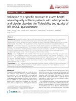
Figure 1
Adenoma plus focal nodular hyperplasia (cases 1, 2 and 3). Liver segments are indicated by roman numbers. FNH – focal nodu-
lar hyperplasia; GGT – gamma glutamyl transpeptidase; Hem: hemorrhagic; HV – hepatic vein; LH – left hepatectomy; LL – left
lobe; MRI – magnetic resonance imaging; N – nodule; OC – oral contraceptives; PV – portal vein; RH – right hepatectomy; RL
– right lobe; T – tumorectomy. (Abbreviations are valid for Figures 2 and 3.)
Case 1 2 3
Age / Sex 45 / F 40 / F 38 / F
OC (duration - in years) + (10) + (15) + (17)
Main clinical signs / disease Fatigue (alcoholism) Increased GGT Increased GGT
MRI location and number of nodules
(size largest one: cm)
VII, 1 (3) VI, 1 (5) VII, 1 (4) LL, 1 (3) LL, 1 (2) RL, 1 made of 2 parts (13)
Clinical, radiological diagnosis Adenoma FNH FNH ? FNH Adenoma Surrounded by FNH
Surgery (years) RH (95)
Plus tumorectomy
(III) for 1
superficial N
Biopsy Biopsy LH (98) RH (98)
Gross macroscopy diagnosis Adenoma FNH (1 cm) Adenoma FNH Adenoma Surrounded by FNH
Liver pathology nodules Adenoma FNH (1 cm) FNH FNH
Adenoma
(steatotic)
FNH + 2
steatotic N
(few mm) ?
Adenoma Surrounded by FNH
Liver pathology of non tumoral liver Normal
Some HV with thickened walls. Some isolated
arteries and abnormal portal tracts
Normal
Follow up (new nodule) 1 (small) probable FNH (99) No change No change
Comparative Hepatology 2003, 2 />Page 3 of 10
(page number not for citation purposes)
Figure 2
Adenoma plus focal nodular hyperplasia (case 4).
Figure 3
A - Adenoma plus focal nodular hyperplasia (case 5) B - Details of contraceptives, for all five cases.
Case 4
Age / Sex 29 / F
OC (duration - in years)
+ (2)
Main clinical signs / disease Abdominal pain + shock
MRI location and number of nodules
(size largest one: cm)
I (intratumural
bleeding), 1 (6)
II, 1 (1) III, 1 (1) VII, 1 (1) VIII, 1 (3) VI, 1 (1)
Clinical, radiological diagnosis Adenoma Adenoma Adenoma Adenoma Adenoma FNH
Many small N
Surgery (years)
LH + I (00)
3 T for superficial N (RL)
Gross macroscopy diagnosis Adenoma (Hem) Adenoma Adenoma Several small N ?
Liver pathology of nodules Adenoma (Hem) Adenoma Adenoma Adenoma - FNH - N? 2 FNH - 1N ?
Liver pathology of non tumoral liver
Some HV with thickened walls
Follow up (new nodule) No change
A
Case 5
Age / Sex 41 / F
OC (duration - in years) + (20)
Main clinical signs / disease Abdominal pain
MRI location and number of nodules (size largest one: cm) RL, 1 (3) RL, 1 (4)
Clinical, radiological diagnosis FNH Adenoma
Surgery (years) RH (02)
Gross macroscopy diagnosis
FNH
Adenoma + several N near the
gallbladder (few mm up to 1 cm)
Liver pathology of nodules FNH Adenoma - FNH - N ?
Liver pathology of non tumoral liver HV thrombosis. Areas of approximation + thrombosis of PV
Follow up (new nodule) No change
B
Oral contraceptives Cases
Levonorgestrel (0.15 - 0.20 mg), Ethinylestradiol (30 µg) Patients 2, 3 and 5
Gestodene (50 µg), Ethinylestradiol (30 µg) Patients 4 and 5
Cytoproterone (2 mg), Ethinylestradiol (35 µg) Patient 2
Norethisterone (1 mg), Ethinylestradiol (30 µg) Patient 5
Gestodene (75 µg), Ethinylestradiol (20 µg) Patient 3
Comparative Hepatology 2003, 2 />Page 4 of 10
(page number not for citation purposes)
Additional small nodules were discovered (cases 2, 4 and
5; Figs. 4,5,6,7) either during surgery or during post-resec-
tion dissection of the specimen. Case 4 was known to
have multiple nodules, one of which, at least, was
identified as FNH (Fig. 6) by pre-operative imaging. Addi-
tional small superficial nodules (in case 4) were inter-
preted as adenomas by the surgeon and resected, and were
solid FNH by light microscopy.
Adenoma and FNH recognized by imaging techniques
were easily identified as such by light microscopy. Con-
versely, some small nodules could not be identified with
certainty because of the presence of biliary structures with-
out ductular reaction (Figs. 4,5,6,7). To achieve a categor-
ical assignment for the purposes of this study, we
classified these small nodules with intralesional arteries
and few/rare biliary cells, better identified by cytokeratin
7 (CK7) immunostaining, as adenoma [10]; and nodules
with some/frequent biliary cells associated with intrale-
sional arteries as FNH.
In one case (case 5) there were additional abnormalities
in the non-nodular liver in the area around the multiple
nodules that were found (Figs. 4, 5). These abnormalities
Figure 4
Case 5, segment IV. (a) shows several small nodules on the surface of segment IV (arrows). Among the nodules (surface and
cut sections) there were several small adenomas (b, c). One nodule contained numerous CK7 positive biliary cells (d); it could
not be classified with certainty, but it could be an early FNH lesion.
ab
cd
CK7 CK7
Comparative Hepatology 2003, 2 />Page 5 of 10
(page number not for citation purposes)
consisted of portal tract approximation, portal venous
and hepatic venous thromboses (totally or partially), por-
tal tract remnants with arteries surrounded with a rim of
fibrosis, and some von Meyenburg complexes were also
observed. In two cases (cases 2 and 4) some large hepatic
veins had thick walls.
In our referral center (3 million inhabitants), we receive
mainly difficult cases either because the nature of the nod-
ule(s) is unknown or because surgery might be technically
difficult. Our patients thus consist of a highly selected
population. Nevertheless, our finding of liver cell ade-
noma together with FNH in 5 out of 30 cases of "multiple
benign hepatocytic nodules", collected in the files of the
Department of Pathology of the University Hospital of
Bordeaux during the last 12 years, suggests the possibility
of a significant association.
If we omit case 1 which could be a co-incidental associa-
tion of a known adenoma and a separate small FNH dis-
covered incidentally during surgery, the four other cases
show features that suggest a close relationship between
adenoma and FNH:
Figure 5
Case 5, segment IV. Around the nodules, there were many abnormalities such as obstruction of portal vein branches (a), areas
of portal tract approximation (b), portal tract remnants (c), and sinusoidal dilatation (d). Some von Meyenburg complexes were
also observed (d).
ab
c
d
Portal vein
von Meyenburg complex
Comparative Hepatology 2003, 2 />Page 6 of 10
(page number not for citation purposes)
Figure 6
Case 4. In addition to the presence of adenomas and FNH seen macroscopically (a), there were many tiny nodules (b, c). In (b)
there were many CK7 positive biliary cells (b') in the vicinity of arteries. This nodule could be an early FNH lesion. In another
nodule (c) CK7 positive cells were faintly stained, isolated and did not form biliary structures. This nodule (c') is more likely an
adenoma than a pre-FNH lesion.
b
c
a
b’
c
Comparative Hepatology 2003, 2 />Page 7 of 10
(page number not for citation purposes)
Figure 7
Case 2. In addition to the presence of several FNH and one adenoma (white solid arrow) (a), there were many small areas that
were difficult to differentiate from the surrounding non-nodular tissue (white dotted arrows) (a). These areas corresponded to
steatotic zones that could not be defined with certainty (b). In the non-nodular tissue there was an isolated hepatic artery (c).
b
c
a
Comparative Hepatology 2003, 2 />Page 8 of 10
(page number not for citation purposes)
- Case 3 is a rare example of the intermingling of the two
lesions, with the FNH surrounding the adenoma [3]. The
two lesions were diagnosed by pre-operative imaging.
- Case 4 demonstrates the presence of FNH in a patient
with multiple adenomas (so-called adenomatosis) [8].
Here, pre-operative imaging identified at least one FNH,
too.
- Case 2 illustrates the converse situation with at least, one
adenoma in a patient with multiple FNH.
- Case 5 is similar to case 2 except that only one large FNH
was picked up by imaging techniques.
It is thought that in FNH, increased arterial flow leads to
secondary hepatocellular hyperplasia. Therefore, FNH is
considered the consequence of a hyperplastic rather than
a neoplastic process [11]. The association of FNH and ade-
noma could be coincidental or secondary to shared causal
mechanisms [12–14]:
a) systemic and local abnormalities of angiogenesis
induced by oral contraceptives (in our series all of the
patients were women on oral contraceptives).
b) neoplastic growth factors inducing a nearby hyperplas-
tic reaction; the surrounding of the adenoma by FNH
(case 3) represents the best example.
c) thrombosis and local arterio-venous shunting [12]; in
three cases, there were obvious abnormalities of veins in
the non-nodular liver.
On the one hand, FNH might occur as a consequence of
adenoma/adenomatosis; on the other hand, vascular
abnormalities (congenital or acquired, and which are the
key factor for the formation of FNH [11,13,14]), could
favor the development of adenomas/adenomatosis (Fig.
8). The risk link to estrogen was thought to decrease with
the use of the third generation of OC (lesser estrogens).
However, the reduction of the dose of estrogens had lim-
ited effect on reducing the risk of venous thrombosis.
Moreover, third generation of progestins in combination
preparation increases the extent of adverse hemostatic
changes and the associated risk of thrombosis [15].
One important finding shown in this study is that the
number of nodules seen on the resected specimen is occa-
sionally much greater than the number of nodules
detected by imaging techniques. Some of these nodules
cannot be identified with certainty [16]. This is certainly a
major limitation of histopathology (and imaging). Clon-
ality assessment [17] and gene analysis [18] could help to
solve the identification of those small nodules. The size of
the nodule (and thus the stage of the development of
either adenoma or FNH) are probably the main reasons
for the diagnostic difficulty which could be used as an
argument to postpone the resection of small yet unidenti-
fied "benign" nodules (as long as their growth remains
slow).
A better knowledge of the association of adenoma and
FNH particularly in the context of multiple nodules,
either adenomas or FNH, should prevent clinicians to
conclude a final diagnosis in patients with multiple liver
lesions of either type, simply on the basis of characterizing
any single lesion. This will be helpful for a better follow-
up of patients thought to have only multiple FNH or only
multiple adenomas.
Conclusions
Liver adenoma and FNH in the context of multiple nod-
ules may be significantly associated. The number of
nodules seen in a resected specimen is occasionally much
greater than nodules detected by imaging techniques.
Small nodules present diagnostic difficulties. A better rec-
ognition of the association of adenoma and FNH, partic-
ularly in the context of multiple nodules, should be useful
in clinical practice.
Methods
In the last 12 years, we collected in our files of the Depart-
ment of Pathology of the University Hospital of Bordeaux,
30 cases of "multiple benign hepatocytic nodules". In our
Institution, resected liver tumors are sliced thinly in the
Department of Pathology. Each lesion detected prior to,
or during resection or slicing is sampled. In addition, non-
lesional liver near to and distant from the nodules are also
sampled. Haematoxylin and eosin, Massons's trichrome,
reticulin, and Perls' stains are performed routinely. For
this study, slides were reviewed (by P.B-S and C.B.), and
additional sections and immunostaining (CK7, alpha
smooth muscle actin and CD 34) were performed when-
ever necessary. Standard criteria were used for the identi-
fication of adenoma and FNH [19]. Liver cell adenoma
together with FNH was found in five cases out of 30 cases
of "multiple benign hepatocytic nodules". Clinical, radio-
logical and surgical data of these cases were reviewed.
Authors' Contributions
C Laurent contributed with clinical data. H Trillaud con-
tributed with radiological data. C Balabaud reviewed
slides and wrote the paper. S Lepreux contributed to the
pathological examination. P Bioulac-Sage did the patho-
logical examination and reviewed the paper. All authors
read and approved the final manuscript.
Comparative Hepatology 2003, 2 />Page 9 of 10
(page number not for citation purposes)
Acknowledgements
The authors wish to thank Jean Saric, Jean Frédéric Blanc, Noureddine
Kerioui, Pierre Henri Bernard, Brigitte Le Bail and Antonio Sá Cunha for
their participation in the patient's diagnosis and care.
References
1. Reichlin B, Stalder GA, Ruedi T and Bianchi L: Co-occurring liver
cell adenoma and focal nodular hyperplasia due to contra-
ceptives. Case report Schweiz Med Wochenschr 1980, 110:873-
874.
2. Defrance R, Zafrani ES, Hannoun S, Saada M, Fagniez PL and Metreau
JM: Association of hepatocellular adenoma and focal nodular
hyperplasia of the liver in a woman on oral contraceptives
Gastroenterol Clin Biol 1982, 6:949-950.
3. Guntz M, Francois H, Ben Bouali A, Joubaud F and Taviaux R: Hepa-
tocellular adenoma within focal nodular hyperplasia Gastroen-
terol Clin Biol 1983, 7:826-827.
4. Friedman LS, Gang DL, Hedberg SE and Isselbacher KJ: Simultane-
ous occurrence of hepatic adenoma and focal nodular
hyperplasia: report of a case and review of the literature
Hepatology 1984, 4:536-540.
5. Grange JD, Guechot J, Legendre C, Giboudeau J, Darnis F and Poupon
R: Liver adenoma and focal nodular hyperplasia in a man
with high endogenous sex steroids Gastroenterology 1987,
93:1409-1413.
6. Marks WH, Thompson N and Appleman H: Failure of hepatic ade-
nomas (HCA) to regress after discontinuance of oral contra-
ceptives. An association with focal nodular hyperplasia
(FNH) and uterine leiomyoma Ann Surg 1988, 208:190-195.
Figure 8
Hypothetical relationships between adenoma/adenomatosis and FNH.
1
Vascular malformations: hereditary hemorrhagic tel-
angiectasia; congenital absence of portal vein; intra hepatic venous shunt.
2
Local vascular disturbances: Budd Chiari; cirrhosis;
tumors (epithelioid haemangio-endothelioma; fibrolamellar hepatocellular carcinoma).
3
The risk linked to estrogens.
Primary arterial malformation
Vascular malformation
1
FNH
Local vascular disturbances
2
Haemangioma
Oral contraceptives
Adenoma/adenomatosis
3
?
?
Publish with BioMed Central and every
scientist can read your work free of charge
"BioMed Central will be the most significant development for
disseminating the results of biomedical research in our lifetime."
Sir Paul Nurse, Cancer Research UK
Your research papers will be:
available free of charge to the entire biomedical community
peer reviewed and published immediately upon acceptance
cited in PubMed and archived on PubMed Central
yours — you keep the copyright
Submit your manuscript here:
/>BioMedcentral
Comparative Hepatology 2003, 2 />Page 10 of 10
(page number not for citation purposes)
7. Ichikawa T, Federle MP, Grazioli L and Nalesnik M: Hepatocellular
adenoma: multiphasic CT and histopathologic findings in 25
patients Radiology 2000, 214:861-868.
8. Grazioli L, Federle MP, Ichikawa T, Balzano E, Nalesnik M and Madar-
iaga J: Liver adenomatosis: clinical, histopathologic, and imag-
ing findings in 15 patients Radiology 2000, 216:395-402.
9. Nguyen BN, Flejou JF, Terris B, Belghiti J and Degott C: Focal nod-
ular hyperplasia of the liver: a comprehensive pathologic
study of 305 lesions and recognition of new histologic forms
Am J Surg Pathol 1999, 23:1441-1454.
10. Libbrecht L, De Vos R, Cassiman D, Desmet V, Aerts R and Roskams
T: Hepatic progenitor cells in hepatocellular adenomas Am J
Surg Pathol 2001, 25:1388-1396.
11. Wanless IR, Mawdsley C and Adams R: On the pathogenesis of
focal nodular hyperplasia of the liver Hepatology 1985, 5:1194-
1200.
12. Wanless IR, Terris B and Bioulac-Sage P: Focal Hyperplasia (FH)
of liver associated with localized hepatic vein thrombosis: a
lesion with some features of "focal nodular hyperplasia" Am J
Surg Pathol 2000:191A.
13. Wanless IR: Vascular disorders In: Pathology of the liver 4th edition.
Edited by: MacSween RNM BA, Portmann BC, Ishak PJ, Scheur PJ, Anthony
PP. London, Churchill Livingstone; 2002:539-573.
14. Wanless IR: Epithelioid hemangioendothelioma, multiple
focal nodular hyperplasias, and cavernous hemangiomas of
the liver Arch Pathol Lab Med 2000, 124:1105-1107.
15. Vandenbroucke JP, Rosing J, Bloemenkamp KW, Middeldorp S, Helm-
erhorst FM, Bouma BN and Rosendaal FR: Oral contraceptives
and the risk of venous thrombosis N Engl J Med 2001, 344:1527-
1535.
16. Bioulac-Sage P, Balabaud C and Wanless IR: Diagnosis of focal nod-
ular hyperplasia: not so easy Am J Surg Pathol 2001, 25:1322-1325.
17. Paradis V, Laurent A, Flejou JF, Vidaud M and Bedossa P: Evidence
for the polyclonal nature of focal nodular hyperplasia of the
liver by the study of X-chromosome inactivation Hepatology
1997, 26:891-895.
18. Bluteau O, Jeannot E, Bioulac-Sage P, Marques JM, Blanc JF, Bui H,
Beaudoin JC, Franco D, Balabaud C, Laurent-Puig P and Zucman-Rossi
J: Bi-allelic inactivation of TCF1 in hepatic adenomas Nat
Genet 2002, 32:312-315.
19. International Working Party: Terminology of nodular hepatocel-
lular lesions Hepatology 1995, 22:983-993.