Báo cáo y học: "Differential expression of copper-associated and oxidative stress related proteins in a new variant of copper toxicosis in Doberman pinscher" potx
Bạn đang xem bản rút gọn của tài liệu. Xem và tải ngay bản đầy đủ của tài liệu tại đây (736.07 KB, 13 trang )
BioMed Central
Page 1 of 13
(page number not for citation purposes)
Comparative Hepatology
Open Access
Research
Differential expression of copper-associated and oxidative stress
related proteins in a new variant of copper toxicosis in Doberman
pinschers
Bart Spee*
1
, Paul JJ Mandigers
1
, Brigitte Arends
1
, Peter Bode
2
, Ted SGAM van
den Ingh
3
, Gaby Hoffmann
1
, Jan Rothuizen
1
and Louis C Penning
1
Address:
1
Department of Clinical Sciences of Companion Animals, Faculty of Veterinary Medicine, Utrecht University, The Netherlands,
2
Interfacultary Reactor Institute, Delft University, The Netherlands and
3
Department of Pathobiology, Faculty of Veterinary Medicine, Utrecht
University, The Netherlands
Email: Bart Spee* - ; Paul JJ Mandigers - ; Brigitte Arends - ;
Peter Bode - ; Ted SGAM van den Ingh - ; Gaby Hoffmann - ;
Jan Rothuizen - ; Louis C Penning -
* Corresponding author
Abstract
Background: The role of copper accumulation in the onset of hepatitis is still unclear. Therefore,
we investigated a spontaneous disease model of primary copper-toxicosis in Doberman pinschers
so to gain insights into the pathophysiology of copper toxicosis, namely on genes involved in copper
metabolism and reactive oxygen species (ROS) defences.
Results: We used quantitative real-time PCR to determine differentially expressed genes within a
target panel, investigating different groups ranging from copper-associated subclinical hepatitis
(CASH) to a clinical chronic hepatitis with high hepatic copper concentrations (Doberman
hepatitis, DH). Furthermore, a non-copper associated subclinical hepatitis group (N-CASH) with
normal hepatic copper concentrations was added as a control. Most mRNA levels of proteins
involved in copper binding, transport, and excretion were around control values in the N-CASH
and CASH group. In contrast, many of these (including ATP7A, ATP7B, ceruloplasmin, and
metallothionein) were significantly reduced in the DH group. Measurements on defences against
oxidative stress showed a decrease in gene-expression of superoxide dismutase 1 and catalase in
both groups with high copper. Moreover, the anti-oxidative glutathione molecule was clearly
reduced in the DH group.
Conclusion: In the DH group the expression of gene products involved in copper efflux was
significantly reduced, which might explain the high hepatic copper levels in this disease. ROS
defences were most likely impaired in the CASH and DH group. Overall, this study describes a new
variant of primary copper toxicosis and could provide a molecular basis for equating future
treatments in dog and in man.
Published: 24 March 2005
Comparative Hepatology 2005, 4:3 doi:10.1186/1476-5926-4-3
Received: 27 January 2005
Accepted: 24 March 2005
This article is available from: />© 2005 Spee et al; licensee BioMed Central Ltd.
This is an Open Access article distributed under the terms of the Creative Commons Attribution License ( />),
which permits unrestricted use, distribution, and reproduction in any medium, provided the original work is properly cited.
Comparative Hepatology 2005, 4:3 />Page 2 of 13
(page number not for citation purposes)
Background
Copper is an imperative molecule in life; in contradiction,
however, it is highly toxic [1]. Like zinc, iron, and sele-
nium, copper is an essential trace element in diets and is
required for the activity of a number of physiologically
important enzymes [2]. Cells have highly specialized and
complex systems for maintaining intracellular copper
concentrations [3]. If this balance is disturbed, excess cop-
per can induce oxidative stress that could lead to chronic
inflammation [4,5]. Copper induced hepatitis has been
described both in humans (Wilson's disease) as well as in
dogs. There are several non-human models of copper tox-
icosis models, such as the Long-Evans Cinnamon rats and
Bedlington terriers. Although the gene underlying Wil-
son's disease (ATP7B) is deficient in Long-Evans Cinna-
mon rats [6-9], in Bedlington terriers it has been excluded
as a candidate for copper toxicosis [10]. The recent discov-
ery of mutations in gene MURR1, responsible for copper
toxicosis in Bedlington terriers, has given rise to the dis-
covery of a new copper pathway [11]. Here, we describe in
Doberman pinschers a copper associated chronic hepati-
tis (also called Doberman hepatitis), characterized by
micro-nodular cirrhosis with elevated hepatic copper con-
centrations [12-15]. Doberman hepatitis accounts for 4 %
of all deaths in a Dutch population of 340 Dobermans
[16]. Until recently, the role of copper in the development
and progression of hepatitis in the Doberman pinscher
had been unclear. Recent studies using intravenous
64
Cu
clearly show an impaired copper excretion in dogs with
hepatitis and elevated copper concentrations [17]. How-
ever, genes ATP7B and MURR1 have been excluded by us
as possible candidates by genotyping (data not shown).
Therefore, Doberman hepatitis can be seen as a separate
form of copper toxicosis and a possible model for other
types of copper toxicosis in humans, such as Indian child-
hood cirrhosis, non-Indian childhood cirrhosis, or idio-
pathic copper toxicosis.
Intracellular copper is always transiently associated with
small copper-binding proteins (Figure 1), denoted copper
chaperones, which distribute copper to specific intracellu-
lar destinations [18]. One of these copper chaperones is
the anti-oxidant protein 1 (ATOX1) [19], which trans-
ports copper to the copper-transporting ATPases ATP7A
and ATP7B [20], located in the trans-Golgi network. Cop-
per can then be bound to liver specific ceruloplasmin
(CP) [21] or MURR1 and transferred outside the cell to
blood and bile, respectively [22]. The second chaperone –
cytochrome c oxidase (COX17) is responsible for deliver-
ing copper to the mitochondria for incorporation into
cytochrome c oxidase [23]. The third chaperone – copper
chaperone for superoxide oxidase (CCS) is responsible for
the incorporation of copper into Cu/Zn superoxide dis-
mutase (SOD1) – one of the most important cytosolic
enzymes in the defence against oxidative stress [24,25].
Also known as ferroxidase or oxygen oxidoreductase, CP
is a plasma metalloprotein which is involved in peroxida-
tion of Fe(II)transferrin to Fe(III)transferrin and forms 90
to 95 % of plasma copper. CP is synthesized in hepato-
cytes and is secreted into the serum with copper incorpo-
rated during biosynthesis. Metallothionein 1A (MT1A) is
a small intracellular protein capable of chelating several
metal ions, including copper. It contains many cysteine
residues, which allow binding and storage of copper. Fur-
thermore, MT1A is inducible, at the transcriptional level,
by metals and a variety of stressors such as reactive oxygen
species (ROS), hypoxia, and UV radiation [26]. MT1A can
donate copper to other proteins, either following degrada-
tion in lysosomes or by exchange via glutathione (GSH)
complexation [27].
High hepatic levels of copper induce oxidative stress.
There are several important proteins and molecules
involved in the defence against oxidative stress. Most of
the anti-oxidants can be grouped into either enzymatic
defences or non-enzymatic defences [28]. The enzymatic
defence against oxidative stress consists of several proteins
that have tight regulations such as SOD1 and catalase
(CAT). Non-enzymatic defences against oxidative stress
consist of molecules such as α-tocopherol, β-carotene,
ascorbate, and a ubiquitous low molecular thiol compo-
nent – the GSH [29]. The present study was undertaken to
investigate the effect of copper toxicosis on expression of
gene-products involved in copper metabolism and oxida-
tive stress in several gradations of hepatic copper toxicosis
in Doberman pinschers.
Results
To gain insight into the pathogenesis of copper toxicosis,
we first measured mRNA levels on several important cop-
per binding gene-products by means of quantitative real-
time PCR (Q-PCR). Because copper toxicity is often asso-
ciated with oxidative stress, we also measured several oxi-
dative stress related gene-products. To determine a
possible damaging effect of the oxidative stress, we inves-
tigated proteins involved in apoptosis and cell-prolifera-
tion.
Gene-expression measurements on copper metabolism
related gene products
Several proteins in the Doberman hepatitis (DH) group
are reduced compared to healthy controls (Figure 2C). In
all groups the copper chaperone ATOX1 is not affected,
whereas COX17 is decreased three-fold in the DH group
and remains unchanged in the non-copper associated
subclinical hepatitis group (N-CASH, Figure 2A) and cop-
per associated subclinical hepatitis group (CASH, Figure
2B). In the DH group, the mRNA levels of both trans-
Golgi copper transporting proteins ATP7A and ATP7B are
decreased, three- and two-fold respectively. Interestingly,
Comparative Hepatology 2005, 4:3 />Page 3 of 13
(page number not for citation purposes)
mRNA levels of ATP7A are decreased in the CASH group
as well (Figure 2B). In contrast, ATP7B is not affected in
the CASH group but is induced two-fold in the N-CASH
group. CP mRNA levels are normal except for the DH
group where it is decreased two-fold. The same observa-
tion was made with measurements on MT1A mRNA,
although this protein is decreased four-fold in the DH
group. The protein MURR1 (that transports copper from
hepatocytes into bile) is unaffected in the N-CASH group
but halved in the CASH and DH groups.
Gene expression measurements on oxidative stress
markers
SOD1 and CAT are reduced 7- and 4-fold (respectively) in
the DH group when compared to healthy controls (Figure
3C). This reduction in mRNA levels can be seen in the
CASH group (Figure 3B), where SOD1 and CAT are
halved, but are not lowered significantly in the N-CASH
group (Figure 3A). One of the GSH synthesis enzymes –
the glutathione synthetase (GSS) is unaffected in the N-
CASH group but reduced 2 to 4-fold in the CASH and DH
group, respectively. The glutathione peroxidase (GPX1)
responsible for converting oxidized glutathione (GSSG)
into its reduced form (GSH) is induced slightly in mRNA
expression in the N-CASH group, and is doubled in the
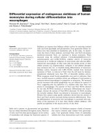
Schematic overview of intra-cellular copper trafficking in hepatocytesFigure 1
Schematic overview of intra-cellular copper trafficking in hepatocytes. Copper uptake is mediated by the receptor
CTR1. In the cell, copper can bind to copper chaperones such as CCS, COX17, and ATOX1 which in turn deploy to SOD1,
the mitochondrial COX, and ATP7A/B, respectively. ATP7A can directly excrete copper or bind it to ceruloplasmin (CP).
ATP7B can excrete copper through CP to blood or via MURR1 to bile. Furthermore, metallothioneins (MT) are present in the
cytoplasm which can bind and sequester metals. [SCO are metallochaperone proteins with essential, but not yet fully under-
stood, roles in copper delivery to mitochondrial COX.]
CTR 1
Cu
+
CCS
COX17
ATOX1
SOD1
SCO
COX
MT
ATP7A
ATP7B
CPMURR1
Bile Blood
Extra-cellular
Intra-cellular
MT
MT
MT
Comparative Hepatology 2005, 4:3 />Page 4 of 13
(page number not for citation purposes)
Quantitative Real-Time PCR of copper metabolism related genesFigure 2
Quantitative Real-Time PCR of copper metabolism related genes. mRNA levels of non-copper associated subclinical
hepatitis (n = 6 dogs) is shown in (A). mRNA levels of copper associated subclinical hepatitis (n = 6 dogs) is shown in (B).
mRNA levels of Doberman hepatitis (n = 6 dogs) is shown in (C). Data represent mean ± 2 SD.
0,00
0,50
1,00
1,50
2,00
2,50
3,00
ATOX1 COX17 ATP7A ATP7B CP MT1A MURR1
mRNA levels (fold change)
Control N-CASH
A
(p=0.468) (p=0.859) (p=0.400)
(p=0.010)
(p=0.335) (p=0.458) (p=0.218)
0,00
0,50
1,00
1,50
2,00
2,50
3,00
ATOX1 COX17 ATP7A ATP7B CP MT1A MURR1
mRNA levels (fold change)
Control DH
C
(p=0.777) (p<0.001) (p=0.001) (p=0.004)
(p=0.032)
(p=0.009)
(p=0.004)
0,00
0,50
1,00
1,50
2,00
2,50
3,00
ATOX1 COX17 ATP7A ATP7B CP MT1A MURR1
mRNA levels (fold change)
Control CASH
B
(p=0.104) (p=0.060) (p<0.001) (p=0.315)
(p=0.098) (p=0.476)
(p=0.004)
Comparative Hepatology 2005, 4:3 />Page 5 of 13
(page number not for citation purposes)
Quantitative Real-Time PCR of oxidative stress markersFigure 3
Quantitative Real-Time PCR of oxidative stress markers. mRNA levels of non-copper associated subclinical hepatitis
(n = 6 dogs) is shown in (A). mRNA levels of copper associated subclinical hepatitis (n = 6 dogs) is shown in (B). mRNA levels
of Doberman hepatitis (n = 6 dogs) is shown in (C). Data represent mean ± 2 SD.
0,00
0,50
1,00
1,50
2,00
2,50
3,00
CCS SOD1 CAT GSS GPX1
mRNA levels (fold change)
Control CASH
B
(p=0.001) (p=0.002) (p=0.018) (p=0.036)
(p<0.001)
0,00
0,50
1,00
1,50
2,00
2,50
3,00
CCS SOD1 CAT GSS GPX1
mRNA levels (fold change)
Control N-CASH
A
(p=0.088) (p=0.291) (p=0.122) (p=0.447)
(p=0.021)
0,00
0,50
1,00
1,50
2,00
2,50
3,00
CCS SOD1 CAT GSS GPX1
mRNA levels (fold change)
Control DH
C
(p<0.001) (p<0.001) (p<0.001) (p<0.001)
(p<0.001)
Comparative Hepatology 2005, 4:3 />Page 6 of 13
(page number not for citation purposes)
CASH and DH groups. The third copper chaperone CCS,
responsible for the transport of copper to SOD1, is inhib-
ited 8-fold in the DH group, 2-fold in the CASH group,
and remained unchanged in the N-CASH group.
Gene expression measurements on apoptosis and cell
proliferation
We measured two anti-apoptotic gene products, viz. Bcl-2,
the frequently described anti-apoptotic protein, and a x-
linked inhibitor of apoptosis (XIAP) recently associated
with MURR1 [30]. Our apoptosis measurements on Bcl-2
showed no reduction in gene expression in the N-CASH
group (Figure 4A), but is inhibited 4-fold in the CASH and
DH groups (Figures 4B and 4C, respectively). XIAP is
halved in all groups. The most dramatic changes were
found in the mRNA levels of the cell-cycle inhibitor
p27KIP which is inhibited 24-fold in the DH group, 12-
fold in the CASH group, and 3-fold in the N-CASH group.
Western blots analysis on metallothionein proteins during
copper toxicosis
Measurements on the mRNA levels of MT1A showed a
marked decrease in gene expression in the DH group. In
order to see whether this decrease was also occurring at
the protein level, Western blots were performed in order
to confirm decreased mRNA levels. Therefore, the total
amount of metallothionein was determined from Dober-
man pinschers with chronic hepatitis and high copper
(DH-group) levels compared to healthy Dobermans. Met-
allothionein was detected in both samples, where it was
present as a single band of 6 kDa (Figure 5). Interestingly,
the immunoreactive band shows no difference in concen-
tration between the two samples.
Total Glutathione measurements during copper toxicosis
In order to determine whether the decrease in mRNA lev-
els of GSS decreases the GSH levels, we measured the total
amount of GSH. Interestingly, in Figure 6, the total
amount of GSH in the high copper group is halved when
compared to healthy controls.
Discussion
In the present study, the expression of a total of 15 gene
products involved in copper metabolism of Doberman
pinschers was measured. This provided insight into the
molecular pathways of a canine copper-associated hepatic
disease model ranging from subclinical hepatitis with ele-
vated copper levels (CASH) to severe chronic hepatitis
with high hepatic copper levels (DH). Furthermore, these
diseases were compared to non-copper associated subclin-
ical hepatitis (N-CASH).
Because of the centrolobular accumulation of copper in
the hepatocytes during copper toxicosis in the Doberman,
a probable defect may be sought in the copper metabo-
lism instead of a secondary effect due to, for instance,
cholestasis. Recent findings by Mandigers et al. [17] indi-
cated that Doberman pinschers with hepatitis and ele-
vated copper concentrations suffer from impaired
64
Cu
bile excretion which is, together with other studies, con-
clusive that copper toxicosis exists in the Doberman pin-
scher. Furthermore, a double blind placebo-controlled
study with the copper chelating agent, D-penicillamine,
on Doberman pinschers with CASH showed a marked
improvement of liver pathology [31]; currently, that agent
is the only treatment option.
If copper is sequestered, in time metallothioneins will
store the copper in lysosomes, as described by Klein et al.
[32]. They found that chronic copper toxicity in Long-
Evans Cinnamon rats involved the uptake of copper-
loaded metallothioneins into lysosomes, where it was
incompletely degraded and polymerized into an insolu-
ble material, which contained reactive copper. This cop-
per initiated a lysosomal lipid peroxidation, which led to
hepatocyte necrosis. Phagocytosis of this reactive copper
by Kupffer cells amplified the liver damage. Histological
examination of the DH (Figure 7) and CASH group sam-
ples revealed copper accumulation in hepatocytes and
copper-laden Kupffer cells similar to that described by
Klein et al. [32]; therefore, that can be denoted as bench-
marks of chronic exposure to copper.
In our study, the gene expression levels of several gene
products involved in copper metabolism seem to be
reduced in the DH and CASH groups when compared to
healthy controls. Short term studies on in vitro models all
show an induction of MT1A or CP indicative of a higher
efflux of copper from hepatocytes [33,34]. The reductions
that are seen in our results could therefore be ascribed to
the prolonged or chronic nature of copper accumulation
as dogs in the high copper or DH group present clinical
signs after 2 years. Therefore, our observations are not
directly comparable with the short-term induced copper
effects in vitro, but are clinically more relevant, showing
the effects of long-term copper accumulation in Dober-
man hepatitis. However, Western blot experiments on
metallothionein, which stores the copper in lysosomes,
did not show any reduction at the protein level. This
observation could be ascribed to the antibody that binds
all metallothioneins, including metallothionein 2
(MT2A), which also is present in the liver. It remains to be
proven if this effect is a compensation for the decrease of
MT1A.
In the earlier stages of copper accumulation, comparable
to the CASH group, higher amounts of copper can still be
excreted. Interestingly, in the N-CASH group, ATP7B is
indeed induced compared to healthy controls, emphasiz-
ing a possible higher efflux of copper. Furthermore, from
Comparative Hepatology 2005, 4:3 />Page 7 of 13
(page number not for citation purposes)
Quantitative Real-Time PCR of apoptosis and cell proliferation related genesFigure 4
Quantitative Real-Time PCR of apoptosis and cell proliferation related genes. mRNA levels of non-copper associ-
ated subclinical hepatitis (n = 6 dogs) is shown in (A). mRNA levels of copper associated subclinical hepatitis (n = 6 dogs) is
shown in (B). mRNA levels of Doberman hepatitis (n = 6 dogs) is shown in (C). Data represent mean ± 2 SD.
0,00
0,50
1,00
1,50
2,00
2,50
3,00
Bcl-2 XIAP p27KIP
mRNA levels (fold change)
Control DH
C
(p=0.003) (p=0.002)
(p<0.001)
0,00
0,50
1,00
1,50
2,00
2,50
3,00
Bcl-2 XIAP p27KIP
mRNA levels (fold change)
Control CASH
B
(p=0.003) (p=0.027) (p<0.001)
0,00
0,50
1,00
1,50
2,00
2,50
3,00
Bcl-2 XIAP p27kip
mRNA levels (fold change)
Control N-CASH
A
(p=0.325) (p=0.011) (p=0.004)
Comparative Hepatology 2005, 4:3 />Page 8 of 13
(page number not for citation purposes)
the two subclinical disease groups, the N-CASH group is
the only one able to recuperate, whereas the CASH group
will eventually turn into clinical hepatitis as seen in the
DH group (data not shown). Taken together, our data sug-
gest that in the Doberman pinchers copper accumulates in
time and, finally, will have its negative effect on copper
metabolism and induce oxidative stress.
Oxidative stress has been ascribed to copper toxicosis as
one of the most important negative effects [35]. We can
confirm this with four different observations: (i) our
measurements showed a decrease in mRNA levels of
SOD1 and CAT, indicative of a reduction in the enzymatic
defence against oxidative stress in all groups with copper
accumulation; (ii) a reduction of GSS mRNA levels
Western blot analysis of the metallothionein proteinsFigure 5
Western blot analysis of the metallothionein proteins. Immunoreactive bands of total metallothionein of pooled frac-
tions of the Doberman hepatitis (DH) group (n = 6 dogs) versus healthy controls (n = 8 dogs).
Total glutathione (GSH) measurements during copper toxicosis in DobermanFigure 6
Total glutathione (GSH) measurements during copper toxicosis in Doberman. Total GSH levels of pooled protein
fractions of the Doberman hepatitis (DH) group (n = 6 dogs) versus healthy controls (n = 8 dogs). Data represent mean ± 2 SD.
Lane: Sample:
1 Doberman hepatitis
2 Healthy controls
3 Protein precision marker
15 kDa
1 2 3
10 kDa
0,00
0,20
0,40
0,60
0,80
1,00
1,20
1,40
1,60
Doberman hepatitis Control
[GSH] (µM/mg protein)
Comparative Hepatology 2005, 4:3 />Page 9 of 13
(page number not for citation purposes)
(glutathione synthesis), indicative for a reduced
glutathione level in these groups which is one of the most
important non-enzymatic molecules against oxidative
stress; (iii) the mRNA levels of GPX1 were significantly
increased, indicating an increase in GSH oxidation; (iv)
the decrease in GSH was confirmed by measuring total
glutathione levels in the DH group towards healthy
Doberman pinschers. A similar decrease in expression of
anti-oxidant enzymes was observed in ApoE-deficient
mice in response to chronic inflammation [36], and
inflammatory bowel disease (IBD) [37]. This indicates
that chronic inflammation (copper toxicosis, atheroscle-
rosis, IBD) is associated with reduced protection against
enhanced exposure to ROS.
Other effects of high copper can also be seen in the meas-
urements on apoptosis and cell-cycle. Measurements on
Bcl-2 and XIAP indicate a decrease of protection against
apoptosis; however, the most affected hepatocytes will go
into necrosis due to the formation of hydroxyl radicals by
the Haber-Weiss reaction, which is catalyzed by copper
[38]. A striking observation was made measuring p27KIP
which was shown to be reduced up to 24-fold in the DH
group. This could indicate an induction of cell-cycle com-
pared to healthy controls. This could be ascribed to the
renewal of hepatocytes, thus managing the total amount
of copper in time.
Whether differential gene expression is cause-or-conse-
quence of hepatitis is unknown. However, it is
conceivable that the reduction in copper processing gene
products might explain copper accumulation and the sub-
sequent oxidative stress. Furthermore, recent Q-PCR
measurements on non-copper related hepatitis and extra
hepatic cholestasis suggest that ATP7A and CP are not
down-regulated by inflammation or cholestasis (data not
shown). Therefore, we can conclude that the decreased
expression of these gene products is a Doberman hepatitis
specific effect. Other important copper associated gene
products such as COX17, ATP7B, and MT1A are probably
down-regulated due to inflammation.
Conclusion
This study is the first to show the effect of prolonged expo-
sure to different copper levels on oxidative stress and cop-
per metabolism in canine livers. Our data supports that:
(i) Doberman hepatitis is a new variant of primary copper
toxicosis; (ii) there is a clear indication of a reduced cop-
per excretion in the Doberman hepatitis group; (iii) there
is a clear correlation between high copper levels and
reduced protection against ROS; (iv) this Doberman hep-
atitis could be a good model to study copper toxicosis and
its effects for several human copper storage diseases such
as Indian childhood cirrhosis, non-Indian childhood cir-
rhosis, and idiopathic copper toxicosis, and provide the
Histological evaluation of Doberman hepatitisFigure 7
Histological evaluation of Doberman hepatitis. (A) Hepatitis characterised by accumulation of pigmented granules
(probably copper) in hepatocytes, and inflammation with lymphocytes and pigmented (probably copper) macrophages. HE
staining. (B) Centrolobular accumulation of copper in hepatocytes and band of fibrous tissue with inflammatory cells and cop-
per-laden macrophages. Rubeanic acid staining. P = Portal area, CV = Central vein area.
A
B
P
CV
Comparative Hepatology 2005, 4:3 />Page 10 of 13
(page number not for citation purposes)
basis for possible future treatments in dog and even in
man.
Methods
Dogs
Doberman pinschers were kept privately as companion
animals. The dogs were presented to the Department of
Clinical Sciences of Companion Animals, Utrecht Univer-
sity, either for a survey investigating the prevalence of
Doberman (chronic) hepatitis, as described by Mandigers
et al. [39] or were referred for spontaneously occurring
liver disease. All samples were obtained after written con-
sent of the owner. The procedures were approved by the
Ethical Committee, as required under Dutch legislation.
Groups
Animals were divided in groups based on histopathologi-
cal examination and quantitative copper analysis. Each
group contained both sexes from four to seven years of
age. [A possible gender effect was later excluded by look-
ing at the individual data.] Liver tissue of all Doberman
pinschers was obtained using the Menghini aspiration
technique [40]. Four biopsies, 2–3 cm in length, were
taken with a 14-gauge Menghini needle for
histopathological examination and quantitative copper
analysis and stored for future quantitative PCR and pro-
tein investigations. The quantitative copper analysis was
performed using instrumental neutron activation analysis
via the determination of
64
Cu [41]. Histopathological
biopsies were fixed in 10% neutral buffered formalin, rou-
tinely dehydrated and embedded in paraffin. Sections (4
µm thick) were stained with haematoxylin-eosin, van Gie-
son's stain, reticulin stain (according to Gordon and
Sweet), and with rubeanic acid. One experienced board
certified veterinary pathologist performed all histological
examinations. All diseased groups contained at least six
animals that were compared with a group of eight age-
matched healthy dogs. Four groups were included in this
study (Table 1):
1) Healthy group (n = 8 dogs), clinically healthy dogs
with normal liver enzymes and bile acids. Histopathology
of the liver did not reveal histomorphological lesions.
Liver copper concentrations were below 200 mg/kg dry
matter.
2) Non-copper associated subclinical hepatitis group (N-
CASH, n = 6 dogs), dogs with liver enzymes and bile acids
within reference values. Although histological examina-
tion showed evidence of a slight hepatitis, hepatic copper
concentrations were within normal levels, i.e., below 300
mg/kg dry matter. The dogs were classified as suffering
from subclinical hepatitis, which most likely was the
result of a different etiological factor, such as infections,
deficiencies, other toxins, deficient immune status or
immune-mediated mechanism [42].
3) Copper associated subclinical hepatitis group (CASH, n
= 6 dogs), dogs with liver enzymes and bile acids within
reference values. At histopathology these dogs showed
centrolobular copper-laden hepatocytes, on occasions
apoptotic hepatocytes associated with copper-laden
Kupffer cells, lymphocytes, plasma cells and scattered
neutrophils. These lesions were classified as subclinical
copper-associated hepatitis [43,44]. Hepatic copper con-
centrations were in all dogs above 600 mg/kg dry matter.
4) Doberman hepatitis group (DH, n = 6 dogs), dogs with
chronic hepatitis and elevated hepatic copper concentra-
tions. All dogs were referred with a clinical presentation of
hepatic failure (apathy, anorexia, vomiting, jaundice, and
in chronic cases sometimes ascites) and died within 2
months after diagnosis from this disease. Heparinized
plasma liver enzymes (alkaline phosphatase and alanine
aminotransferase) and fasting bile acids were, at least,
three times elevated above normal reference values.
Abdominal ultrasound revealed small irregular shaped
echo dense liver, as performed with a high definition
Ultrasound system – HDI 3000 ATL (Philips) – with a 4–
7 MHz broad band Faced-array transducer. Histopathol-
ogy showed chronic hepatitis (Figure 7A) with histologi-
cal features of fibrosis / micronodular cirrhosis, etc. These
lesions are comparable to chronic hepatitis in man [42].
Rubeanic acid staining revealed copper accumulation in
hepatocytes and Kupffer cells / macrophages (Figure 7B).
Table 1: Doberman pinscher group description
Group n Hepatic copper Copper concentrations
(mg/kg dry matter)
Clinical observation
Healthy 8 Normal 100 – 200 No abnormalities
N-CASH 6 Normal < 300 Subclinical hepatitis
CASH 6 Elevated copper levels > 600 Subclinical hepatitis
DH 6 Highly elevated copper
levels
> 1500 Chronic hepatitis
Comparative Hepatology 2005, 4:3 />Page 11 of 13
(page number not for citation purposes)
Hepatic copper concentrations were in all cases above
1500 mg/kg dry matter.
RNA isolation and reverse-transcription polymerase chain
reaction
Total cellular RNA was isolated from each frozen Dober-
man liver tissue in duplicate, using Qiagen RNeasy Mini
Kit (Qiagen, Leusden, The Netherlands) according to the
manufacturer's instructions. The RNA samples were
treated with Dnase-I (Qiagen Rnase-free DNase kit). In
total 3 µg of RNA was incubated with poly(dT) primers at
42°C for 45 min, in a 60 µl reaction volume, using the
Reverse Transcription System from Promega (Promega
Benelux, Leiden, The Netherlands).
Q-PCR of oxidative-stress proteins, copper metabolism
and other related signaling molecules
Q-PCR was performed on a total of 17 genes involved in
oxidative stress and copper metabolism. Real-time PCR
was based on the high affinity double-stranded DNA-
binding dye SYBR green I (SYBR
®
green I, BMA, Rockland,
ME) and was performed in triplicate in a spectrofluoro-
metric thermal cycler (iCycler
®
, BioRad, Veenendaal, The
Netherlands). For each PCR reaction, 1.67 µl (of the 2×
diluted stock) of cDNA was used in a reaction volume of
50 µl containing 1× manufacturer's buffer, 2 mM MgCl
2
,
0.5 × SYBR
®
green I, 200 µM dNTP's, 20 pmol of both
primers, 1.25 units of AmpliTaq Gold (Applied Biosys-
tems, Nieuwerkerk a/d IJssel, the Netherlands), on 96-
well iCycler iQ plates (BioRad). Primer pairs, depicted in
Table 2, were designed using PrimerSelect software
(DNASTAR Inc., Madison, WI). All PCR protocols
included a 5-minute polymerase activation step and con-
tinued with for 40 cycles (denaturation) at 95°C for 20
sec, annealing for 30 sec, and elongation at 72°C for 30
sec with a final extension for 5 min at 72°C. Annealing
temperatures were optimized at various levels ranging
from 50°C till 67°C (Table 2). Melt curves (iCycler, Bio-
Rad), agarose gel electrophoresis, and standard sequenc-
ing procedures were used to examine each sample for
purity and specificity (ABI PRISM 3100 Genetic Analyser,
Applied Biosystems). Standard curves constructed by plot-
ting the relative starting amount versus threshold cycles
were generated using serial 4-fold dilutions of pooled
cDNA fractions from both healthy and diseased liver tis-
sues. The amplification efficiency, E (%) = (10
(1/-s)
-1)·100
(s = slope), of each standard curve was determined and
appeared to be > 95 %, and < 105 %, over a wide dynamic
range. For each experimental sample the amount of the
gene of interest, and of the endogenous references glycer-
aldehyde-3-phosphate dehydrogenase (GAPDH) and
hypoxanthine phosphoribosyl transferase (HPRT) were
determined from the appropriate standard curve in auton-
omous experiments. If relative amounts of GAPDH and
HPRT were constant for a sample, data were considered
valid and the average amount was included in the study
(data not shown). Results were normalized according to
the average amount of the endogenous references. The
normalized values were divided by the normalized values
of the calibrator (healthy group) to generate relative
expression levels.
Western blot analysis
Pooled liver tissues (n = 6 dogs) were homogenized in
RIPA buffer containing 1 % Igepal, 0.6 mM
Phenylmethylsulfonyl fluoride, 17 µg/ml aprotinine and
1 mM sodium orthovanadate (Sigma chemical Co., Zwi-
jndrecht, The Netherlands). Protein concentrations were
obtained using a Lowry-based assay (DC Protein Assay,
BioRad). Thirty five µg of protein of the supernatant was
denatured in Leammli-buffer supplemented with Dithio-
threitol (Sigma Chemical Co.) for 3 min at 95°C and
electrophoresed on 10 % Tris-HCl SDS PAGE polyacryla-
mide gels (BioRad). Proteins were transferred onto
Hybond-C Extra Nitrocellulose membranes (Amersham
Biosciences Europe, Roosendaal, The Netherlands) using
a Mini Trans-Blot
®
Cell blot-apparatus (BioRad). The pro-
cedure for immunodetection was based on an ECL west-
ern blot analysis system, performed according to the
manufacturer's instructions (Amersham Biosciences
Europe). The membranes were incubated with 4 % ECL
blocking solution and 0.1 % Tween 20 (Boom B.V., Mep-
pel, The Netherlands) in TBS for 1 hour under gentle shak-
ing. The incubation of the primary antibody was
performed at room temperature for one hour, with a
1:2000 dilution of mouse anti-horse metallothionein
(DakoCytomation B.V., Heverlee, Belgium). After wash-
ing, the membranes were incubated with horseradish per-
oxidase-conjugated chicken anti-mouse (Westburg B.V.,
Leusden, The Netherlands) at room temperature for one
hour. Exposures were made with Kodak BioMax Light-1
films (Sigma chemical Co.).
Total GSH assay
The total amount of GSH was determined by a modified
version of a total GSH Determination Colorimetric Micro-
plate Assay according to Allen et al. [45], based on the
original Tietze macro assay [46]. Protein samples from
Doberman hepatitis (n = 6 dogs) and healthy controls (n
= 8 dogs) were isolated as described in Western blot anal-
ysis and subsequently pooled. Total protein concentration
was measured using a Lowry-based assay (DC Protein
Assay, BioRad). In short, 50 µl of the cell-lysate (1 mg/ml)
was used in triplicate in a 96-wells plate. The lysates were
incubated for 5 minutes with 50 µl of 1.3 mM
5,5'dithiobis-2-nitrobenzoic acid (DTNB), and 50 µl GSH
reductase (1.5 U/ml). To start the reaction 50 µl of
NADPH (0.7 mM) was added to the wells. Absorbance at
450 nm was measured at start and after 5 minutes. The
rate of 2-nitro-5-thiobenzoic acid production (yellow
Comparative Hepatology 2005, 4:3 />Page 12 of 13
(page number not for citation purposes)
product) was measured in delta absorbance per minute
and is directly proportionate with the amount of GSH in
the samples. A standard curve was added with known con-
centrations GSH (0 to 20 µM) in order to determine the
GSH concentrations in the samples.
Statistical analysis
A Kolmogorov-Smirnov test was performed to confirm
normal distribution of every group, and a Levene's test
checked the homogeneity of variances across groups. After
both verifications, the statistical significance of the
difference between the control group and each particular
non-healthy group was determined by using the Student's
t-Test. The significance level (α) was set at 0.05.
Authors' contributions
BS performed all Q-PCR measurements and wrote the
manuscript. PM participated in its design and coordina-
tion and helped to draft the manuscript. BA performed the
GSH assays and participated with Western blotting. PB
performed the Copper measurements on which our
groups are based. TI histochemically examined all sam-
ples described in this manuscript. GH performed genotyp-
ing on Dobermans and provided theoretical background.
JR and LP, conceived of the study, and participated in its
design and coordination and helped to draft the manu-
script. All authors read and approved the final
manuscript.
Acknowledgements
We thank Clare Rusbridge for thoroughly reading this manuscript.
References
1. Hamza I, Faisst A, Prohaska J, Chen J, Gruss P, Gitlin JD: The metal-
lochaperone Atox1 plays a critical role in perinatal copper
homeostasis. Proc Natl Acad Sci U S A 2001, 98:6848-6852.
2. Mertz M: The essential trace elements. Science 1981,
213:1332-1336.
Table 2: Nucleotide Sequences of Dog-Specific Primers for Quantitative Real-Time PCR
Gene Primer Sequence (5'-3') Tm (°C) Product size (bp) Accession number
GAPDH Forward TGT CCC CAC CCC CAA TGT ATC 58 100 AB038240
Reversed CTC CGA TGC CTG CTT CAC TAC CTT
HPRT Forward AGC TTG CTG GTG AAA AGG AC 56 100 L77488 /
Reversed TTA TAG TCA AGG GCA TAT CC L77489
SOD1 Forward TGG TGG TCC ACG AGA AAC GAG ATG 64 99 AF346417
Reversed CAA TGA CAC CAC AAG CCA AAC GAC T
CAT Forward TGA GCC CAG CCC TGA CAA AAT G 62 119 AB012918
Reversed CTC GAG CCC GGA AAG GAC AGT T
GSS Forward CTG GAG CGG CTG AAG GAC A 62 131 AY572226
Reversed AGC TCT GAG ATG CAC TGG ACA
GPX1 Forward GCA ACC AGT TCG GGC ATC AG 62 123 AY572225
Reversed CGT TCA CCT CGC ACT TCT CAA AA
CCS Forward TGT GGC ATC ATC GCA CGC TCT G 64 96 AY572228
Reversed GGG CCG GCC TCG CTC CTC
p27KIP Forward CGG AGG GAC GCC AAA CAG G 60 90 AY455798
Reversed GTC CCG GGT CAA CTC TTC GTG
Bcl-2 Forward TGG AGA GCG TCA ACC GGG AGA TGT 61 87 AB116145
Reversed AGG TGT GCA GAT GCC GGT TCA GGT
ATOX1 Forward ACG CGG TCA GTC GGG TGC TC 67 137 AF179715
Reversed AAC GGC CTT TCC TGT TTT CTC CAG
COX17 Forward ATC ATT GAG AAA GGA GAG GAG CAC 60 127 AY603041
Reversed TTC ATT CTT CAA GGA TTA TTC ATT TAC A
ATP7A Forward CTA CTG TCT GAT AAA CGG TCC CTA AA 50 99 AY603040
Reversed TGT GGT GTC ATC ATC TTC CCT GTA
ATP7B Forward GGT GGC CAT CGA CGG TGT GC 56 136 AY603039
Reversed CGT CTT GCG GTT GTC TCC TGT GAT
CP Forward AAT TCT CCC TTC TGT TTT TGG TT 62 97 AY572227
Reversed TTG TTT ACT TTC TCA GGG TGG TTA
MT1A Forward AGC TGC TGT GCC TGA TGT G 64 130 D84397
Reversed TAT ACA AAC GGG AAT GTA GAA AAC
MURR1 Forward GAC CAA GCT GCT GTC ATT TCC AA 58 122 AY047597
Reversed TTG CCG TCA ACT CTC CAA CTC A
XIAP Forward ACT ATG TAT CAC TTG AGG CTC TGG TTT C 54 80 AY603038
Reversed AGT CTG GCT TGA TTC ATC TTG TGT ATG
Comparative Hepatology 2005, 4:3 />Page 13 of 13
(page number not for citation purposes)
3. Dijkstra M, Vonk RJ, Kuipers F: How does copper get into bile?
New insights into the mechanism(s) of hepatobiliary copper
transport. J Hepatol 1996, 24:109-120.
4. Sternlieb I: Copper and the liver. Gastroenterology 1980,
78:1615-1628.
5. Thornburg LP: A perspective on copper and liver disease in the
dog. J Vet Diagn Invest 2000, 12:101-110.
6. Haywood S, Fuentealba IC, Kemp SJ, Trafford J: Copper toxicosis
in the Bedlington terrier: a diagnostic dilemma. J Small Anim
Pract 2001, 42:181-185.
7. Hultgren BD, Stevens JB, Hardy RM: Inherited, chronic, progres-
sive hepatic degeneration in Bedlington terriers with
increased liver copper concentrations: clinical and patho-
logic observations and comparison with other copper-associ-
ated liver diseases. Am J Vet Res 1986, 47:365-377.
8. Owen CA Jr, Bowie EJ, McCall JT, Zollman PE: Hemostasis in the
copper-laden Bedlington terrier: a possible model of Wil-
son's disease. Haemostasis 1980, 9:160-166.
9. Twedt DC, Sternlieb I, Gilbertson SR: Clinical, morphologic, and
chemical studies on copper toxicosis of Bedlington Terriers.
J Am Vet Med Assoc 1979, 175:269-275.
10. Sluis van de BJ, Breen M, Nanji M, Wolferen van M, Jong de P, Binns
MM, Pearson PL, Kuipers J, Rothuizen J, Cox DW, Wijmenga C, Oost
van BA: Genetic mapping of the copper toxicosis locus in Bed-
lington terriers to dog chromosome 10, in a region syntenic
to human chromosome region 2p13-p16. Hum Mol Genet 1999,
8:501-507.
11. Sluis van de BJ, Rothuizen J, Pearson PL, Oost van BA, Wijmenga C:
Identification of a new copper metabolism gene by positional
cloning in a purebred dog population. Hum Mol Genet 2002,
11:165-173.
12. Crawford MA, Schall WD, Jensen RK, Tasker JB: Chronic active
hepatitis in 26 Dobermann pinschers. J Am Vet Med Assoc 1985,
187:1343-1350.
13. Ingh van den T, Rothuizen J, Cupery R: Chronic active hepatitis
with cirrhosis in the Dobermann pinscher. Vet Q 1988,
10:84-89.
14. Thornburg LP: Histomorphological and immunohistochemical
studies of chronic active hepatitis in Dobermann Pinschers.
Vet Pathol 1998, 35:380-385.
15. Rolfe DS, Twedt DC: Copper-associated hepatopathies in dogs.
Vet Clin North Am Small Anim Pract 1995, 25:399-417.
16. Mandigers PJ, Senders T, Rothuizen J: Morbidity and mortality in
a Dutch Dobermann population born between 1993 and
1999. Vet Rec 2005 in press.
17. Mandigers PJ, Ingh van den T, Spee B, Penning LC, Bode P, Rothuizen
J: Chronic hepatitis in Doberman pinschers. A review. Vet Q
2004, 26:98-106.
18. Lippard SJ: Free copper ions in the cell? Science 1999,
284:748-749.
19. Klomp LW, Lin SJ, Yuan DS, Klausner RD, Culotta VC, Gitlin JD:
Identification and functional expression of HAH1, a novel
human gene involved in copper homeostasis. J Biol Chem 1997,
272:9221-9226.
20. Vulpe C, Levinson B, Whitney S, Packman S, Gitschier J: Isolation of
a candidate gene for Menkes disease and evidence that it
encodes a copper-transporting ATPase. Nat Genet 1993,
3:7-13.
21. Bull PC, Thomas GR, Rommens JM, Forbes JR., Cox DW: The Wil-
son disease gene is a putative copper transporting P-type
ATPase similar to the Menkes gene. Nat Genet 1993, 5:327-337.
22. Wijmenga C, Klomp LW: Molecular regulation of copper excre-
tion in the liver. Proc Nutr Soc 2004, 63:31-9.
23. Cox DW: Genes of the copper pathway. Am J Hum Genet 1995,
56:828-834.
24. Valentine JS, Gralla EB: Delivering copper inside yeast and
human cells. Science 1997, 278:817-818.
25. Harris ED: Cellular copper transport and metabolism. Annu Rev
Nutr 2000, 20:291-310.
26. Yagle MK, Palmiter RD: Coordinate regulation of mouse metal-
lothionein I and II genes by heavy metals and
glucocorticoids. Mol Cell Biol 1985, 5:291-294.
27. Ferenci P, Zollner G, Trauner M: Hepatic transport systems. J
Gastroenterol Hepatol 2002:105-112.
28. Gaetke LM, Chow CK: Copper toxicity, oxidative stress, and
antioxidant nutrients. Toxicology 2003, 189:147-163.
29. Uhlig S, Wendel A: Glutathione enhancement in various mouse
organs and protection by glutathione isopropyl ester against
liver injury. Biochem Pharmacol 1990, 39:1877-1881.
30. Burstein E, Ganesh L, Dick RD, Sluis van de B, Wilkinson JC, Klomp
LW, Wijmenga C, Brewer GJ, Nabel GJ, Duckett CS: A novel role
for XIAP in copper homeostasis through regulation of
MURR1. EMBO J 2004, 14:244-254.
31. Mandigers PJ, Ingh van den T, Bode P, Rothuizen J: Improvement in
liver pathology after 4 months of D-penicillamine in 5 Dober-
man pinschers with subclinical hepatitis. J Vet Intern Med 2005,
19:40-43.
32. Klein D, Lichtmannegger J, Heinzman U, Muller-Hocker J, Michaelsen
S, Summer KH: Association of copper to metallothionein in
hepatic lysosomes of Long-Evans cinnamon (LEC) rats dur-
ing the development of hepatitis. Eur J Clin Invest 1998,
28:302-310.
33. Daffada AA, Young AP: Coordinated regulation of ceruloplas-
min and metallothionein MRNA by interleukin-1 and copper
in HepG2 cells. Febs Lett 1999, 457:214-218.
34. Mattie MD, Freedman JH: Copper-inducible transcription: regu-
lation by metal- and oxidative stress-responsive pathways.
Am J Physiol Cell Physiol 2004, 286:293-301.
35. Ozcelik D, Ozaras R, Gurel Z, Uzun H, Aydin S: Copper-mediated
oxidative stress in rat liver. Biol Trace Elem Res 2003, 96:209-215.
36. Hoen 't PA, Lans van der CA, Eck van M, Bijsterbosch MK, Berkel van
TJ, Twisk J: Aorta of ApoE-deficient mice responds to athero-
genic stimuli by a prelesional increase and subsequent
decrease in the expression of antioxidant enzymes. Circ Res
2003, 8:262-269.
37. Kruidenier L, Kuiper I, Duijn van W, Marklund SL, Hogezand van RA,
Lamers CB, Verspaget HW: Differential mucosal expression of
three superoxide dismutase isoforms in inflammatory bowel
disease. J Pathol 2003, 201:7-16.
38. Klotz LO, Kroncke KD, Buchczyk DP, Sies H: Role of copper, zinc,
selenium and tellurium in the cellular defense against oxida-
tive and nitrosative stress. J Nutr 2003, 133:1448-1451.
39. Mandigers PJ, Ingh van den T, Bode P, Teske E, Rothuizen J: Associ-
ation between liver copper concentration and subclinical
hepatitis in Doberman pinschers. J Vet Intern Med 2004,
18:647-650.
40. Osborne CA, Hardy RM, Stevens JB, Perman V: Liver biopsy. Vet
Clin North Am 1974, 4:333-350.
41. Bode P: Automation and Quality Assurance in the Neutron
Activation Facilities in Delft. J Radioanal Nucl Chem 2000,
245:127-132.
42. Sterczer A, Gaal T, Perge E, Rothuizen J: Chronic hepatitis in the
dog – a review. Vet Q 2001, 23:148-152.
43. Speeti M, Eriksson J, Saari S, Westermarck E: Lesions of subclinical
Dobermann hepatitis. Vet Pathol 1998, 35:361-369.
44. Fuentealba I, Haywood S, Trafford DJ: Variations in the intralob-
ular distribution of copper in the livers of copper-loaded rats
in relation to the pathogenesis of copper storage diseases. J
Comp Pathol 1989, 100:1-11.
45. Allen S, Shea JM, Felmet T, Gadra J, Dehn PF: A kinetic microassay
for glutathione in cells plated on 96-well microtiter plates.
Methods Cell Sci 2000, 22:305-312.
46. Teitze F: Enzymatic method for quantitative determination of
nanogram amounts of total oxidized glutathione: Applica-
tions to mammalian blood and other tissues. Anal Biochem
1969, 27:502-522.