Báo cáo y học: "Comparison of tissue pressure and ablation time between the LeVeen and cool-tip needle methods" doc
Bạn đang xem bản rút gọn của tài liệu. Xem và tải ngay bản đầy đủ của tài liệu tại đây (229.11 KB, 5 trang )
BioMed Central
Page 1 of 5
(page number not for citation purposes)
Comparative Hepatology
Open Access
Research
Comparison of tissue pressure and ablation time between the
LeVeen and cool-tip needle methods
Makoto Nakamuta
1
, Motoyuki Kohjima
1
, Shusuke Morizono
1
,
Tsuyoshi Yoshimoto
1
, Yuzuru Miyagi
1
, Hironori Sakai
2
, Munechika Enjoji*
1
and Kazuhiro Kotoh
1
Address:
1
Department of Medicine and Bioregulatory Science, Graduate School of Medical Sciences, Kyushu University, Japan and
2
Department
of Gastroenterology, National Hospital Organization Kyushu Medical Center, Fukuoka, Japan
Email: Makoto Nakamuta - ; Motoyuki Kohjima - ;
Shusuke Morizono - ; Tsuyoshi Yoshimoto - ;
Yuzuru Miyagi - ; Hironori Sakai - ;
Munechika Enjoji* - ; Kazuhiro Kotoh -
* Corresponding author
Abstract
Background: Radio frequency ablation (RFA) has been accepted clinically as a useful local
treatment for hepatocellular carcinoma (HCC). However, intrahepatic recurrence after RFA has
been reported which might be attributable to increase in intra-tumor pressure during RFA. To
reduce the pressure and ablation time, we developed a novel method of RFA, a multi-step method
in which a LeVeen needle, an expansion-type electrode, is incrementally and stepwise expanded.
We compared the maximal pressure during ablation and the total ablation time among the multi-
step method, single-step method (a standard single-step full expansion with a LeVeen needle), and
the method with a cool-tip electrode. Finally, we performed a preliminary comparison of the
ablation times for these methods in HCC cases.
Results: A block of pig liver sealed in a rigid plastic case was used as a model of an HCC tumor
with a capsule. The multi-step method with the LeVeen electrode resulted in the lowest pressure
as compared with the single-step or cool-tip methods. There was no significant difference in the
ablation time between the multi-step and cool-tip ablation methods, although the single-step
methods had longer ablation times than the other ablation procedures. In HCC cases, the multi-
step method had a significantly shorter ablation time than the single-step or cool-tip methods.
Conclusion: We demonstrated that the multi-step method was useful to reduce the ablation time
and to suppress the increase in pressure. The multi-step method using a LeVeen needle may be a
clinically applicable procedure for RFA.
Background
Hepatocellular carcinoma (HCC) is one of the most com-
mon cancers worldwide. Most of HCC patients suffer
from virus-induced liver injury and most have underlying
liver cirrhosis [1]. Percutaneous ethanol injection therapy
(PEIT) has been used widely for the treatment of unresect-
Published: 21 December 2006
Comparative Hepatology 2006, 5:10 doi:10.1186/1476-5926-5-10
Received: 06 July 2005
Accepted: 21 December 2006
This article is available from: />© 2006 Nakamuta et al; licensee BioMed Central Ltd.
This is an Open Access article distributed under the terms of the Creative Commons Attribution License ( />),
which permits unrestricted use, distribution, and reproduction in any medium, provided the original work is properly cited.
Comparative Hepatology 2006, 5:10 />Page 2 of 5
(page number not for citation purposes)
able HCC [2]. Many reports showed that the efficacy of
PEIT for small HCC tumors was comparable to that of
hepatic resection; however, PEIT demands multiple ses-
sions to achieve complete necrosis, resulting in protracted
hospitalization [3]. Furthermore, many patients suffer
from local recurrence after PEIT, which is attributable to
intra-tumor septa that prevent the injected ethanol from
infiltrating the entire tumor [4,5]. We reported that local
recurrence after PEIT should be prevented as much as pos-
sible because it is one of the most important negative
prognostic factors for HCC patients [6].
It has been reported that radio frequency ablation (RFA)
is an effective procedure for hepatocellular carcinoma
(HCC) as well as for metastatic liver tumors [7,8]. How-
ever, it has also been shown that it is not uncommon for
RFA to cause various complications [9,10]. During or just
after the procedure, peritoneal bleeding, hepatic abscess,
hemothorax, perforation of the gastrointestinal wall, and
rapid hepatic decompensation can occur. In addition to
these acute complications, intrahepatic recurrence occurs
at a relatively high rate following RFA [11,12] and can
appear as either a local recurrence or a multiple scattered
recurrence. Seki et al. described a case of rapid progression
of numerous tumors around the treated area after RFA for
a small HCC [13]. Takada et al. and Nicoli et al. reported
cases of bilobular multiple recurrence that occurred 6
months after RFA [14,15]. More recently, Ruzzenente et
al. reported on patients with HCC who suffered from rap-
idly spreading recurrence after RFA, which was observed
in 4.5% of patients [16]. We also reported the clinical
study of scattered and rapid intrahepatic recurrences [17].
The common characteristics of these recurrences were
rapid growth and scattered location, and they were found
to occur around the ablated tumor or throughout the
liver. We presumed that scattered recurrence could be
attributable to an increase in intra-tumor pressure during
ablation and a subsequent explosion of the ablated
tumor. In a previous study using an in vitro porcine liver
model [18], we demonstrated that the RFA procedure
could produce an extreme increase in pressure. Because
the scattered pattern of recurrence was associated with a
poorer prognosis, we also developed a novel multi-step,
incremental expansion (multi-step) using a modified
expansion-type electrode technique, which was shown to
result in significantly lower pressures. In addition to the
LeVeen needle, the cool-tip needle (Radionics, Burling-
ton, MA, USA), a non-expansion-type electrode, has been
accepted clinically as a useful local treatment for HCC. In
this study, we evaluated the maximal pressure during
ablation and the total ablation time under the multi-step,
single-step, or cool-tip method.
Results
As a model of an HCC tumor with a capsule, we prepared
blocks of pig liver tissue packed into a rigid plastic case
and the blocks were used in this study (see Methods). Fig-
ure 1A shows the peak pressure with the LeVeeen elec-
trode (multi-step or single-step) and cool-tip electrode
procedures (40 W ablation). With the LeVeen electrode
procedure, the peak pressure produced during ablation
was significantly lower than that with the cool-tip proce-
dure. The peak pressure of 40 W was the highest among
the various procedures; cool-tip 40 W, 416.3 ± 108.4 kPa
> single-step method, 279.1 ± 29.6 kPa > multi-step
method, 27.4 ± 13.8 kPa. The data are presented as mean
± standard deviation (SD).
In contrast, there was no significant difference in the abla-
tion time between the multi-step (118.3 ± 16.4 s) and
cool-tip ablation methods (123.7 ± 67.0 s), although the
single-step method had longer ablation time (172.0 ±
26.9 s) than the other ablation procedures (Figure 1B).
Although the multi-step method required ten times 'roll-
off', the ablation was completed within less than 10 s
before the fifth step. As a result, the cumulative ablation
time was significantly shorter than that of the single-step
method, and there was no significant difference in the
total ablation time between the cool-tip (40 W) and
multi-step methods.
In a preliminary clinical trial, we compared ablation time
among the multi-step (n = 14), single-step (n = 13), and
cool-tip (n = 13) methods. The multi-step method
showed a significantly shorter ablation time than the sin-
gle-step or cool-tip method (Table 1), and there was no
significant difference in the area ablated by RF. Rapid and
scattered recurrence after RFA occurred in some patients
treated by the cool-tip or single-step method, but we
found no cases of scattered recurrence associated with the
multi-step procedure (Table 1). There were no other
adverse events during treatment in the patients.
Discussion
In this study, we demonstrated that the multi-step
method with a LeVeen electrode resulted in the lowest
pressure as compared with the single-step or cool-tip
method (Figure 1A). There was no significant difference in
the ablation time between the multi-step and cool-tip
ablation methods, although the single-step method had
longer ablation times than the other ablation procedures
(Figure 1B). The difference in pressure during the ablation
is probably attributable to the differences in the region of
ablation. With the cool-tip and single-step procedures, all
ablated cells in the entire targeted region would expand
simultaneously to generate high pressure, while the abla-
tion of a limited region in the multi-step procedure would
result in a lower level of intra-tumor pressure. In our pre-
Comparative Hepatology 2006, 5:10 />Page 3 of 5
(page number not for citation purposes)
Table 1: Comparison of clinical backgrounds, ablation time, and RFA-treated "area" size among the single-step, multi-step, and cool-
tip methods in clinical HCC cases.
LeVeen needle Cool-tip needle
Single-step Multi-step
Number 13 14 13
Sex (male/female) 9/4 10/4 8/5
Age 55.8 ± 2.7 56.7 ± 2.5 54.1 ± 3.3
Child A/Child B 10/3 11/3 9/4
Tumor location (r/l lobe) 11/2 10/4 10/3
Tumor size (mm) 22.0 ± 2.3 21.5 ± 1.9 20.8 ± 3.1
Ablation time (min) 22.1 ± 1.9 10.9 ± 2.5
b, c
15.4 ± 1.1
b
Ablated "area" (mm) 32.1 ± 2.6 34.2 ± 2.7 35.8 ± 3.7
Scattered recurrence
a
102
Tumor size and ablated area by RFA are expressed as the diameter (mm). χ
2
-test, ANOVA, and Scheffe's test showed no significant background
difference among the single-step, multi-step, and cool-tip methods.
a
Number of the patients in whom scattered intrahepatic recurrence occurred
after radio frequency ablation (RFA).
b
p < 0.001 vs. single-step;
c
p < 0.01 vs. cool-tip.
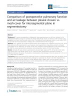
The maximal pressure (A) and total ablation time (B) on the porcine liver model, compared for the various tested methodsFigure 1
The maximal pressure (A) and total ablation time (B) on the porcine liver model, compared for the various tested methods.
The multi-step method with a LeVeen needle resulted in a significantly lower pressure than the cool-tip procedure or the sin-
gle-step method, and a total ablation time equal to that of the cool-tip procedure. All measurements were performed four
times, and the results are expressed as mean ± SD. Statistical comparisons for the maximal pressure and the total ablation time
were made using ANOVA and Scheffe's test.
a
p < 0.05 vs. cool-tip;
b
p < 0.05 vs. single-step.
Comparative Hepatology 2006, 5:10 />Page 4 of 5
(page number not for citation purposes)
vious report [18], based on histological findings, we con-
cluded that the cell density of the internal region was
higher than that of the outer region, and that some of the
pressure caused by ablation during the second and subse-
quent steps could escape into the internal region.
Although an in vitro porcine liver model was accepted in
this study, it may be necessary to confirm the phenome-
non in an in vivo model because the impedance during
ablation in vitro differs from that observed during the
treatment of patients. Under the condition of porcine liver
blocks covered entirely with hard plastic, the pressure near
the ablated area during RFA increased to over 100 kPa,
whereas the measured pressure in normal porcine liver in
vivo was much lower in our previous study [19]. A sub-
stantial increase in pressure should be necessary for the
tumor to explode during RFA. It is considered that, in nor-
mal liver in vivo, the pressure generated can easily escape
to the surrounding parenchyma or blood vessels. On the
other hand, the different conditions exist in most of HCC
patients, a fibrotic capsule around the tumor and paren-
chymal fibrosis surrounding the tumor accompanied by
cirrhosis. Therefore, during clinical RFA treatment for
HCC in cirrhotic liver, the escape of pressure is more or
less blocked and the pressure in the ablated area may pos-
sibly reach the level sufficient to cause an explosion.
Although our in vitro pig liver block model is artificial and
without blood flow, we assume the situation in our model
to be that in a tumor with poor arterial flow and a thick
capsule.
Because the reported scattered recurrence after RFA would
be attributable to an increase in intra-tumor pressure dur-
ing ablation and a subsequent explosion in the ablated
tumor [13-16], for clinical application, our aim should be
to reduce intra-tumor pressure during RFA. We showed
that our multi-step method using a LeVeen needle
resulted in much lower pressure than the cool-tip or the
standard single-step method, both of which might entail
a risk of extreme increase in intra-tumor pressure under
some conditions. We should also aim to shorten the abla-
tion time in order to reduce the patients' discomfort dur-
ing treatment. We demonstrated that the multi-step
procedure takes the same amount of time as the cool-tip
method. We are now applying our multi-step method in
clinical RFA treatment, and preliminary results indicated
that the multi-step method consumed significantly
shorter ablation times than the single-step or cool-tip
method (Table 1). We will collect more clinical data in
order to evaluate the appropriateness of this procedure for
clinical use, and to confirm whether the intratumor pres-
sure created by the sudden heating of RFA contributes to
the spreading or local recurrence of HCC.
Conclusion
Critical complication of rapid and scattered recurrence
after RFA may possibly be avoided by the use of modified
protocols. We consider that the multi-step method using
a LeVeen needle may be one of the clinically applicable
procedures for RFA.
Methods
Measurement of ablation time and pressure in vitro model
We measured the pressure in a block of porcine liver
sealed in a rigid plastic case using a pressure sensor
(model P303-01, M0101D; SSK. Co., Ltd., Tokyo, Japan)
as previously reported [17]. Two blocks of liver tissue (5 ×
5 × 4 cm) were cut and packed into a rigid 5 × 5 × 8 cm
plastic case with the pressure sensor mounted at one end.
We used two different systems, a LeVeen™ multipolar
array needle (3.0 cm diameter type) in combination with
an RF 2000 generator™ (Radio Therapeutics Corporation),
and a Cool-tip™ RF System (3.0 cm exposure length type)
(Radionics). The electrode needle was inserted from the
opposite end of the apparatus to the pressure sensor, until
the tip of the needle reached 3 cm from the sensor.
The LeVeen needle was used with either a standard proto-
col (single-step method) or a modified protocol (multi-
step method). For the single-step method, the tines were
fully expanded after the needle was inserted to the target
position. RF energy was then applied to the tissue at an
initial power setting of 40 W and was subsequently
increased at increments of 10 W per minute to a maxi-
mum power of 75 W. The power setting was left at this
point until power 'roll-off' occurred; tissue impedance (an
increase in tissue resistance caused by decreased conduc-
tivity of electrical current due to protein denaturation and
loss of intracellular fluids) rose to over 200, at which time
the power passively decreased to less than 10 W. If no roll-
off occurred, a total of 15 min elapsed. Using the same
device, the multi-step method involved expanding one-
tenth of the length of the electrode tines at first step, and
the current was delivered until power roll-off occurred. At
the second step, immediately following roll-off, two-
tenths of the length of the tines was expanded and a cur-
rent was supplied. With stepwise expansion of the tines,
the ablation was repeated until the tines were fully
expanded. RF energy was applied with an initial power
setting of 30 W in first step. When power roll-off occurred
within 30 s at a given step, the ablation at the next step
was started at the same electrical power. If roll-off took
more than 30 s, the next step was started with the power
set 10 W up to 75 W. If the stepwise increase in power
reached the maximum level before the final step, the abla-
tion at the subsequent step was performed at maximum
power. For the cool-tip electrode method, RF energy deliv-
ery was started at 40 W. The electric power was then
increased by 10 W every minute. The maximum electrical
Comparative Hepatology 2006, 5:10 />Page 5 of 5
(page number not for citation purposes)
power was 120 W, and the RF energy delivery was contin-
ued until the impedance increased beyond the limit of the
generator.
Measurement of ablation time in clinical HCC cases
We performed a preliminary comparison of ablation
times for the single-step, multi-step, and cool-tip methods
in 36 HCC cases with liver cirrhosis (Table 1). For the sin-
gle-step method (n = 13), ablation was started at 50 W,
and the electrical power was increased by 10 W per minute
in the subsequent ablation until 90 W was reached. For
the multi-step method (n = 14), ablation was started at 50
W. The electrical power was increased to 70 W at the fifth
step and to 90 W at the final step. For the cool-tip method
(n = 13), RF energy delivery was started at 40 W. The elec-
tric power was then increased by 10 W every minute. The
maximum electrical power was 120 W, and the RF energy
delivery was continued until the impedance increased
beyond the limit of the generator. Tumor location, tumor
size, and the area ablated by RFA were determined by
computed tomography (CT) examination.
Statistical analysis
Baseline characteristics of the patients prior to RFA treat-
ment are shown as mean ± SD, and the statistical compar-
isons were performed using the χ
2
-test for categorical data
and the non-paired t-test for numeric data. Regarding the
in vitro model, all measurements were performed four
times and the results are shown as mean ± SD. Statistical
comparisons for the cumulative ablation time and the
pressure at the programmed endpoint in vitro were made
using ANOVA and the Scheffe's test, via the Statview soft-
ware (SAS Institute, Cary, NC, USA).
Competing interests
The author(s) declare that they have no competing inter-
ests.
Authors' contributions
MN, ME, and HS participated in the experimental design,
and performed most of the analyses and writing of the
manuscript. MK, SM, TY, YM, and KK measured the abla-
tion time and pressure. All authors read and approved the
final manuscript.
References
1. Tang ZY: Hepatocellular carcinoma-cause, treatment and
metastasis. World J Gastroenterol 2001, 7:445-454.
2. Livraghi T, Festi D, Monti F, Salmi A, Vettori C: US-guided percu-
taneous alcohol injection of small hepatic and abdominal
tumors. Radiology 1986, 161:309-312.
3. Kotoh K, Sakai H, Sakamoto S, Nakayama S, Satoh M, Morotomi I,
Nawata H: The effect of percutaneous ethanol injection ther-
apy on small solitary hepatocellular carcinoma is compara-
ble to that of hepatectomy. Am J Gastroenterol 1994, 89:194-198.
4. Kotoh K, Sakai H, Morotomi I, Nawata H: The use of percutane-
ous ethanol injection therapy for recurrence of hepatocellu-
lar carcinoma. Hepatogastroenterology 1995, 42:197-200.
5. Yamamoto J, Okada S, Shimada K, Okusaka T, Yamasaki S, Ueno H,
Kosuge T: Treatment strategy for small hepatocellular carci-
noma: comparison of long-term results after percutaneous
ethanol injection therapy and surgical resection. Hepatology
2001, 34:707-713.
6. Arimura E, Kotoh K, Nakamuta M, Morizono S, Enjoji M, Nawata H:
Local recurrence is an important prognostic factor of hepa-
tocellular carcinoma. World J Gastroenterol 2005, 11:5601-5606.
7. Caturelli E: Percutaneous ablative therapies for small hepato-
cellular carcinoma: radio-frequency or percutaneous etha-
nol injection? Radiology 2000, 216:304-306.
8. Livraghi T, Goldberg SN, Lazzaroni S, Meloni F, Ierace T, Solbiati L,
Gazelle GS: Hepatocellular carcinoma: radio-frequency abla-
tion of medium and large lesions. Radiology 2000, 214:761-768.
9. Felekouras E, Kontos M, Pissanou T, Drakos E, Pikoulis E, Papalois A,
Bramis J: Radio-frequency tissue ablation in liver trauma: an
experimental study. Am Surg 2004, 70:989-993.
10. Livraghi T, Solbiati L, Meloni MF, Gazelle GS, Halpern EF, Goldberg
SN: Treatment of focal liver tumors with percutaneous
radio-frequency ablation: complications encountered in a
multicenter study. Radiology 2003, 226:441-451.
11. Poon RT, Ng KK, Lam CM, Ai V, Yuen J, Fan ST: Radiofrequency
ablation for subcapsular hepatocellular carcinoma. Ann Surg
Oncol 2004, 11:281-289.
12. Solbiati L, Livraghi T, Goldberg SN, Ierace T, Meloni F, Dellanoce M,
Cova L, Halpern EF, Gazelle GS: Percutaneous radio-frequency
ablation of hepatic metastases from colorectal cancer: long-
term results in 117 patients. Radiology 2001, 221:159-166.
13. Seki T, Tamai T, Ikeda K, Imamura M, Nishimura A, Yamashiki N,
Nakagawa T, Yamashiki N, Nakagawa T, Inoue K: Rapid progres-
sion of hepatocellular carcinoma after transcatheter arterial
chemoembolization and percutaneous radiofrequency abla-
tion in the primary tumour region. Eur J Gastroenterol Hepatol
2001, 13:291-294.
14. Takada Y, Kurata M, Ohkohchi N: Rapid and aggressive recur-
rence accompanied by portal tumor thrombus after radiof-
requency ablation for hepatocellular carcinoma. Int J Clin
Oncol 2003, 8:332-335.
15. Nicoli N, Casaril A, Hilal MA, Mangiante G, Marchiori L, Ciola M,
Invernizzi L: A case of rapid intrahepatic dissemination of
hepatocellular carcinoma after radiofrequency thermal
ablation. Am J Surg 2004, 188:165-167.
16. Ruzzenente A, Manzoni GD, Molfetta M, Pachera S, Genco B, Dona-
taccio M, Guglielmi A: Rapid progression of hepatocellular car-
cinoma after radiofrequency ablation. World J Gastroenterol
2004, 10:1137-1140.
17. Kotoh K, Enjoji M, Arimura E, Morizono S, Kohjima M, Sakai H,
Nakamuta M: Scattered and rapid intrahepatic recurrences
after radio frequency ablation for hepatocellular carcinoma.
World J Gastroenterol 2005, 11:6828-6832.
18. Kotoh K, Nakamuta M, Morizono S, Kohjima M, Arimura E, Fuku-
shima M, Enjoji M, Sakai H, Nawata H: A multi-step, incremental
expansion method for radio frequency ablation: optimiza-
tion of the procedure to prevent increases in intra-tumor
pressure and to reduce the ablation time. Liver Int 2005,
25:542-547.
19. Kotoh K, Morizono S, Kohjima M, Enjoji M, Sakai H, Nakamuta M:
Elevation of liver parenchymal pressure and portal endothe-
lium damage during radio frequency ablation: in an in vivo
porcine model. Liver Int 2005, 25:1217-123.