Báo cáo y học: "Plasma redox status is impaired in the portacaval shunted rat – the risk of the reduced antioxidant ability" doc
Bạn đang xem bản rút gọn của tài liệu. Xem và tải ngay bản đầy đủ của tài liệu tại đây (698.55 KB, 8 trang )
BioMed Central
Page 1 of 8
(page number not for citation purposes)
Comparative Hepatology
Open Access
Research
Plasma redox status is impaired in the portacaval shunted rat
– the risk of the reduced antioxidant ability
Maria-Angeles Aller*
1
, Maria-Inmaculada García-Fernández
2
,
Fernando Sánchez-Patán
1
, Luis Santín
2
, José Rioja
2
, Raquel Anchuelo
1
,
Jaime Arias
1
and Jorge-Luis Arias
3
Address:
1
Surgery I Department, School of Medicine, Complutense University of Madrid, Spain,
2
Human Physiology Department, School of
Medicine, University of Malaga, Spain and
3
Psychobiology Laboratory, School of Psychology, University of Oviedo, Asturias, Spain
Email: Maria-Angeles Aller* - ; Maria-Inmaculada García-Fernández - ; Fernando Sánchez-
Patán - ; Luis Santín - ; José Rioja - ; Raquel Anchuelo - ;
Jaime Arias - ; Jorge-Luis Arias -
* Corresponding author
Abstract
Background: Portacaval shunting in rats produces a reduction of hepatic oxidant scavenging
ability. Since this imbalance in hepatic oxidant/antioxidant homeostasis could coexist with systemic
changes of oxidant stress/antioxidant status, plasma oxidants and antioxidant redox status in
plasma of portacaval shunted-rats were determined.
Results: Male Wistar male: Control (n = 11) and with portacaval shunt (PCS; n = 11) were used.
Plasma levels of the oxidant serum advanced oxidation protein products (AOPP), lipid
hydroperoxides (LOOH), the antioxidant total thiol (GSH) and total antioxidant status (TAX)
were measured. Albumin, ammonia, Aspartate-aminotransferase (AST), Alanine-aminotransferase
(ALT), thiostatin and alpha-1-acid glycoprotein (α
1
-AGP) were also assayed 4 weeks after the
operation. AOPPs were significantly higher (50.51 ± 17.87 vs. 36.25 ± 7.21 μM; p = 0.02) and TAX
was significantly lower (0.65 ± 0.03 vs. 0.73 ± 0.06 mM; p = 0.007) in PCS compared to control rats.
Also, there was hypoalbuminemia (2.54 ± 0.08 vs. 2.89 ± 0.18 g/dl; p = 0.0001) and
hyperammonemia (274.00 ± 92.25 vs. 104.00 ± 48.05 μM; p = 0.0001) and an increase of thiostatin
(0.23 ± 0.04 vs. 0.09 ± 0.01 mg/ml; p = 0.001) in rats with a portacaval shunt. The serum
concentration of ammonia is correlated with albumin levels (r = 0.624; p = 0.04) and TAX
correlates with liver weight (r = 0.729; p = 0.017) and albumin levels (r = 0.79; p = 0.007)
Conclusion: These findings suggest that in rats with a portacaval shunt a systemic reduction of
oxidant scavenging ability, correlated with hyperammonemia, is principally produced. It could be
hypothesized, therefore, that the reduced antioxidant defences would mediate a systemic
inflammation.
Published: 5 February 2008
Comparative Hepatology 2008, 7:1 doi:10.1186/1476-5926-7-1
Received: 1 June 2007
Accepted: 5 February 2008
This article is available from: />© 2008 Aller et al; licensee BioMed Central Ltd.
This is an Open Access article distributed under the terms of the Creative Commons Attribution License ( />),
which permits unrestricted use, distribution, and reproduction in any medium, provided the original work is properly cited.
Comparative Hepatology 2008, 7:1 />Page 2 of 8
(page number not for citation purposes)
Background
Portosystemic collateral circulation is a frequent compli-
cation of chronic liver disease [1,2]. The portacaval
shunted rat is an experimental model of great interest for
studying the metabolic alterations related to a portosys-
temic shunt [3]. Particularly, in this model it has been
described that, portal blood flow deprivation (long-term
ischemia) may make the atrophic liver more susceptible
to oxidant-induced injury because the oxidant scavenging
system of the liver decreases [4].
However, recent evidence has shown that the altered
redox status in liver disease is not confined to the diseased
liver, but that it is a systemic phenomenon involving ext-
rahepatic tissues [5]. So, the determination of oxidant and
antioxidant plasma levels in portacaval shunted rats could
broaden the knowledge of the systemic pathophysiologi-
cal mechanisms, which are activated by the systemic
bypass of the portal blood flow.
This study has been carried out to determine serum
advanced oxidation protein products (AOPP), lipid
hydroperoxides (LOOH), total serum antioxidants (TAX),
total thiols and albumin as markers of the plasma redox
status.
Results
Body and liver weights
Rats with portacaval shunt (PCS) show a body weight
(BW) decrease (p < 0.001) during the 4 weeks of postop-
erative evolution. Liver weight (LW) and LW/FBW ratio
are also inferior (p < 0.001) in rats with PCS in relation-
ship to control rats (Table 1).
Hepatic liver function assays
Aspartate-aminotransferase (AST) (p = 0.004), alanine-
aminotransferase (ALT) (p = 0.0001), ammonia (p =
0.0001) and thiostatin (p = 0.0001) serum levels are
higher in PCS-rats compared to control rats. On the con-
trary, albumin (p = 0.0001) and α
1
-acid glycoprotein (α
1
-
AGP) (p = 0.04) are lower in PCS-rats (Table 2).
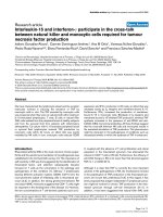
Redox status
The serum advanced oxidation protein product (AOPP)
level increases (p = 0.02) whereas total antioxidant status
(TAX) decreases (p = 0.007) in portacaval shunted rats in
relation to control rats. The serum concentrations of lipid
hydroperoxides (LOOH) and total thiols do not change in
PCS-rats (Figure 1).
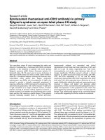
Correlation between liver function parameters and serum
redox status
The serum concentration of ammonia correlates with
albumin levels (r = 0.624; p = 0.04) and TAX correlates
with liver weight (r = 0.729; p = 0.017) and albumin levels
(r = 0.79; p = 0.007) (Figure 2).
Discussion
The results reported in this study show a significant
decrease of the TAX, associated with an increased AOPP
plasmatic level of portacaval-shunted rats. The considera-
ble decrease in TAX levels in long-term (4 weeks) porta-
caval shunted-rats suggest that a weakening of the
antioxidative barrier of the body exists, perhaps as a con-
sequence of the increased systemic oxidative stress pro-
duced by the portosystemic shunting in this experimental
model.
Oxidative stress, in general, is the overpowering of the
antioxidative defence system by the oxidative system [6].
A number of diseases, including liver disease [5], are asso-
ciated with an imbalance between oxidant stress and anti-
oxidative defence mechanisms that favour the former
[5,7]. Oxidative stress is produced by free radicals, i.e.,
reactive oxygen species (ROS) and reactive nitroxy species
(RNOS) and if they are not removed or neutralized, react
with lipids, proteins, and nucleic acids, damaging the cel-
lular functions and eventually causing cell death [5,6,8].
Both the excessive oxidative stress and the reduced anti-
oxidant ability could participate in the imbalance
between the oxidant stress and antioxidative defence
mechanism, which is produced in the rats with portacaval
shunt.
Chronic liver ischemia derived from the portal blood flow
bypass in the rat impairs oxidant scavenging, but does not
impair the oxidant generating systems of the liver [4].
However, the sources of ROS and RNOS in liver diseases
can be subdivided into intrahepatic and extrahepatic. Par-
ticularly, the extrahepatic oxidative stress is considered a
systemic phenomenon involving extrahepatic tissues [5]
and mainly portal circulation [5,9]. In rats with portal
Table 1: Body parameters. Initial body weight (IBW), final body weight (FBW), body weight increase (BWI), liver weight (LW) and
liver weight/body weight ratio (LW/FBW) in control rats and in rats with portacaval shunt (PCS) at 4 weeks of evolution.
Group IBW (g) FBW (g) BWI (g) LW (g) LW/FBW × 100
Control (n = 11) 221.55 ± 5.01 267.82 ± 5,69 46.82 ± 6.35 7.48 ± 0,40 2.79 ± 0.14
PCS (n = 11) 237.64 ± 12.96 212.00 ± 22.19*** -25.09 ± 25.05*** 4.03 ± 0.63*** 1.90 ± 0.24***
Data presented as: Mean ± SD. ***Statistically different from the control group (p < 0.001).
Comparative Hepatology 2008, 7:1 />Page 3 of 8
(page number not for citation purposes)
vein stenosis and portosystemic collateral circulation, the
existence of a causal relationship between oxidative stress
and the hyperdynamic circulation developed has been
accepted [9]. Since the hepatocellular injury is not a fea-
ture of this animal model it has been proposed that oxida-
tive stress originates from the portal circulation and not
the diseased liver [5,9]. Furthermore, portacaval shunted
rats also develop a hyperdynamic splanchnic circulation
related to portosystemic shunting [10-12]. Therefore, in
this experimental model the hyperdynamic splanchnic
Table 2: Biochemical data. Aspartate-aminotransferase (AST), alanine-aminotransferase (ALT), AST/ALT ratio, ammonia, albumin,
thiostatin and α
1
acid glycoprotein (α
1
-AGP) serum concentrations in control rats and in rats with portacaval shunt (PCS), 4 weeks
after the operation.
Group Control (n = 11) PCS (n = 11) p-value
AST (IU/L) 65.72 ± 11.19 134.50 ± 67.60 p = 0.004
ALT (IU/L) 29.36 ± 5.90 65.33 ± 27.27 p = 0.0001
AST/ALT 2.35 ± 0.08 2.38 ± 1.58 NS
Ammonia (μmol/L) 104.00 ± 48.05 274.00 ± 92.25 p = 0.0001
Albumin (g/dl) 2.89 ± 0.18 2.54 ± 0.08 p = 0.0001
Thiostatin (mg/ml) 0.09 ± 0.01 0.23 ± 0.04 p = 0.001
Data presented as: Mean ± SD. NS = non-significant difference.
Redox status in control rats and in rats with portacaval shunt at 4 weeks of evolutionFigure 1
Redox status in control rats and in rats with portacaval shunt at 4 weeks of evolution. Duplicate (TAX, AOPP and
THIOLS) and triplicate (LOOH) assays in control (n = 11) and portocaval shunt (PCS) (n = 11) rats, except for TAX in which
one PCS value was excluded. The results are expressed as mean ± SD. AOPP: serum advanced oxidation protein product;
LOOH: serum lipid hydroperoxides; TAX: serum total antioxidant; THIOLS: total plasma thiols.
Comparative Hepatology 2008, 7:1 />Page 4 of 8
(page number not for citation purposes)
circulation or mesenteric hyperemia could also be associ-
ated with intestinal oxidative stress. It has been proposed
that the chronic hypoxemia of the intestinal mucosa
related to vascular congestion could be an etiologic key
factor in the production of bacterial translocation because
the enterocytes would suffer injury by oxidative stress
[13,14]. Moreover, NO-overproduction could represent
an adaptive mechanism of the endothelium in response
to chronic increases in flow-induced shear stress [15-18].
NO reacting with ROS, such as O
2
-
., can also induce the
peroxynitrite ion (ONOO
-
) hyperproduction [19]. Intesti-
nal oxidative stress could participate through this mecha-
nism in the production of increased plasmatic levels of
AOPP in rats with a portosystemic shunt.
Protein oxidation products have increasingly been used as
markers instead of lipid peroxidation products in demon-
strating oxidative stress [20]. A novel oxidative stress
marker of protein, referred to as AOPP was developed in
plasma [21]. Furthermore, AOPP oxidation of plasma
thiol groups, termed "thiol stress," is quantitatively the
major manifestation of protein oxidation [22]. Since
AOPP is not only a marker of oxidative stress, but also acts
as an inflammatory mediator [23-27] the knowledge of
AOPP pathophysiology in this experimental model could
provide valuable information with respect to the relation-
ship between oxidative stress and the inflammatory
response related to a portosystemic shunt. In this regard,
since the liver and the spleen play important roles in the
elimination of AOPP [28], the apoptosis and liver atrophy
after portacaval shunting in the rat [29-31] could induce
its decreased plasma clearance, thus favouring its
increased plasmatic levels.
The marked plasmatic levels increase of thiostatin and the
hypoalbuminemia in rats with a portosystemic shunt may
be involved in the acute phase changes associated with a
systemic inflammatory response [32,33]. The proteins act-
ing as acute phase proteins differ from humans to animals
and from one species to another. In the rat, thiostatin and
α
1
-acid glycoprotein (α
1
-AGP) are among the major posi-
tive acute phase proteins while albumin reacts as a nega-
tive acute phase protein [34]. Thiostatin is a plasma
proteinase inhibitor protecting against proteolytic auto-
degradation [33]. Therefore, the synthesis of thiostatin
benefits from the metabolic priority during decreased
functional liver mass caused by the portosystemic shunt.
However, α
1
-AGP does not increase in these animals.
Since it is considered that α
1
-AGP prevents gram-negative
infections [34] and has anti-inflammatory functions [35],
rats with portacaval anastomoses would lose an essential
component in nonspecific resistance to infection and
inflammation.
Albumin plasma levels correlate with the TAX and with
the hyperammonemia in portosystemic shunted rats.
Albumin is a powerful extracellular antioxidant [36] and
its decreased liver synthesis after portacaval shunt reduces
Ammonia and total antioxidant statusFigure 2
Ammonia and total antioxidant status. Ammonia and
serum total antioxidant (TAX) status are correlated with
albumin serum levels. TAX also correlated with hepatic atro-
phy in portacaval shunted-rats 4 weeks after the operation.
Comparative Hepatology 2008, 7:1 />Page 5 of 8
(page number not for citation purposes)
its antioxidant functions. However, albumin synthesis
increases when ammonia levels are higher. This could rep-
resent an attempt of compensating the deleterious meta-
bolic effects caused by ammonium.
In rats with a portosystemic shunt, the acute-phase
response could be associated with oxidative stress, as well
as with inflammation. Particularly, IL-6, the major stimu-
lator of most acute phase proteins, is primarily produced
by Kupffer cells [37,38]. Upregulation of this cytokine
may be related to the enhanced respiratory burst activity
of Kupffer cells leading to the redox activation of NF-κB
[39-41]. This compensatory response has already been
described in order to re-establish homeostasis in the liver
and extrahepatic tissues exhibiting oxidative stress [38].
Another metabolic feature that has been shown to be
upregulated, though not always, due to a lack of oxygen or
oxidative stress, is the antioxidant system [42]. It has been
shown that the portosystemic bypass in the rat reduces the
oxidant scavenging system of the liver with a significant
reduction of superoxide dismutase and xanthine-dehy-
drogenase [4]. Furthermore, in the present study, the TAX
(i.e., the fraction of antioxidant pool available for further
anti-ROS activity) is significantly lower in portacaval
shunted rats compared to control rats. These results may
mean that a portosystemic shunt, including hyperdy-
namic circulatory syndrome and acute-phase response,
has its own effect on lowering TAX. Since oxidative stress
exhausts the antioxidative pool of the body, TAX could
also decrease [8,42]. However, the ROS overproduction
after portacaval shunting is not excessive, and indeed a
plasmatic increase of lipid peroxidation is not produced,
therefore it can be suspected that the novo antioxidant
synthesis is reduced. If so, the reduction of the systemic
antioxidant activity makes the organism susceptible to
oxidant-induce multi-organ injury because a normal ROS
production could be indeed a potential cause of oxidative
stress when an antioxidative deficit coexists [6,42-44].
Since the existence of an anti-inflammatory redox-oxidant
revolving axis has been suggested [43], in rats with porto-
systemic shunt, it could also be considered that the reduc-
tion of antioxidant ability would represent the mediator
signal for the evolution and perpetuation of the inflam-
matory process that is often associated with the condition
of oxidative stress, which involves gene regulation
[43,45]. Thus, the altered redox homeostasis in this exper-
imental model would be one of the hallmarks of the proc-
esses that regulate gene transcription in oxidative-stress-
mediated inflammation [8,43,45]. If so, we could call it:
"reduced antioxidative defence-mediated inflammation."
The decrease of the antioxidant protection in rats with
portacaval shunt, evidenced by lower TAX and hypoalbu-
minemia, is noteworthy since it is correlated with hyper-
ammonemia. This correlation suggests that, in this
experimental model, the grade of insufficient antioxidant-
mediated inflammation would be involved in a particular
metabolic alteration related to the portosystemic shunt, as
is the ammonia hyperproduction. Hyperammonemia is
considered a key etiopathogenic factor in the develop-
ment of hepatic encephalopathy [46-49]. Although,
ammonia is believed to be responsible for the neurologi-
cal abnormalities associated with hepatic encephalopa-
thy, growing evidence supports the view that glutamine,
synthesized from glutamic acid and ammonia, plays a
major role in the deleterious effects of ammonia [49] and
induce oxidative stress [46-48]. In turn, L-glutamic acid is
also a precursor of the antioxidant glutathione
[5,42,50,51]. Thus, hyperammonemia could be added as
an etiopathogenic factor of the oxidative stress-mediated
inflammation pathway that induces the portosystemic
shunt [48,49].
Conclusion
The decreased liver antioxidant activity in portacaval
shunted rats could potentiate the oxidative stress. In turn,
the increased synthesis of acute phase proteins by the
liver, since their anti-enzymatic ability, would attempt to
balance the enzymatic stress in this experimental model.
Methods
Animals
Male Wistar rats, with weights ranging from 230 to 270 g,
from the Vivarium of the Complutense University of
Madrid, were used. The animals were fed a standard labo-
ratory rodent diet (rat/mouse A04 maintenance diet, Pan-
lab, Spain) and water ad libitum. They were housed in a
light/dark-controlled room, with an average temperature
(22 ± 2°C) and humidity (65–70%) in groups of three to
four animals.
The experimental procedures and facilities complied with
the requirements of Commission Directive 86/609/EEC
(The Council Directive of the European Community) con-
cerning the protection of animals used for experimental
and other scientific purposes. The National legislation, in
agreement with this Directive, is defined in Royal Decree
n° 1202/2005.
Surgical technique of portacaval shunt
The animals were anesthetized by i.m. injection of keta-
mine (100 mg/Kg) and xylacine (12 mg/Kg). The end-to-
side portacaval anastomoses (PCA) was performed
according to a modified [29] Lee's technique [52,53]. In
brief, the intestinal loops are retracted to the animal's left
and covered with saline wet gauze to expose the inferior
vena cava (IVC) and the portal vein (PV). The dissection
and vascular anastomoses were done by a microsurgical
Comparative Hepatology 2008, 7:1 />Page 6 of 8
(page number not for citation purposes)
technique with the aid of an operative microscopy (Zeiss,
OPMI-1; 12 × 5). The IVC was dissected between the
hepatic parenchyma and the right renal vein. The PV was
individualized from the proper hepatic artery and the gas-
troduodenal vein was dissected and sectioned between
ligatures (silk 7/0). The infrahepatic IVC was clamped
with two microclips and an elliptical venotomy (3 × 2
mm) was performed on its anterior wall. The PV was then
ligated and sectioned in the liver hilum and clamped in its
confluence with the splenic vein. Nylon (9-10/0) was
used to perform the end-to-side portacaval anastomoses.
The midline abdominal incision was closed in two layers
using a continuous running technique with an absorbable
suture (polyglycolic acid) and 3-0 silk. Analgesia was
maintained with buprenorphine (0.05 mg/kg/8 h s.c.)
during the first 48 hours after the operation.
The animals were sacrificed by exsanguination 4 weeks
after the operation. Hepatic tissue was excised and rapidly
frozen in liquid nitrogen. Frozen livers were stored in
labelled containers at -80°C for posterior molecular stud-
ies and metabolic determinations.
Biochemical blood assays
Serum levels of albumin, total proteins, AST and ALT were
determined by routine laboratory methods using a
COBAS MIRA autoanalyzer according to the manufac-
turer's instructions (HORIBA ABX diagnostic, Montpel-
lier, France). Plasma ammonia was immediately
measured by glutamate dehydrogenase enzyme assay on a
clinical analyzer (COBAS MIRA autoanalyzer; Products:
BIOLABO SA, Maizy, France). Rat alpha-1-Acid Glycopro-
tein (alpha-1-AGP) and thiostatin serum levels were
assayed by ELISA (Life Diagnostics, Inc, USA)
Total antioxidant status
The total antioxidant capacity of serum was estimated in
duplicate using the commercial kit 'Total Antioxidant Sta-
tus' (Randox, UK), adapted to the Cobas Mira autoana-
lyser, which measures at 600 nm the formation of the
radical ABTS
+
using the Reagent ABTS
®
in the presence of
H
2
O
2
and peroxidase [54]. The method was calibrated
using the TROLOX standard included in the kit.
Determination of plasma sulfhydryl groups
Plasma sulfhydryl (-SH) groups were measured in dupli-
cate by using Ellman's reagent, 5,5'-dithiobis-(2-nitroben-
zoate) (DTNB), adapted to Cobas Mira [55]. Ten μl of
plasma were mixed with 200 μL of 0.1 M Tris buffer, con-
taining 10 mM EDTA, pH 8.2. The absorbance at 405 nm,
given by the plasma alone, was subtracted from that
obtained from the same sample 10 minutes after adding 8
μL of 10 mM DTNB. A blank containing only DTNB was
also included, and -SH concentration was calculated by
using a standard curve of glutathione. Thiol levels were
expressed in μmol/L plasma. Intra- and inter-assay varia-
tion coefficients were 1.2% and 6%, respectively.
Evaluation of plasma AOPP
Plasma AOPP were evaluated in duplicate by using a
microassay adapted to Cobas Mira according to Matteucci
et al [56] and based on the original method of Witko-Sar-
sat et al. [21]. Briefly, 10 μl of plasma or chloramine-T (ch-
T) standard solutions (400 – 6.25 μmol/l) were placed in
each well of the Cobas Mira autoanalyser. Then 200 μl of
the reaction mixture was added, consisting of 81% phos-
phate buffer solution (PBS), 15% acetic acid and 4% 1.16
mM potassium iodide. The absorbance was read at 340
nm (the blank contained PBS instead of plasma). AOPP
concentration was expressed as ch-T equivalents. Intra-
and inter-assay variation coefficients were 1% and 5%,
respectively.
Evaluation of plasma lipid hydroperoxides
Lipid hydroperoxides (LOOH) were evaluated in tripli-
cate by the FOX2 reagent (Ferrous Oxidation) automated
by Arab & Steghens [57] and adapted to Cobas Mira
(wavelength 600 nm) for studying lipid peroxidation in
serum samples. Xylenol orange (180 μl – 167 μM), the
first reagent, was added after to the sample (25 μl). The
first optical reading was recorded before adding 45 μl of
833 μM iron II D-gluconate. LOOH was calculated using
a standard curve of tert-butylhydroperoxide and LOOH
levels were expressed in μmol/L serum. Intra- and inter-
assay variation coefficients were 3% and 8%, respectively.
Statistical analysis
Statistical analyses were performed using SPSS software
(Statistical Package for the Social Sciences, version 14.00).
The results are expressed as mean ± standard deviation
(SD). Student's t test for independent data was used to
compare the different variables between the two groups of
animals. The relationship between the biochemical serum
parameters were verified using the Pearson coefficient cor-
relation. A p-value of less than 0.05 was considered signif-
icant.
Abbreviations
AST: aspartate-aminotransferase ; ALT: alanine-ami-
notransferase; α
1
-AGP: alpha-1 acid glycoprotein; AOPP:
advanced oxidation protein products; BW: body weight;
ch-: chloramine-T; DTNB: 5,5'-dithiobis-(2-nitroben-
zoate); FBW: final body weight; FOX2: ferrous oxidation;
GSH: reduced glutathione; IL: interleukin ; IVC: inferior
vena cava ; LOOH: lipid hydroperoxides; LW: liver weight;
LW/BW: liver weight to body weight ratio; NF-κB: nuclear
factor kappa beta; NO: nitric oxide; ONOO
-
: peroxynitrite
ion; PBS: phosphate buffer solution; PCA: portacaval
anastomoses; PCS: portacaval shunt; PV: portal vein; ROS:
reactive oxygen species; RNOS: reactive nitroxy species;
Comparative Hepatology 2008, 7:1 />Page 7 of 8
(page number not for citation purposes)
TAX: total antioxidant status/capacity of the serum; TNF-
α: tumor necrosis factor alpha.
Competing interests
The author(s) declare that they have no competing inter-
ests.
Authors' contributions
MIGF, FSP, LS, JR, RA, MAA and JLA performed most of
the experiments and provided assistance for the prepara-
tion of the manuscript. MAA, MIGF, JLA and JA partici-
pated in the design of the study and prepared the
manuscript. All authors have read and approved the con-
tent of the manuscript.
Acknowledgements
This work was supported in part with a Grant from MEC.SEJ 2004/07445
and the Department of Health. Castilla-La Mancha Regional Council (Ref.
04047-00). We would like to acknowledge the excellent secretarial assist-
ance of Maria-Elena Vicente, as well as Elizabeth Mascola for translating the
manuscript into English.
References
1. Rodríguez-Vilarrupla A, Fernández M, Bosch J, Garcia-Pagán JC: Cur-
rent concepts on the pathophysiology of portal hyperten-
sion. Ann Hepatol 2007, 6:28-36.
2. De Francis R, Dell'Era A: Non-invasive diagnosis of cirrhosis and
the natural history of its complications. Best Pract Res Clin Gas-
troenterol 2007, 21:3-18.
3. Herz R, Sautter V, Robert F, Bircher J: The Eck fistula rat: defini-
tion of an experimental model. Eur J Clin Invest 1972, 2:390-397.
4. Benoit JN, Grisham MB, Mash CL, Korthuis RJ, Granger DN:
Hepatic oxidant and antioxidant systems in portacaval-
shunted rats. J Hepatol 1992, 14:253-258.
5. Bomzon A, Ljubuncic P: Oxidative stress and vascular smooth
muscle cell function in liver disease. Pharmacol Ther 2001,
89:295-308.
6. Chauhan V, Chauhan A: Oxidative stress in Alzheimer's disease.
Pathophysiology 2006, 13:195-208.
7. Jain SK: Oxidative stress and metabolic diseases: Introduc-
tion. Pathophysiology 2006, 13:127-128.
8. Ott M, Gogvadze V, Orrenius S, Zhivotovsky B: Mitochondira, oxi-
dative stress and cell death. Apoptosis 2007, 12:913-922.
9. Fernando B, Marley R, Holt S, Anand R, Harry D, Sanderson P, Smith
R, Hamilton G, Moore K: N-acetylcysteine prevents develop-
ment of the hyperdynamic circulation in the portal hyper-
tensive rat. Hepatology 1998, 28:689-694.
10. Romeo JM, Lopez-Farre A, Martin-Paredero V, Lopez-Novoa JM:
Effect of portacaval surgical anastomosis on systemic and
splanchnic hemodynamics in portal hypertensive, cirrhotic
rats. Can J Physiol Pharmacol 1988, 66:1493-1498.
11. Srivastava A, Gottstein J, Blei AT: Cerebral blood flow and the
hyperdynamic circulation of rats after portacaval anastomo-
sis. J Hepatol 1993, 17:15-19.
12. Wong J, Zhang Y, Lee SS: Effects of portacaval shunting on
hyperdynamic circulation in bile duct-ligated cirrhotic rats. J
Hepatol
1997, 26:369-375.
13. Schimpl G, Pesendorfer P, Steinwender G, Feierl G, Ratschek M, Hol-
lwarth ME: Allopurinol reduces bacterial translocation, intes-
tinal mucosal lipid peroxidation, and neutrophil-derived
myeloperoxydase activity in chronic portal hypertensive and
common bile duct-ligated growing rats. Pediatr Res 1996,
40:422-428.
14. Schimpl G, Pesendorfer P, Steinwender G, Feierl G, Ratschek M, Hol-
lwarth ME: Allopurinol and glutamine attenuate bacterial
translocation in chronic portal hypertensive and common
bile duct-ligated growing rats. Gut 1996, 39:48-53.
15. Pateron D, Tazi KA, Sogni P, Heller J, Chagneau C, Poirel O, Philippe
M, Moreau R, Lebrec D: Role of aortic nitric oxide synthase
3(eNOS) in the systemic vasodilation of portal hypertension.
Gastroenterology 2000, 119:196-200.
16. Fernandez M, Mejias M, Angermayer B, Garcia-Pagan JL, Rodes J,
Bosch J: Inhibition of VEGF receptor-2 decreases the develop-
ment of hyperdynamic splanchnic circulation and portal-sys-
temic collateral vessels in portal hypertensive rats. J Hepatol
2005, 43:98-103.
17. Hori N, Wiest R, Groszman RJ: Enhanced release of nitric oxide
in response to changes in flow and shear stress in the supe-
rior mesenteric arteries of portal hypertensive rats. Hepatol-
ogy 1998, 28:1467-1473.
18. Wiest RW, Groszmann RJ: Nitric oxide and portal hyperten-
sion: its role in the regulation of intrahepatic and splanchnic
vascular resistance. Semin Liver Dis 1999, 19:411-426.
19. Beckman JS, Koppenol WH: Nitric oxide, superoxide, and per-
oxynitrite: the good, the bad, and ugly. Am J Physiol 1996,
271(5):1424-1237.
20. Dalle-Donne I, Rossi R, Giustarini D, Milzani A, Colombo R: Protein
carbonyl groups as biomarkers of oxidative stress. Clin Chim
Acta 2003, 329:23-38.
21. Witko-Sarsat V, Frielander M, Capeillere-Blandin C, Nguyen-Khoa T,
Nguyen AT, Zingraff J, Jungers P, Descamps-Latscha B: Advanced
oxidation protein products as a novel marker of oxidative
stress in uremia. Kidney Int
1996, 49:1304-13.
22. Himmelfarb J, McMonagle E, McMenamin E: Plasma protein thiol
oxidation and carbonyl formation in chronic renal failure.
Kidney Int 2000, 58:2571-2578.
23. Alderman ChJ, Shah S, Foreman JC, Katz DR: The role of advanced
oxidation protein products in regulation of dendritic cell
function. Free Radical Biol Med 2002, 32(5):377-385.
24. Witko-Sarsat V, Gausson V, Nguyen AT, Touam M, Drüeke T, San-
tangelo F, Descamps-Latscha B: AOPP-induced activation of
human neutrophil and monocyte oxidative metabolism: a
potential target for N-acetylc-cysteine treatment in dialysis
patients. Kidney Int 2003, 64:82-91.
25. Yazici C, Köse K, Calis M, Kuzugüden S, Kirnap M: Protein oxida-
tion status in patients with ankylosing spondylitis. Rheumatol-
ogy 2004, 43:1235-1239.
26. Baskol G, Demir H, Baskol M, Kilic E, Ates F, Karakukcu C, Ustdal M:
Investigation of protein oxidation and lipid peroxidation in
patients with rheumatoid artritis. Cell Biochem Funct 2006,
24:307-311.
27. Fialova L, Malbohan I, Kalousova M, Soukupova J, Krofta L, Stipek S,
Zima T: Oxidative stress and inflammation in pregnancy.
Scand J Clin Lab Invest 2006, 66:121-127.
28. Iwao Y, Anraku M, Hiraike M, Hawai K, Nakajou K, Kai T, Suenaga A,
Otagiri M: The structural and pharmacokinetic properties of
oxidized human serum albumin, advanced oxidation protein
products (AOPP). Drug Metab Pharmacokinet 2006, 21:140-146.
29. Lee S: Abdominal large blood vessels anastomoses. I. Porta-
caval shunt. In Manual of Microsurgery Volume 10. CRC Press Inc.
Florida USA; 1985:69-76.
30. Gandhi CR, Murase N, Subbotin VM, Uemura T, Nalesnik M, Deme-
tris AJ, Fung JJ, Starzl TE: Portacaval shunt causes apoptosis and
liver atrophy in rats despite increases in endogenous levels of
major hepatic growth factors. J Hepatol 2002, 37:340-348.
31. Zaitoun AA, Apelqvist G, Al-Mardini H, Gray T, Bengtsson F, Record
CO: Quantitative studies of liver atrophy after portacaval
shunt in the rat. J Surg Res 2006, 131:
225-232.
32. Gabay C, Kushner I: Acute-phase proteins and other systemic
responses to inflammation. N Engl J Med 1999, 340:448-454.
33. Schreiber G, Tsykin A, Aldred AR, Thomas T, Fung WP, Dickson PW,
Cole T, Birch H, De Jong FA, Milland J: The acute phase response
in the rodent. Ann N Y Acad Sci 1989, 557:61-85.
34. Hochepied T, Van Mole W, Berger FG, Baumann H, Libert C:
Involvement of the acute phase protein α
1
-acid glycoprotein
in nonspecific resistance to a lethal gram-negative infection.
J Biol. Chemistry 2000, 275:14903-14909.
35. Hochepied T, Berger FG, Baumann H, Libert C: Alpha (1)-acid
glycoprotein: an acute phase protein with inflammatory and
immunomodulating properties. Cytokine Growth Factor Rev 2003,
14:25-34.
36. Bourdon E, Loreau N, Blache D: Glucose and free radicals impair
the antioxidant properties of serum albumin. FASEB J 1999,
13:233-244.
Publish with BioMed Central and every
scientist can read your work free of charge
"BioMed Central will be the most significant development for
disseminating the results of biomedical research in our lifetime."
Sir Paul Nurse, Cancer Research UK
Your research papers will be:
available free of charge to the entire biomedical community
peer reviewed and published immediately upon acceptance
cited in PubMed and archived on PubMed Central
yours — you keep the copyright
Submit your manuscript here:
/>BioMedcentral
Comparative Hepatology 2008, 7:1 />Page 8 of 8
(page number not for citation purposes)
37. Ramadori G, Christ B: Cytokines and the hepatic acute-phase
response. Semin Liver Dis 1999, 19:141-155.
38. Tapia G, Fernández V, Pino C, Ardiles R, Videla LA: The acute-
phase response of the liver in relation to thyroid hormone-
induced redox signaling. Free Radic Biol Med 2006, 40:1628-1635.
39. Tapia G, Pepper I, Smok G, Videla LA: Kupffer cell function in thy-
roid hormone-induced liver oxidative stress in the rat. Free
Radic Res 1997, 26:267-279.
40. Lander HM: An essential role for free radicals and derived spe-
cies in signal transduction. FASEB J 1997, 11:118-124.
41. Kunsch Ch, Medford RM: Oxidative stress as a regulator of gene
expression in the vasculature. Circ Res 1999, 85:753-766.
42. Blokhina O, Virolainen E, Fagerstedt KV: Antioxidants, oxidative
damage and oxygen deprivation stress: a review. Ann Bot
(Lond) 2003, 91:179-194.
43. Ryter SW, Kim HP, Hoetzel A, Park JW, Nakahira K, Wang X, Choi
AMK: Mechanisms of cell death in oxidative stress. Antioxid
Redox Sign 2007, 9:49-89.
44. Haddad JJ, Fahlman CS: Redox- and oxidant-mediated regula-
tion of interleukin-10: an antiinflammatory, antioxidant
cytokine? Biochem Biophys Res Commun 2002, 297:163-176.
45. Haddad JJ, Land SC: The differential expression of apoptosis
factors in the alveolar epithelium is redox sensitive and
requires NF-kappaB (ReIA)-selective targeting. Biochem Bio-
phys Res Commun 2000, 271:257-267.
46. Norenberg MD, Rao KVR, Jayakumar AR: Ammonia neurotoxic-
ity and the mitochondrial permeability transition. J Bioenerg
Biomembr 2004, 36:303-307.
47. Ahl B, Weissenborn K, Van den Hoff J, Fischer-Wasels D, Köstler H,
Hecker H, Burchert W: Regional differences in cerebral blood
flow and cerebral ammonia metabolism in patients with cir-
rhosis. Hepatology 2004, 40:73-79.
48. Shawcross D, Jalan R: The pathophysiologic basis of hepatic
encephalopathy: central role for ammonia and inflamma-
tion. Cell Mol Life Sci 2005, 62:2295-2304.
49. Arias JL, Aller MA, Sánchez-Patán F, Arias J: The inflammatory
bases of hepatic encephalopathy. Eur J Gastroenterol Hepatol
2006, 18:1297-1310.
50. Jayakumar AR, Rao KVR, Norenberg MD: Glutamine-induced free
radical production in cultured astrocytes. Glia 2004,
46:296-301.
51. Li Y, Wei G, Chen J: Glutathione: a review on biotechnological
production. Appl Microbiol Biotechnol 2004, 66:233-242.
52. Arias J, Andres-Trelles F, Alsasua A: Simplified technique for por-
tocaval shunt in rats. Arch Farmacol Toxicol 1977, 3:205-214.
53. Lee SH, Fisher B: Portacaval shunt in the rat. Surgery 1961,
50:668-672.
54. Miller NJ, Rice-Evans C, Davies MJ, Gopinathan V, Milner A: A novel
method for measuring antioxidant capacity and its applica-
tion to monitoring the antioxidant status in premature
neonates. Clin Sci (Lond) 1993, 84:407-412.
55. Halliwell B, Hu ML, Louie S, Duvall TR, Tarkington BK, Motchnik P,
Cross CE: Interaction of nitrogen dioxide with human plasma
antioxidant depletion and oxidative damage. FEBS Lett 1992,
313:62-66.
56. Matteucci E, Biasci E, Giampietro O: Advanced oxidation protein
products in plasma: stability during storage and correlation
with other clinical characteristics. Acta Diabetol 2001,
38:187-189.
57. Arab K, Steghens JP: Plasma lipid hydroperoxides measure-
ment by an automated xylenol orange method. Anal Biochem
2004, 325:158-163.