Báo cáo y học: "Effects of ischemic pre- and postconditioning on HIF-1a, VEGF and TGF-b expression after warm ischemia and reperfusion in the rat liver" ppsx
Bạn đang xem bản rút gọn của tài liệu. Xem và tải ngay bản đầy đủ của tài liệu tại đây (558.63 KB, 6 trang )
RESEA R C H Open Access
Effects of ischemic pre- and postconditioning on
HIF-1a, VEGF and TGF-b expression after warm
ischemia and reperfusion in the rat liver
Anders R Knudsen
1*
, Anne-Sofie Kannerup
1
, Henning Grønbæk
3
, Kasper J Andersen
1
, Peter Funch-Jensen
1
,
Jan Frystyk
2
, Allan Flyvbjerg
2
and Frank V Mortensen
1
Abstract
Background: Ischemic pre- and postconditioning protects the liver against ischemia/reperfusion injuries. The aim
of the present study was to examine how ischemic pre- and postconditioning affects gene expression of hypoxia
inducible factor 1a (HIF-1a), vascular endothelial growth factor A (VEGF-A) and transforming growth factor b (TGF-
b) in liver tissue.
Methods: 28 rats were randomiz ed into five groups: control; ischemia/reperfusion; ischemic preconditioning (IPC);
ischemic postconditioning (IPO); combined IPC and IPO. IPC consisted of 10 min of ischemia and 10 min of
reperfusion. IPO consisted of three cycles of 30 sec. reperfusion and 30 sec. of ischemia.
Results: HIF-1a mRNA expression was significantly increased after liver ischemia compared to controls (p = 0.010).
HIF-1a mRNA expression was significantly lower in groups subjected to IPC or combined IPC and IPO when
compared to the ischemia/reperfusion group (p = 0.002). VEGF-A mRNA expression increased in the ischemia/
reperfusion or combined IPC and IPO groups when compared to the control group (p < 0.05).
Conclusion: Ischemic conditioning seems to prevent HIF-1a mRNA induction in the rat liver after ischemia and
reperfusion. This suggests that the protective effects of ischemic conditioning do not involve the HIF-1 system. On
the other hand, the magnitude of the HIF-1a response might be a marker for the degree of I/R injuries after liver
ischemia. Further studies are needed to clarify this issue.
Background
Colorectal cancer is a leading form of cancer in the
Western world. Approximately 50% of patients with this
disease have, or will eventually develop, live r metastases.
Surgical removal of those metastases remains the treat-
ment of choice, with a five year survival rate of 37%-
58% a fter resection [1-3]. Major hemorrhage and blood
transfusion during liver resection is related to an
increase in morbidity and mortality [4-6]. Vascular
clamping is a frequently used method for reducing
blood loss [7]. Several studies have shown that the nor-
mal livers tolerate periods of continuous warm ischemia
up to 90 min and intermittent warm ischemia up to 120
min [8-10].
However, ischemia/reperfusion (I/R) injury of the liver
is an unfortunate side effect of this method, ranging
from slightly elevated liver enzymes to acute liver failure
[11]. Ischemic pre- or postconditioning (IPC or IPO),
defined as brief periods of ischem ia and reperfusion
before or after sustained ischemia, have proven to
increase the ability of organs to tolerate I/R injury
[12-16]. The precise mechanisms responsible for the
hepatoprotection from ischemic injuries are only par-
tially known. Focus has been on a system of h ypoxia
inducible factors (HIF), where especially HIF-1 appea rs
to have a major role in cellular adaptation to hypoxia.
HIF-1 mediates essential homeostatic responses to cellu-
lar hypoxia by up-regulating gene transc ript ion, via spe-
cific DNA motif called hypoxia response elements, and
* Correspondence:
1
Department of Surgical Gastroenterology L, Aarhus University Hospital,
Aarhus, Denmark
Full list of author information is available at the end of the article
Knudsen et al. Comparative Hepatology 2011, 10:3
/>© 2011 Knudsen et al; licensee BioMed Central Ltd. This is an Open Access article distributed under the terms of the Creative
Commons Attribution License (http://creativecommons. org/licenses/b y/2. 0), which permits unrestricted use, distribution, and
reproduction in any medium, provided the original work is properly cited.
activating target genes. HIF-1 is a heterodimer protein
consisting of an a and b-subunit. T he b-subunit is
expressed ubiquitously in most cells, whereas expres sion
of the a-subunit is controlled by cellular oxygen tension.
Under normal conditions the H IF-1a protein is
degraded via an oxygen dependent system. By contrast,
hypoxia inactivates the degradation causing stabilization
of the HIF-1a protein, which then translocate to the
nucleus and forms dimers with the b-subunit [17]. The
active form of HIF-1 transactivates other genes as vascu-
lar endothelial growth factor (VEGF) and t ransforming
growth factor b1(TGF-b1) [18,19]. VEGF is an impor-
tan t growth factor invol ved in angiogenesis. It is a mul-
tifunctional protein, with several effects on endothelial
cells to promote the formation of new vessels. Further-
more, it stimulates the production of hepatocyte growth
factor (HGF), which is regarded as an initiator of liver
regeneration [20]. TGF-b1 is a member of the superfam-
ily of cytokines. In the liver, TGF-b1 has anti-inflamma-
tory properties and stimulates cell proliferation as well
as differentiation [20].
Besides I/R injuries, another possible drawback of liver
ischemia in cancer surgery could be growth stimulation
of micrometastases. Several studies indicate that the out-
growth of micrometastases is stimulated by I/R injuries
during hepatic resections [21-23]. Outgrowth of these
micro m etastases may at least in part, be stimulated by
an increased HIF-1a stabilization [22]. As mentioned
above, HIF-1a activates other genes such as VEGF and
TGF-b. Especially VEGF is an important growth factor
involved in angiogenesis [24-26]. In this sense a stimula-
tion of HIF-1a, via liver ischemia, could be a double-
edged sword; i.e., it pro tects the liver against I/R inju-
ries, but a side effect could be the growth stimulation of
micrometastases through angiogenesis.
Theaimofthepresentstudywastoexaminehow
ischemia, with or without IPC and IPO, affects the
expression of HIF-1a and the target genes VEGF and
TGF-b1, in rodent liver.
Methods
The surg ical and experimental protocols were approved
by the Danish Animal Research Committee, Copenha-
gen, Denmark according to license number 2007/561-
1311 and followed the GuidefortheCareandUseof
Laboratory Ani mals published by the National Institute
of Health. Twenty-eight adult male Wistar rats weighing
300-350 g (M&B Taconic, Eiby, Denmark) were used for
the experiment. Animals were housed in standard ani-
mal laboratories with a temperature maintained at 23°C
and an artificial 12-hour light-dark cycle, with food and
water ad libitum, until the time of the experiment. The
rats were randomly divided into five groups as follows:
sham operated control (CG) (n = 4); pure ischemia and
reperfusion (IRI) (n=6); IPC (n=6); IPO (n=6); and
IPC+IPO (n=6) (Figure 1). All animals were anaesthe-
tized with 0.75 ml/kg Hypnorm s.c. (Fentanyl/Fluani-
sone, Jansen Pharma, Birkerød, Denmark) and 4 mg/kg
Midazolams.c.(Dormicum,LaRoche,Basel,Switzer-
land) and placed on a heated pad. A mid line laparotomy
was performed and total hepatic ischemia was accom-
plished using a microvascular clamp placed on the hepa-
toduodenal ligament, i.e., performing the Pringle
maneuver. Reflow was initiated by removal of the clamp.
Discoloration of the liver was used as a positive marker
for hepatic ischemia. Reperfusion was ascertained by the
return of the normal brown/reddish color of the liver.
The experimen tal protocol was performed as described
in Figure 1. At the end of each experiment after 30 mi n
of reperfusion, a biopsy was taken from the right liver
lobe, immediately frozen in liquid nitrogen and stored at
-80°C for further analysis. Blood samples wer e collected
from the common iliac artery in tu bes for measurement
of alanine aminotransferase (ALAT), alkaline phosphates
and bilirubin, and analyzed immediately hereafter. All
rats were subsequently killed with an overdose of
pentobarbital.
Quantitative Real-Time PCR (RT-PCR)
After homogenization of liver tissue by the use of a
MM301 Mixer Mill (Retsch, Haan, Germany), total cel-
lular RNA was extracted from the liver tissue using a
6100 Nucleic Acid PrepStation (Applied Biosystems,
Foster City, CA, USA). The quality of rRNA was esti-
mated by agarose gel electrophoresis by the appearance
of two distinct bands visible by fluorescence of ethide
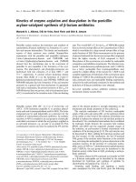
Figure 1 Experimental protocol of t he five groups.Blackareas
represent periods of hepatic ischemia; white areas represent periods
of normal hepatic blood perfusion. Liver biopsies were collected at
the end of each experiment. CG, Control group. IRI, 30 min of
ischemia. IPC, ischemic preconditioning + 30 min of ischemia. IPO,
30 min ischemia + ischemic postconditioning. IPC+IPO, ischemic
preconditioning + 30 min of ischemia + ischemic postconditioning.
Knudsen et al. Comparative Hepatology 2011, 10:3
/>Page 2 of 6
bromide representing intact rRNA. The amounts of
RNA extracted were quantified by measuring the absor-
bance by spectrophotometry, at 260 nm. Reverse tran-
scription from RNA to DNA was performed with a
Multiscribe Reverse Transcriptase kit from Applied Bio-
system at 25°C for 10 min, a t 48°C for 30 min and at
94°C for 29 sec. The PCR was performed in triplicates
of each sample in a volume of 25 μL in each well con-
taining RNA, TaqMan Universal PCR MasterMix and a
primer of the target, i.e., HIF-1a (Rn00577560_m1),
TGF-b (Rn00572010_m1) and VEGF-A (Rn4331348),
and a primer of the housekeeping gen e, 18S (4319 413),
all purchased from Applied Biosystems. Each RT-PCR
reaction ran at 50°C for 2 min, at 95°C for 10 min and
in 40 cycles changing between 95°C for 15 sec. and 60°C
for 1.30 min [27].
PCR Data analysis
Data was analyzed with the ABI Prism 7000 Sequence
Detector Software from Applied Biosystems. The output
of amplification was measured in the exponential phase
of the reaction as the threshold cycle/Ct-value, which is
defined as the cycle number at which amplification pro-
ducts are detected corresponding to the point where
fluorescent intensity exceeds the background fluorescent
intensity, which is 10 × the standard deviation of the
baseline. The average of triplicates from each sampl e
was used. The relative quantification of target g ene was
calculated using the formula: (1/2)
Ct-target gene- Ct-house-
keeping gene
, which is described in the Users Bulletin 2,
1997 from Perkin- Elmer (Perkin-Elmer Cetus, Norwalk,
CT, USA) [27].
Statistical analysis
Statistical analysis were performed by SPSS
®
11.0 pro-
grams (SPSS Inc., Chicago, Illinois, USA). All data is
expressed as mean ± SEM. Comparisons of data betwe en
groups were performed by non-paramet ric Kruskal-
Wallis (ANOVA) test followed by the Mann-Whitney U-
test. A p value < 0.05 was considered significant.
Results
Liver parameters
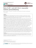
Blood samples showed a significant incre ase in ALAT in
group IRI (334 ± 135 U/L), IPC (377 ± 104 U/L), IPO
(1177 ± 379 U/L) and IPC+IPO (710 ± 199 U/L) com-
pared to the control group (40 ± 2 U/L) (CG vs. IRI,
IPC, IPO, and IPC+IPO, p = 0.01). No significant differ-
ences were found in ALAT between groups IRI, IPC,
IPO and IPC+IPO. Alkaline phosphates and bilirubin
were comparable between groups (Figure 2).
HIF-1a expression
In the IRI group the expression of HIF-1a mRNA was
sig nificantly increased after 30 min of reperfusion com-
pared to the control group (p ≤ 0.01). In the IPC group
HIF-1a mRNA expression was significantly lower than
the IRI group (p ≤ 0.01). In rats subjected to IPO there
was a tendency towards lower HIF-1a mRNA expres-
sion compared to the IRI group (p = 0.065). In the IPC
+IPO group HIF-1a mRNA expression was significantly
lower compared to the IRI group (IRI vs. IPC+IPO, p ≤
0.01). The HIF-1a mRNA levels were comparable
between group CG, IPC, IPO and IPC+IPO (Figure 3)
VEGF expression
As shown in Figure 4, VEGF mRNA expression was signif-
icantly increased in the IRI group compared to the control
group (p ≤ 0.01). When applying IPC+IPO VEGF mRN A
expression was also increased compared to the control
group (p ≤ 0.038). No significant differences were observed
between groups IPC, IPO and the control group (IPC vs.
CG, p ≤ 0.067) and (IPO vs. CG, p ≤ 0.067).
TGF-b1 expression
No differences in TGF-b1 mRNA expression were
observed between the five groups (Figure 5).
Discussion
As expected HIF-1a mRNA expression was increased
significantly in rats subjected to 30 minutes of warm
Figure 2 Blood samples including ALAT (A), alkaline phosphatase (AP) (B) and bilirubin (C) levels. Samples 30 min after reperfusion in CG,
Control group. IRI, 30 min of ischemia. IPC, ischemic preconditioning + 30 min of ischemia. IPO, 30 min ischemia + ischemic postconditioning.
IPC+IPO, ischemic preconditioning + 30 min of ischemia + ischemic postconditioning. * indicates p ≤ 0.01 compared to the control group.
Knudsen et al. Comparative Hepatology 2011, 10:3
/>Page 3 of 6
liver ischemia and 30 minutes of reperfusion c ompared
to the control group. The main finding of this study was
an absent of HIF-1 a induction in IPC or IPC+IPO trea-
ted animals. In both of these groups, the expression
levels were similar t o that of CG. In the IPO group the
same tendency towards an absent induction of HIF-1a
was observed although not significant. VEGF mRNA
expression increased significantly when applying 30 min
of ischemia without ischemic condi tioning compared to
sham operated controls. IPC+IPO also showed increased
VEGF mRNA expression compared to sham operated
controls, whereas neither ischemia nor ischemic condi-
tioning affected hepatic TGF-b expression.
The cytoprotective effects of IPC, defined as brief peri-
ods of ischemia and reperfusion prior to prolonged
ischemia, on I/R injuries to the liver have become indis-
putable with an increasing number of studies supporting
this fact [12-14]. The IPC protocol used in this study
has previously been shown to induce hepatoprotection
against I/R injuries. We choose circulating ALAT as
marker of hepac ellular injuries, as this parameter is wel l
established and known to correlate to the degree of
injury [28-30]. However, we were unable to see any
hepatoprotective effects as assessed by changes in liver
parameters. In previous s tudies with the same I PC pro-
toco l, longer periods of ischemia and longer reperfu sion
periods were utilized [12,14,31]. This might explain why
we were not able to demonstrate protective effects of
IPC and IPO as judged by liver parameters, i.e., the
duration of ischemia was too short. Furthermore, 30
min of reperfusion might be too short follow up t o
demonstrate the full extent of the I/R injuries. The cyto-
protective effect of IPO, defined as brief periods of
ischemia and reper fusion after liver ischemia, is less well
established [15,16]. In the present study, we could not
demonstrate any hepatoprotective effects of IPO
assessed by liver parameters, and we speculate that the
explanation may be the same as above. We choose the
Figure 3 Expression of HIF-1a mRNA. Expression after 30 min of
reperfusion. CG, Control group. IRI, 30 min of ischemia. IPC, IPC +
30 min of ischemia. IPO, 30 min ischemia + IPO. IPC+IPO, IPC + 30
min of ischemia + IPO. * indicates p ≤ 0.01 compared to group IRI.
¤ indicates p = 0.065 compared to group IRI.
Figure 4 Expression of VEGF mRNA. Expression after 30 min of
reperfusion. CG, Control group. IRI, 30 min of ischemia. IPC, IPC +
30 min of ischemia. IPO, 30 min ischemia + IPO. IPC+IPO, IPC + 30
min of ischemia + IPO. *indicates p ≤ 0.01 compared to group CG.
**indicates p ≤ 0.038 compared to group CG.
Figure 5 Expression of TGF-b1mRNA. Expressio n after 30 min of
reperfusion. CG, Control group. IRI, 30 min of ischemia. IPC, IPC +
30 min of ischemia. IPO, 30 min ischemia + IPO. IPC+IPO, IPC + 30
min of ischemia + IPO.
Knudsen et al. Comparative Hepatology 2011, 10:3
/>Page 4 of 6
actual time protocol with 30 minutes of ischemia
because we wanted to create a setting relevant for nor-
mal clinics. Even though longer periods of liver ischemia
have been safely applied, most su rgeons would be reluc-
tant to induce more than 30 minutes of ischemia on the
liver.
The mechanisms responsible for the protective effects
of IPC and IPO are only partially understood. In the
present study, IPC result ed in a significantly lower
expression of HI F-1a mRNA compared with rats sub-
jected to liver ischemia without IPC. This leads us to
conclude that HIF-1a, in our model of modest I/R-inju-
ries, does not seem to be a mediator of the cyto-protec-
tive effects of IPC. In r ats subjected to IPO there was a
tendency towards lower HIF-1a mRNA expression,
although not significant, when compared to the sheer
liver ischemia group. This indicates that HIF 1a is not
involved in the cytoprotective effects of IPO. In this
sense, the HIF-1a mRNA response could to be a marker
of the degr ee of I/R injury, i.e ., the higher HIF-1a
mRNA response after ischemia, the more pronounced I/
R injuries. Further studies need to be performed to
address this issue, but it is first and fore most supported
in a study by Cursio et al., where they showed that the
expression of HIF-1 and the degree of apoptosis was
increased in rats subjected to 120 min of warm liver
ischemi a compared to non-ischemia [32]. Another study
supporting the conclusion in the present paper is that
by Feinman et al. [33]. They used partially HIF-1 defi-
cient mice in a hemorrhagic shock model and concluded
that HIF-1 activation was necessary for ischemic gut
mucosal injury.
The expression of VEGF mRNA was regulated
upwards by the ischemic episodes in the group sub-
ject ed to sustained ischemia and in the IPC+IPO group.
A higher expression of VEGF in the group with liver
ischemia only, correl ates with the elevated HIF-1a
expression in this gro up. TGF-b expression levels were
not affected in any of the groups. Both VEGF and TGF-
b are, as previously described, genes that are regulated
downstream of HIF-1a.However,asthisstudyonly
focuses on the expression levels after 30 min o f reperfu-
sion,wecannotbesurethatwearemeasuringthefull
effect of the changed HIF-1a levels. If we had followed
the expression levels over time, we might have seen a
more direct correlation, as already reported [34].
Conclusions
Ischemic conditioning seems to prevent HIF-1 a mRNA
induction in the rat liver after ischemia and reperfusion.
This suggests that the protective effects of ischemic con-
ditioning do not involve the HIF-1 system. On the other
hand, the magnitude of the HIF-1a response might be a
marker for the degree of I/R injuries after liver ischemia.
Further studies need to be performed to elucidate this
matter.
Acknowledgements
The excellent technical assistance by Karen Mathiassen and Kirsten Nyborg is
highly appreciated. The work was supported by the Health Research Fund of
Central Denmark Region, Danish Medical Research Council, the Eva and
Henry Frænkels Memorial Foundation and the Clinical Institute, University of
Aarhus, Denmark.
Author details
1
Department of Surgical Gastroenterology L, Aarhus University Hospital,
Aarhus, Denmark.
2
The Medical Research Laboratories, Clinical Institute,
Aarhus University Hospital, Aarhus, Denmark.
3
Department of Medicine V,
Aarhus University Hospital, Aarhus, Denmark.
Authors’ contributions
Study conception and design: ARK, A-SK, FVM. Acquisition of data: ARK, A-SK,
KJA. Analysis and interpretation of data: ARK, A-SK, HG, KJA, PF-J, JF, AF, FVM.
Drafting of manuscript: ARK, A-SK, KJA, FVM. Critical revision of manuscript:
ARK, A-SK, HG, KJA, PF-J, JF, AF, FVM. All authors read and were in
accordance with the final manuscript.
Competing interests
The authors declare that they have no competing interests.
Received: 10 January 2011 Accepted: 19 July 2011
Published: 19 July 2011
References
1. Fong Y, Fortner J, Sun RL, Brennan MF, Blumgart LH: Clinical score for
predicting recurrence after hepatic resection for metastatic colorectal
cancer: analysis of 1001 consecutive cases. AnnSurg 1999, 230:309-318.
2. Abdalla EK, Vauthey JN, Ellis LM, Ellis V, Pollock R, Broglio KR, Hess K,
Curley SA: Recurrence and outcomes following hepatic resection,
radiofrequency ablation, and combined resection/ablation for colorectal
liver metastases. AnnSurg 2004, 239:818-825.
3. Pawlik TM, Scoggins CR, Zorzi D, Abdalla EK, Andres A, Eng C, Curley SA,
Loyer EM, Muratore A, Mentha G, et al: Effect of surgical margin status on
survival and site of recurrence after hepatic resection for colorectal
metastases. AnnSurg 2005, 241:715-722, discussion.
4. Kooby DA, Stockman J, Ben-Porat L, Gonen M, Jarnagin WR, DeMatteo RP,
Tuorto S, Wuest D, Blumgart LH, Fong Y: Influence of transfusions on
perioperative and long-term outcome in patients following hepatic
resection for colorectal metastases. AnnSurg 2003, 237:860-869.
5. Jarnagin WR, Gonen M, Fong Y, DeMatteo RP, Ben-Porat L, Little S,
Corvera C, Weber S, Blumgart LH: Improvement in perioperative outcome
after hepatic resection: analysis of 1,803 consecutive cases over the past
decade. AnnSurg 2002, 236:397-406.
6. Rosen CB, Nagorney DM, Taswell HF, Helgeson SL, Ilstrup DM, van
Heerden JA, Adson MA: Perioperative blood transfusion and
determinants of survival after liver resection for metastatic colorectal
carcinoma. AnnSurg 1992, 216:493-504.
7. van der Bilt JD, Livestro DP, Borren A, van HR, Borel RI: European survey on
the application of vascular clamping in liver surgery. Dig Surg 2007,
24:423-435.
8. Delva E, Camus Y, Nordlinger B, Hannoun L, Parc R, Deriaz H, Lienhart A,
Huguet C: Vascular occlusions for liver resections. Operative management
and tolerance to hepatic ischemia: 142 cases. Ann Surg 1989, 209:211-218.
9. Hannoun L, Borie D, Delva E, Jones D, Vaillant JC, Nordlinger B, Parc R: Liver
resection with normothermic ischaemia exceeding 1 h. Br J Surg 1993,
80:1161-1165.
10. Belghiti J, Noun R, Malafosse R, Jagot P, Sauvanet A, Pierangeli F, Marty J,
Farges O: Continuous versus intermittent portal triad clamping for liver
resection: a controlled study. AnnSurg 1999, 229:369-375.
11. Jaeschke H: Molecular mechanisms of hepatic ischemia-reperfusion
injury and preconditioning. Am J Physiol Gastrointest Liver Physiol 2003,
284:G15-G26.
Knudsen et al. Comparative Hepatology 2011, 10:3
/>Page 5 of 6
12. Koti RS, Seifalian AM, Davidson BR: Protection of the liver by ischemic
preconditioning: a review of mechanisms and clinical applications. Dig
Surg 2003, 20:383-396.
13. Clavien PA, Selzner M, Rudiger HA, Graf R, Kadry Z, Rousson V, Jochum W:
A prospective randomized study in 100 consecutive patients
undergoing major liver resection with versus without ischemic
preconditioning. Ann Surg 2003, 238:843-850.
14. Lee WY, Lee SM: Ischemic preconditioning protects post-ischemic
oxidative damage to mitochondria in rat liver. Shock 2005, 24:370-375.
15. Sun K, Liu ZS, Sun Q: Role of mitochondria in cell apoptosis during
hepatic ischemia-reperfusion injury and protective effect of ischemic
postconditioning. World J Gastroenterol 2004, 10:1934-1938.
16. Wu BQ, Chu WW, Zhang LY, Wang P, Ma QY, Wang DH: Protection of
preconditioning, postconditioning and combined therapy against
hepatic ischemia/reperfusion injury. Chin J Traumatol 2007, 10:223-227.
17. Schofield CJ, Ratcliffe PJ: Oxygen sensing by HIF hydroxylases.
NatRevMolCell Biol 2004, 5:343-354.
18. Lario S, Mendes D, Bescos M, Inigo P, Campos B, Alvarez R, Alcaraz A,
Rivera-Fillat F, Campistol JM: Expression of transforming growth factor-
beta1 and hypoxia-inducible factor-1alpha in an experimental model of
kidney transplantation. Transplantation 2003, 75:1647-1654.
19. Semenza G: Signal transduction to hypoxia-inducible factor 1.
BiochemPharmacol 2002, 64:993-998.
20. Michalopoulos GK: Liver regeneration. JCell Physiol 2007, 213:286-300.
21. van der Bilt JD, Kranenburg O, Nijkamp MW, Smakman N, Veenendaal LM,
Te Velde EA, Voest EE, van Diest PJ, Borel RI: Ischemia/reperfusion
accelerates the outgrowth of hepatic micrometastases in a highly
standardized murine model. Hepatology 2005, 42:165-175.
22. van der Bilt JD, Soeters ME, Duyverman AM, Nijkamp MW, Witteveen PO,
van Diest PJ, Kranenburg O, Borel RI: Perinecrotic hypoxia contributes to
ischemia/reperfusion-accelerated outgrowth of colorectal
micrometastases. AmJPathol 2007, 170:1379-1388.
23. Nicoud IB, Jones CM, Pierce JM, Earl TM, Matrisian LM, Chari RS, Gorden DL:
Warm hepatic ischemia-reperfusion promotes growth of colorectal
carcinoma micrometastases in mouse liver via matrix metalloproteinase-
9 induction. Cancer Res 2007, 67:2720-2728.
24. Carmeliet P, Jain RK: Angiogenesis in cancer and other diseases. Nature
2000, 407:249-257.
25. Drixler TA, Vogten MJ, Ritchie ED, van Vroonhoven TJ, Gebbink MF,
Voest EE, Borel RI: Liver regeneration is an angiogenesis- associated
phenomenon. AnnSurg 2002, 236:703-711.
26. Los M, Voest EE, Borel RI: VEGF as a target of therapy in gastrointestinal
oncology. DigSurg 2005, 22:282-293.
27. Jensen LJ, Denner L, Schrijvers BF, Tilton RG, Rasch R, Flyvbjerg A: Renal
effects of a neutralising RAGE-antibody in long-term streptozotocin-
diabetic mice. JEndocrinol 2006, 188:493-501.
28. Schmidt E, Schmidt FW: Enzyme diagnosis of liver diseases. Clin Biochem
1993, 26:241-251.
29. Scheig R: Evaluation of tests used to screen patients with liver disorders.
Prim Care 1996, 23:551-560.
30. Giannini EG, Testa R, Savarino V: Liver enzyme alteration: a guide for
clinicians. CMAJ 2005, 172:367-379.
31. Peralta C, Hotter G, Closa D, Gelpi E, Bulbena O, Rosello-Catafau J:
Protective effect of preconditioning on the injury associated to hepatic
ischemia-reperfusion in the rat: role of nitric oxide and adenosine.
Hepatology 1997, 25:934-937.
32. Cursio R, Miele C, Filippa N, Van OE, Gugenheim J: Liver HIF-1 alpha
induction precedes apoptosis following normothermic ischemia-
reperfusion in rats. TransplantProc 2008, 40:2042-2045.
33. Feinman R, Deitch EA, Watkins AC, Abungu B, Colorado I, Kannan KB,
Sheth SU, Caputo FJ, Lu Q, Ramanathan M, et al: HIF-1 mediates
pathogenic inflammatory responses to intestinal ischemia-reperfusion
injury. Am J Physiol Gastrointest Liver Physiol 2010, 299:G833-843.
34. Wang YQ, Luk JM, Ikeda K, Man K, Chu AC, Kaneda K, Fan ST: Regulatory
role of vHL/HIF-1alpha in hypoxia-induced VEGF production in hepatic
stellate cells. BiochemBiophysResCommun 2004, 317:358-362.
doi:10.1186/1476-5926-10-3
Cite this article as: Knudsen et al.: Effects of ischemic pre- and
postconditioning on HIF-1a, VEGF and TGF-b expression after warm
ischemia and reperfusion in the rat liver. Comparative Hepatology 2011
10:3.
Submit your next manuscript to BioMed Central
and take full advantage of:
• Convenient online submission
• Thorough peer review
• No space constraints or color figure charges
• Immediate publication on acceptance
• Inclusion in PubMed, CAS, Scopus and Google Scholar
• Research which is freely available for redistribution
Submit your manuscript at
www.biomedcentral.com/submit
Knudsen et al. Comparative Hepatology 2011, 10:3
/>Page 6 of 6