Báo cáo y học: "Morphological changes in diabetic kidney are associated with increased O-GlcNAcylation of cytoskeletal proteins including a-actinin " docx
Bạn đang xem bản rút gọn của tài liệu. Xem và tải ngay bản đầy đủ của tài liệu tại đây (16.91 MB, 16 trang )
RESEARCH Open Access
Morphological changes in diabetic kidney are
associated with increased O-GlcNAcylation of
cytoskeletal proteins including a-actinin 4
Yoshihiro Akimoto
1*
, Yuri Miura
2
, Tosifusa Toda
2
, Margreet A Wolfert
3
, Lance Wells
3,4,5
, Geert-Jan Boons
3,4
,
Gerald W Hart
6
, Tamao Endo
2
and Hayato Kawakami
1
* Correspondence: yakimoto@ks.
kyorin-u.ac.jp
1
Department of Anatomy, Kyorin
University School of Medicine,
Mitaka, Tokyo 181-8611, Japan
Full list of author information is
available at the end of the article
Abstract
Purpose: The objective of the present study is to identify proteins that change in
the extent of the modification with O-linked N-acetylglucosamine (O-GlcNAcylation)
in the kidney from diabetic model Goto-Kakizaki (GK) rats, and to discuss the relation
between O-GlcNAcylation and the pathological condition in diabetes.
Methods: O-GlcNAcylated proteins were identified by two-dimensional gel
electrophoresis, immunoblotting and peptide mass fingerprinting. The level of
O-GlcNAcylation of these proteins was examined by immunoprecipitation,
immunoblotting and in situ Proximity Ligation Assay (PLA).
Results: O-GlcNAcylated proteins that changed significantly in the degree of
O-GlcNAcylation were identified as cytoskeletal proteins (a-actin, a-tubulin, a-actinin 4,
myosin) and mitochondrial proteins (ATP synthase b, pyruvate carboxylase). The extent
of O-GlcNAcylation of the above proteins increased in the diabetic kidney.
Immunofluorescence and in situ PLA studies revealed that the levels of O-GlcNAcylation
of actin, a-actinin 4 and myosin were significantly increased in the glomerulus and the
proximal tubule of the diabetic kidney. Immunoelectron microscopy revealed that
immunolabeling of a-actinin 4 is disturbed and increased in the foot process of
podocytes of glomerulus and in the microvilli of proximal tubules.
Conclusion: These results suggest that changes in the O-GlcNAcylation of
cytoskeletal proteins are closely associated with the morphological changes in the
podocyte foot processes in the glomerulus and in microvilli of proximal tubules in
the diabetic kidney. This is the first report to show that a-actinin 4 is O-GlcNAcylated.
a-Actinin 4 will be a good marker protein to examine the relation between O-
GlcNAcylation and diabetic nephropathy.
Keywords: O-GlcNAc modification, Hexosamine biosynthetic pathway, Kidney, Glomeru-
lus, Cytoskeleton, a?α?-actinin, GK Rat, Mass spectrometry, Proximity Ligation Assay
Introduction
O-linked N-acetyl-b-D-glucosamine, termed O-GlcNAc, is a post-translational modifi-
cation involved in m odulation of signaling and transcription in response to cellular
nutrients or stress by interplay with O-phosphorylation [1-3]. O-GlcNAc serves as a
glucose sensor via the hexosamine biosynthetic pathw ay. Elevated O-GlcNAc modifica-
tion (O-GlcNAcylation) of proteins by increased flux through the hexosamine
Akimoto et al. Clinical Proteomics 2011, 8:15
/>CLINICAL
PROTEOMICS
© 2011 Akimoto et al; lice nsee BioMed Central Ltd. This is an Open Access article distributed under the terms of the Creative
Commons Attribution License ( whic h permits unrestricted use, distribution, and
reproduction in any medium, provided the original work is properly cited.
biosynthetic pathway has been implicated in the development of insulin resistance and
diabetic complications and in the up-regulated gene expression of transforming growth
factor-beta1, plasminogen activator inhibitor 1, and upstream stimulatory factor
proteins in mesangial cells, leading to mesangial matrix expansion and diabetic glomer-
ulosclerosis [2,4-9]. We previously demonstrated increased O-GlcNAcylation in the
kidney and pancreas of the Goto-Kakizaki (GK) rat, which is an animal model of type
2 diabetes [10,11]. Also, altered O-GlcNAcylation and O-GlcNAc transferase (OGT)
expression were recently reported in the kidney from diabetic patients [12].
In this study we carried out proteomic analysis, especially focused on the proteins with
remarkable change of the O-GlcNAc level in the kidney from GK rats, and suggested the
potential of O-GlcNAcylation as a biomarker of diabetic nephropathy. Total kidney pro -
teins from Wistar and GK rats were separated by two-dimensional gel electrophoresis.
O-GlcNAcylated proteins were detected by immunoblotting using anti-O-GlcNAc anti-
body. Selected proteins that changed markedly in their extent of O-GlcNAcylation were
identified by Mass Spectrometry (MS) analysis. MS sequencing of tryptic peptides identi-
fied some cytoskeletal proteins, including a-tubulin and a-actinin 4. Immunoprecipita-
tion and immunoblot findings demonstrated that O-GlcNAcylation of these identified
proteins was increased in the diabetic rats. To examine the localization of the identified
cytoskeletal proteins, we conducted an immunohistochemical study using confocal scan-
ning microscopy and immuno-electron microscopy. The localization and quantity of
these O-GlcNAcylated proteins were further examine d by performing the in situ Proxi-
mity Ligation Assay (PLA), which was developed to examine protein- to-protein interac-
tion and post-translational modification of proteins [13,14].
Methods
Animals and tissues
Kidney tissues were obtained by dissecting 15-week-old male (n = 3) Wistar rats (as con-
trols) and GK rats, which are a nonobese model of non-insulin-dependent diabetes mel-
litus and had been developed by the selective breading of glucose-intolerant Wistar rats.
Both rats were obtaine d from CLEA (Tokyo, Japan). All experimental procedures us ing
laboratory animals were approved by the Animal Care and Use Committee of Kyorin
University School of Medicine.
Reagents
Rabbit polyclonal anti-a-actinin 4 antibody was obtained from LifeSpan BioSciences
(Seattle, WA). Rabbit polyclonal anti-myos in antibody was obtained from Biomedical
Technologies (Stoughton, MA). Rabbit monoclonal anti-actin antibody (clone EP184E)
and rabbit monoclonal anti-a-tubulin antibody (clone EP1332Y) were obtained from
Epitomics (Burlingame, CA). Mouse monoclonal anti-O-GlcNAc antibodies
(CTD110.6, 18B10.C7 [3]) were used. The generation of CTD110.6, 18B10.C7(3) was
previously described [15,16].
Two-dimensional gel electrophoresis (2D-PAGE) and immunoblotting
Protein extraction and 2D-PAGE were performed as previously reported [17-19]. Three
nondiabetic and 3 diabetic rat kidneys were used simultaneously from prot ein extrac-
tion to gel matching. Five-hundred micrograms of total protein prepared from normal
Akimoto et al. Clinical Proteomics 2011, 8:15
/>Page 2 of 16
and diabetic kidneys was loaded onto the gel for isoelectric focusing, which was per-
formed by using pre-cast immobilize d pH gr adient (IPG) strips (18 cm long, pH4-7,
GE Healthcare Science). After equilibration in reducing solution and then in alkylating
solution, second-dimensional gel electro phoresis was performed by 10% SDS-PAGE.
Separated protein spots on polyacrylamide gels were electroblotted onto PVDF mem-
branes. Then tot al protein spots were stained with BODIPY FL-X (Invitrogen). The
membranes were first blocked for 1 h at room temperature with 0.3% BSA in TBS-T
and then incubated with mouse monoclonal anti-O-GlcNAc antibody (CTD 110.6,
Covance) at a dilution of 1:5,000 for 1 hr at room temperature, followed by incubation
with biotin-goat anti mouse IgM (Vector) at a dilution of 1:2,000 and then Qdot 625-
conjugated streptavidin (Invitrogen) at a dilution of 1:2,000, each for 1 hr at room
temperature. The total spots and immunoreactive spots were scanned by using a Mole-
cular Imager FX laser scanning fluorometer (BioRad Laboratories, Hercules, CA). The
intensities of spots were analyzed by using PDQuest software ver.8.0 (BioRad Lab).
The search for protein spots whose O-GlcNAcylation had changed in the GK sample
was performed as described previously [17]. The abundance of spots was presented as
parts per million of the total spots integrated by using the ‘ total quantity i n analysis
set’ feature of the PDQuest software . When the abundanc e of spots on 2D gels of dia-
betic kidneys was > 2-fold or less than 0.5-fold compared with that for the control
nondiabetic kidneys, we regarded the spots as proteins with a changed O-GlcNAcyla-
tion level.
In-gel protein digestion and peptide mass fingerprinting
The selected spots were cut from the second dimensional gel stained with SYPRO-
Ruby. In-gel protein digestion of selected gel spots was performed according to the
protocol described P eptide-mass
fingerprinting data were acquired by using a MALDI-TOF-MS spectrometer (AXIMA-
CFR, Shimazu Biotech). Proteins were identified with the Mascot search engine (Matrix
Science, London, UK) search algorithms by using the Swiss-Prot protein databases
(Version 57.6).
Immunoprecipitation, immuno-blotting and immunohistochemical study of actin, a-
actinin 4, a-tubulin, and myosin
Immunoprecipitation, immuno-blot analysis and immunohistochemical study were car-
ried out as described previously [11].
In situ PLA analysis
In situ PLA analysis was performed according to the manufacturer’s instructions with
an HRP/NovaRed detection kit from Olink Bioscience (Uppsala, Sweden) [20]. Cryostat
sections of kidney were cut and placed onto MAS-coated slides (Matsunami Glass,
Tokyo, Japan). The slides were then incubated at 60°C in LAB solution for 5 min.
These antigen-retrieved tissues were next washed with PBS, incubated for 15 min in
15 ml/L hydrogen peroxide in PBS, washed, and blocked with 1% BSA-PBS. For the in
situ ligation assay, we used mouse monoclonal anti-O-GlcNAc antibodies (clone:
18B10.C7 [3]) . The sections were incubated with this mouse anti-O-GlcNAc antibody
in combination with rabbit anti-actin, anti-a-actinin 4, anti-a-tubulin or anti-myosin
Akimoto et al. Clinical Proteomics 2011, 8:15
/>Page 3 of 16
antibody overnight at 4°C. After having been washed with TBS-0.1% Tween20 (TBS-T),
the sections were incubated with a mixture of MINUS secondary PLA probe against
mouse immunoglobulins and PLUS secondary PLA probe against rabbit immunoglobu-
lins for 1 h at 37°C. Then they were washed with TBS-T, and subsequently incubated
in hybridization solution for 15 min at 37°C and washed with TBS-T once for 1 min.
The slides were next incubated with the lig ation mix for 30 min at 37°C and washed
with TBS-T twice for 1 min each time. Then the sections were incubated with the
amplification mix for 90 min at 37°C and washed 3 times for 5 min each time with
TBS-T. The slides were thereafter incubated with the HRP-label ed hybridization probe
for 30 min at room temperature. After 2 washes with TBS for 2 min each time, the
slides were incubated with DAB staining mix for 5 min and then washed with water.
Nuclei were stained with hematoxyline. The slides were examined with a bright-field
microscope equipped with a CCD camera Pro600ES (Pixera, San Jose, CA). In the
negative controls in which the primary antibodies had been replaced with normal rab-
bit IgG and normal mouse IgG or omitted, signals were scarcely observed (data not
shown). Signals were quantified with Blo bFinderBright software, which is available on
BlobFinder Website />Statistics
Data were compiled from 3 independent experiments. Student’s t-test was used for sta-
tistical analysis, and for all cases P < 0.05 was considered to indicate statistical
significance.
Results
Identification of O-GlcNAcylated proteins in normal and diabetic kidneys
To identify O-GlcNAcylated proteins, we employed two-dimensional electrophoresis
and immunoblotting using the CTD110.6 anti-O-GlcNAc antibody. Total proteins of
diabetic GK rat kidney or nondiabetic Wistar rat kidney were electrophoresed on a
two-dimensional gel. Approximatel y 1,000 protein spots were detected in each SYPRO
Ruby-stained gel (Figure 1A, B). Approximately 100 protein spots were detected in
each Qdot 625-stained PVDF membrane (Figure 1C, D). The immunofluorescence
intensity of each spot was quantified and compared between the nondiabetic kidney
and the diabetic one. This comparison revealed enhanced O-GlcNAcylation in many
proteins from the diabetic kidney (Figure 1C-P). Twenty-seven spots that indicated a
significant difference in the relative quantity of O-GlcNAcylated protein were applied
to MALDI-TOF MS for identification of the proteins. The identified proteins included
various cytoskeletal prot eins (a-tubulin, a-actinin 4, myosin, actin) and mitochondrial
proteins (ATP synthase b pyruvate carboxylase), which were temporarily referred to as
spots “a"-"f” (Figure 1, Table 1). The intensity of these spots increased in the d iabetic
kidneys (Table 1). Almost the same results were obtained among the 3 GK rats as well
as the 3 control rats. These proteins except for a-actinin 4 have already been reported
to be O-GlcNAcylated.
In the next step we focused on the cytoskeletal proteins, as they play important roles
in maintaining the morphology of kidney tissue. To further confirm that actin, a-acti-
nin 4, myosin, and a-tubulin were substantially O-GlcNAcylated and to examine both
the protein level and the level of O-GlcNAcylation of these proteins in the diabetic
Akimoto et al. Clinical Proteomics 2011, 8:15
/>Page 4 of 16
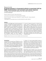
Figure 1 Comparison of total protein map and O-GlcNAcylation map of kidney between
nondiabetes and diabetes. (A, B) Representative total protein map of 2D PAGE for nondiabetic (A) and
diabetic (B) rat kidneys detected by SYPRO-Ruby. (C, D) Typical O-GlcNAcylated protein map of 2D PAGE
for nondiabetic (C) and diabetic (D) rat kidneys detected by O-GlcNAc antibody. Spots indicated by arrows
represent the identified proteins that had changed in terms of O-GlcNAc level. (E-L) Regions around spots
“a,”“b,”“c,”“d,”“e,” and “f” in the maps “C” and “D” are enlarged to facilitate the identification of each spot,
indicated by arrows, in the non-diabetic (E, G, I, K, M, O) and diabetic (F, H, J, L, N, P) kidneys.
Akimoto et al. Clinical Proteomics 2011, 8:15
/>Page 5 of 16
Table 1 Proteins showing an increase in the O-GlcNAcylation level in the diabetic kidney
Spot Protein Theoretical mass (kDa)/pI Score
(a)
Peptides Peptides
matched
(a)
Fold change
b)
Sequence
searched Wistar(control) GK Coverage (%)
a Actin 42.1/5.29 70 20 8 1 2.15 22
b a-actinin 4 105.0/5.27 118 40 17 1 2.05 21
c myosin heavy chain 222.5/5.69 54 18 12 1 2.22 9
d a-tubulin 50.6/4.95 59 51 9 1 2.75 33
e ATP-synthase b 56.3/5.19 62 27 9 ND
c)
↑
d)
23
f pyruvate carboxylase 130.4/6.34 73 30 14 1 4.32 16
Spots a-f correspond to those shown in Figure 1.
a)
Values of score and peptides matched were determined according to the Mascot search engine on the Swiss-Prot web server (Database: SwissProt 57.6).
b)
Fold changes were calculated by using the mean values for the spots of diabetic GK rat kidney relative to those of normal control Wistar rat kidney, which are indicated as “1.”
c)
ND: not detected.
d)
Up-arrow: This O-GlcNAcylated spot appeared only in the diabetic kidney.
Akimoto et al. Clinical Proteomics 2011, 8:15
/>Page 6 of 16
kidney, we performed immunoprecipitation using antibodies against these proteins and
compared relative protein expression and O-GlcNAcylation by immunoblotting using
anti-protein or anti-O-GlcNAc, respectively. As shown in Figure 2, the level of these
proteins did not change except in the case of a-actinin 4, whose level increased in the
diabetic kidney. In contrast, the O-GlcN Acylation level of each protein from the dia-
betic kidney relative to that of each from the nondiabetic one was significantly higher
(Figure 2). There might be other proteins in the immunoprecipitants which interacted
with or bound to the target proteins. If such proteins were present, it is important to
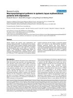
Figure 2 Anal ysi s of O-GlcNAcylation level of cytoskeletal proteins by immunoprecipitation and
immunoblotting. Total lysates of nondiabetic and diabetic kidneys were immunoprecipitated with anti-
actin (A), anti-a-actinin 4 (B), anti-a-tubulin antibody (C) or anti-myosin (D). The immunocomplexes were
separated by SDS-PAGE, transferred to PVDF membranes, and probed with O-GlcNAc antibody (a, upper
panel). The membrane was then stripped and reprobed with the antibody used for immunoprecipitation
(a, lower panel). Results shown are representative of 3 independent experiments. Intensity of bands
recognized by the antibody used for immunoprecipitation was quantified by scanning densitometry (b).
Relative intensities of the O-GlcNAc reactive bands to bands reactive with each antibody used for
immunoprecipitation were then determined (c). Data are the means ± SEM from 3 different rats. (□), Wistar
rats; (■), GK rats. *P < 0.05 and **p < 0.01 vs. control Wistar rat.
Akimoto et al. Clinical Proteomics 2011, 8:15
/>Page 7 of 16
determine the expression level and the O-GlcNAcylation level of these p roteins
between the diabetic and nondiabetic kidney in the future.
Immunohistochemical study on cytoskeletal proteins
To examine the localization of the identified cytoskeletal proteins (actin, a-actinin 4,
a-tubulin, and myosin), we carried out immunohistochemical analyses of the glomeruli
and proximal tubules (Figure 3).
The intensity of immunoreactivity for actin in t he glomerulus from the diabetic kid-
ney was increased, especially in its mesangi al cells and podocytes (Figure 3B). This
result is consistent with an earlier study on the GK rat [21]. The immunoreactivity
against actin was also detected in the brush border in the proximal tubules. However,
its intensity did not change in the diabetic kidney (Figure 3B)
The immunofluorescence indicating a-actinin 4 w as observed as a linear pattern
around the lumen of the glomerular capillary, and its i ntensity in the glomerulus was
Figure 3 Immunohistochemi cal analysis of cyto skeletal proteins by confocal la ser scanning
microscope. Localization of actin, a-actinin 4, myosin, and a-tubulin in glomeruli (1) and tubules (2). 1)
Although the intensity of immunoreactivity of a-tubulin did not change, that of immunoreactivity of actin, a-
actinin 4 and myosin was increased in the diabetic glomerulus. 2) Whereas the intensity of immunoreactivity of
actin and myosin did not change, that of immunoreactive a-actinin 4 was increased and that of
immunoreactive a-tubulin were decreased in the diabetic tubules. Scale bars, 1)-A, 50 μm, 2)-A, 20 μm.
Akimoto et al. Clinical Proteomics 2011, 8:15
/>Page 8 of 16
increased in the diabetic kidney (Figure 3D). a-Actinin 4 was also localized in the
brush border area at the luminal side of tubules, and the immunoreactivity was greater
in the sections from the diabetic kidney (Figure 3D).
In the glomerulus the immunoreactivity of a-tubulin was weak and did not change
in the section from the diabetic kidney (Figure 3F). In the sections showing
the proximal tubules of the nondiabetic kidney, a striated immunoreactivity pattern
was observed; whereas a diffuse one was noted i n the case of the diabetic kidney
(Figure 3F).
Whereas weak immunofluorescence was observed in the glomerulus from the normal
kidney (Figure 3-1G), intense immunoreactivity was observed in that from the diabetic
kidney (Figure 1C-P). Myosin immunoreactivity was also observed in th e brush border
area of the proximal tubules in sections from both normal and diabetic kidneys, but no
difference in intensity was observed between normal and diabetic proximal tubules
(Figure 3H).
Immuno-electron microscopy of a-actinin 4
Because a-actinin 4 has been identified as the causal gene for familial focal segmental glo-
merulosclerosis and is considered to play an important role in the maintenance of podo-
cyte morphology [22,23], we further examined the precise localizatio n o f a-actinin 4 in
the glomerulus and tubules by performing immuno-electron microscopy.
As we had reported previously [10], in the diabetic kidney of the GK rat thickened
basement membranes of the capillaries in the glomerulus and fused foot processes of
podocytes were observed (Figure 4A, B). Immuno-electron microscopy revealed that in
the normal kidney the immunogold particles labeling a-actinin 4 were localized in the
cortical area of foot pr ocesses of podocytes except beneath the basal plasma membrane
(Figure 4C) but that the localization was different in the diabetic kidney; i.e., a-actinin 4
was distri buted not only in the cortical area but also in the inner area of foot processes
(Figure 4D), and the number of immuno-gold particles was greater in the sections from
the diabetic kidney (Figure 4E).
Microvilli at the luminal side of proximal tubules from the diabetic GK rats were occa-
sionally swollen and had become bulbous, wh ereas those from the non-diabetic Wistar
rats were regularly shaped and closely packed (Figure 5 A, B). Immuno-electron micr o-
scopy of sections from the diabetic kidney revealed that colloidal gold par ticles labeling
a-actinin 4 were localized along the full length of the microvilli of proximal tubules of
diabetic kidney, whereas in those from the normal kidney the particles tended to be
localized near the bottom of the microvilli (Figure 5C, D). The colloidal gold particle
density indicating a-actinin 4 in the microvilli was increased significantly in the sections
from the diabetic kidney (Figure 5E). The gold label was also observed in the adherence
junctions of proximal tubule cells (Figure 5F, G), but no signif icant differenc e in the
labeling density or localization of a-actinin 4 in these junctions was observed between
normal and diabetic kidney sections.
In situ proximity ligation assay of O-GlcNAcylated cytoskeletal proteins
O-G lcNAcylation of proteins is a common type of post translat ional modi fication. The
in situ PLA was developed to image protein-to-protein interactions and posttransla-
tional modifications in cells and tissues [13,14]. Using this in assay, we examined the
Akimoto et al. Clinical Proteomics 2011, 8:15
/>Page 9 of 16
localization of O-GlcNAcylated cytoskeletal proteins (actin, a-actinin4, a-tubulin, and
myosin) and quantified their signals.
Signals of O-GlcNAcylated-actin, a-actinin 4, a-tubulin, and myosin were observed
in the glomerulus (Figure 6-1) and tubules (Figure 6-2) in sections of normal kidney.
The number of signals of all these O-GlcNAcylated-proteins was significantly increased
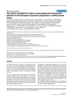
Figure 4 Morphological change of foot processes of podocytes in glomerulus is correlated with the
change of a-actinin 4. Electron microscopic appearance of glomerular capillary (A, B) and immuno-
electron microscopic localization of a-actinin 4 in glomerular capillaries (C, D). (A, B) Comparison of
electron micrographs of the capillary wall of the glomerulus from nondiabetic (A) and diabetic (B) kidneys
revealed fused foot processes of podocytes and a thickened basement membrane in the sections from the
diabetic rat. En, endothelial cell; GBM, glomerular basement membrane; P, foot process of podocyte. Scale
bar, 200 nm. (C, D) Sections of nondiabetic and diabetic kidneys were immunolabeled for a-actinin 4 (12-
nm gold particles). Whereas the gold particles were localized mainly in the periphery of the foot processes
in the nondiabetic kidney, they were found not only in the periphery but also in the inner aspect of foot
processes in the diabetic kidney. (E) Density (number/μm
2
) of colloidal gold particles representing a-actinin
4 in the foot processes of podocytes from nondiabetic (open bar) and diabetic (closed bar) kidneys. Data
are the means ± SEM from 3 different rats. *P < 0.01 vs. control (Wistar rat).
Akimoto et al. Clinical Proteomics 2011, 8:15
/>Page 10 of 16
in both the glomeruli and tubules in sections of the diabetic kidney (Figure,6-1, -2). In
the diabetic kidney sections, the signals of O-GlcNAcyl ated a-actinin 4 were increased
especially in the podocytes of the glomeruli (Figure 6-1D). The localizations of O-
GlcNAcylated actin, a-actinin 4, and myosin shown by in si tu PLA were almost the
same as those observed in the conventional immunohistochemical study (Figure 3).
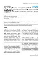
Figure 5 Morphological change of microvilli in proximal tubule is correlated with the change of a-
actinin 4. Electron microscopic appearance of microvilli of the tubules (A, B) and immuno-electron
microscopic localization of a-actinin 4 in microvilli (C, D) and in the adherins junction (F, G) of tubules. (A,
B) Comparison of electron micrographs of proximal tubules from nondiabetic rat (A) and diabetic rat (B)
revealed that microvilli of the diabetic rat were swollen and had become bulbous, whereas those of the
Wistar rat were regularly shaped. (C, D, F, G) Sections of nondiabetic and diabetic kidneys were
immunolabeled for a-actinin 4 (12-nm gold particles). Whereas only a few gold particles were localized in
the microvilli of proximal tubules in the nondiabetic kidney, many gold particles were detected in the
microvilli of proximal tubules in the diabetic kidney (C, D). (E) Density (number/μm
2
) of colloidal gold
particles representing a-actinin 4 in the microvilli from nondiabetic (open bar) diabetic kidney (closed bar)
kidneys. Data are the means ± SEM from 3 different rats. *P < 0.01 vs. control (Wistar rat). (F, G) The
colloidal gold labels indicating a-actinin 4 were also observed in the adherins junction of proximal tubules
of both nondiabetic (F) and diabetic (G) kidneys. Scale bars: 200 nm (A, C) and 100 nm (F).
Akimoto et al. Clinical Proteomics 2011, 8:15
/>Page 11 of 16
However, with respect to the O-GlcNAcylated a-tubulin, the striated signal pattern in
the normal kidney revealed by the conventional immunohistochemistry (Figure 3-2E)
was not observed (Figure 6-2E); rather, the signals were diffusely distributed in the
tubules in sections of normal and diabetic k idney (Figure 3F). This observation indi-
cates that polymerized tubulin may not be O-GlcNAcylated.
Discussion
In this study using proteomic analysis, we identified several O-GlcNAcylated proteins
including cytoskeletal proteins, actin, a-actinin 4, a-tubulin, and myosin and we
demonstrated that their extent of O-GlcNAcylation was elevated in the diabetic kidney.
For the first time, in situ PLA studies demonstrated the localization of O-GlcNAcylated
cytoskeletal proteins and confirmed their increase in O-GlcNAcylation.
Actin is an important cytoskeletal protein and is O-GlcNAcylated [24]. O-GlcNAcyla-
tion of actin may modulate the actin-tropomyosin interaction and be involved in the poly-
merization of myosin heavy chains [24]. High glucose alters protein kinase C-dependent
actin assembly in both cultured glomerular mesangial cells and podocytes [25]. Mapping
of O-GlcNAcylation sites revealed that the Ser
201
residue of actin is both O-GlcNAcylated
Figure 6 Analysis of O-GlcNAcylated cytoskeletal proteins by using the in situ PLA assay.
Localization of O-GlcNAcylated actin (A, B), O-GlcNAcylated a-actinin 4 (C, D), O-GlcNAcylated a-tubulin (E,
F) and O-GlcNAcylated myosin (G, H) in the glomerulus (1) and tubule (2) of normal (A, C, E, G) and
diabetic (B, D, F, H) rats. Arrows in D indicate podocytes. I-L) Relative number of signals per cell. Ten
different glomeruli and tubules were obtained from each sample. Signals were analyzed by Blob-Finder
software. Values represent means ± SEM from 3 different rats. *P < 0.05 vs. control Wistar rat (W).
Akimoto et al. Clinical Proteomics 2011, 8:15
/>Page 12 of 16
and phosphorylated (Table 2) [26,27]. The increased O-GlcNAcylation of actin may cause
the abnormal assembly of the cytoskeleton, which might have led to the morpho logical
changes in the foot processes and microtubules in the diabetic kidney. It is important to
note that if even a small percentage of actin monomers were O-GlcNAcylated at a key site
regulating filament assembly, this could significantly alter polymerization of the actin
network.
The second O-GlcNAcylated prote in that we identified was a-actinin 4. This is the
first report to show that a -actinin 4 is O-GlcNAcylated. a-actinin 4 is thought to play
an important role in the maintenance of morphology of podocytes by cross-linking
with the co rtical actin network, which serves as an anchor for a variety of intracellular
stru ctures [28,29]. a-actinin 4 is also involved in the bundling of F-act in and vesicular
trafficking and has also been identified as the causal gene for familial focal segmental
glomerulosclerosis [22]. The present results suggest that abnormal O-GlcNAcylation of
a-actinin 4 and actin may affect the crosslinking to actin and cause the morphological
changes seen in the podocyte foot processes in the glomeruli and in the microvilli of
tubules. a-Actinin 4 is reported to be phosphorylated at its Tyr/Ser/Thr residues
[27,30], but the O-GlcNAcylation sites have not been reported yet. The predicted O-
GlcNAcylation sites Ser
262
and Ser
269
are located near the phosphoryl ation site Tyr
265
,
where the phosphorylation at this site re gulates the interac tion of a-actinin 4 with
actin and alters its intracellular location and conformation (Table 2) [31]. Further
study remains to be done to clarify the precise O-GlcNAcylation sites of a-actinin 4
and their role in the normal and diabetic kidneys.
a-Tubulin is a component of microtubules, which are involved in reabsorption of
substances in kidney tubules via the transport of vesicles from the luminal surface to
the basal surface of the tubules. It was reported that O-GlcNAcylation of miro and
milton (OIP106 and GRIF-1) plays a crucial role in the transportation of vesicles and
mitochondria via microtubules [32]. The present in situ PLA study showed that the O-
GlcNAcylation of a-tubulin was increased in both the glomerulus and tubule. It has
bee n reported that a-tubulin is O-GlcNAcylated [33] and phosphorylated [34-36], but
the O-GlcNAcylation site s in it have not been determine d yet. Thr
271
residue, one of
the predicted O-GlcNAcylation sites, is located next to the phosphorylation sites
Tyr
272
(Table 2). By inhibiting t he phosphorylation of Tyr
272
, O-GlcNAcylation of
Thr
271
may regulate microtubule formation and/or interaction with ligands, such as
tau, microtubule-associated proteins 1, 2 (MAPs-1, 2), which are also O-GlcNAcylated.
Table 2 Comparison between phosphorylation sites and O-GlcNAcylation sites
Protein Phosphorylation sites O-GlcNAcylation sites
actin S35, S201
27
S54, S157, S201, S234, S370
26
a-actinin 4 Y265, S423, S621, K625, T667
27, 30
S262, S269, T612, S613, S656*
a-tubulin Y103, Y224, S232, Y272, T334
35, 36
S6, S158, S178, T271, S419**
myosin
heavy
chain
Y389, Y410, S949, S1038, Y1375
Y1415, Y1852, S1956, T1960
39, 40
S172, S179, S196, S392, S622, S626, S643, S644, S749, S880,
S1038, S1102, S1148, S1159, S1189, S1200, S1299, S1308,
S1336, S1470, S1471, S1596, S1597, S1600, S1606, S1710,
S1711, S1777, S1916
26
Boldface type indicates the site at which O-GlcNAcylation and O-phosphorylation compete or their respective sites that
are close to each other.
* Predicted by YinOYang1.2 prediction server />* * Predicted by dbOGAP prediction server />Akimoto et al. Clinical Proteomics 2011, 8:15
/>Page 13 of 16
Recently it was demonstrated that O-GlcNAcylation of tubulin inhibits its polymeriza-
tion and negatively regulates microtubule formation [37]. The present results suggest
that the reabsorption of certain substances in the proximal tubules by transportation
via microtubules may be hampered as polymerization of tubulin is inhibited by its
abnormal O -GlcNAcylation.
Myosin is another important cytoskeletal protein, and it is also O-GlcNAcylated
[24,38]. In situ PLA revealed that the O-GlcNAcylation of myosin was increased in
both glomeruli and tubules, where myosin plays an important role in the maintenance
of the morphology of glomerular cells and micro villi of tubules. Mapping of O-GlcNA-
cylation sites revealed that S er
1038
residue is O-GlcNAcylated(Table2)[26]andthis
same residue has also been shown to be phosphorylated (Table 2) [39,40]. The role of
O-GlcNAcylation and phosphorylation of Ser
1038
residue remains to be clarified.
In conclusion these results suggest that in thediabetickidneythemorphological
changes in the glomerulus and tubules may be ascribed to the abnormal O-GlcNAcylation
of cytoskeletal proteins including a-actinin 4, which O-GlcNAcylation is induced by
hyperglycemia-enhanced flux through the hexosamine biosynthetic pathway. a-Actinin 4
will be a good maker to examine the relation between O-GlcNAcylation and diabetic
nephropathy. In situ PLA method could be used for the clinical diagnosis to localize the
O-GlcNAcylated proteins and quantify them when the antibody against O-GlcNAcylated
protein is not available. Further studies ne ed to be carried out to clarify the roles of
O-GlcNAcylation of cytoskeletal proteins in the maintenance of cell morphology and the
relationships between O-GlcNAcylation of proteins and the etiology of diabetes complica-
tions by the glycomic approaches [41].
List of Abbreviations
O-GlcNAc: O-linked N-acetylglucosamine; O-GlcNAcylation: modification of proteins by O-GlcNAc; PLA: in situ proximity
ligation assay; GK rat: Goto-Kakizaki rat; OGT: O-GlcNAc transferase; MS: Mass Spectrometry; LAB solution: liberate
antibody binding solution; TBS-T: Tris-buffered saline-0.1% Tween 20; MAPs-1, 2: microtubule-associated proteins 1, 2.
Acknowledgements
The authors thank Ms. Sachie Matubara, Ms. Miki Kanai, Ms. Tomoko Miura and Ms. Sayuri Koroishi (Laboratory for
Electron Microscopy and Department of Anatomy, Kyorin University School of Medicine) for technical assistance.
This study was supported in part by Grant-in-Aid for Scientific Research from the Japanese Ministry of Education,
Culture, Sports, Science and Technology (C-20590198 to YA), from Japan Diabetes Foundation (to YA), from the Kazato
Research Foundation (to YA), from Kyorin University School of Medicine, Kyorin Medical Research Award 2010 (to YA)
and by NIH R01 DK61671 (to GWH).
Author details
1
Department of Anatomy, Kyorin University School of Medicine, Mitaka, Tokyo 181-8611, Japan.
2
Research Team for
Mechanism of Aging, Tokyo Metropolitan Institute of Gerontology, Itabashi, Tokyo 173-0015, Japan.
3
Complex
Carbohydrate Research Center, University of Georgia, Athens, GA 30602, USA.
4
Department of Chemistry, University of
Georgia, Athens, GA 30602, USA.
5
Department of Biochemistry and Molecular Biology, University of Georgia, Athens,
GA 30602, USA.
6
Department of Biological Chemistry, Johns Hopkins University School of Medicine, Baltimore, MD
21205, USA.
Authors’ contributions
YA performed all experiments, contributed to discussion, and drafted the manuscript. YM, TT and TE performed MS,
analyzed data, contributed to discussion and edited the manuscript. HK contributed to discussion and edited the
manuscript. MAW, LW, GJB contributed to antibodies and discussion. GWH contributed to discussion, and reviewed
and edited the manuscript. All authors read and approved the final manuscript.
Conflicts of interests
The authors declare that they have no competing interests.
Received: 2 September 2011 Accepted: 21 September 2011 Published: 21 September 2011
Akimoto et al. Clinical Proteomics 2011, 8:15
/>Page 14 of 16
References
1. Wells L, Vosseller K, Hart GW: Glycosylation of nucleocytoplasmic proteins: signal transduction and O-GlcNAc. Science
2001, 291:2376-2378.
2. Hart GW, Housley MP, Slawson C: Cycling of O-linked β-N-acetylglucosamine on nucleocytoplasmic proteins. Nature
2007, 446:1017-1022.
3. Butkinaree C, Park K, Hart GW: O-linked β-N-acetylglucosamine (O-GlcNAc): Extensive crosstalk with phosphorylation
to regulate signaling and transcription in response to nutrients and stress. Biochim Biophys Acta 2010, 1800:96-106.
4. Yang X, Ongusaha PP, Miles PD, et al: Phosphoinositide signaling links O-GlcNAc transferase to insulin resistance.
Nature 2008, 451:964-969.
5. Teo CF, Wollaston-Hayden EE, Wells L: Hexosamine flux, the O-GlcNAc modification, and the development of insulin
resistance in adipocytes. Mol Cell Endocrinol 2010, 318:44-53.
6. Daniels MC, Kansal P, Smith TM, Paterson AJ, Kudlow JE, McClain DA: Glucose regulation of transforming growth
factor-alpha expression is mediated by products of the hexosamine biosynthesis pathway. Mol Endocrinol 1993,
7:1041-1048.
7. Kolm-Litty V, Sauer U, Nerlich A, Lehmann R, Schleicher ED: High glucose-induced transforming growth factor β1
production is mediated by the hexosamine pathway in porcine glomerular mesangial cells. J Clin Invest 1998,
101:160-169.
8. Goldberg HJ, Scholey J, Fantus IG: Glucosamine activates the plasminogen activator inhibitor 1 gene promoter
through Sp1 DNA binding sites in glomerular mesangial cells. Diabetes 2000, 49:863-871.
9. Weigert C, Brodbeck K, Sawadogo M, Häring HU, Schleicher ED: Upstream stimulatory factor (USF) proteins induce
human TGF-β1 gene activation via the glucose-response element-1013/-1002 in mesangial cells: up-regulation of
USF activity by the hexosamine biosynthetic pathway. J Biol Chem 2004, 279:15908-15915.
10. Akimoto Y, Yamamoto K, Munetomo E, et al: Elevated post-translational modification of proteins by O-linked N-
acetylglucosamine in various tissues of diabetic Goto-Kakizaki rats accompanied by diabetic complications. Acta
Histochem Cytochem 2005, 38
:131-142.
11.
Akimoto
Y, Hart GW, Wells L, et al: Elevation of the post-translational modification of proteins by O-linked N-
acetylglucosamine leads to deterioration of the glucose-stimulated insulin secretion in the pancreas of diabetic
Goto-Kakizaki rats. Glycobiology 2007, 17:127-140.
12. Degrell P, Cseh J, Mohás M, et al: Evidence of O-linked N-acetylglucosamine in diabetic nephropathy. Life Sci 2009,
84:389-393.
13. Fredriksson S, Gullberg M, Jarvius J, et al: Protein detection using proximity-dependent DNA ligation assays. Nat
Biotechnol 2002, 20:473-477.
14. Söderberg O, Gullberg M, Jarvius M, et al: Direct observation of individual endogenous protein complexes in situ by
proximity ligation. Nat Methods 2006, 3:995-1000.
15. Comer FI, Vosseller K, Wells L, Accavitti MA, Hart GW: Characterization of a mouse monoclonal antibody specific for
O-linked N-acetylglucosamine. Anal Biochem 2001, 293:169-177.
16. Teo CF, Ingale S, Wolfert MA, et al: Glycopeptide-specific monoclonal antibodies suggest new roles for O-GlcNAc.
Nat Chem Biol 2010, 6:338-343.
17. Miura Y, Kano M, Yamada M, et al: Proteomic study on X-irradiation-responsive proteins and ageing: search for
responsible proteins for radiation adaptive response. J Biochem 2007, 142:145-155.
18. Toda T, Kimura N: Standardization of protocol for immobiline 2-D PAGE and construction of 2-D PAGE protein
database on world wide web home page. Jpn J Electroph 1997, 41:13-19.
19. Toda T, Kaji K, Kimura N: TMIG-2DPAGE: a new concept of two-dimensional gel protein database for research on
aging. Electrophoresis 1998, 19:344-348.
20. Zieba A, Wählby C, Hjelm F, et al: Bright-field microscopy visualization of proteins and protein complexes by in situ
proximity ligation with peroxidase detection. Clin Chem 2010, 56:99-110.
21. Janssen U, Riley SG, Vassiliadou A, Floege J, et al: Hypertension superimposed on type II diabetes in Goto Kakizaki
rats induces progressive nephropathy.
Kidney Int 2003, 63:2162-2170.
22.
Kaplan
JM, Kim SH, North KN, et al: Mutations in ACTN4, encoding α-actinin-4, cause familial focal segmental
glomerulosclerosis. Nat Genet 2000, 24:251-256.
23. Kos CH, Le TC, Sinha S, et al: Mice deficient in α-actinin-4 have severe glomerular disease. J Clin Invest 2003,
111:1683-1690.
24. Hédou J, Bastide B, Page A, Michalski J-C, Morelle W: Mapping of O-linked β-N-acetylglucosamine modification sites
in key contractile proteins of rat skeletal muscle. Proteomics 2009, 9:2139-2148.
25. Zhou X, Hurst RD, Templeton D, Whiteside CI: High glucose alters actin assembly in glomerular mesangial and
epithelial cells. Lab Invest 1995, 73:372-383.
26. Ramirez-Correa GA, Jin W, Wang Z, et al: O-linked GlcNAc modification of cardiac myofilament proteins: a novel
regulator of myocardial contraction function. Circ Res 2008, 103:1354-1358.
27. Huang S-Y, Tsai M-L, Chen G-Y, Wu C-J, Chen SH: A systematic MS-based approach for identifying in vitro substrates
of PKA and PKG in rat uteri. J Proteome Res 2007, 6:2674-2684.
28. Nabet B, Tsai A, Tobias JW, Carstens RP: Identification of a putative network of actin-associated cytoskeletal proteins
in glomerular podocytes defined by co-purified mRNAs. PLOS ONE 2009, 4:e6491.
29. Ichimura K, Kurihara H, Sakai T: Actin filament organization of foot processes in rat podocytes. J Histochem Cytochem
2003, 51:1589-1600.
30. Chen Y, Choong L-Y, Lin Q, et al: Differential expression of novel tyrosine kinase substrates during breast cancer
development. Mol Cell Proteomics 2007, 6:2072-2087.
31. Shao H, Wu C, Wells A: Phosphorylation of α-actinin 4 upon epidermal growth factor exposure regulates its
interaction with actin. J Biol Chem 2010, 285:2591-2600.
32. Glater EE, Megeath LJ, Stowers RS, Schwarz TL: Axonal transport of mitochondria requires milton to recruit kinesin
heavy chain and is light chain independent. J Cell Biol
2006, 173:545-557.
33.
Walgren
JLE, Vincent TS, Schey KL, Buse MG: High glucose and insulin promote O-GlcNAc modification of proteins,
including α-tubulin. Am J Physiol Endocrinol Metab 2003, 284:E424-E434.
Akimoto et al. Clinical Proteomics 2011, 8:15
/>Page 15 of 16
34. Nogales E: Structural insights into microtubule function. Annu Rev Biochem 2000, 69:277-302.
35. Jørgensen C, Sherman A, Chen GI, et al: Cell-specific information processing in segregating populations of Eph
receptor ephrin-expressing cells. Science 2009, 326:1502-1509.
36. Ballif BA, Carey GR, Sunyaev SR, Gygi SP: Large-scale identification and evolution indexing of tyrosine
phosphorylation sites from murine brain. J Proteome Res 2008, 7:311-318.
37. Ji S, Kang JG, Park SY, Lee J, Oh YJ, Cho JW: O-GlcNAcylation of tubulin inhibits its polymerization. Amino Acids 2011,
40:809-818.
38. Cieniewski-Bernard C, Bastide B, Lefebvre T, Lemoine J, Mounier Y, Michalski JC: Identification of O-linked N-
acetylglucosamine proteins in rat skeletal muscle using two-dimensional gel electrophoresis and mass
spectrometry. Mol Cell Proteomics 2004, 3:577-585.
39. Molina H, Horn DM, Tang N, Mathivanan S, Pandey A: Global proteomic profiling of phosphopeptides using electron
transfer dissociation tandem mass spectrometry. Proc Natl Acad Sci USA 2007, 104:2199-2204.
40. Zanivan S, Gnad F, Wickström SA, et al: Solid tumor proteome and phosphoproteome analysis by high resolution
mass spectrometry. J Proteome Res 2008, 7:5314-5326.
41. Wang Z, Hart GW: Glycomic approaches to study GlcNAcylation: Protein identification, site-mapping, and site-
specific O-GlcNAc quantitation. Clin Proteom 2008, 4:5-13.
doi:10.1186/1559-0275-8-15
Cite this article as: Akimoto et al.: Morphological changes in diabetic kidney are associated with increased O-
GlcNAcylation of cytoskeletal proteins including a-actinin 4. Clinical Proteomics 2011 8:15.
Submit your next manuscript to BioMed Central
and take full advantage of:
• Convenient online submission
• Thorough peer review
• No space constraints or color figure charges
• Immediate publication on acceptance
• Inclusion in PubMed, CAS, Scopus and Google Scholar
• Research which is freely available for redistribution
Submit your manuscript at
www.biomedcentral.com/submit
Akimoto et al. Clinical Proteomics 2011, 8:15
/>Page 16 of 16