Báo cáo y học: " Research Upregulation of CD23 (FcεRII) Expression in Human Airway Smooth Muscle Cells (huASMC) in Response to IL-4, GM-CSF, and IL-4/GM-CSF" pot
Bạn đang xem bản rút gọn của tài liệu. Xem và tải ngay bản đầy đủ của tài liệu tại đây (481.3 KB, 12 trang )
BioMed Central
Page 1 of 12
(page number not for citation purposes)
Clinical and Molecular Allergy
Open Access
Research
Research Upregulation of CD23 (FcεRII) Expression in Human
Airway Smooth Muscle Cells (huASMC) in Response to IL-4,
GM-CSF, and IL-4/GM-CSF
Joseph T Belleau
†
, Radha K Gandhi
†
, Holly M McPherson and D Betty Lew*
Address: Department of Pediatrics, Children's Foundation Research Center at the Le Bonheur Children's Medical Center, University of Tennessee
Health Science Center, 50 North Dunlap Street, Rm401, WPT, Memphis, TN 38103, USA
Email: Joseph T Belleau - ; Radha K Gandhi - ;
Holly M McPherson - ; D Betty Lew* -
* Corresponding author †Equal contributors
Abstract
Background: Airway smooth muscle cells play a key role in remodeling that contributes to airway
hyperreactivity. Airway smooth muscle remodeling includes hypertrophy and hyperplasia. It has
been previously shown that the expression of CD23 on ASMC in rabbits can be induced by the IgE
component of the atopic serum. We examined if other components of atopic serum are capable
of inducing CD23 expression independent of IgE.
Methods: Serum starved huASMC were stimulated with either IL-4, GM-CSF, IL-13, IL-5, PGD2,
LTD4, tryptase or a combination of IL-4, IL-5, IL-13 each with GM-CSF for a period of 24 h. CD23
expression was analyzed by flow cytometry, western blot, and indirect immunofluorescence.
Results: The CD23 protein expression was upregulated in huASMC in response to IL-4, GM-CSF,
and IL-4/GM-CSF. The percentage of cells with increased fluorescence intensity above the control
was 25.1 ± 4.2% (IL-4), 15.6 ± 2.7% (GM-CSF) and 32.9 ± 13.9% (IL-4/GMCSF combination)(n = 3).
The protein content of IL-4/GMCSF stimulated cells was significantly elevated. Expression of CD23
in response to IL-4, GM-CSF, IL-4/GM-CSF was accompanied by changes in cell morphology
including depolymerization of isoactin fibers, cell spreading, and membrane ruffling. Western blot
revealed abundant expression of the IL-4Rα and a low level expression of IL-2Rγc in huASMC.
Stimulation with IL-4 resulted in the phosphorylation of STAT-6 and an increase in the expression
of the IL-2Rγc.
Conclusion: CD23 on huASMC is upregulated by IL-4, GM-CSF, and IL-4/GM-CSF. The
expression of CD23 is accompanied by an increase in cell volume and an increase in protein content
per cell, suggesting hypertrophy. Upregulation of CD23 by IL-4/GM-CSF results in phenotypic
changes in huASMC that could play a role in cell migration or a change in the synthetic function of
the cells. Upregulation of CD23 in huASMC by IL-4 and GM-CSF can contribute to changes in
huASMC and may provide an avenue for new therapeutic options in asthma targeting ASMC.
Published: 20 May 2005
Clinical and Molecular Allergy 2005, 3:6 doi:10.1186/1476-7961-3-6
Received: 30 August 2004
Accepted: 20 May 2005
This article is available from: />© 2005 Belleau et al; licensee BioMed Central Ltd.
This is an Open Access article distributed under the terms of the Creative Commons Attribution License ( />),
which permits unrestricted use, distribution, and reproduction in any medium, provided the original work is properly cited.
Clinical and Molecular Allergy 2005, 3:6 />Page 2 of 12
(page number not for citation purposes)
Background
Chronic inflammation and airway smooth muscle dys-
function are consistent features of asthma responsible for
disease progression and airway remodeling [1]. The
increase in bronchial smooth muscle, both hypertrophy
[2] and hyperplasia [3], plays a critical role in the develop-
ment of airway hyperreactivity (AHR), the hallmark of
asthma. Airway smooth muscle cells (ASMC) may also
play a secretory or immunomodulatory role by producing
pro-inflammatory cytokines, chemokines, polypeptide
growth factors, extracellular matrix proteins, cell adhesion
receptors, and co-stimulatory molecules, which perpetu-
ate submucosal inflammation [4,5]. These mediators may
act on the ASM itself in an autocrine manner as well to fur-
ther contribute to the asthma phenotype [6]. Therefore,
smooth muscle itself may be capable of initiating and
maintaining airway inflammation. Also, ASMC have been
shown to undergo cell migration, which could contribute
to airway remodeling [7]. Thus, regulation of airway
smooth muscle hypertrophy and migration may be a new
target for treatment of asthma [7,8].
It is well known that IgE plays a critical role in the patho-
genesis of asthma in the early and late phases by interact-
ing with its two receptors, the high affinity receptor
(FcεRI) and the low affinity receptor (FcεRII) [9]. IgE plays
a key role in bronchial hyperresponsiveness and smooth
muscle hyperreactivity [8]. Crosslinking of the high affin-
ity IgE receptor (FcεRI) on mast cells leads to cellular
degranulation and the release of various proinflammatory
mediators and cytokines contributing to bronchoconstric-
tion. The low affinity IgE receptor (CD23) (FcεRII) has
been identified on B cells, monocytes, follicular dendritic
cells, Langerhan's cells, eosinophils, and platelets [10].
Upregulation of the CD23 receptor is thought to increase
allergic responses in the bronchial mucosa through the
enhancement of antigen uptake and presentation [8]. The
receptor has two isoforms that differ only in their cyto-
plasmic domains [11]. CD23a is constitutively expressed
on B cells and is associated with endocytosis of IgE coated
particles, and CD23b is induced by IL-4 and is also found
on non- B cells such as T cells, Langerhan's cells, mono-
cytes, macrophages, platelets, and eosinophils [12,13]. IL-
4 causes CD23 induction on B cells through CD40 [12].
CD23b mediates phagocytosis of soluble IgE complexes.
An autocatalytic process involving cleavage of membrane
bound CD23 by a matrix metalloprotease yields a series of
soluble elements (sCD23) which increase IgE production
via the CD21 receptor on B cells [13,14].
The CD23 receptor has been shown to be upregulated on
monocytes and alveolar macrophages in a T helper cell
type 2 (TH2) environment and may contribute to chronic
inflammation in asthma through this mechanism [15]. It
has been shown that IL-4 and GM-CSF induce CD23
expression on monocytes, and GM-CSF primes mono-
cytes for cellular activation and secretion of IL-1 upon
subsequent exposure to IgE-containing immune com-
plexes [8]. CD23 is also involved in antigen presentation
to B cells as well as cellular interactions between B and T
cells [12].
In previous studies, Hakonarson et al. [16] demonstrated
the expression of CD23 (FcεRII) messages and a low level
of the protein in airway smooth muscle cells. In two
patients who died of status asthmaticus, CD23 expression
was also markedly upregulated on ASMC. The CD23
expression was inducible with human atopic sera or IgE
immune complexes in naïve (control) ASMC, and this
upregulation was blocked when pretreated with anti-
CD23 blocking antibody. The authors concluded that IgE
coupled activation of CD23 contributes largely to its
upregulation [5,14]. In a corresponding experiment with
rabbit ASMC subjected to control and atopic serum, they
were able to demonstrate, through Western blot analysis,
a markedly enhanced expression of CD23 in the ASMC
sensitized with atopic serum or IgE immune complexes.
They were able to achieve significant inhibition of upreg-
ulation by pretreatment with anti-CD23 mAb. They
hypothesized that IgE was responsible for the upregula-
tion of the low affinity IgE receptor [17]. Hakonarson, et
al. have also demonstrated that ASMC in vitro exposed to
human atopic sera results in an initial increase in TH2
cytokines including IL-5 and GM-CSF followed hours
later by production of IL-1 and TH1 cytokines [18,19].
Recently, phase I trials have been completed on IDEC-
152, an IgG1 anti-CD23 antibody, for patients with mild
to moderate persistent asthma. The drug was well toler-
ated by participants, and a dose-dependent decrease in
mean IgE values was reported [20].
T helper cell type 2 (TH2) mediated inflammatory
cytokines, such as IL-4, IL-13, and IL-5, as well as other
enzymes and chemokines are active in the asthmatic
patient. GM-CSF has been shown to be involved in
asthma pathogenesis and in vivo can induce TH2 differen-
tiation independent of IL-4 [21]. Tryptase and prostagla-
din D2 (PGD2) were chosen as major mast cell mediators,
and more recently the PGD2 receptor gene (PTGDR) has
been shown to be an asthma susceptibility gene [22-24].
It is possible that many factors are responsible for the
upregulation of the low affinity IgE receptor in addition to
and independent of IgE. The purpose of this study was to
identify specific mediators released in the asthmatic
patient that are responsible for the upregulation of CD23
on human airway smooth muscle cells independent of
IgE.
Clinical and Molecular Allergy 2005, 3:6 />Page 3 of 12
(page number not for citation purposes)
Methods
Cell culture and flow cytometry
Alpha-smooth muscle isoactin positive Human ASMC
(Cambrex, Walkersville, MD) in T-75 flasks were starved
for 24 hours in 0.1% (vol/vol) fetal bovine serum (FBS)
containing medium M199 (Cellgro, Herndon, VA) sup-
plemented with 1% (vol/vol) antibiotic/antimycotic solu-
tion (Sigma Chemical Co., St Louis, MO). The cells were
then stimulated with either vehicle (bovine serum albu-
min, BSA, vehicle for cytokines (1 mg/ml), in M199; eth-
anol (EtOH), vehicle for LTD4 (6% final concentration)
and PGD2 (0.001–0.01% final concentration); M199,
vehicle for tryptase), an individual mediator, or a media-
tor in combination with GM-CSF at their optimum con-
centrations for 24 hours. The doses of cytokines used were
up to four time ED50 including: IL-4 (0.04–1 nM), GM-
CSF (0.07–0.8 nM), IL-13 (0.4 nM), IL-5 (0.01–0.07 nM),
IL-13 (0.3–2.2 nM), PGD2 (1–10 µM), LTD4 (1–10 µM),
tryptase (30 nM, a concentration sufficient to induce
ASMC proliferation) (Sigma). Dose ranging studies were
performed to determine the optimum concentration of
IL-4 and GM-CSF on the expression of CD23, and the
doses chosen were IL-4 (0.5 nM) and GM-CSF (0.4 nM)
(Figure 1). All cytokines were obtained from R & D Sys-
tems Inc. Minneapolis, MN except GM-CSF which was
obtained from Sigma. The cells were then harvested with
a soft rubber edged scraper, centrifuged for 5 minutes at
1000 rpm (200 g), washed and resuspended in 1% BSA in
phosphate buffered saline (PBS) and fixed with 70%
ETOH. After washing twice more, the cells were resus-
pended in 1% BSA in PBS. Finally, they were filtered
through a 40 µm nylon mesh to obtain single cell suspen-
sion and stained with (20 µl) of PE (phycoerythrin)-CD23
(EBVCS-5, BD Biosciences, San Jose, CA) or PE-mouse
IgG
1
for 15 minutes in the dark to facilitate staining for
flow cytometry.
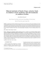
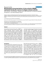
Upregulation of CD23 by IL-4 and GM-CSFFigure 1
Upregulation of CD23 by IL-4 and GM-CSF. Dose-ranging studies were performed to determine the optimum concen-
trations of IL-4 and GM-CSF. Alpha-smooth muscle isoactin positive Human ASMC (Clonetics) in T-75 flasks were starved for
24 h in 0.1% FBS containing medium M199. The cells were then stimulated with BSA (1 µg/ml), IL-4 (0.125. 0.25, 0.5, or 1.0 nM)
or GM-CSF (0.1, 0.2, 0.4, or 0.8 nM) for 24 h. The cell lysates in RIPA buffer were subjected to western blot analysis for CD23.
Mouse anti-human CD23 monoclonal antibody (clone M-L233, BD Biosciences, 1 µg/5 ml) was used as the primary antibody
and anti-mouse horseradish peroxidase linked antibody as the secondary antibody (Amersham). The immunoreactive protein
bands were detected by enhanced chemiluminescence light (ECL) (Amersham).
IL
IL
-
-
4(
4(
nM
nM
) 0.125 0.25 0.5 1.0
) 0.125 0.25 0.5 1.0
24hr
24hr
GM
GM
-
-
CSF (
CSF (
nM
nM
) 0.1 0.2 0.4 0.8_
) 0.1 0.2 0.4 0.8_
-
-
CD23 (45
CD23 (45
kD
kD
)
)
-
-
CD23 (45
CD23 (45
kD
kD
)
)
Clinical and Molecular Allergy 2005, 3:6 />Page 4 of 12
(page number not for citation purposes)
Protein analysis
A commercially available bicinchoninic acid (BCA) kit
(Pierce, Rockford, IL) was used for protein analysis
according to the manufacturer's instructions. The optical
densities were read using a Bio-Kinetics EL-312 Micro-
plate reader.
Indirect immunofluorescence
Indirect immunofluorescence stainings were performed
with anti-smooth muscle-α isoactin antibody (Sigma)
and anti-human CD23 antibody (M-L233, 1 µg/ml, BD
Biosciences), which are specific monoclonal antibodies
and either a FITC or TRITC fluorochrome, conjugated sec-
ond antibody. Fixed huASMC were incubated with the
above antibodies diluted in PBS with 3% BSA for 60 min
at room temperature. The cells were then washed three
times with PBS for 10 minutes for each wash. Non-specific
binding was blocked by incubating cells with 3% BSA in
PBS for 60 minutes. The blocking solution was then
removed and cells were incubated with FITC- or TRITC-
fluorochrome conjugated antibody for 45 minutes in the
dark to facilitate staining. Cells were then washed with
PBS three times. Finally, one drop of Fluoromount-G
(Southern Biotechnology Inc., Birmingham, AL) was
added.
Western blot
Standard Western blot analyses were performed to detect
anti-STAT6 (1:500, Calbiochem, San Diego, CA) polyclo-
nal rabbit, anti-p-STAT-6 (1:500, Calbiochem) polyclonal
rabbit. Human ASMC lysates in radio-immunoprecipita-
tion assay (RIPA) buffer were transferred onto Hybond-
ECL nitrocellulose membranes and were immunoblotted
with monoclonal anti-human CD23 (1:500 dilution,
clone M-L233, 1 µg/5 ml, BD Biosciences), polyclonal
anti-IL-4Rα (1:500 dilution, Santa Cruz), monoclonal
anti-IL-2Rγc (1:250 dilution, R&D Systems, Inc.). The
nitrocellulose membranes were incubated with a 1:1,000
dilution of anti-rabbit or anti-mouse horseradish peroxi-
dase linked whole antibody (Amersham, Piscataway, NJ)
in PBS-T for 1 hour at room temperature. Paxillin mono-
clonal antibody (1:500 dilution, Transduction Laborato-
ries) was used as a positive isotype control for CD23, and
fibronectin polyclonal antibody (1:250, Sigma) was used
as a positive control for the remaining antibodies. The
immunoreactive protein bands were detected by
enhanced chemiluminescence light (ECL) (Amersham).
Statistical analysis
Data were analyzed with Prism 4 software (GraphPad, San
Diego, CA). One-way analysis of variance (ANOVA) was
used. Results are expressed as mean ± SEM. A P value less
than 0.05 was considered statistically significant.
Results
CD23 protein expression is upregulated in huASMC by IL-
4, GM-CSF, or IL-4/GM-CSF
Previous studies have shown that IgE immune complexes
in atopic serum caused an increase in CD23 expression in
ASMC [16]. To determine if other humoral factors in
atopic serum effect CD23 expression in human ASMC, we
have tested the effect of the relevant cytokines, arachi-
donic acid metabolites, and the mast cell enzyme tryptase.
Flow cytometry was performed to evaluate differences in
cell populations after stimulation of the huASMC for 24
hours with either individual mediators IL-4 (0.5 nM),
GM-CSF (0.4 nM), IL-13 (0.4 nM), IL-5 (0.4 nM), PGD2
(10 µM), LTD4 (10 µM), tryptase (30 nM) or a combina-
tion of IL-4, IL-5, and IL-13 each with GM-CSF. Within the
huASMC stimulated by IL-4, GM-CSF or the combination
of IL-4/GM-CSF, two populations of cells were detected
distinguishable by cell size. While the smaller cells did not
show a significant expression of CD23, many of the larger
cells showed increased expression of CD23. In the exam-
ple in Figure 2, 66% of the larger cells (gate D) showed an
increase in cell expression of CD23 when compared to the
controls. As stated previously, the functions of ASMC are
heterogeneous including proliferation and synthesis. Pre-
vious studies have shown, on flow cytometry of ASMC
stimulated in vitro with IL-1β and TNF-α, only 20–60% of
ASMC produce GM-CSF. The ASMC producing GM-CSF
include some which also have increased proliferative
properties. This suggests that considerable heterogeneity
exists in the phenotypic expression of the ASMC in culture
[25].
In addition to the combination of IL-4/GM-CSF inducing
increased expression of CD23, both IL-4 and GM-CSF
alone independently increased the expression of CD23 in
huASMC. The percentage of cells with increased fluores-
cence intensity above the control was 25.1 ± 4.2% (IL-4),
15.6 ± 2.7% (GM-CSF) and 32.9 ± 13.9% (IL-4/GM-CSF
combination). On the other hand, IL-5, IL-13, cysteinyl
leukotrienes, and tryptase did not induce CD23 expres-
sion (Table 1).
Expression of CD23 in response to IL-4, GM-CSF, IL-4/GM-
CSF is accompanied by changes in huASMC morphology
Western blot analysis of huASMC stimulated with IL-4,
GM-CSF, or Il-4/GM-CSF for 24 h showed an increase in
CD23 expression compared to BSA vehicle control (Figure
3). Indirect immunofluorescence was used also to identify
any morphological changes associated with the cytokine
stimulation and upregulation of CD23 (Figure 4A–D).
Those cells stimulated with the combination of IL-4/GM-
CSF demonstrated CD23 expression along with changes
in cell morphology including depolymerization of isoac-
tin fibers, cell spreading, and membrane ruffling (Figure
4B). These changes in phenotype are consistent with flow
Clinical and Molecular Allergy 2005, 3:6 />Page 5 of 12
(page number not for citation purposes)
cytometry results in that the larger cells expressed CD23
(Figure 4D). In contrast, the control BSA stimulated pop-
ulation showed no changes in cell cytoskeletal structure
and morphology (Figure 4A) or specific staining for CD23
(Figure 4C).
To confirm activity of protein synthesis, the protein con-
tent of the control and the experimental groups of cells
were compared using a BCA protein analysis kit. Human
ASMC were starved for 24 hours in 0.1% FBS containing
medium M199 and then stimulated with BSA (1 µg/mL),
IL-4 (0.5 nM), GM-CSF (0.4 nM), or IL-4 (0.5 nM)/GM-
CSF (0.4 nM) for 24 hours. The protein content was
increased by 19% in the IL-4/GM-CSF treated cells above
that of the control (Table 2). The increase in protein con-
centration with IL-4 alone was not statistically significant.
Stimulation of huASMC with IL-4 induces phosphorylation
of STAT-6 and expression of IL-2R
γ
c
IL-4 binds the IL-4R with high affinity, and signaling
through IL-4 causes enhanced expression of IL-4R [21].
The induction of these genes is mediated through signal
transduction molecules including signal transducer acti-
vator of transcription (STAT-6). The binding of IL-4 to its
receptor complex induces the formation of an IL-4 recep-
tor complex which consists of IL-4Rα and the common
gamma chain (γc) of the receptors for IL-2, IL-4, IL-7, IL-
9, IL-15, and IL-21 [21]. It has not been previously
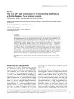
Upregulation of CD23 by IL-4, GM-CSF and IL-4/GM-CSFFigure 2
Upregulation of CD23 by IL-4, GM-CSF and IL-4/GM-CSF. Alpha-smooth muscle isoactin positive Human ASMC
(Clonetics) in T-75 flasks were starved for 24 h in 0.1% FBS containing medium M199. The cells were then stimulated with IL-
4 (0.5 nM)/GM-CSF (0.4 nM) for 24 h. The FACS analysis showed that the smaller cells passed through Gate A and larger cells
passed through Gate D. Background noise was eliminated using the BSA-stimulated control cells that were labeled with PE-
anti-CD23 (EBVCS), represented by C in Gate A, and F in Gate D. The FACS results of a representative experiment showed
66% of the larger cells (Gate D) had an increase in cell expression of CD23 when compared to the controls.
Fluorescence Intensity Fluorescence Intensity
Gate A
Gate D
Gate A
Gate D
Clinical and Molecular Allergy 2005, 3:6 />Page 6 of 12
(page number not for citation purposes)
Table 1: Il-4, GM-CSF, and IL-4/GM-CSF Increase CD23 Expression on ASMC (n = 3)
Cytokine (24 hours) #Cells in Gate D
(n = 3)
%Cells with Increased CD23
Expression Above the Control
BSA (1 µg/ml) 1,343 ± 122 0
IL-4 (0.5 nM) 1,413 ± 197 25.1 ± 4.2*
GM-CSF (0.4 nM) 1,346 ± 243 15.6 ± 2.7*
IL-4/GM-CSF (0.4/0.5 nM) 1,324 203 32.9 ± 13.9*
IL-5 (0.4 nM) 1,130 ± 251 0
IL-13 (0.4 nM) 1,316 ± 269 0
PGD2 (1 µM) 1,521 ± 123 0
PGD2 (10 µM) 1,159 ± 204 0
LTD4 (10 µM) 2,037 ± 375 0
Ethanol (6% vol/vol) 2,507 ± 200 0
Tryptase (10 µM) 2,385 ne ± 405 0
Alpha-smooth muscle isoactin positive Human ASMC (Clonetics) in T-75 flasks were starved for 24 h in 0.1% FBS containing medium M199. The
cells were then stimulated with BSA (1 µg/ml), IL-4 (0.5 nM), GM-CSF (0.4 nM), IL-4 (0.5 nM)/GM-CSF (0.4 nM), IL-13 (0.4 nM), IL-5 (0.4 nM),
PGD2 (10 µM), LTD4 (10 µM), tryptase (30 nM) for 24 h. Results are mean ± SEM of the percentage of cells with an increased fluorescence
intensity above the control (n = 3). Control values: BSA-stimulated, PE-anti-CD23 labeled, 10.9 ± 1.4 %; BSA-stimulated, PE-mouse IgG
1
, non-
immune, 0.5 ± 0.1 % (n = 3). *denotes significant increase in CD23 expression above the BSA control value.
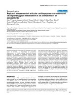
Western blot analysis of CD23 after stimulation of IL-4, GM-CSF, IL-4/GM-CSFFigure 3
Western blot analysis of CD23 after stimulation of IL-4, GM-CSF, IL-4/GM-CSF. Alpha-smooth muscle isoactin pos-
itive huASMC (Clonetics) in T-75 flasks were starved for 24 h in 0.1% FBS containing medium M199. The cells were then stim-
ulated with BSA (1 µg/ml) (vehicle control), IL-4 (0.5 nM), GM-CSF (0.4 nM), or IL-4/GM-CSF (0.5 nM/0.4 nM) for 24 h. The
cell lysates in RIPA buffer were subjected to western blot analysis for CD23. Mouse anti-human CD23 monoclonal antibody
(clone M-L233, BD Biosciences, 1 µg/5 ml) was used as the primary antibody and anti-mouse horseradish peroxidase linked
antibody as the secondary antibody (Amersham). The immunoreactive protein bands were detected by enhanced chemilumi-
nescence light (ECL) (Amersham). Paxillin mouse monoclonal IgG
1
(Transduction Laboratories) was used as an irrelevant iso-
type control.
BSA IL-4 GM-CSF IL-4/GM-CSF
(1 µg/ml) (0.5) (0.4) (0.5/0/4) nM (24 h)
-CD23
−paxillin
45 kD -
68 kD -
Clinical and Molecular Allergy 2005, 3:6 />Page 7 of 12
(page number not for citation purposes)
reported that airway smooth muscle cells express the IL-
2Rγc, the signaling unit of the IL-4 receptors.
Western blot analysis of IL-4Rα and IL-2Rγc in huASMC
lysates showed the presence of these receptor components
on huASMC. Figure 5 shows abundant expression of IL-
4Rα and a low level expression of IL-2Rγc protein on
huASMC. After stimulation of huASMC with IL-4 (0.4
nM) for 24 h, a two fold increase in γc expression was
observed compared to the BSA vehicle control (Figure 6).
To confirm that IL-4 was activating the IL-4Rα during the
stimulation of huASMC, we examined the phosphoryla-
tion of downstream STAT-6 by western blot. Human
ASMC were starved for 24 h and then stimulated with IL-
4 (0.4 nM) for fifteen minutes. Results of Western blot
revealed an approximately a four fold increase in intensity
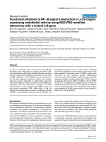
Expression of CD23 in response to IL-4/GM-CSF is accompanied by changes in huASMC morphologyFigure 4
Expression of CD23 in response to IL-4/GM-CSF is accompanied by changes in huASMC morphology. Alpha-
smooth muscle isoactin positive huASMC (Clonetics) in T-75 flasks were starved for 24 h in 0.1% FBS containing medium
M199. The cells were then stimulated with either BSA or IL-4 (0.5 nM)/GM-CSF (0.4 nM) for 24 h, and stained with either anti-
smooth muscle-isoactin (A & B) or anti-CD23 antibody (C & D). Those cells stimulated with the combination of IL-4/GM-CSF
demonstrated CD23 expression (D) and changes in cell morphology including depolymerization of isoactin fibers, cell spread-
ing, and membrane ruffling (B). Cells stimulated with BSA (vehicle for IL-4/GM-CSF) alone did not increase the expression of
CD23 (C) nor changes in phenotype (A). These findings were confirmed by three independent observers.
BSA
IL-4/
GM-CSF
A
A
B
B
C
C
D
D
α-isoactin CD23
α-isoactin CD23
BSA
IL-4/
GM-CSF
Table 2: IL-4/GM-CSF Combination Increases Protein Content in
huASMC.
Cytokine (nM) mg/10
6
cells (n = 3)
BSA (vehicle) 1.17 ± 0.08
IL-4 (0.5) 1.18 ± 0.01
GM-CSF 1.12 ± 0.03
IL-4 (0.5)/GM-CSF (0.4) 1.39 ± 0.02 *
Alpha-smooth muscle isoactin positive huASMC (Clonetics) in T-75
flasks were starved for 24 h in 0.1% FBS containing medium M199.
The cells were then stimulated with BSA (1 µg/ml)), IL-4 (0.5 nM),
GM-CSF (0.4 nM), or IL-4/GM-CSF (0.4/0.5 nM) for 24 h. The cell
lysates in RIPA buffer were analyzed for protein content using a
commercially available BCA kit (Pierce). The optical density was read
using a Bio-Kinetics EL-312 Microplate reader. Results are mean ±
SEM (n = 3). *denotes value significantly different from the BSA
vehicle treated control.
Clinical and Molecular Allergy 2005, 3:6 />Page 8 of 12
(page number not for citation purposes)
of the band for phosphorylated-STAT-6 in IL-4 stimulated
cells when compared to BSA control (Figure 7). This sup-
ports the role of an IL-4 mediated signal transduction
pathway involvement in CD23 upregulation in huASMC.
Discussion
In our study, we have demonstrated that CD23, the low
affinity IgE receptor, is upregulated on human airway
smooth muscle cells by the cytokines IL-4, GM-CSF, and
the combination of IL-4/GM-CSF. This upregulation of
CD23 by the combination of IL-4 and GM-CSF was
accompanied by an increase in cell volume and protein
content, cytoskeletal depolymerization, cell spreading
and membrane ruffling. Because ASMC require a dou-
bling time of 48 hours, the increase in protein content
could not be attributed to an increase in cell number.
Also, the increase in cell number in gate D seen with LTD4
was likely secondary to the effects of ethanol (Table 1).
Stimulation of huASMC by IL-4 caused an activation of
STAT-6 and an increase in γc expression. Collectively, our
findings suggest that CD23 expression can be stimulated
by IL-4 and GM-CSF cytokines independent of IgE in
huASMC and the upregulation of CD23 may play a role in
cell migration and hypertrophy.
Previous studies have demonstrated an increase in CD23
expression in alveolar monocytes after stimulation with
IL-4 and GM-CSF [10]. In that study, the use of the indi-
vidual mediators alone did not increase the CD23 levels
to that of asthmatic patients suggesting a possible syner-
gistic role between IL-4 and GM-CSF. Our findings are
consistent with these in that the combination of IL-4 and
GM-CSF was most effective in upregulating CD23 in
huASMC. Not all TH2 cytokines are involved in this
process; IL-5 (0.4 nM) and IL-13 (0.4 nM) had no effect
on CD23 expression. Cysteinyl leukotriene LTD4 (10
µM), prostaglandin PGD2 (10 µM) and tryptase (30 nM)
did not induce CD23 expression on huASMC [26].
IL-4Rα and IL-2Rγc Expression in huASMCFigure 5
IL-4Rα and IL-2Rγc Expression in huASMC. A: IL-4Rα and IL-2Rγc expression in huASMC at baseline. Unstarved
huASMC lysates were subjected to western blot analysis for IL-4Rα and IL-2Rγc using polyclonal anti-IL-4Rα (1:500 dilution,
Santa Cruz), or monoclonal anti-IL-2Rγc (1:125 dilution, R&D Systems, Inc.). The nitrocellulose membranes were incubated
with a 1:1,000 dilution of anti-rabbit or anti-mouse horseradish peroxidase linked whole antibody (Amersham). The immuno-
reactive protein bands were detected by ECL (Amersham). IL-2Rγc is minimally expressed in huASMC while IL-4Rα is
expressed abundantly in huASMC.
1 M.W. (kD) 2
IB: anti-IL-4Rα anti-IL-2Rγc
-140
53 -
IL-4Rα -
-IL-2Rγc
Clinical and Molecular Allergy 2005, 3:6 />Page 9 of 12
(page number not for citation purposes)
We evaluated the effect of stimulation of huASMC with IL-
4 on phosphorylation of STAT-6 via the IL-4R which
would confirm the presence of the receptor in huASMC.
STAT-6 is a critical mediator of IL-4 stimulated gene acti-
vation, and it is regulated by both tyrosine and serine
kinases [27]. It has been shown in a mouse model that
STAT-6 binds the CD23a murine promoter, and STAT -/-
mice stimulated with IL-4 are unable to upregulate CD23.
This suggests STAT-6 is a critical mediator for IL-4 induced
upregulation of CD23 [28]. IL-4 along with CD40 medi-
ated signals are responsible for upregulation of CD23 on
B cells [14]. In this study, we have confirmed the
expression of the IL-4Rα and a low level of common
gamma chain in huASMC and that after stimulation of
huASMC with IL-4, there was a two fold increase in γc
chain expression (Figure 6). Phosphorylation of STAT-6
Upregulation of IL-2Rγc Expression in huASMC by IL-4 and IL-4/GM-CSFFigure 6
Upregulation of IL-2Rγc Expression in huASMC by IL-4 and IL-4/GM-CSF. Alpha-smooth muscle isoactin positive
Human ASMC (Clonetics) in T-75 flasks were starved for 24 h in 0.1% FBS containing medium M199. The cells were then
either stimulated with BSA (vehicle) (1 µg/ml), IL-4 (0.5 nM), GM-CSF (0.4 nM), or IL-4/GM-CSF (0.5 nM/0.4 nM) for 24 hours.
The IL-4 and IL-4/GM-CSF stimulated cells had increased IL-2Rγc expression compared to the BSA (vehicle) group. Fibronectin
polyclonal rabbit antibody (Sigma) (1:250) was used as an irrelevant isotype control.
M.W. BSA IL-4 GM-CSF IL-4/GM-CSF
kD (1 µg/ml) (0.5 nM) (0.4 nM) (0.5/0.4 nM)
-IL-4Rα
−IL-2Rγc
-fibronec tin
140 -
53 -
220 -
Clinical and Molecular Allergy 2005, 3:6 />Page 10 of 12
(page number not for citation purposes)
after stimulation with IL-4 for 15 min confirms that IL-4
has bound and activated the IL-4 receptor complex (Fig-
ure 7). It has been shown that the common gamma chain
is a functional β chain of the IL-4 receptor complex in cer-
tain cells [27], and our data suggest that this is the case in
huASMC. Interestingly, IL-13 (4 nM, a concentration suf-
ficient to simulate huASMC proliferation, unpublished
observation) did not upregulate CD23. For proliferation
of ASMC by IL-13, IL-4Rα and IL-13Rα1 are required for
signal transduction and downstream activation of p44/42
extracellular regulated kinases (ERK, unpublished data).
Apparently, IL-4Rα and IL-13Rα1 engagement is not suf-
ficient for CD23 expression, further supporting the role of
γc chain in CD23 expression by IL-4. The signs of signal
transduction in response to IL-4, and the increase in pro-
tein content of the cell in response to IL-4 and GM-CSF
combination (Table 2) represent activation of transcrip-
tion and translation of CD23 in this case. Coupling of
GM-CSF and its receptor complex is known to activate
ERK that may have contributed to the synergistic effect of
GM-CSF on CD23 expression.
CD23 expression was associated with changes in cell mor-
phology including depolymerization of isoactin fibers,
Phosphorylation of STAT-6 by IL-4 and IL-4/GM-CSF in huASMCFigure 7
Phosphorylation of STAT-6 by IL-4 and IL-4/GM-CSF in huASMC. Starved huASMC were incubated with either BSA
vehicle control (1 µg/ml), IL-4 (0.5 nM), GM-CSF (0.4 nM), or IL-4/GM-CSF (0.5 nM/0.4 nM) for 15 minutes. Standard Western
blot analyses were performed to detect STAT-6 and phosphorylated-STAT-6 (p-STAT-6) using a anti-STAT-6 polyclonal rabbit
antibody (Calbiochem) and anti-p-STAT-6 polyclonal rabbit antibody (Calbiochem). Anti-rabbit horseradish peroxidase linked
antibody was used as the secondary antibody. Protein bands were detected by ECL. STAT-6 was abundantly expressed by all
four groups, while p-STAT-6 was only expressed in the IL-4 and IL-4/GM-CSF groups. Fibronectin polyclonal rabbit antibody
(Sigma) was used as an irrelevant isotype control and was abundantly expressed in all four groups.
MW BSA IL-4 GM-CSF IL-4/GM-CSF
(kD) (1 µg/ml) (0.5nM) (0.4nM) (0.5/0.4nM)
15min Incubation
-p-stat 6
-stat-6
-fibronectin
110 -
110 -
220 -
Clinical and Molecular Allergy 2005, 3:6 />Page 11 of 12
(page number not for citation purposes)
cell spreading, and membrane ruffling (Figure 4B &4D).
Actin in ASMC is in a dynamic state and undergoes
polymerization-depolymerization during the contraction-
relaxation cycle [29,30]. Membrane ruffling and cell
migration involve signaling pathways including PI3-
kinase, Rac and other Rho family G protein members in a
variety of cell types, including vascular smooth muscle
cells. Rac has an essential role in cell migration and regu-
lation of the actin cytoskeleton [31,32]. Moreover, ASMC
are capable of switching their phenotypes from contractile
to synthetic phenotype that is mediated by Rho kinases
[32,33].
In summary, we have demonstrated that the low affinity
IgE receptor can be induced on huASMC by specific
cytokines including IL-4, GM-CSF, and the combination
of IL-4/GM-CSF. The combination of IL-4/GM-CSF also
induced morphologic changes in the ASMC that may
contribute to the synthetic function or migration. In addi-
tion, IL-4 and IL-4/GM-CSF stimulation of huASMC
increased the protein content of the cell, suggesting
hypertrophy.
Conclusion
T helper type 2 cytokines including IL-4 have major role
in asthma pathogenesis. GM-CSF is a hemopoetic growth
factor, mostly released by activated monocytes and T cells.
Additional sources of GM-CSF include epithelium of asth-
matic airways [34] and human airway smooth muscle
cells [6,35]. Therefore, the effect of GM-CSF on CD23
expression can be both via paracrine and autocrine mech-
anisms. Previous studies by Hakonarson et al. [16,17]
have shown that upregulation of the CD23 receptor has
been associated with proasthmatic changes in agonist-
mediated ASM constrictor and relaxant responsiveness.
Our study suggests that CD23 expression is associated
with elements of hypertrophy (i.e. an increase in cell vol-
ume and protein content), thus consistent with their find-
ings. Inhaled corticosteroids, the mainstay in treatment of
asthma, effectively reduce inflammation and remodeling
of the epithelium and basement membrane. However, no
agents have been proven effective in reducing smooth
muscle mass in asthmatic patients. Recent study results on
anti-CD23 therapy showed decrease in serum IgE. Further
studies to intervene the upregulation of CD23 expression
by cytokines IL-4 and GM-CSF may open a new avenue to
target smooth muscle hypertrophy, an important element
of severe asthma [2].
Competing interests
The author(s) declare that they have no competative
intrests.
List of abbreviations
huASMC: Human airway smooth muscle cells
FceRI: High-affinity receptor for IgE
FceRII (CD23): Low-affinity receptor for IgE
AHR: Airway hyperreactivity
PGD2: Prostaglandin D2
LTD4: Leukotriene D4
PE: Phycoerythrin
FBS: Fetal bovine serum
BSA: Bovine serum albumin
PBS: Phosphate bufferred saline
BCA: Bicinchoninic acid
FITC: Fluorescein Isothiocyanate
TRITC: Tetramethyl Rhodamine Iso-Thiocyanate
RIPA: Radio-immunoprecipitation assay
ECL: Enhanced chemiluminescence light
Rα: Receptor alpha
γc: Common gamma chain
FACS: Fluorescent Activated Cell Sorter
STAT: Signal Transducer Activator of Transcription
TH2: T helper cell type 2
ERK: Extracellular regulated kinases
Authors' contributions
JTB participated in designing experiments and performing
CD23 analysis by flow cytometry.
RKG carried out flow cytometry, immunofluorescence
studies, and drafted the manuscript.
HM carried out western blot analyses.
DBL supervised all aspects of the project.
All authors have read and approved the final manuscript.
Acknowledgements
This work was supported in part by the grant from the Le Bonheur Chil-
dren's Medical Center, NIH-HL56812, Children's Foundation Research
Publish with Bio Med Central and every
scientist can read your work free of charge
"BioMed Central will be the most significant development for
disseminating the results of biomedical research in our lifetime."
Sir Paul Nurse, Cancer Research UK
Your research papers will be:
available free of charge to the entire biomedical community
peer reviewed and published immediately upon acceptance
cited in PubMed and archived on PubMed Central
yours — you keep the copyright
Submit your manuscript here:
/>BioMedcentral
Clinical and Molecular Allergy 2005, 3:6 />Page 12 of 12
(page number not for citation purposes)
Center, Molecular Resource Center, and American Academy of Allergy,
Asthma and Immunology. The authors would like thank Jan Aldrich for her
technical assistance.
References
1. Vignola AM: Effects of inhaled corticosteroids, leukotriene
receptor antagonists, or both, plus long-acting beta2-ago-
nists on asthma pathophysiology: a review of evidence. Drugs
2003, 63:35-51.
2. Benayoun L, Druihe A, Dombret M-C, Aubier M, Pretolani M: Air-
way structural alterations selectively associated with severe
asthma. Am J Respir Crit Care Med 2003, 167:1360-8.
3. Woodruff PG, Dolganov GM, Ferrando RE, Donnelly S, Hays SR, Sol-
berg OD, Carter R, Wong HH, Cadbury PS, Fahy JV: Hyperplasia of
smooth muscle in mild to moderate asthma without changes
in cell size or gene expression. Am J Respir Crit Care Med 2004,
169:1001-6.
4. Hirst SJ: Regulation of airway smooth muscle cell immu-
nomodulatory function: role in asthma. Respir Physiol Neurobiol
2003, 137:309-26.
5. Busse W, Banks-Schlegel S, Wenzel S: Pathophysiology of severe
asthma, Nihlbi workshop summary. J Allerg Clin Immunol 2000,
106:1033-42.
6. Hakonarson H, Grunstein M: Autocrine regulation of airway
smooth muscle responsiveness. Respir Physiol & Neurobiol 2003,
137:263-76.
7. Madison M: Migration of airway smooth muscle cells:. Am J
Respir Cell Mol Biol 2003, 29:8-11.
8. Schmidt D, Rabe K: Immune mechanisms of smooth muscle
hyperreactivity in asthma. J Allerg Clin Immunol 2000, 105:673-83.
9. Broide D: Novel therapies in allergic disease: molecular and
cellular mechanisms of allergic disease. J Allerg Clin Immunol
2001, 108(Suppl):65-71.
10. Matz J, Williams J, Rosenwasser L, Borish L: Granulocyte-macro-
phage colony-stimulating factor stimulates macrophages to
respond to IgE via the low affinity IgE receptor (CD23). J
Allerg Clin Immunol 1994, 93:650-7.
11. Visan I, Goller M, Berberich I, Kneitz C, Tony HP: Pax-5 is a key
regulator of the B cell restricted expression of the CD23a
isoform. Eur J Immunol 2003, 33:1163-1173.
12. Novak N, Kraft S, Bieber T: IgE receptors. Curr Opinion in Immunol
2001, 13:721-6.
13. Ewart MA, Ozanne BW, Cushley W: The CD23a and CD23b
proximal promoters display different sensitivities to exoge-
nous stimuli in B lymphocytes. Genes Immunol 2003, 3:158-64.
14. Oettgen H, Geha R: IgE regulation and roles in asthma
pathogenesis. J Allerg Clin Immunol 2001, 107:429-40.
15. Harkins MS, Moseley PL, Iwamoto GK: Regulation of CD23 in the
chronic inflammatory response in asthma: a role for inter-
feron-gamma and heat shock protein 70 in the TH2
environment. Ann Allergy Asthma Immunol 2003, 6:567-74.
16. Hakonarson H, Carter C, Kim C, Grunstein M: Altered expression
and action of the low-affinity IgE receptor FCRII (CD23) in
asthmatic airway smooth muscle. J Allerg Clin Immunol 1999,
104:575-84.
17. Hakonarson H, Grunstein MM: Autologously up-regulated Fc
receptor expression and action in airway smooth muscle
mediates its altered responsiveness in the atopic asthmatic
sensitized state. Proc Nat Acad Sci 1998, 95:5257-62.
18. Hakonarson H, Maskeri N, Carter C, Grunstein M: Regulation of
TH1- and TH2-tye cytokine expression and action in atopic
asthmatic sensitized airway smooth muscle. J Clin Invest 1999,
103:1077-87.
19. Hakonarson H, Maskeri N, Kim C, Grunstein M: Autocrine inter-
action between IL-5 and IL-1 mediates altered responsive-
ness of atopic asthmatic sensitized airway smooth muscle. J
Clin Invest 1999, 104:657-67.
20. Rosenwasser L, Busse W, Lizambri R, Olejnik T, Totoritis : Allergic
asthma and anti-CD23 mAb (IDEC-152): Results of a phase
1, single-dose, dose-escalating clinical trial. J Allerg Clin Immunol
2003, 112:563-70.
21. Ritz SA, Cundall M, Gajewska B, Alvarez D, Gutierrez-Ramos J, Coyle
A, McKenzie A, Stampfli M, Jordana M: Granulocyte macrophage
colony-stimulating factor-driven respiratory mucosal sensiti-
zation induces Th2 differentiation and function independ-
ently of interleukin-4. Am J Respir Cell Mol Biol 2002, 27:428-435.
22. Schwartz LB, Bradford TR: Regulation of tryptase from human
lung mast cells by heparin. Stabilization of the active
tetramer. J Biol Chem 261(16):7372-9. 1986 Jun 5
23. Brightling CE, Bradding P, Symon FA, Holgate ST, Wardlaw AJ, Pavord
ID: Mast-cell infiltration of airway smooth muscle in asthma:.
N Engl J Med 346(22):1699-705. 2002 May 30
24. Oguma T, Palmer LJ, Birben E, Sonna LA, Asano K, Lilly CM: Role of
prostanoid DP receptor variants in susceptibility to asthma:.
N Engl J Med 351(17):1752-63. 2004 Oct 21
25. Sukkar MB, Stanley AJ, Blake AE, Hodgkin PD, Johnson PR, Armour
CL, Hughes JM: 'Proliferative' and 'synthetic' airway smooth
muscle cells are overlapping populations. Immunol Cell Biol
2004, 82(5):471-8.
26. Brown JK, Jones CA, Rooney LA, Caughey GH, Hall IP: Tryptase's
potent mitogenic effects in human airway smooth muscle
cells are via nonproteolytic actions. Am J Physiol Lung Cell Mol
Physiol 2002, 282:L197-206.
27. Jiang H, Harris M, Rothman P: IL-4/IL-13 signaling beyond JAK/
STAT. J Allerg Clin Immunol 2000, 105:1063-70.
28. Tinnell SB, Jacobs-Helber SM, Sterneck E, Sawyer ST, Conrad DH:
STAT6, NF-kappaB and C/EBP in CD23 expression and IgE
production:. Int Immunol 1998, 10(10):1529-38.
29. Gunt SJ, Tang DD, Saez AO: Cytoskeletal remodeling of the air-
way smooth muscle cell: a mechanism for adaptation to
mechanical forces in the lung. Respir Physiol Neurobiol 2003,
137:151-168.
30. Hirshman CA, Zhu D, Panettieri RA, Emala CW: Actin depolymer-
ization via beta-adrenoreceptor in airway smooth muscle
cells: a novel PKA-independent pathway. Am J Physiol Cell Physiol
2001, 281:C1468-76.
31. Halayko AJ, Solway J: Molecular mechanisms of phenotypic
plasticity in smooth muscle cells. J Appl Physiol 2001, 90:358-68.
32. Nishiyama T, Sasaki T, Takaishi K, Kato M, Yaku H, Araki K, Matsuura
Y, Takai Y: rac p21 is involved in insulin-induced membrane
ruffling and rho p21 is involved in hepatocyte growth factor-
and 12-O-tetradecanoylphorbol-13-acetate (TPA)-induced
membrane ruffling in KB cells. Mol Cell Biol 1994, 14:2447-56.
33. Okamoto H, Takuwa N, Yokomizo T, Sugimoto N, Sakurada S, Shige-
matsu H, Takuwa Y: Inhibitory Regulation of Rac Activation,
Membrane Ruffling, and Cell Migration by the G Protein-
Coupled Sphingosine-1-Phosphate Receptor EDG5 but Not
EDG1 or EDG3. Mol Cell Biol 2000, 20:9247-61.
34. Fish J, Peters S: Airway remodeling and persistent airway
obstruction in asthma. J Allerg Clin Immunol 1999, 104:509-16.
35. Oltman U, Issa R, Sukkar MB, John M, Chung KF: Role of c-jun N-
terminal kinase in the induced release of GM-CSF, RANTES
and IL-8 from human airway smooth muscle cells. Br J
Pharmacol 2003, 139:1228-34.