Báo cáo y học: "Protective effect of the DNA vaccine encoding the major house dust mite allergens on allergic inflammation in the murine model of house dust mite allergy" ppsx
Bạn đang xem bản rút gọn của tài liệu. Xem và tải ngay bản đầy đủ của tài liệu tại đây (706.92 KB, 9 trang )
BioMed Central
Page 1 of 9
(page number not for citation purposes)
Clinical and Molecular Allergy
Open Access
Research
Protective effect of the DNA vaccine encoding the major house dust
mite allergens on allergic inflammation in the murine model of
house dust mite allergy
Nacksung Kim
1
, Soon Seog Kwon
2
, Jaechun Lee
3,4
, Sohyung Kim
4
and
Tai June Yoo*
4
Address:
1
Medical Research Center for Gene Regulation, Chonnam National University Medical School, Gwangju, Korea,
2
Department of Internal
Medicine, Catholic University Medical School, Seoul, Korea,
3
Department of Internal Medicine, College of Medicine, Cheju National University,
Jeju, Korea and
4
Division of Allergy/Immunology, Department of Medicine, University of Tennessee, Memphis, TN, USA
Email: Nacksung Kim - ; Soon Seog Kwon - ; Jaechun Lee - ;
Sohyung Kim - ; Tai June Yoo* -
* Corresponding author
Abstract
Background: Vaccination with naked DNA encoding antigen induces cellular and humoral
immunity characterized by the activation of specific Th1 cells.
Objective: To evaluate the effects of vaccination with mixed naked DNA plasmids encoding Der
p 1, Der p 2, Der p 3, Der f 1, Der f 2, and Der f 3, the major house dust mite allergens on the allergic
inflammation to the whole house dust mites (HDM) crude extract.
Methods: Three hundred micrograms of these gene mixtures were injected into muscle of BALB/
c mice. Control mice were injected with the pcDNA 3.1 blank vector. After 3 weeks, the mice
were actively sensitized and inhaled with the whole house dust mite extract intranasally.
Results: The vaccinated mice showed a significantly decreased synthesis of total and HDM-specific
IgE compared with controls. Analysis of the cytokine profile of lymphocytes after challenge with
HDM crude extract revealed that mRNA expression of interferon-γ was higher in the vaccinated
mice than in the controls. Reduced infiltration of inflammatory cells and the prominent infiltration
of CD8+ T cells were observed in histology of lung tissue from the vaccinated mice.
Conclusion: Vaccination with DNA encoding the major house dust mite allergens provides a
promising approach for treating allergic responses to whole house dust mite allergens.
Background
It has been reported that IgE-mediated inflammation to
mites is associated with diseases such as asthma, allergic
rhinitis, and atopic dermatitis and that the relatively
minor determinants of house dust mite (HDM) allergens
show IgE binding with 40% or more of allergic sera [1-4].
The most frequently implicated allergens are derived from
Dermatophagoides pteronyssinus (Der p) and Dermatopha-
goides farinae (Der f) [5]. Targeting specific T lymphocytes
that induce or regulate the allergic inflammation is one of
the therapeutic goals in allergic disorders. Specific immu-
notherapy with crude extracts has been used mainly in
Published: 20 February 2006
Clinical and Molecular Allergy 2006, 4:4 doi:10.1186/1476-7961-4-4
Received: 11 October 2005
Accepted: 20 February 2006
This article is available from: />© 2006 Kim et al; licensee BioMed Central Ltd.
This is an Open Access article distributed under the terms of the Creative Commons Attribution License ( />),
which permits unrestricted use, distribution, and reproduction in any medium, provided the original work is properly cited.
Clinical and Molecular Allergy 2006, 4:4 />Page 2 of 9
(page number not for citation purposes)
treating HDM-induced allergy. However, it has limited
efficacy. Recently, vaccinations with naked DNA encoding
antigen were reported to induce long-lasting cellular and
humoral immune tolerance [6,7]. Injection of plasmid
DNA encoding T cell epitopes could suppress allergic reac-
tion [8,9]. However, the potential barrier to T cell recep-
tor-based immunotherapy for allergy is the apparent
complexity of the allergen-specific T cell response in terms
of epitope usage in individuals [10]. A recent literature
search showed that injection of plasmid DNA encoding
Der p 5 in rat not only inhibits Der p 5-specific IgE anti-
body production but also an allergic response such as his-
tamine release and airway hyperresponsiveness to Der p 5
[11]. However, Der p 5 is not one of the major HDM aller-
gens; thus, it has limited clinical application [3]. In this
study, we investigated immune responses by gene vaccina-
tion with plasmid DNA encoding major HDM allergens
(Der p 1,2, and 3, and Der f 1,2, and 3) to challenges with
whole HDM crude extract in sensitized mice.
Methods
Animals
Twenty female BALB/c mice 6–8 weeks old were pur-
chased from Jackson Laboratory (Bar Harbor, ME) and
bred in the animal facility of the University of Tennessee
Health Science Center. This study was performed in
accordance with the PHS Policy on Humane Care and Use
of Laboratory Animals and the NIH Guide for the Care
and Use of Laboratory Animal Welfare Act (7 U.S.C. et
seq.). The animal use protocol was approved by the Insti-
tutional Animal Care and Use Committee (IACUC) of the
University of Tennessee.
Plasmid construction
Total mRNA was isolated from Der p and Der f HDM,
respectively. By using murine leukemia virus reverse tran-
scriptase and random hexanucleotide primer following
the instructions of the Perkin Elmer Gene Amp RNA PCR
kit (Perkin Elmer, Branchberg, NJ), first-strand cDNA was
generated from 1 µg of total RNA and subjected to reverse
transcriptase polymerase chain reaction (RT-PCR). The
cDNA was used in PCR with Taq polymerase with primers
specific for Der p 1 (5'- CCGGAATTCGCCGCCACCAT-
GGAAACTAACGCCTGCAGTATCAATGGA -3' and 5'-
TGCTCTAGATTAGAGAATGACAACATATGGATATTC -3'),
Der p 2 (5'- CCGGAATTCGCCGCCACCATGGAT-
CAAGTCGATGTCAAAGATTGTGCC -3' and 5'-
TGCTCTAGATTAATCGCGGATTTTAGCATGAGTAG-
CAAT -3'), Der p 3 (5'- CCGGAATTCGCCGCCACCAT-
GATTGTTGGTGGTGAAAAAGCATTAGCTG -3' and 5'-
TGCTCTAGATTACTGTGAACGTTTTGATTCAATCCAATC-
GATA -3'), Der f 1 (5'- CCGGAATTCGCCGCCACCAT-
GGAAACAAGCGCTTGCCGTATCAATTCG -3' and 5'-
TGCTCTAGATTAGAGGTTGTTTCCGGCTT-
GGAAATATCCG -3'), Der f 2 (5'-
CCGGAATTCGCCGCCACCATGGATCAAAGTCGATGT-
TAAAGATTGTGCC -3' and 5'- TGCTCTAGATTAATCACG-
GATTTTACCATGGGTAGCAAT -3'), and Der f 3 (5'-
CCGGAATTCGCCGCCACCATGATTGTTGGTGGTGT-
GAAAGCACAAGCC -3' and 5'- TGCTCTAGATTACTGT-
GAACGTTTTGATTCAATCCAATCGAC -3'). These primers
cover the mature excreted region of each gene and include
EcoR1 and Xb1 sites for cloning. The amplified PCR prod-
ucts were subcloned into pcDNA3.1 eukaryotic expression
vector (Invitrogen, San Diego, CA) and then sequenced to
verify the insertion of the correct gene with the appropri-
ate open reading frame.
DNA preparation and vaccination
Each plasmid construct was prepared using Maxi prep
(Quiagen, Chatsworth, CA). Mice were vaccinated by
injection with 300 µg of pcDNA3.1 blank vector in 100 µl
of phospate-buffered saline (PBS) (the control group) or
the same amount of the mixed naked DNA encoding the
major HDM allergens (the vaccination group) three times
at weekly intervals into muscle (week 0, 1, and 2).
Immunization and inhalation of allergen
Mice were sensitized with HDM crude extract previously
described [12]. HDM crude extract was emulsified with an
equal volume of complete Freund adjuvant (CFA) for
immunization. Three weeks after the last vaccination,
mice were sensitized subcutaneously at the base of the tail
with 100 µg of HDM extract in CFA. The mice were also
given an intraperitoneal dose of 300 ng of purified pertus-
sis toxin at 24 and 72 h after first immunization. Seven
days later, the mice were boosted again with the same
amount of antigen in incomplete Freund adjuvant. Under
inhaled anesthesia with methoxyflurane, mice were chal-
lenged with 10 µg of HDM crude extract through one nos-
tril six times at weekly intervals after immunization.
Determining total IgE, HDM-specific IgE, and HDM-
specific IgG
The blood from the six mice in two groups was collected
six times at week 0 (first vaccination), 3, 5 (first immuni-
zation), 7, 9, and 11. The HDM-specific IgG was deter-
mined by ELISA. One hundred microliter of HDM (5 µg/
ml in 0.1 M carbonate buffer, pH 9.6) were dispensed in
each well of a polystyrene microtiter plate (Cost, Cam-
bridge, MA) and incubated overnight at 4°C. The concen-
tration of HDM was determined by the preliminary
experiments. The antigen-coated plates were washed three
times in 0.05% PBS-Tween 20 buffer (washing buffer) and
incubated with mice sera overnight at 4°C. The plates
were washed five times with washing buffer and incu-
bated with peroxidase-conjugated anti-mouse IgG anti-
body (Sigma, St. Louis, MO) overnight at 4°C. The plates
were washed five times before adding citric acid-phos-
phate buffer (pH 5.0) containing 0.15 mg/ml of O-phe-
Clinical and Molecular Allergy 2006, 4:4 />Page 3 of 9
(page number not for citation purposes)
nylenediamine (Sigma, St. Louis, MO). The color was
developed at room temperature, and the reaction was
stopped by 2.5 M sulfuric acid. The color was measured at
492 nm (Bio-Rad, Richmond, CA).
The total IgE level was determined by ELISA. One hundred
microliter of anti-mouse IgE capture monoclonal anti-
body (mAb) (clone R35–72; Pharmingen, San Diego, CA)
were added in each well to plates and incubated overnight
at 4°C. After washing, 200 µL of 10% fetal calf serum were
incubated at room temperature for 30 min. The plates
were washed five times with washing buffer and incu-
bated with the diluted mouse serum overnight at 4°C, fol-
lowed by adding 100 µL of HRP-conjugated anti-mouse
IgE detection mAb (clone R35–118; Pharmingen, San
Diego, CA) overnight at 4°C. After washing, color was
developed following the procedure for IgG. The purified
mouse serum IgE (BD Biosciences, Palo Alto, CA) was
used for total IgE standard. To measure HDM-specific IgE,
the plate was coated with 25 µg/ml HDM in 0.1 M carbon-
ate buffer (pH 9.6), and serum samples were diluted five-
fold in 10% FCS. The concentration of HDM was
determined by the preliminary experiments. The proce-
dure after this point was the same as that for measuring
HDM-specific IgG. The level of HDM-specific IgE was ref-
erenced to the standard serum pooled from six mice that
were immunized with 100 µg of HDM twice and inhaled
with 10 µg of antigen six times. The standard serum was
calculated as 100 ELISA units/ml.
Immunohistochemical staining for CD4+ and CD8+ T cells
in lung tissue
The lung tissues from the vaccination and control groups
were removed immediately after the final intranasal inha-
lation. Tissues were fixed with periodate-lysine-parafor-
maldehyde solution for 24 h at 4°C. The specimens were
rinsed with 0.01 M of PBS (pH 7.4) containing 10% to
20% sucrose for 36 h at 4°C, embedded in OCT com-
pound (Miles Laboratories Inc., Elkhart, IN), and imme-
diately frozen. The lung specimens were immersed in 10%
EDTA and decalcified for 10 days at 4°C. Frozen sections
cut at 4 to 6 µm in thickness were dehydrated and rinsed
in cold PBS. The endogenous pseudoperoxidase was
blocked with absolute methanol containing 0.5% hydro-
gen peroxide for 20 min at room temperature. The sec-
tions were treated with 10% normal goat serum in PBS to
reduce nonspecific binding. Biotin conjugated rat anti-
mouse CD8 or CD4 mAb (Pharmingen, San Diego, CA)
diluted to 1:200 in PBS containing 0.5% bovine serum
albumin was applied to the sections and incubated over-
night at 4°C. After rinsing, the sections were incubated
with avidin-biotin peroxidase complexes (Vectastain Elite
ABC Kit, Vector Laboratories Inc., Burlingame, CA) for 30
min at room temperature and rinsed sufficiently with PBS.
The reaction was developed with 0.02% 3,3'-diaminoben-
zidine in 0.05 M of Tris buffer (pH 7.6) with 0.005%
hydrogen peroxidase for 7 min. The sections were dehy-
drated, cleared in xylene, and mounted.
Histological examination of lung tissue
Mice were anesthetized with a mixture of ketalar (35 mg/
ml), rompun (0.6%/ml) and atropine (0.1 mg/ml), of
which 0.2 ml was injected intramuscularly. The vascular
bed of the lungs was perfused with 0.01 M PBS and then
with 4% paraformaldehyde 0.1 M PBS buffers. Whole
lungs were taken out and stored in 4% paraformaldehyde
for 24 h at 4°C. After fixation, these tissues were dehy-
drated and embedded in paraffin. Frozen sections cut at 3
µm in thickness were stained by hematoxylin and eosin.
After coding, the sections were evaluated by two observers
using light microscopy. The amount of inflammation per
section was scored using the method described by Hessel
et al. [13]. Lungs that showed no focal inflammation were
scored as grade 0. Those that showed one or two centrally
located microscopic foci of inflammatory infiltrate were
graded as 1. In grade 2, a dense inflammatory infiltrate
was seen in a perivascular and peribronchial distribution
originating in the center of the lung. In grade 3, the
perivascular and peribronchial infiltrates extended to the
periphery of the lung.
Measuring cytokine mRNA expression
Measuring the expression level was done as previousy
described [9]. Briefly, four mice from each group were sac-
rificed 10 days postboost. The lymph nodes were removed
from the mice and minced to create single cell suspen-
sions. Cells were cultured in RPMI for 18 h with no anti-
gen as a negative control, recombinant Der p 1 (100 µg/
ml), or HDM crude extract (100 µg/ml). Cells were
washed with PBS buffer and mRNAs prepared (Biotech,
Houston, TX). By using murine leukemia virus reverse
transcriptase and random hexanucleotide primer follow-
ing the instructions of the Perkin Elmer Gene Amp RNA
PCR kit (Perkin Elmer, Branchber, NJ), first-strand cDNA
was generated from 1 µg of total RNA and subjected to RT-
PCR analysis. We used the primers specific for β-actin (5'-
GTGGGCCGCTCTAGGCACCAA -3' and 5'- CTCTTTGAT-
GTCACGCACGATTTC -3') as control primer, IL-2 (5'-
TTCAAGCTCCACTTCAAGCTCTACAGCGGAAG -3' and
5'- GACAGAAGGCTATCCATCTCCTCAGAAAGTCC -3'),
IFN-γ (5'- TGCATCTTGGCTTTGCAGCTCTTCCTCATGGC
-3' and 5'- TGGACCTGTGGGTTGTTGACCTCAAACT
TGGC -3') (Clonetech, Palo Alto, CA), IL-4 (5'-
CAGCTAGTTGTCATCCTGCTCTTC -3' and 5'- GTGATGT-
GGACTTGGACTCATTCATGG -3'), or IL-5 (5'- TGTCT-
GGGCCACTGCCATGGAGATTC -3' and 5'-
CCATTGCCCACTCTGTACTCATCACAC -3') in the RT-
PCR analysis. The amplified DNAs of β-actin, IFN-γ, IL-2,
IL-4, and IL-5 were 540, 365, 413, 354, and 349 base
pairs, respectively.
Clinical and Molecular Allergy 2006, 4:4 />Page 4 of 9
(page number not for citation purposes)
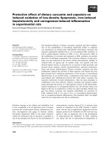
Effect of vaccination on immunoglobulin productionFigure 1
Effect of vaccination on immunoglobulin production. The total IgE antibody levels (1a), HDM-specific IgE antibody lev-
els (1b), and HDM-specific IgG antibody levels (1c) in sera of each mouse were detected by ELISA every 2 weeks after immuni-
zation with HDM. The data are expressed as means ± SD (n = 6 per group). *P < .05 compared with the control group.
Clinical and Molecular Allergy 2006, 4:4 />Page 5 of 9
(page number not for citation purposes)
Statistical analysis
Data in immunoglobulin response were analyzed by Stu-
dent's paired t test for comparisons between control and
vaccination groups. Histological grades were analyzed by
a nonparametric Mann-Whitney U test. Data were
expressed as mean ± SD. A p-value of < 0.05 was consid-
ered significant.
Results
Downregulation of allergen specific IgE production by
DNA vaccination
DNA vaccination with the major HDM allergen gene, Der
p 1, 2, and 3, and Der f 1, 2, and 3 showed about 50%
reduction of HDM-specific IgE and more than 70% reduc-
tion of total IgE compared with the control group at 6
weeks after immunization (Fig. 1a and 1b). However, pro-
duction of HDM-specific IgG antibody showed no differ-
ence (Fig. 1c). Thus, in vivo total and allergen-specific IgE
synthesis might be efficiently inhibited by DNA vaccina-
tion.
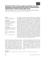
Histological and immunohistochemical study
To investigate whether the DNA vaccination affects
inflammation of lung, we stained lung tissue by histolog-
ical and immunohistochemical methods. The lungs from
the control group showed much more infiltration of
inflammatory cells in the submucosa of airways than did
those lungs from the vaccination group. The inflamma-
tion grades were scored as 1.64 ± 0.52 (mean ± SD) in the
control group and 0.68 ± 0.48 in the vaccination group
(Fig. 2a, 2b, and 2c). Also, eosinophils were detected in
the lungs of the control mice (Fig. 2d). In the immunohis-
Effect of genetic vaccination on lung histopathology in an animal model of allergyFigure 2
Effect of genetic vaccination on lung histopathology in an animal model of allergy. A and B, Light microscopic
examinations of lung tissue from control group mouse (×100 and ×200). C, From vaccination group mouse (×200). D, Inflam-
matory cells including eosinophils (indicated with arrow) were observed in the peribronchial area in lung tissue from control
group (×600). All tissue samples were stained with hematoxylin and eosin.
Clinical and Molecular Allergy 2006, 4:4 />Page 6 of 9
(page number not for citation purposes)
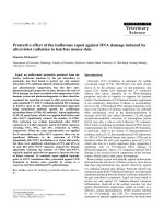
tochemical stain for CD4 and CD8 molecules, the more
CD8+ cells were infiltrated in the submucosa and mucosa
of airway from the vaccination group compared with the
control group (Fig. 3). But no difference in CD4+ cells was
shown between the two groups. We considered whether
the DNA vaccination might have an effect on the cellular
response and the CD8+ T cells, which might protect
against subsequent allergen challenges.
Cytokines expressed by antigen stimulation
Lymphocytes were harvested from lymph nodes of the
two groups of mice and stimulated with recombinant Der
p 1 or HDM crude extract to determine the Th1 or Th2
cytokines involved in the DNA vaccination. Significantly
elevated expression of IFN-γ mRNA was detected in the
vaccination group compared with that in the control
group. However, mRNA expression of IL-2, 4, 5, and 10
showed no difference from the control group (Fig. 4).
Discussion
Diseases such as allergic asthma, rhinitis, and atopic der-
matitis are all characterized by elevated levels of serum
IgE. Total and specific IgE levels also show a close relation-
ship with clinical symptoms in atopic allergy [14]. A vari-
ety of approaches targeting the suppression of IgE have
been proposed such as synthetic peptides and T cell vac-
cine. However, synthetic peptides have substantial limita-
tions because of poor immunogenecity [15,16]. Recently,
in an animal model of allergic disorders, DNA vaccine
encoding one of the major birch pollen allergens has been
shown to be allergen-specifically protective and therapeu-
tic [17]. DNA vaccination with plasmid encoding Der p 5,
Immunohistochemical examination for CD8+ T cells in lung tissueFigure 3
Immunohistochemical examination for CD8+ T cells in lung tissue. In the lung tissue from the vaccination group,
more CD8+ T cells were infiltrated along the airway than in control group. A and B, Lung tissue from control group mouse
(×100 and ×200). C and D, Vaccination group (×100 and ×200). Immunohistochemical staining with rat anti-mouse CD8 mon-
oclonal antibody.
Clinical and Molecular Allergy 2006, 4:4 />Page 7 of 9
(page number not for citation purposes)
one of the minor HDM allergens, was reported to prevent
induction of specific IgE synthesis [11]. Vaccination with
pDNA encoding Der p 5 was shown to induce Th1
immune response to the encoded antigens. However,
these results have some limitations on the clinical appli-
cation for treating allergic disorders. Each allergen that
causes allergic disorders in humans contains various kinds
of protein that have their own epitopes and are complex.
The Der p 5 allergen reacts with about only 40% of allergic
sera to HDM, and the Der p 1 and 2 allergens react with
about 80% of allergic sera [3,5]. To evaluate the effect of
the gene vaccination with DNA fragments encoding major
allergen on the allergic response to whole HDM extract,
we used plasmid with cDNAs encoding the major six
HDM allergens (Der p 1, 2, and 3, and Der f 1, 2, and 3)
for vaccination. We showed about 50% reduction of
HDM-specific IgE and more than 70% inhibition of total
IgE at 6 weeks after immunization compared with control
group. DNA vaccine with plasmid encoding the major
HDM allergens might inhibit IgE synthesis more effi-
ciently than encoding one of the HDM allergens.
In animal models of allergic disease, it has been estab-
lished that Th2 responses are mediated by T helper cells
that secret cytokines such as IL-4 and IL-5, which induce
antibody production in B cells, and IgE plays a central role
in allergic responses [18]. IFN-γ is the Th1 cytokine
responsible for inhibiting IL-4-mediated IgE responses
and promoting the formation of IgG2a [19]. Plasmid vec-
tor containing DNA that encodes allergens has been
reported to decrease Th2-mediated responses, enhance
Th1 responses, and suppress the allergic response
[11,20,21]. In this investigation, mRNA expression of
IFN-γ in lymphocytes from the vaccination group
increased significantly relative to that from the control
group, and less production of total and specific IgE in the
vaccinated group was detected than in the control group,
suggesting that the gene vaccination might successfully
redirect the immune response from Th2 into Th1 to the
encoded antigen or allergen.
Allergic asthma is characterized as a chronic inflammatory
disease of the bronchi. It is well established that a variety
of cells including mast cells, eosinophils, and lym-
phocytes play a role in this process [22,23]. After inhala-
tion challenges, the inflammatory cells migrate from the
peripheral blood to the site of inflammation in the bron-
chial mucosa, and Th2 type cytokines are dominantly
detected in bronchoalveolar lavage fluid [24,25]. How-
ever, our investigation showed that DNA vaccination suc-
cessfully reduced the recruitment of inflammatory cells in
lung tissues. The effect of DNA vaccination in allergic
inflammation might be elicited through not only
humoral immune responses but also cellular responses.
T lymphocytes have been suggested to play a key role in
orchestrating the interaction of the participating cells
since they are able to release an array of cytokines that can
attract, prime, and activate other cell types [25]. A success-
ful outcome of immunotherapy is known to be associated
with the development of regulatory T cells, which can
downregulate the allergic response [26-30]. It is also
known that functionally distinct subsets of CD8+ T cells
may play an important regulatory role in IgE production
[30-32]. However, there are some different explanations
regarding the mechanisms of DNA vaccine. Manickan et
al. [33] demonstrated the mechanism of genetic immuni-
zation against herpes simplex virus principally by CD4+ T
cells, not by CD8+ T cells. Lee et al. [22] reported that both
CD4+ and CD8+ subsets of T cells from pDNA vaccinated
mice can suppress IgE antibody production by affecting
the primary response or by propagating the Th1 memory
Cytokine expression in lymphocytesFigure 4
Cytokine expression in lymphocytes. Lymphocytes from
the control group and vaccination group were cultured in the
presence of no antigen (N), recombinant Der p 1 (P1), and
HDM crude extract (HDM) for 18 h. mRNAs of each indi-
cated cytokine (interferon-gamma, IL-2, IL-4, IL5, IL-10) were
measured by RT-PCR and that of beta-actin was measured
for control.
Clinical and Molecular Allergy 2006, 4:4 />Page 8 of 9
(page number not for citation purposes)
response in a passive cell transfer system. Draghi et al. [34]
investigated whether DNA vaccination leads to the gener-
ation of a distinct population of noncytotoxic/regulatory
CD8+ T cells. In the authors' immunohistochemical
investigation, more CD8+ T cells were more infiltrated in
the lung tissue of the vaccination group than that of the
control group.
Peptides derived from extracellular molecules are pre-
sented to CD4+ T cells by MHC (histocompatibility com-
plex) class II molecules normally generated by antigen-
presenting cells, whereas peptides derived from intracellu-
lar proteins are generally presented to CD8+ T cells by
MHC class I molecules, which are expressed on virtually
all somatic cells [35]. We injected mixed naked DNA into
the muscle of the murine model of allergic disorder. Some
of the injected DNA might be postulated to stay in the
nuclei of cells or be integrated in the host DNA and elicit
the endogenous production of an allergenic protein.
MHC class I molecule, might induce CD8+ T cells that
protectively function in immune response against a subse-
quent allergic challenge in sensitized host cells. The CD8+
T cells might be capable of conferring protection from
allergic inflammation. DNA vaccination, which contains
plasmid and DNA encoding specific allergen, might pro-
vide a more efficient therapeutic method for intervening
allergic responses than conventional specific immuno-
therapy with allergen extracts.
Abbreviations
HDM : house dust mite
pDNA : plasmid DNA
Authors' contributions
N Kim and SS Kwon carried out animal and molecular
experiments and initial draft. J Lee and S Kim handled
images of the draft and final draft. TJ Yoo supported the
whole step of experiment and submission.
References
1. Platts-Mills TAE, Chapman MD: Dust mites, immunology, aller-
gic disease, and environmental control. J Allergy Clin Immunol
1987, 80:755-775.
2. Lin KL, Hsieh KH, Thomas WR, Chiang BL, Chua KY: Allergens,
IgE, mediators, inflammatory mechanisms. Characteriza-
tion of Der p 5 allergen, cDNA analysis, and IgE-mediated
reactivity to the recombinant protein. J Allergy Clin Immunol
1994, 94:989-996.
3. International workshop report: Dust mite allergens and
asthma: a world wide problem. WHO Bulletin 1988, 66:769-780.
4. Van der, Zee JS, Van Swieten P, Janse HM, Aalberse RC: Skin tests
andhistamine release with P1-depleted Dermatophagoides
pteronyssinus body extracts and purified P1. J Allergy Clin Immu-
nol 1988, 81:884-895.
5. Thomas WR: Mite allergens groups I-VII. A catalogue of
enzymes. Clin Exp Allergy 1993, 23:350-353.
6. Raz E, Carson DA, Parker SE, Par TB, Abai AM, Aichinger G: Intra-
dermalgene immunization: the possible role of DNA uptake
in the induction of cellular immunity to viruses. Proc Natl Acad
Sci U S A 1994, 91:9519-9523.
7. Reyes-Sandoval A, Ertl HC: DNA vaccines. Curr Mol Med 2001,
1:217-243.
8. Ruiz PJ, Garren H, Ruiz IU, Hirschberg DL, Nguyen LV, Karpuj MV, et
al.: Suppressive immunization with DNA encoding a self-pep-
tide prevents autoimmune disease: modulation of T cell cos-
timulation. J Immunol 1999, 162:3336-3341.
9. Kwon SS, Kim N, Yoo TJ: The effect of vaccination with DNA
encoding murine T-cell epitopes on the Der p 1 and 2
induced immunoglobulin E synthesis. Allergy 2001, 56:741-748.
10. Lewis DB: Allergy immunotherapy and inhibition of Th2
immune responses: a sufficient strategy? Curr Opin Immunol
2002, 14:644-651.
11. Hsu CH, Chua KY, Tao MH, Lai YL, Wu HD, Huang SK, et al.: Immu-
noprophylaxis of allergen-induced immunoglobulin E synthe-
sis and airway hyperresponsiveness in vitro by genetic
immunization. Nature Med 1996, 2:540-544.
12. Cheng KC, Lee KM, Krug MS, Watanabe T, Suzuki M, Choe IS, et al.:
House dust mite-induced sensitivity in mice. J Allergy Clin Immu-
nol 1998, 101:51-59.
13. Hessel EM, Van Oosterhout AJ, Hofstra CL, De Bie JJ, Garssen J, Van
Loveren H, et al.: Bronchoconstriction and airway hyperre-
sponsiveness after ovalbumin inhalation in sensitized mice.
Eur J Phamacol 1995, 93:2401-412.
14. Droste JH, Kerkhof M, de Monchy Jan GR, Schouten Jan P, Rijcken B,
Dutch ECRHS group: Association of skin test reactiviy, specific
IgE, total IgE, and eosinophils with nasal symptoms in a com-
munity-based population study. J Allergy Clin Immunol 1996,
97:922-932.
15. Bot A, Bot S, Karjalainen K, Bona C: Kinetics of generation and
persistence on membrane class II molecules of a viral pep-
tide expressed on foreign and self proteins. J Immunol 1996,
157:3436-3442.
16. Demotz S, Grey HM, Sette A: The minimal number of class II
MHC-antigen complexes needed for T cell activation. Science
1990, 249:1028-1030.
17. Hartl A, Hochreiter R, Stepanoska T, Ferreira F, Thalhamer J: Char-
acterization of the protective and therapeutic efficiency of a
DNA vaccine encoding the major birch pollen allergen Bet v
1a. Allergy 2004, 59:65-73.
18. Maggi E: The TH1/TH2 paradigm in allergy. Immunotechnology
1998, 3:233-244.
19. Snapper CM, Paul WE: Interferon-γ and B cell stimulatory fac-
tor-1 reciprocally regulate Ig isotype production. Science
1987, 236:944-947.
20. Lai WC, Bennett M, Johnston SA, Barry MA, Pakes SP: Protection
against Mycoplasma pulmois infection by genetic vaccina-
tion. DNA Cell Biol 1995, 14:643-651.
21. Huygen K, Content J, Denis O, Montgomery DL, Yawman AM, Deck
RR, et al.: Immunogenicity and protective efficacy of a tuber-
culosis DNA vaccine. Nature Medicine 1996, 2:893-898.
22. Lee DJ, Tighe H, Corr M, Carson DA, Spiegelberg HL, Raz E: Inhibi-
tion of IgE antibody formation by plasmid DNA immuniza-
tion is mediated by both CD4+ and CD8+ T cells. Int Arch
Allergy Immunol 1997, 113:227-230.
23. Krug N, Tshernig T, Holgate S, Pabst R: How do lymphocytes get
intothe asthmatic airways? Lymphocyte traffic into and
within the lung in asthma. Clin Exp Allergy 1998, 28:10-18.
24. Ying S, Durham SR, Cirrogan CJ, Hamid Q, Kay AB: Phenotype of
cellsexpressing mRNA for Th2-type (interleukin 4 and inter-
leukin 5) and Th1-type (interleukin 2 and interferon γ)
cytokines in bronchoalveolar lavage and bronchial biopsies
from atopic asthmatic and normal control subjects. Am J
Respir Cell Mol Biol 1995, 12:477-487.
25. Humbert M, Durham SR, Ying S: IL-4 and IL-5 mRNA and protein
in bronchial biopsies from patients with atopic and nonat-
opic astham: evidence against 'intrinsic' asthma being a dis-
tinct immunopathologic entity. Am J Respir Crit Care Med 1996,
154:1497-1504.
26. Hsieh KH: Changes of lymphoproliferative responses of T cell
subsets to allergen and mitogen after hyposensitization in
asthmatic children. J Allergy Clin Immunol 1984, 74:34-40.
27. Hsieh KH, Lue KH, Chiang CF: Immunological changes after
hyposensitization in house-dust-sensitive asthmatic children.
J Asthma 1987, 24:19-27.
Publish with BioMed Central and every
scientist can read your work free of charge
"BioMed Central will be the most significant development for
disseminating the results of biomedical research in our lifetime."
Sir Paul Nurse, Cancer Research UK
Your research papers will be:
available free of charge to the entire biomedical community
peer reviewed and published immediately upon acceptance
cited in PubMed and archived on PubMed Central
yours — you keep the copyright
Submit your manuscript here:
/>BioMedcentral
Clinical and Molecular Allergy 2006, 4:4 />Page 9 of 9
(page number not for citation purposes)
28. Akbari O, Stock P, DeKruyff RH, Umetsu DT: Role of regulatory
T cells in allergy and asthma. Curr Opin Immunol 2003,
15:627-633.
29. Akbari O, Freeman GJ, Meyer EH, Greenfield EA, Chang TT, Sharpe
AH, et al.: Antigen-specific regulatory T cells develop via the
ICOS-ICOS-ligand pathway and inhibit allergen-induced air-
way hypersensitivity. Nat Med 2002, 8:1024-1032.
30. Kemeny DM, Diaz-Sanchez S: Role of CD8+T cells in rat IgE
responses. Int Arch Allergy Appl Immunol 1991, 94:99-101.
31. McMenamin C, Holt PG: The natural immune response to
inhaled soluble protein antigens involves major histocom-
patibility complex (MHC) class 1 restricted CD8+ T cell-
mediated but MHC class II-restricted CD4+T cell-dependent
immune deviation resulting in selective suppression of
immunoglobulin production. J Exp Med 1993, 178:889-899.
32. Renz H, Lack G, Salog J, Schwinzer R, Bradley K, Loader J, et al.: Inhi-
bition of IgE production and normalization of airways
responsiveness by sensitized CD8+ T cells in a mouse model
of allergen-induced sensitization. J Immunol 1994, 152:351-360.
33. Manickan E, Rouse RJD, Yu Z, Wire WS, Rouse BT: Genetic immu-
nization against herpes simplex virus: protection is mediated
by CD4+ T lymphocytes. J Immunol 1995, 155:259-265.
34. Draghi M, Jarman ER, Grifantini R, Galli-Stampino L, Lamb JR, Valiante
NM, et al.: Different profile of CD8+ effector T cells induced in
Der p1-allergic and naïve mice by DNA vaccination. Eur J
Immunol 2002, 32:3720-3728.
35. Germain RN, Margulies DH: The biochemistry and cell biology
of antigen processing and presentation. Annu Rev Immumol
1993, 11:403-450.