Báo cáo y học: "A multi-modal treatment approach for the shoulder: A 4 patient case series" docx
Bạn đang xem bản rút gọn của tài liệu. Xem và tải ngay bản đầy đủ của tài liệu tại đây (309.85 KB, 9 trang )
BioMed Central
Page 1 of 9
(page number not for citation purposes)
Chiropractic & Osteopathy
Open Access
Case report
A multi-modal treatment approach for the shoulder: A 4 patient
case series
Mario Pribicevic
1
and Henry Pollard*
2
Address:
1
Macquarie Injury Management Group Department of Health and Chiropractic Macquarie University, 2109, Sydney Australia and
2
Macquarie Injury Management Group Department of Health and Chiropractic Macquarie University, 2109, Sydney Australia
Email: Mario Pribicevic - ; Henry Pollard* -
* Corresponding author
ShoulderImpingement SyndromeMulti-modal TreatmentChiropractic
Abstract
Background: This paper describes the clinical management of four cases of shoulder impingement
syndrome using a conservative multimodal treatment approach.
Clinical Features: Four patients presented to a chiropractic clinic with chronic shoulder pain,
tenderness in the shoulder region and a limited range of motion with pain and catching. After
physical and orthopaedic examination a clinical diagnosis of shoulder impingement syndrome was
reached. The four patients were admitted to a multi-modal treatment protocol including soft tissue
therapy (ischaemic pressure and cross-friction massage), 7 minutes of phonophoresis (driving of
medication into tissue with ultrasound) with 1% cortisone cream, diversified spinal and peripheral
joint manipulation and rotator cuff and shoulder girdle muscle exercises. The outcome measures
for the study were subjective/objective visual analogue pain scales (VAS), range of motion
(goniometer) and return to normal daily, work and sporting activities. All four subjects at the end
of the treatment protocol were symptom free with all outcome measures being normal. At 1
month follow up all patients continued to be symptom free with full range of motion and complete
return to normal daily activities.
Conclusion: This case series demonstrates the potential benefit of a multimodal chiropractic
protocol in resolving symptoms associated with a suspected clinical diagnosis of shoulder
impingement syndrome.
Background
Practitioners of manual therapy commonly encounter
patients presenting with shoulder pain and symptoms
associated with rotator cuff pathology. Shoulder pain is
the most common extraspinal complaint encountered in
primary care clinics, and in clinical frequency is exceeded
only by low back and neck pain [1]. Many shoulder con-
ditions are associated with dysfunction of the rotator cuff
[2-4].
Rotator cuff disorders represent a complex clinical entity
requiring a thorough understanding and knowledge of
shoulder anatomy, biomechanics and the functional
Published: 16 September 2005
Chiropractic & Osteopathy 2005, 13:20 doi:10.1186/1746-1340-13-20
Received: 06 September 2005
Accepted: 16 September 2005
This article is available from: />© 2005 Pribicevic and Pollard; licensee BioMed Central Ltd.
This is an Open Access article distributed under the terms of the Creative Commons Attribution License ( />),
which permits unrestricted use, distribution, and reproduction in any medium, provided the original work is properly cited.
Chiropractic & Osteopathy 2005, 13:20 />Page 2 of 9
(page number not for citation purposes)
relationship of the shoulder to nearby spinal structures
including the cervical and thoracic spines.
Rotator cuff disorders commonly occur secondary to
repetitive overuse (occupational or overhead throwing
sports), which contributes to micro traumatic changes
within rotator cuff tissue [5]. In addition, a single macro
traumatic episode (fall on outstretched hand) can cause
injury to rotator cuff tissue [5]. The normal aging process
will also negatively influence the rotator cuff mechanism
[2].
The most common source of shoulder pain originates
from the rotator cuff tendons, with the most prevalent
clinical diagnosis being impingement syndrome of the
supraspinatus tendon [2-4,6].
Before discussing our case series it is important to review
some important elements of taking a history and perform-
ing a shoulder physical examination. Certain clinical fea-
tures may alert the practitioner to potentially serious
causes (red flags) of shoulder pain, which constitute pos-
sible contra-indication to manual therapy [7,8] (Table 1).
Other (yellow flag) features of the clinical history may
affect the outcome of manual therapy and therefore recov-
ery [7,8] (Table 2). A differential diagnosis list for shoul-
der pain [9] is seen in Table 3.
Table 4[9] shows sources of shoulder pain mostly derived
from local structures within the shoulder, whether due to
trauma, overuse, arthritides or disease.
This paper will discuss a common cause of shoulder pain
and its largely unreported multi-modal conservative man-
agement in a chiropractic setting. This management will
include pertinent aspects of the patient history, physical
examination, differential diagnosis for shoulder pain as
well as its management in 4 cases.
Case Presentations
Four presentations
A case of shoulder pain in a fit 42-year-old Caucasian
male is presented. The pain was located diffusely in the
postero-lateral aspect of the right shoulder and started
gradually 4–6 weeks prior to presentation. No causative
event was reported, although workplace activities
required the patient to repetitively lift files above the
shoulder level onto a shelf. Of note was the mention of a
particularly busy period (increased intensity and dura-
tion) at work prior to the onset of pain.
The patient described the pain as being of a constant nag-
ging and aching sensation with an intensity of 3/10 on the
visual analogue scale (VAS). He also reported an intermit-
tent sharp and catching sensation in the same location on
shoulder abduction, with an intensity of 6/10 (VAS scale).
No referred pain, or other neurological symptoms were
reported, although he did report subjective weakness of
the shoulder during elevation above shoulder level and
inability to use the right arm comfortably.
Holding his arm on top of the steering wheel aggravated
the pain when driving, as did sleeping on his right side,
and also combing his hair. He described that heat packs
provided short-term relief of pain. The patient reported no
prior shoulder problems, no use of medication, and his
medical, family and social history were otherwise
unremarkable.
Table 1: Alerting features of a possible serious condition (red flag), which may present with shoulder pain [7,8].
POSSIBLE SERIOUS CAUSES OF SHOULDER PAIN (RED FLAGS)
Signs of infection (fever) Violent trauma
History of drug abuse Swelling
Weight loss Pain at rest
Age over 50 Night sweats
History of previous malignancy History of fall
Constant, non mechanical pain No precipitating event (for onset)
Palpable deformities of bone/tissue HIV
Widespread neurological symptoms/signs
Table 2: Possible features that may affect manual therapy
outcome and ultimate patient recovery for patients presenting
with shoulder pain (yellow flags) [7,8].
YELLOW FLAGS
Previous history of shoulder pain
Personal problems (alcohol, financial, marital)
Compensable injury
Unrealistic expectation of therapy
Long term absence from sport work
Belief that shoulder pain is dangerous
Dissatisfaction
Chiropractic & Osteopathy 2005, 13:20 />Page 3 of 9
(page number not for citation purposes)
Physical examination of the right arm produced pain and
restriction of movement at 50 degrees of right external
rotation in the neutral position, with restriction and pain
at 90 degrees of abduction. Both movements were
guarded. An impingement sign was present, as confirmed
by a positive Hawkins test. Hawkins test involves posi-
tioning the arm at 90 degrees of flexion with subsequent
internal rotation. In addition Neers impingement test
gave slight discomfort. Neer's impingement test is per-
formed with the patient sitting as the practitioner stands
behind the patient with one hand supporting the scapula
to prevent scapula rotation and the other hand holding
the forearm. The shoulder is brought into maximum flex-
ion with a small degree of internal rotation. The test is
considered positive if there is pain in the last 10–15
degrees of flexion. Pain is produced because the greater
tuberosity is compressed against the anterior acromion or
coracoacromial ligament, hence this test may aggravate an
inflamed bursa (subacromial), the supraspinatus tendon
or the anterior structures of the coracoacromial arch [10].
Muscle testing revealed slight weakness of the right infra-
spinatus muscle (Grade lV of V) and also right latissimus
dorsi. Other routine shoulder tests revealed no abnormal
findings (including instability testing, glenoid labrum
testing, lateral slide test and muscle tests).
On palpation muscle spasm was noted in the right infra-
spinatus muscle and to a lesser extent the right rhomboid,
supraspinatus and upper trapezius when compared to the
other side. Significant focal tenderness was palpated over
the rotator cuff insertion on the greater tuberosity of the
humerus. Specific joint motion palpation revealed likely
lateral flexion restriction of the right C5/6 lower cervical
facet joint and left T2/3 thoracic facet joint with immobil-
ity of the right acromio-clavicular joint in an inferior
direction.
The patient presented with X-rays, revealing no
abnormalities.
A likely working diagnosis of a Primary Grade 2 Postero-
lateral Rotator Cuff Impingement (Neer classification-
Table 5[11]) was determined.
A second patient presenting was a slightly overweight 32
years old caucasian female with right-sided shoulder pain
located superior, and in the postero-lateral aspect of the
shoulder. The pain started 2 weeks prior to presentation
after practising certain manual therapy manoeuvres of the
lumbar spine at university. The patient was practising
lumbar spine and sacro-iliac pisiform contact posterior-
anterior manipulation. During this the shoulder is placed
repetitively in a combined position of adduction, flexion
and internal rotation. The patient described the pain as
being a sharp, shooting sensation, intermittent, depend-
ent on motion, with an intensity of 7/10 (VAS scale).
A diffuse aching sensation was also reported in the right
upper deltoid region (so-called "military badge"). The
pain was aggravated by elevation of the arm and sleeping
on the right side. Relief was obtained by applying ice and
taking anti-inflammatory/analgesic medication (Ibupro-
fen). The patient reported no prior shoulder problems, no
general use of medication; her medical, family and social
history were otherwise unremarkable.
Table 3: Describes the differential diagnosis for shoulder pain [9].
Referred pain from musculoskeletal sources Cervical facet joints
Thoracic facet joints
Myofascial pain syndromes
Referred pain from visceral sources Lungs
Gallbladder
Heart
Diaphragm
Neuropathies Brachial plexus neuropathies
Peripheral neuropathies
Radicular pain Cervical nerve root compression
Table 4: Describes the sources of shoulder pain derived from
local structures [9].
Trauma Fracture
Dislocation
Tendon rupture
Overuse Inflammation (tendinitis, bursitis)
Capsular sprains
Arthritides Osteoarthritis
Rheumatoid variants
Other Infection
Neoplasm
Chiropractic & Osteopathy 2005, 13:20 />Page 4 of 9
(page number not for citation purposes)
Physical examination of the right shoulder revealed slight
postero-lateral pain in the shoulder on external rotation
and abduction. External rotation was restricted at 60
degrees and abduction at 90 degrees. Impingement was
elicited with the Hawkins test and with the Neer's test.
Other routine shoulder tests revealed no abnormal
findings.
On palpation muscle spasm was notionally present in the
right rhomboid major, upper trapezius, supraspinatus
and particularly the infraspinatus. Trigger points were
noted in the infraspinatus muscle with reproduction of
the upper arm pain upon specific pressure. Motion palpa-
tion revealed likely right acromio-clavicular and sterno-
clavicular joint fixation, left T3/4 and right C5/6 lateral
flexion restriction. The patient presented with plain film
radiographs, which revealed no abnormality.
A likely working diagnosis of Grade 2 Primary Impinge-
ment of the rotator cuff (Neer classification-Table 5[11])
was determined. The working diagnosis also included the
presence of an active infraspinatus myofascial pain
syndrome.
The third patient was a slightly apprehensive 29-year-old
Caucasian male with right-sided diffuse anterior and
superior shoulder pain. The pain started gradually over an
8–10 week period, with the intensity being most prevalent
during the 2 weeks prior to presentation. The patient was
employed as a factory worker; a job that required com-
bined repetitive shoulder movements and periods of
administrative keyboard work.
The pain was described as a constant, deep, dull and nag-
ging ache with an intensity of 5/10 (VAS scale). No neuro-
logical symptoms were reported, there were no
dermatomal/sclerotomal pain referral patterns, although
a slight diffuse aching sensation was mentioned in the
right elbow and more prominently right "military badge"
area. Together with the shoulder pain the patient reported
a less intense (4/10) dull sensation specifically at the base
of the cervical spine on the right and a vague headache
like sensation at the base of the skull.
The right shoulder felt subjectively weaker with inability
to lift the arm above shoulder level without pain. The pain
was aggravated by specific arm postures and lying on the
right side. There was no pertinent medical/family/social
history.
Examination revealed a painful arc with onset of pain at
70 degrees abduction, external rotation being restricted at
70 degrees with a catching sensation at the end of motion.
Reproduction of the pain was elicited with a Hawkins test
and on supraspinatus muscle testing ("Empty can" test)
revealing a grade 4 weakness and pain. Other routine
shoulder tests revealed no abnormal findings.
Right cervical rotation restriction (65 degrees) was noted
on the right, with a right Kemps joint stress test (com-
bined right cervical rotation, lateral flexion and exten-
sion) reproducing the low cervical pain but no shoulder
pain.
Palpation revealed muscle tenderness in the right suprasp-
inatus, upper trapezius, levator scapulae and infraspinatus
muscle groups. A trigger point was palpated in the infra-
spinatus muscle, which upon applying pressure
reproduced the right upper arm diffuse ache. Palpating
the rotator cuff insertion on the humerus and coracoacro-
mial ligament caused significant tenderness. Motion pal-
pation revealed likely joint restriction at the right C5/6
cervical facet joint, T2/3 and acromio-clavicular joint. Of
interest was the postural presentation of a "rounded
shoulder" and increased thoracic kyphosis.
A likely primary working diagnosis of a Grade ll Primary
Rotator Cuff Impingement (Neer classification-Table
5[11]) with Supraspinatus tendonosis was determined,
with secondary involvement of the cervical and thoracic
spines.
The fourth patient presenting was a 40-year-old Caucasian
female. She presented with right-sided anterior shoulder
pain, which was nagging, aching and accompanied by a
catching sensation on specific movements. The aching
pain was constant with an intensity of 6.5/10 (VAS scale),
while the catching pain was slightly more intense at 8/10.
No neurological sensations were reported. The patient
reported a diffuse aching pain in the posterior aspect of
the shoulder over the scapula. Nothing relieved the pain,
while arm elevation, driving, prolonged sitting behind the
computer and poor posture made the pain worse.
The pain started 4 days prior to presentation after spend-
ing most of the weekend cleaning the walls at home with
a sponge prior to painting. The patient had not had this
pain before although due to her work (accountant) she
often complains of posterior shoulder tension. The
patient had been treated previously for an unrelated
Table 5: Neer classification of impingement [11].
STAGE l Involving oedema and haemorrhage
STAGE ll Involving fibrosis and tendonitis
STAGE lll Involving degeneration (bone spurs) and tendon
rupture
Chiropractic & Osteopathy 2005, 13:20 />Page 5 of 9
(page number not for citation purposes)
complaint (right sided sacroiliac area pain). The medical,
family and social histories were unremarkable.
The physical examination revealed restriction in external
rotation (60 degrees), and abduction with pain/catching
at 90 degrees. Internal rotation was also tight and sore
especially with the Hawkins test. The impingement sign
was present with reproduction of the anterior pain with a
Hawkins and Neers test.
Scapula dysfunction was also noted with a positive right-
sided lateral glide test. It should be noted that no major
difference was seen with the lateral glide test on the previ-
ous 3 patients.
Of importance was the postural presentation of anteriorly
rotated shoulders, increased thoracic kyphosis and for-
ward head carriage. A scoliotic curve was also noted with
an apex convex to the right in the mid thoracic region. Pal-
pation revealed muscle spasm in the right posterior shoul-
der girdle muscles with increased muscular tension and
sensitivity to palpation in the right supraspinatus and
infraspinatus compared to the left. Infraspinatus palpa-
tion revealed local muscle spasm with a reproduction of
the posterior ache on specific pressure. Increased tender-
ness was noted whilst palpating the coracoacromial liga-
ment and supraspinatus insertion on the humerus.
Specific joint motion palpation revealed likely restriction
in the right C5/6 joint, T3/4 and acromioclavicular joint.
A likely working diagnosis of a Grade ll, Primary Shoulder
Rotator Cuff Impingement (Neer classification- refer to
Table 5[11]) was determined. Of note was the secondary
contribution of the scapula to this process. The working
diagnosis also included the presence an active infraspina-
tus myofascial pain syndrome.
The interventions
The 4 patients were admitted to a multimodal treatment
protocol, which included the following interventions: soft
tissue therapy, ultrasound phonophoresis, manipulation
and exercise.
All of the patients received soft tissue therapy that
involved the application of ischaemic pressure to the
supraspinatus and infraspinatus muscles, as well as the
rhomboids, upper trapezius and levator scapulae. The
application involved palpating the muscle bellies and
applying a sustained pressure into areas of muscle spasm
until a release of the barrier of resistance was felt. Release
meaning the relaxation of the point of muscle spasm with
a decrease in the sensitivity and muscle tone after re-pal-
pating the area. The pressure was applied repetitively,
using a myofascial T-bar (a plastic, T shaped hand held
tool with a rubber tip attached to the end in contact with
the skin). Care was taken not to cause increased discom-
fort to the patient (to the level of pain tolerance).
Longitudinal and transverse friction massage was applied
to the posterior tenomuscular junction of the infraspina-
tus muscle, the coracoacromial ligament (postero-inferior
aspect) and the insertion of the supraspinatus on the
greater tuberosity of the humerus. The friction massage
application was achieved by palpating the capsular or
tendinous adhesions and frictioning over its surface with
the practitioner's index finger. This was maintained until
friction anaesthesia was achieved and the patient could
not feel any discomfort. A new point was then chosen and
the process repeated. Once again care was taken to not
cause excessive discomfort to the patient. At the end of the
treatment sessions ice application was advised at a fre-
quency of three applications of 15 minutes with two 20-
minute breaks.
Ultrasound phonophoresis was applied to the areas that
previously underwent friction massage with a topical cor-
ticosteroid [1% sigmacort]. Ultrasound was applied with
a continuous wave form for 7 minutes at a setting of 2.2
W/cm
2
to the rotator cuff insertion on the anterior-inferior
aspect of the humerus and posterior inferior aspect of the
acromioclavicular joint.
Peripheral thrust manual manipulation was applied to the
glenohumeral joints in external rotation (progressive)
and inferiorly to the acromioclavicular joint and anterior
to posterior to the sternoclavicular joint in all of the
patients where a likely motion restriction was detected.
Mechanically assisted manipulations were also used with
the Activator 2 apparatus in humeral external rotation or
inferior through the AC joint. This particular technique
was chosen for one of the female patients (fourth patient
as an alternative) who expressed concern with peripheral
manual manipulation after the first treatment session as
an alternative technique.
Diversified spinal manipulations were used to manipulate
the thoracic and cervical spines at the level of T3/4 and
C5/6. All patients were given a basic exercise program
with initial emphasis on isometric strengthening of the
supraspinatus and infraspinatus muscles. This was imple-
mented once a reduction in pain and improved range of
motion was noted at a frequency of 4 sets of 10 repeti-
tions, 2–3 times per day. Theraband (extendable elastic)
exercises were also implemented at the same frequency
after the initial isometric strengthening period. This also
included shoulder shrugs, wall push-ups and scapula
retraction exercises.
Chiropractic & Osteopathy 2005, 13:20 />Page 6 of 9
(page number not for citation purposes)
Patient 1 was treated for a total of 5 visits, patient 2 was
treated 4 times, patient 3 was treated 5 times, and patient
4 was treated 4 times.
At the end of the last treatment session (5 and 4 treat-
ments respectively) a repeat physical examination
revealed a full and painless range of motion with no sub-
jective symptoms, and negative orthopaedic testing
(Hawkin's and Neer's). Patient 1 was seen 4 weeks later
for a new and unrelated complaint, who after questioning
reported no shoulder complaints (pain). Full range of
motion was maintained. Patient 2 was contacted via the
phone and upon questioning also reported no subjective
pain and full return to normal activities at 1 month post
treatment. Patient 3 was followed at 4 and 8 weeks after
the last treatment revealing no subjective and objective
symptoms. Patient 4 was seen at 4 and 8 weeks with no
symptoms of impingement reported and no objective
findings.
Discussion and Conclusions
Rotator cuff impingement and or tendonosis is a common
disorder encountered in a primary health care setting [12-
15]. Perhaps, less in chiropractic practises as opposed to
medical and physiotherapy. To date, there are no data
investigating the prevalence of shoulder pain in the chiro-
practic setting. This may be due to the lack of general pub-
lic awareness about the scope and capabilities of
chiropractors to be involved in management of non-spi-
nal disorders or simply the public making another choice.
This condition presents a challenge to the chiropractor
due to its prevalence, and its possible close interrelation-
ship with the spine.
A major reason for documenting this treatment protocol
is to encourage the development of future clinical guide-
lines for chiropractors and to encourage the expansion of
their treatment range to include peripheral disorders.
Another goal of this report is to highlight that multimodal
management is often required to address the painful
shoulder and not to determine or show which treatment
approach or particular therapy was more effective. The
four patients in this paper were managed with a treatment
protocol that included a number of therapies. The litera-
ture [16-22] suggests that the multimodal approach is an
appropriate method for the successful conservative man-
agement of shoulder problems.
The cervical and thoracic spines should be reviewed as a
possible factor associated with rotator-cuff dysfunction.
As an example consider the slumping posture in a com-
petitive swimmer. Others and we hypothesise that the
rounded shoulders and increased thoracic kyphosis places
increased demands on the rotator cuff and contributes to
the impingement process [23]. A possible mechanism for
this hypothesis is as follows: the posture may alter the
mechanical function (orientation) of the scapula and
humerus, leading to muscular imbalances, abnormal
movement patterns during glenohumeral elevation with
associated weakness of the posterior cuff muscles. There-
fore this may lead to a loss of force couple at the gleno-
humeral joint with resultant repetitive humeral head
impingement [23-25].
The outcome measures for the study included improve-
ment of pain, return to pre-treatment activities, and resto-
ration of full active and passive movements. The outcome
measures were mainly subjective in nature and dependent
on the response of the patients and the practitioner's skill
in conducting the orthopaedic reassessment, therefore
allowing an element of examination bias. This particular
shortcoming may be improved by using more sensitive
scoring systems that can be accurately reproduced by dif-
ferent observers such as the subjective shoulder rating sys-
tem [26], UCLA scoring system [27], or the highly
sensitive Constant/Murley functional assessment of the
shoulder [28].
Although frequently advocated for outcomes based
assessment, goniometric measurement for the shoulder
remains questionable. Williams et al [29] studied 22
observers who used three different types of goniometers
to assess the range of abduction and visual estimation.
The results demonstrated visual estimation to be the most
reliable method. Moderate inter-observer reliability was
also demonstrated in a study by Bostrom et al [30] where
range of motion was measured using a goniometer.
This report presents an approach that combines aspects of
traditional forms of chiropractic, physiotherapy and med-
icine in the conservative management of certain shoulder
pain.
The individual therapies that were used in this multimo-
dal treatment protocol have been shown to be useful in
the management of shoulder pain both singularly and in
combination [18,19,31-36].
Of the electro-modalities the apparatus used was ultra-
sound. Some authors routinely advocate the usage of
ultrasound in conjunction with other modalities and
report positive outcomes [3,16,35]. The physiologic ben-
efits of ultrasound have been attributed to its thermal
actions; these involve an increase in peripheral blood
flow, increased tissue metabolism and greater tissue
extensibility [37].
The use of ultrasound for shoulder pain and its effect on
soft tissue structures of the shoulder has been studied
Chiropractic & Osteopathy 2005, 13:20 />Page 7 of 9
(page number not for citation purposes)
extensively in the literature. A recent study by Nykanen
[36] investigating pulsed ultrasound treatment of the
painful shoulder in a randomised, double blind and pla-
cebo controlled study, showed no differences in outcome
between the treatment and placebo groups at the end of
the trial period. However, when the ultrasound was used
to complement treatment the patients reported a signifi-
cant subjective improvement in symptoms. A further
study by Downing [35], and Perron et al [38], also showed
no apparent benefit from ultrasound therapy. None of
these studies demonstrated statistically significant results
supporting ultrasound therapy. A recent review of the lit-
erature conducted by Van der Windt [39] also concluded
that there is little evidence that ultrasound therapy is effec-
tive for soft tissue disorders of the shoulder. By contrast to
the above studies the subjects in this paper were treated
with a 3MHz setting plus phonophoresis that may have
influenced the outcome measures. Nevertheless the effi-
cacy and effectiveness of ultrasound for shoulder pain
remains in doubt.
In this study the subjects were also treated with an ultra-
sound technique known as phonophoresis. Phonophore-
sis involves the movement of a medication through intact
skin into the underlying soft tissue, by ultrasonic pertuba-
tion [37]. By using ultrasound a topical corticosteroid
cream can be successfully delivered across the skin with a
view to reducing the inflammation and pain associated
with the more superficial soft tissue injuries and disorders
[40]. Davick [40] showed in his study corticosteroid med-
ication penetration through to the epidermal layer of skin,
and further into the stratum corneum. The medication
used to treat the subjects was a topical corticosteroid – Sig-
macort 1%. This approach combined with the therapeutic
effects of ultrasound appeared subjectively to have a ben-
eficial effect as a treatment adjunct.
There is some evidence reporting the positive effects of
phonophoresis. Griffin et al [41] conducted a double
blind study comparing the effects of phonophoresis and
ultrasound in 102 patients with various shoulder com-
plaints. Of the subjects receiving phonophoresis 68%
showed significant improvement in range of motion and
pain as opposed to 28% in the ultrasound group.
In 1999 one paper by chiropractors investigated the bene-
fits of phonophoresis. Gimblet et al [16], reported treating
two subjects with calcific tendonitis by using soft tissue
therapy, phonophoresis and manipulation. Both subjects
at the end of the treatment protocol experienced complete
resolution of symptoms.
Transverse friction massage has been advocated by a
number of authors in the management of shoulder disor-
ders [19,34]. Hammer describes friction massage as a
technique where an involved muscle, tendon or ligament
is massaged by applying pressure with a reinforced finger
[19,34]. The transverse motion across the involved tissue
and the resultant hyperaemia are said to be the chief heal-
ing factors of friction massage [19,34]. The transverse
action is said to prevent the formation of scar tissue while
longitudinal friction effects the transportation of blood
and lymph [19].
The traumatic hyperaemia is postulated to release hista-
mine and bradykinins resulting in vasodilation and reduc-
tion of oedema [34]. Friction massage is said to stimulate
the proliferation of fibroblasts and collagen fibre realign-
ment with cross linkages [39].
It is reported that up to two weeks are required for mature
cross-links to form [24]. In the acute stage a light friction
is suggested while in the chronic condition, a stronger
pressure may be required [34]. Hammer [19] also
describes the successful management of a chronic bursitis
by the use of soft tissue friction massage.
The management of the subjects in this paper also
included orthopaedic, motion assessment and treatment
of spinal structures including the cervical and thoracic
spines. Diversified spinal adjustments were directed at the
identified hypo mobile motion segments of the cervical
and thoracic spines. This included assessment and
adjustment of the glenohumeral joint in restricted planes
of motion.
It is postulated that abnormal thoracic and cervical spine
postural alignment (with any associated spinal joint fixa-
tion) may alter the resting position of the scapula contrib-
uting to problems of the rotator cuff musculature [23]. In
our cases changes in the lateral spinal curves were particu-
larly noted in the third and fourth patients [23].
Abnormal spinal curves can result from chronic poor pos-
ture which may result in shoulder girdle muscle imbal-
ance, altered muscle length tension relationships, joint
incongruity, ligamentous laxity, changes in arthrokine-
matics and gross shoulder motion [23].
As noted by many clinicians a commonly related postural
condition is that associated with anterior head carriage
associated with rounded shoulders [19,23]. This type of
postural deviation often causes a compensatory extension
at the atlanto-occipital articulation, reversal or flattening
of the cervical lordosis, thoracic kyphosis, protraction of
the scapulae with the inferior angle of the scapula moving
medially whilst the glenoid fossa moves anterior and infe-
rior, and finally internal rotation of the humerus.
Chiropractic & Osteopathy 2005, 13:20 />Page 8 of 9
(page number not for citation purposes)
As a result, muscle imbalances of the shoulder girdle may
occur. These potentially include parascapular muscle
weakness, winging of the scapula, altered scapula posi-
tion, and scapula dysrhythmia [10,23]. Also, weakness of
the posterior rotator cuff muscles may influence the force
couple mechanism at the glenohumeral joint causing a
resultant upward shear of the humeral head during eleva-
tion of the arm.
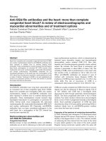
During shoulder elevation the dominant force vector is
provided by the deltoid muscle and in a superior direc-
tion. Under normal circumstances the cuff muscles will
counter this superior shear in the opposite direction, cre-
ating a stabilizing and compressive action of the humeral
head with respect to the glenoid during elevation. A dia-
grammatic representation of the gleno-humeral force cou-
ple [42] is seen in Figure 1. With cuff weakness (even
slight) the force couple may be altered enabling an abnor-
mal upward displacement of the humeral head and the
impingement of the subacromial structures and the
humeral head against the under surface of the acromion
[10,23].
Repetition of this process may cause irritation of pain pro-
ducing structures creating shoulder pain syndromes. In
order to address the abnormal force couple and its poten-
tially causative mechanism, specific exercises were intro-
duced to help restore strength and muscular functioning
of the glenohumeral joint and scapula articulations. (That
is, once motion was normalised).
It is acknowledged that a significant weakness in this case
series is the lack of imaging using diagnostic ultrasound to
confirm the diagnosis of impingement or indeed some
other cause for the pain. We encourage a further study of
the treatment protocol described above. This study should
be a randomised controlled trial and include diagnostic
ultrasound confirmed impingement.
Successful management of rotator cuff impingement and
related shoulder pain syndromes should include the con-
sideration of potential sources of shoulder pain. Also the
function of the implicated structures in global shoulder
function should be reviewed. This should include the
associated structures of the scapulohumeral, scapulotho-
racic articulations, the cervical and the thoracic spine.
This paper highlights a successful outcome for 4 subjects
with clinically diagnosed shoulder impingement syn-
drome after receiving a multimodal treatment approach
in a chiropractic setting.
Authors' contributions
MP provided treatment to the subjects, participated in the
design and helped draft the manuscript.
HP conceived of the study, participated in its design and
helped to draft and edit the manuscript. All authors read
and approved the manuscript.
Acknowledgements
No source of funding was used in the preparation of this manuscript. The
authors have no conflict of interest that is directly relevant to the content
of this manuscript.
References
1. Calliet R: Shoulder Pain 3rd edition. FA Davis Company-Philadelphia;
1991:1-50.
2. Morison DS, Greenbaum BS, Einhorn A: Shoulder impingement.
Orthop Clin Nth Am 2000, 31(2):285-293.
3. Almekinders LC: Impingement syndrome. Clin Sports Med 2001,
20:491-504.
4. Michener LA, McClure PW, Kardune AR: Anatomical and biome-
chanical mechanisms of subacromial impingement
syndrome. Clin Biomech 2003, 18:369-379.
5. Iannoti JP: Evaluation of the painful shoulder. J Hand Therapy
1994, 7:77-83.
6. Dalton S: Clinical examination of the shoulder. Baillieres Clinical
Rheumatology 1989, 3:453-474.
7. Brox J: Shoulder pain: Best Practise and Research. Clinical
Rheumatology 2003, 17:33-56.
8. Australian Cochrane Collaboration: Acute Shoulder Pain. In Evi-
dence Based Management of Acute Musculoskeletal Pain Australasian
Acute Pain Guidelines Group. Australian Academic Press; Brisbane;
2003:119-155.
9. Souza TA: Shoulder girdle complaints. In Differential Diagnosis for
the Chiropractor Gaithersberg. Aspen; 1997:141-172.
10. Hammer WI: The Shoulder. In Functional soft tissue examination and
treatment by manual methods Gaithersberg. Aspen; 1999:35-135.
11. Neer CS: Impingement lesions. Clin Orthop 1983, 173:70-77.
The glenohumeral force coupleFigure 1
The glenohumeral force couple. The resultant force (action)
of the rotator cuff muscles results in compression and infe-
rior glide of the humeral head during elevation. (RA = result-
ant action, Deltoid, SS = Supraspinatus, SSc = Subscapularis,
IS = Infraspinatus and TM = Teres Minor) [42].
Publish with BioMed Central and every
scientist can read your work free of charge
"BioMed Central will be the most significant development for
disseminating the results of biomedical research in our lifetime."
Sir Paul Nurse, Cancer Research UK
Your research papers will be:
available free of charge to the entire biomedical community
peer reviewed and published immediately upon acceptance
cited in PubMed and archived on PubMed Central
yours — you keep the copyright
Submit your manuscript here:
/>BioMedcentral
Chiropractic & Osteopathy 2005, 13:20 />Page 9 of 9
(page number not for citation purposes)
12. Gupta R, Leggin BG, Ianotti JP: Results of surgical repair of full
thickness tears of the rotator cuff. Orthop Clin North Am 1997,
28(2):241-248.
13. Van der Windt , Koes BW, de jong BA, Bouter LM: Shoulder disor-
ders in general practise: incidence, patient characteristics,
and management. Ann Rheum Dis 1995, 54(12):959-964.
14. Almekinders LC: Impingement syndrome. Clin Sports Med 2001,
20:491-504.
15. Michener LA, McClure PW, Kardune AR: Anatomical and biome-
chanical mechanisms of subacromial impingement
syndrome. Clin Biomech 2003, 18:369-379.
16. Gimblet PA, Saville J, Ebrall P: A conservative management pro-
tocol for calcific tendinitis of the shoulder. J Manipulative Physiol
Ther 1999, 22(9):622-627.
17. Pink MM, Tibone JE: The painful shoulder in the swimming
athlete. Orthop Clin North Am 2000, 31(2):247-261.
18. Shrode LW: Treating shoulder impingement using the
supraspinatus synchronization exercise. J Manipulative Physiol
Ther 1994, 17(1):43-53.
19. Hammer WI: The use of transverse friction massage in the
management of chronic bursitis of the hip or shoulder. J
Manipulative Physiol Ther 1993, 16:107-111.
20. Bang MD, Deyle GD: Comparison of supervised exercise with
and without manual physical therapy for patients with shoul-
der impingement syndrome. J Orthop Sports Phys Ther 2000,
30(3):126-137.
21. Conroy DE, Hayes KW: The effect of joint mobilization as a
component of comprehensive treatment for primary shoul-
der impingement syndrome. J Orthop Sports Phys Ther 1998,
28(1):3-14.
22. Leahy PM: Altered biomechanics of the shoulder and the
subscapularis. Chiropractic Sports Med 1991, 5(3):62-66.
23. Greenfield B, Catlin P, Coats P, Green E, McDonald J, North C: Pos-
ture in patients with shoulder overuse injuries and healthy
individuals. J Orthop Sports Phys Ther 1995, 21(5):287-295.
24. Culham EC, Peat M: Functional anatomy of the shoulder
complex. J Orthop Sports Phys Ther 1993, 18:342-350.
25. Kelly BT, Kadrmas WR, Speer KP: The manual muscle examina-
tion for rotator cuff strength: an electromyographic
investigation. Am J Sports Med 1996, 24:581-588.
26. Kohn D, Geyer M: The subjective shoulder rating system. Arch
Orthop Trauma Surg 1997, 116:324-328.
27. Soldatis JJ, Moseley JB, Etminan M: Shoulder symptoms in healthy
athletes: A comparison of outcome scoring systems. J Shoul-
der Elbow Surg 1997, 6:265-271.
28. Constant CR, Murley AHG: A clinical method of functional
assessment of the shoulder. Clin Orthop Rel Res 1987,
214:160-164.
29. Williams JG, Callaghan M: Comparison of visual estimation and
goniometry in determination of a shoulder joint angle. Phys-
iotherapy 1990, 76:655-657.
30. Bostrom Charms-Ringdahl K, Nordemar R: Clinical reliability of
shoulder function assessment in patients with rheumatoid
arthritis. Scand J Rheumatol 1991, 20:36-48.
31. Roodman WU: Etiologies of shoulder impingement syndrome
in competitive swimmers. Chiropractic Sports Med 1989,
3(2):27-31.
32. Frieman BG, Albert TJ, Fenlin JM: Rotator cuff disease: a review
of diagnosis pathophysiology, and current trends in
treatment. Arch Phys Med Rehabil 1994, 75(5):604-609.
33. Morrison DS, Frogameni AD, Woodworth P: Non-operative
treatment of Subacromial Impingement Syndrome. J Bone Jt
Surg 1997, 79(5):732-737.
34. Hammer WI: Friction massage; from Functional soft tissue
examination and treatment by manual methods. Gaithers-
berg: Aspen; 1999:463-478.
35. Downing DS, Weinstein A: Ultrasound therapy of subacromial
bursitis. Phys Ther 1986, 66(2):194-199.
36. Nykanen M: Pulsed ultrasound treatment of the painful shoul-
der a randomised, double-blind, placebo controlled study.
Scan J Rehab Med 1995, 27(2):105-108.
37. Mantone JK, Burkhead WZ, Noonan J Jr: Non-operative treat-
ment of rotator cuff tears. Orthop Clin North Am 2000,
31(2):295-311.
38. Perron M, Malouin F: Acetic acid iontophoresis and ultrasound
for the treatment of calcifying tendinitis of the shoulder: a
randomised controlled trial. Arch Phys Med Rehabil 1997,
78:379-384.
39. Van der Windt D, Van der Heijden G, Van der Berg S, Gerben ter R,
de Winter AF, Bouter LM: Ultrasound therapy for musculoskel-
etal disorders: a systemic review. Pain 1999, 81:257-271.
40. Davick JP, Martin RK, Albright JP: Distribution and deposition of
tritated cortisol using phonophoresis. Phys Ther 1988,
68(11):1672-1675.
41. Griffin JE, Echternach JL, Price RE, Touchstone JC: Patients treated
with ultrasonic driven hydrocortisone and with ultrasound
alone. Phys Ther 1967, 47:594-601.
42. Donatelli RA: Impingement syndrome and impingement
related instability. In Physical therapy of the shoulder 3rd edition.
Churchill Livingstone; 1997:229-256.