Báo cáo y học: "Case report of right hamate hook fracture in a patient with previous fracture history of left hamate hook: is it hamate bipartite" pps
Bạn đang xem bản rút gọn của tài liệu. Xem và tải ngay bản đầy đủ của tài liệu tại đây (1.95 MB, 7 trang )
BioMed Central
Page 1 of 7
(page number not for citation purposes)
Chiropractic & Osteopathy
Open Access
Database
Case report of right hamate hook fracture in a patient with previous
fracture history of left hamate hook: is it hamate bipartite?
Marion W Evans Jr*
1
, Micheal L Gilbert
2
and Sandra Norton
3
Address:
1
Parker College of Chiropractic Research Institute, 2500 Walnut Hill Lane, Dallas, TX 75229, USA,
2
Resident, Parker College of
Chiropractic Department of Radiology, 2500 Walnut Hill Lane, Dallas, TX 75229, USA and
3
Chair – Parker College of Chiropractic Department
of Radiology, 2500 Walnut Hill Lane, Dallas, TX 75229, USA
Email: Marion W Evans* - ; Micheal L Gilbert - ; Sandra Norton -
* Corresponding author
Abstract
Background: Hamate hook fracture is a common fracture in golfers and others who play sports
that involve rackets or sticks such as tennis or hockey. This patient had a previous hamate fracture
in the opposing wrist along with potential features of hamate bipartite.
Case presentation: A 19 year old male presented with a complaint of right wrist pain on the
ulnar side of the wrist with no apparent mechanism of injury. The pain came on gradually one week
before being seen in the office and he reported no prior care for the complaint. His history includes
traumatic left hamate hook fracture with surgical excision.
Conclusion: The patient was found to have marked tenderness over the hamate and with a prior
fracture to the other wrist, computed tomography of the wrist was ordered revealing a fracture
to the hamate hook in the right wrist. He was referred for surgical evaluation and the hook of the
hamate was excised. Post-surgically, the patient was able to return to normal activity within eight
weeks. This case is indicative of fracture rather than hamate bipartite. This fracture should be
considered in a case of ulnar sided wrist pain where marked tenderness is noted over the hamate,
especially after participation in club or racket sports.
Background
Wrist pain is often seen in chiropractic practices [1]. While
fracture to the scaphoid or navicular is the most prevalent
of wrist fractures [2], hamate hook fracture is the most fre-
quent fracture in golfers [3]. In most cases, the lead wrist,
which is the left wrist in a right handed golfer, is most
commonly fractured when the player strikes the ground,
root or rock prior to striking the ball. This leads to twisting
of the butt of the club against the hamate hook resulting
in a fracture, typically of the lead wrist which is the left
wrist in a right-handed golfer [3].
Occasionally, conservative care heals the fracture [4].
However, in many cases the hook must be surgically
removed before normal function will be restored without
pain [5]. Commonly, the diagnosis is delayed due to ini-
tial radiographs being read as negative, a more prominent
injury being seen at the time of initial presentation or the
stoic nature of the athlete who may delay evaluation [6].
Case presentation
The patient was a 19 year old male who was 204.2 cm in
height and weighted 145.15 kg. He was afebrile and had a
blood pressure of 128/80, left arm, seated. Otherwise
Published: 12 October 2006
Chiropractic & Osteopathy 2006, 14:22 doi:10.1186/1746-1340-14-22
Received: 13 June 2006
Accepted: 12 October 2006
This article is available from: />© 2006 Evans Jr et al; licensee BioMed Central Ltd.
This is an Open Access article distributed under the terms of the Creative Commons Attribution License ( />),
which permits unrestricted use, distribution, and reproduction in any medium, provided the original work is properly cited.
Chiropractic & Osteopathy 2006, 14:22 />Page 2 of 7
(page number not for citation purposes)
healthy, he experienced gradual right wrist pain over the
hamate and did not report a traumatic golf injury,
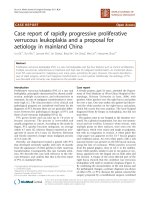
although he does play golf. He had a previous fracture to
the left hamulus over one year prior [Fig 1] that appar-
ently occurred on an attempt at ball strike with a sand
wedge while playing golf, in which he struck a rock just
behind the ball. In that case, immediate pain was noted
and the condition was misdiagnosed by a sports medicine
clinic prior to evaluation by the chiropractic clinician in
his chiropractic office [7]. The left hook had to be excised
due to failure of fragment fusion after plaster splinting,
which was applied for six weeks for the treatment of a sus-
pected scaphoid fracture. The patient's wrist injury healed
post-surgically with some complications involving a sub-
sequent navicular-lunate ligament tear and the patient
was eventually able to return to golf.
Since there was a previous misdiagnosed fracture to the
left wrist in this case, the patient called the chiropractic
office first. Due to his history, computed tomography [CT]
was ordered immediately following an examination
which demonstrated mild to moderate pain on all right
wrist movements, point tenderness over the hamate and
previous difficulty in obtaining a carpal tunnel view on
plain film x-ray.
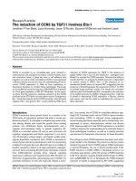
The CT scan revealed a complete, slightly displaced frac-
ture of the hook of the right hamate with associated soft
tissue edema [Fig 2]. A referral to an orthopedic surgeon
was made to assess the need for excision of the hook of the
hamate. Because of prior history, the patient elected to
have the hook excised without conservative therapy.
Bilateral fracture of the hamate is uncommon. In a case-
report by Bray, Swafford and Brown in 1985 [8], their
search of the literature found only 19 cases prior to 1977.
In this case, the left wrist had an apparent mechanism of
injury classic for fracture at this site, as it is the left hamate
that contacts the butt of the club in a right-handed golfer
[3]. However, the right wrist did not have this mechanism
of injury. This may suggest some preexisting condition of
the hamate in this patient. A condition known as hamate
bipartite affects the hamate in some patients [9,10]. This
condition, which is thought to be the result of fibrocarti-
CT of left wrist indicating hamate hook fractureFigure 1
CT of left wrist indicating hamate hook fracture.
Chiropractic & Osteopathy 2006, 14:22 />Page 3 of 7
(page number not for citation purposes)
lagenous union between the body and hook of the
hamate, typically causes symptoms in this part of the wrist
and is characterized by a weak or ununited appearance of
the hook that can be detected on CT or plain film radio-
graphs [10].
Hamate bipartite tends to be suspected in cases where
there is no history of trauma or surgery to the wrist [10].
In our case, there was a denial of traumatic golf injury to
the right wrist but was apparent in the left.
Features of hamate bipartite
Features of hamate bipartite according to Pierre-Jerome &
Roug [10] include;
• Bilaterally similar bipartite hamulus
• No sign or history of traumatic wrist injury or edema or
soft tissue changes suggestive of un-united fracture
• Equal size and uniform signal intensity on MRI evalua-
tion of each part
• Absence of progressive degenerative changes between
the two components of the hamate or elsewhere in the
wrist
• Smooth, well corticated and rounded margins of the
hamate and un-united hook.
Symptoms were noted in the case of the left fracture
immediately upon the patient's dubbed ball strike and
only surgical excision relieved the pain [7]. Additionally,
the original radiological report accompanying the images
of the right wrist was indicative of fracture and not other-
wise. The attending radiologist noted degenerative
changes within the right wrist, separation of the naviculol-
unate interspace and 1–2 mm of separation of the hook
from the body of the hamate. There was also some indica-
CT of right wrist indicating hamate hook fractureFigure 2
CT of right wrist indicating hamate hook fracture.
Chiropractic & Osteopathy 2006, 14:22 />Page 4 of 7
(page number not for citation purposes)
tion of a possible previous fracture of the capitate noted
by the radiologist, which would further indicate possible
previous trauma to the right wrist. However, the patient
denied previous trauma.
Diagnostic Imaging Considerations
Hamate fractures represent approximately 2–4% of all
fractures involving the carpal bones [11]. Fractures involv-
ing the hamulus, or hook, represent one of the two groups
of fractures of the hamate [12]. Norman and others docu-
mented three radiographic signs suggestive of fracture of
the hamulus [13]. According to their criteria, the most fre-
quently encountered and most important feature is the
lack of visualization of the hook. On the dorsovolar view
the hamulus is seen en face super-imposed over the
hamate and demonstrates a cortical ring shadow known
as the "eye sign" [12]. A blurry or indistinguishable
appearance of the "eye", as well as sclerosis of the hamu-
lus, seen associated with nonunion, represents the other
two radiographic features that suggest fracture of the hook
[12,13].
Various radiographic positioning techniques can prove
useful in the evaluation of potential hamate fractures.
These may not always be of diagnostic quality due to lim-
ited range of motion experienced by the patient as a result
of pain, especially in acute or subacute fractures [12].
Moreover, these fractures are commonly overlooked on
standard radiographic studies of the wrist due to the lack
of specific physical exam findings and a low index of sus-
picion [14]. Conventional radiographic examination of
the wrist usually consists of the dorsovolar view, which
demonstrates the radiographic signs first described by
Norman and colleagues as discussed previously, as well as
the lateral and medial oblique projections [15].
The use of the carpal tunnel view [Fig 3] has increased in
an attempt to better elucidate the presence of a hamulus
fracture [15]. Originally described by Gaynor and Hart
[13,15], it is set with the patient positioned such that the
flexor surface of their forearm lies against the film with
slight radial rotation and the long axis of their hand is
made as vertical as possible. The central ray is directed
toward the palmar surface distal to the base of the third
metacarpal with 25–30° of tube angulation. The patient
may use their other hand or some other appropriate
device to hold their wrist in this extended position [16].
The "radial-deviated, thumb-abducted lateral view" [Fig
4] is considered by some authors, an underused technique
that adequately demonstrates the hamate between the
thumb and index finger and clearly displays fractures of
the hamulus [15]. This radiograph is performed by posi-
tioning the patient with their forearm in neutral and the
medial aspect of their wrist against the film cassette. Their
thumb is fully extended and abducted and their wrist
deviated radially. This position results in maximum wid-
ening of the index finger-thumb web space [15]. The cen-
tral ray is directed at the center portion of the index finger
and thumb web. Alternately, the radial deviation can be
excluded and a 15° tube angulation, oriented toward the
wrist, can be used [15]. Bhalla and colleagues consider
this view a cost-effective and time-saving adjunct to tradi-
tional wrist series when fracture of the hook of the hamate
is suspected.
The hamulus, which develops from its own ossification
center, may fail to fuse with the body of the hamate [17].
This normal variant is referred to as the os hamuli pro-
prium and may be difficult to differentiate from avulsion
fractures of the hamate hook [17]. In equivocal cases,
computed tomography [CT] of the wrist is an effective
advanced imaging technique to confirm the diagnosis of
hamulus fractures due to its ability to provide an image
that is orthogonal to the plane of the hamulus base frac-
ture, while avoiding the possibility of superimposed ana-
tomical structures [12,15]. In fact, it has been suggested
that it is pointless to obtain plain films when hamulus
fracture is suspected clinically and that CT should be the
initial imaging modality chosen [18].
Moreover, with the newer generation spiral CT multiple
imaging planes can be obtained after a single scan [18].
With complete fractures, CT clearly reveals an osseous
fragment demonstrating indistinct and irregular apposing
cortical margins separated from the parent bone [14].
Incomplete fractures exhibit partial cortical disruption
without osseous fragment separation. Additionally, inter-
nal joint derangements, such as injuries to the triangular
fibrocartilage complex, may be found in association with
fracture of the hamulus depending on the mechanism of
injury such as a fall on the outstretched, pronated arm.
Physical examination of these patients reveals tenderness
between the pisiform and ulnar styloid on the ulnar bor-
der of the wrist [19]. Typically, however, there would be
no indication of fracture in these patients and imaging
would make the differential diagnosis [20]. Further, mag-
netic resonance imaging [MRI] is, in the opinion of the
authors, most effective in determining the presence and
extent of these injuries.
Conclusion
We propose that in spite of no known mechanism of
injury to the right wrist in the patient, the left wrist was
traumatically fractured, as he felt immediate pain that
completely resolved after surgical excision of the hook.
We also suggest one other possibility in this patient. This
is a young man who is very large for his age. His height
and anthropometric features would suggest that his wrist
bones are very large as well. This could make the hook of
Chiropractic & Osteopathy 2006, 14:22 />Page 5 of 7
(page number not for citation purposes)
the hamate longer and therefore, weaker where the hook
extends from the body of the hamate. Perhaps this made
his bone more vulnerable to fracture. Perhaps, in spite of
the patient's denial of traumatic injury, the fracture is
related to his golf playing, as he is an avid player who
spends quite a bit of time on the course. Further, the cor-
responding author has observed the swing of the young
man while playing golf and he has a powerful swing as
one might imagine in someone his size. The forces exerted
on the wrist would speculatively, be above average.
While a case of hamate bipartite may difficult to rule out
in some cases, it is rather curious to us that one wrist was
apparently fractured while playing golf and the other not,
approximately one year apart. We conclude that this is a
case of bilateral fracture of the hamate, although they
clearly occurred in separate events. Pain on the ulnar side
of the wrist in those who participate in racket or club
sports should be evaluated for fracture and hamate hook
fracture should be given diagnostic consideration.
Hamate bipartite should be considered in case of persist-
ent pain where no prior history of trauma is noted.
List of abbreviations
CT-computed tomography, MRI-magnetic resonance
imaging
Competing interests
The authors declare that they have no competing interests.
Authors' contributions
ME treated the case and contributed to the sequence align-
ment and drafted the primary manuscript. MG contrib-
uted to the sequence alignment of the manuscript and
coordinated additional material on diagnostic imaging
considerations. SN contributed to the sequence alignment
Radiographic position for carpal tunnel viewFigure 3
Radiographic position for carpal tunnel view.
Chiropractic & Osteopathy 2006, 14:22 />Page 6 of 7
(page number not for citation purposes)
of the manuscript. All authors read and approved the final
manuscript.
Acknowledgements
We wish to thank the Parker College of Chiropractic Department of Diag-
nostic Imaging for scanning films used in this manuscript and our subject
who gave his informed consent so this article could be published.
References
1. Christensen M, Kollasch MW: Job analysis of chiropractic: a
project report, survey analysis, and summary of the practice
of chiropractic within the United States. Greeley (CO):
National Board of Chiropractic Examiners; 2005:67.
2. Hoppenfeld S: Physical examination of the spine and extremi-
ties. Norwalk (CT): Appleton-Century-Crofts; 1976:67-71.
3. Stover C, McCarroll J, Mallon W: Feeling up to par: medicine
from tee to green. Philadelphia: FA Davis Company; 1994:158.
4. Fujioka H, Tsunoda M, Noda M, Matsui N, Mizuno K: Treatment of
ununited fracture of the hook of the hamate by low-intensity
pulsed ultrasound: a case report. J Hand Surg [Am] 2000,
25:77-9.
5. Geissler W: Carpal fracture in athletes. Clin Sports Med 2001,
20:167-88.
6. Walsh J, Bishop A: Diagnosis and management of hamate hook
fractures. Han Clin 2000, 16:397-403.
7. Evans MW: Hamate hook fracture in a 17- year old golfer:
Importance of matching symptoms to clinical evidence. J
Manipulative Physiol Ther 2004, 27:516-18.
8. Bray TJ, Swafford AR, Brown RL: Bilateral fracture of the hook of
the hamate. J Trauma 1985, 25:174-5.
9. Green MH, Hadied AM: Bipartite hamulus with ulnar tunnel
syndrome: Case report and literature review. J Hand Surg [Am]
1981, 6:605-9.
10. Pierre-Jerome C, Roug IK: MRI of bilateral bipartite hamulus: a
case report. Surg Radiol Anat 1998, 20:299-302.
11. Resnick D: Diagnosis of bone and joint disorders. Philadelphia
(PA): WB Saunders; 2002:2847-2848.
12. Greenspan A: Orthopaedic Imaging: A practical approach.
Philadelphia (PA): Lippincott-Williams-Wilkins; 2004:191-195.
13. Norman A, Nelson J, Green S: Fractures of the hook of hamate:
Radiographic signs. Radiology 1985, 154:49-53.
14. McCue F, Faltaous A, Baumgarten T: Bilateral hook of the hamate
fractures. Orthopedics 1997, 20(5):
470-472.
15. Bhalla S, Higgs P, Gilula L: Utility of the radial-deviated, thumb-
abducted lateral radiographic view for the diagnosis of
hamate hook fractures: Case report. Radiology 1998,
209:203-207.
16. Ballinger P, (Ed): Merrill's Atlas of Radiographic positions and
radiologic procedures. St. Louis (MO): Mosby; 1991:83, 94-97.
17. Freyschmidt J, Brossmann J, Wiens J, Sternberg A: Borderlands of
Normal and Early Pathological Findings in Skeletal Radiog-
raphy. New York: Thieme; 2003:145.
Radiographic position for radial-deviated, thumb-abducted viewFigure 4
Radiographic position for radial-deviated, thumb-abducted view.
Publish with BioMed Central and every
scientist can read your work free of charge
"BioMed Central will be the most significant development for
disseminating the results of biomedical research in our lifetime."
Sir Paul Nurse, Cancer Research UK
Your research papers will be:
available free of charge to the entire biomedical community
peer reviewed and published immediately upon acceptance
cited in PubMed and archived on PubMed Central
yours — you keep the copyright
Submit your manuscript here:
/>BioMedcentral
Chiropractic & Osteopathy 2006, 14:22 />Page 7 of 7
(page number not for citation purposes)
18. Kato H, Nakamura R, Horri E, Nakao E, Yajima H: Diagnostic imag-
ing for fractures of the hook of the hamate. Hand Surg 2000,
5(1):19-24.
19. Rettig AC: Athletic injuries of the wrist and hand: Part I: Trau-
matic injuries of the wrist. Am J Sports Med 2003,
31(6):1038-1048.
20. Shih JT, Lee HM, Tan CM: Early isolated triangular fibrocarti-
lage complex tears: management by arthoscopic repair. J
Trauma 2002, 53(5):9222-927.