Báo cáo y học: "A diagnosis-based clinical decision rule for spinal pain part 2: review of the literature" pps
Bạn đang xem bản rút gọn của tài liệu. Xem và tải ngay bản đầy đủ của tài liệu tại đây (681.81 KB, 17 trang )
BioMed Central
Page 1 of 17
(page number not for citation purposes)
Chiropractic & Osteopathy
Open Access
Review
A diagnosis-based clinical decision rule for spinal pain part 2: review
of the literature
Donald R Murphy*
1,2,3
, Eric L Hurwitz
4
and Craig F Nelson
5
Address:
1
Rhode Island Spine Center, 600 Pawtucket Avenue, Pawtucket, RI, 02860, USA,
2
Department of Community Health, Warren Alpert
Medical School of Brown University, USA,
3
Research Department, New York Chiropractic College, USA,
4
Department of Public Health Sciences
and Epidemiology, John A. Burns School of Medicine, University of Hawaii at Mânoa, Honolulu, Hawaii, 96822, USA and
5
American Specialty
Health, San Diego, CA, USA
Email: Donald R Murphy* - ; Eric L Hurwitz - ; Craig F Nelson -
* Corresponding author
Abstract
Background: Spinal pain is a common and often disabling problem. The research on various
treatments for spinal pain has, for the most part, suggested that while several interventions have
demonstrated mild to moderate short-term benefit, no single treatment has a major impact on
either pain or disability. There is great need for more accurate diagnosis in patients with spinal pain.
In a previous paper, the theoretical model of a diagnosis-based clinical decision rule was presented.
The approach is designed to provide the clinician with a strategy for arriving at a specific working
diagnosis from which treatment decisions can be made. It is based on three questions of diagnosis.
In the current paper, the literature on the reliability and validity of the assessment procedures that
are included in the diagnosis-based clinical decision rule is presented.
Methods: The databases of Medline, Cinahl, Embase and MANTIS were searched for studies that
evaluated the reliability and validity of clinic-based diagnostic procedures for patients with spinal
pain that have relevance for questions 2 (which investigates characteristics of the pain source) and
3 (which investigates perpetuating factors of the pain experience). In addition, the reference list of
identified papers and authors' libraries were searched.
Results: A total of 1769 articles were retrieved, of which 138 were deemed relevant. Fifty-one
studies related to reliability and 76 related to validity. One study evaluated both reliability and
validity.
Conclusion: Regarding some aspects of the DBCDR, there are a number of studies that allow the
clinician to have a reasonable degree of confidence in his or her findings. This is particularly true
for centralization signs, neurodynamic signs and psychological perpetuating factors. There are other
aspects of the DBCDR in which a lesser degree of confidence is warranted, and in which further
research is needed.
Background
Accurate diagnosis or classification of patients with spinal
pain has been identified as a research priority [1]. We pre-
sented in Part 1 the theoretical model of an approach to
diagnosis in patients with spinal pain [2]. This approach
incorporated the various factors that have been found, or
Published: 11 August 2008
Chiropractic & Osteopathy 2008, 16:7 doi:10.1186/1746-1340-16-7
Received: 25 March 2008
Accepted: 11 August 2008
This article is available from: />© 2008 Murphy et al; licensee BioMed Central Ltd.
This is an Open Access article distributed under the terms of the Creative Commons Attribution License ( />),
which permits unrestricted use, distribution, and reproduction in any medium, provided the original work is properly cited.
Chiropractic & Osteopathy 2008, 16:7 />Page 2 of 17
(page number not for citation purposes)
in some cases theorized, to be of importance in the gener-
ation and perpetuation of neck or back pain into an
organized scheme upon which a management strategy can
be based. The authors termed this approach a diagnosis-
based clinical decision rule (DBCDR). The DBCDR is not
a clinical prediction rule. It is an attempt to identify
aspects of the clinical picture in each patient that are rele-
vant to the perpetuation of pain and disability so that
these factors can be addressed with interventions designed
to improve them. The purpose of this paper is to review
the literature on the methods involved in the DBCDR
regarding reliability and validity and to identify those
areas in which the literature is currently lacking.
The Three Essential Questions of Diagnosis
The DBCDR is based on what the authors refer to as the 3
essential questions of diagnosis [2]. The answers to these
questions supply the clinician with the most important
information that is required to develop an individualized
diagnosis from which a management strategy can be
derived. The 3 questions are:
1. Are the symptoms with which the patient is presenting reflective
of a visceral disorder or a serious or potentially life-threatening
disease?
In seeking the answer to this question, history and exam-
ination and, when indicated, special tests, are used to
detect or raise the level of suspicion for the presence of
pathological disorders for which spinal pain may be the
first or only symptom. Some examples are gastrointestinal
or genitourinary disorders, fracture, infection and malig-
nancy. Potentially serious or life-threatening conditions
are sometimes referred to as "red flags" [3].
2. From where is the patient's pain arising?
In seeking the answer to this question, four signs are
searched for: (1) centralization signs, (2) segmental pain
provocation signs, (3) neurodynamic signs, and (4) mus-
cle palpation signs.
3. What has gone wrong with this person as a whole that would
cause the pain experience to develop and persist?
In seeking the answer to this question, perpetuating fac-
tors are searched for: (1) dynamic instability (impaired
motor control), (2) central pain hypersensitivity, (3) ocu-
lomotor dysfunction (in cervical trauma patients), (4)
fear, (5) catastrophizing, (6) passive coping, and (7)
depression. These latter psychological factors are some-
times referred to as "yellow flags" [4].
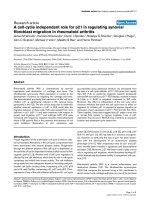
An algorithm illustrating the diagnostic strategy of the
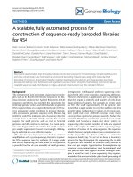
DBCDR is presented in figure 1. The recommended man-
agement strategy based on the DBCDR is presented in fig-
ure 2.
The purpose of this paper is to review the literature on the
reliability and validity of the detection of the individual
diagnostic factors included in the DBCDR, and to present
the evidence as it currently exists, for the various aspects of
this approach.
Methods
Literature search and selection
The following databases were searched up to December
22, 2006: Medline, Cinahl, Embase and MANTIS.
Searches of the authors' own libraries were also con-
ducted. Finally, citation searches of relevant articles and
texts were conducted manually. The following search
terms were used:
Diagnosis AND "low back pain"
Diagnosis AND "neck pain"
Diagnosis AND "low back pain" AND palpation
Diagnosis AND "neck pain" AND palpation
Diagnosis AND "low back pain" AND McKenzie
Diagnosis AND "neck pain" AND McKenzie
Diagnosis AND "low back pain" AND neurodynamics
Diagnosis AND "neck pain" AND neurodynamics
Diagnosis AND "low back pain" AND radiculopathy
Diagnosis AND "neck pain" AND radiculopathy
Diagnosis AND "low back pain" AND trigger points
Diagnosis AND "neck pain" AND trigger points
Diagnosis AND "low back pain" AND muscle
Diagnosis AND "neck pain" AND muscle
Diagnosis AND "low back pain" AND instability
Diagnosis AND "neck pain" AND instability
Diagnosis AND "low back pain" AND "motor control"
Diagnosis AND "neck pain" AND "motor control"
Diagnosis AND "low back pain" AND "central sensitiza-
tion"
Chiropractic & Osteopathy 2008, 16:7 />Page 3 of 17
(page number not for citation purposes)
Diagnosis AND "low back pain" AND "central pain hyper-
sensitivity"
Diagnosis AND "neck pain" AND "central sensitization"
Diagnosis AND "neck pain" AND "central pain hypersen-
sitivity"
Diagnosis AND "neck pain" AND oculomotor
Diagnostic algorithm for the application of the DBCDRFigure 1
Diagnostic algorithm for the application of the DBCDR.
Chiropractic & Osteopathy 2008, 16:7 />Page 4 of 17
(page number not for citation purposes)
Diagnosis AND "low back pain" AND fear
Diagnosis AND "neck pain" AND fear
Diagnosis AND "low back pain" AND catastrophizing
Diagnosis AND "neck pain" AND catastrophizing
Diagnosis AND "low back pain" AND coping
Diagnosis AND "neck pain" AND coping
Diagnosis AND "low back pain" AND depression
Diagnosis AND "neck pain" AND depression
Studies were included if they were in English and pro-
vided original, statistically analyzed data regarding the
reliability and validity of clinic-based diagnostic proce-
dures used for the identification of relevant factors in the
causation or perpetuation of spinal pain. Included studies
had to contain data on the assessment of patients with cer-
vical or lumbar pain, including headache related to the
cervical spine and spine-related upper or lower extremity
pain. Non-English language studies were excluded, as
were studies that did not present data on reliability and
validity. The search focused on diagnostic procedures that
Management algorithm for the application of the DBCDRFigure 2
Management algorithm for the application of the DBCDR.
Chiropractic & Osteopathy 2008, 16:7 />Page 5 of 17
(page number not for citation purposes)
are potentially useful in answering the second or third
question of diagnosis. Studies that were potentially useful
in answering question 1 were not considered for the pur-
pose of this paper. Diagnostic studies that require special
equipment not typically found in the clinic (such as MRI)
or that require a laboratory (such as blood tests) were
excluded because the purpose of the study was to evaluate
clinic-based means by which the DBCDR may be applied.
It is recognized that imaging or laboratory tests are often
useful in the diagnosis of spinal pain, but the presentation
of these procedures was beyond the scope of this paper. In
cases in which systematic reviews of the literature were
found, the individual studies included in the reviews were
not reviewed separately, unless this was necessary to clar-
ify information that was not readily apparent from the
systematic review.
Each study was reviewed by two authors (DRM and CFN)
and deemed relevant or irrelevant. A study was considered
relevant if the information contained in the study indi-
cated that it met the above inclusion/exclusion criteria.
Results
The search strategy identified 1769 articles, and of these,
138 were deemed relevant. Additional files 1 and 2 pro-
vide a breakdown of the number of studies in each area of
consideration. Additional files 3 and 4 present the data
from those studies that met the inclusion criteria. We have
divided the presentation of the literature into those stud-
ies that apply to patients with neck pain and those that
relate to patients with low back pain (LBP).
Neck Pain
Question 1. Are the symptoms with which the patient is presenting
reflective of a visceral disorder or a serious or potentially life-
threatening disease?
A detailed review of the literature related to this question
is beyond the scope of this paper. However, in general,
history, focusing on the presence of symptoms such as GI
distress, fever or previous history of cancer, and examina-
tion, focusing on vital signs, abdominal examination and
examination of peripheral pulses, are useful in raising the
level of suspicion as to the presence of a visceral disorder
or a serious or potentially life-threatening disease [5].
Imaging and/or special tests such as sedimentation rate
can be utilized for further confirmation [5]. Details can be
found elsewhere [5-7].
Question 2. From where is the patient's pain arising?
Centralization signs
Centralization signs are detected through methods origi-
nally developed by McKenzie [8,9]. The examination pro-
cedure involves moving the spine to end range in various
directions and monitoring the mechanical and sympto-
matic response to these movements.
Reliability
Clare, et al [10] used 2 physical therapists trained in the
McKenzie method to examine 25 patients with cervical
pain. They found good inter-examiner reliability (IER)
(kappa, [k] = 0.63 and 93% agreement) for the assessment
procedure.
Validity
No studies were identified that have addressed the validity
of centralization signs in the cervical spine.
Segmental pain provocation signs
A number of studies have examined segmental mobility
assessment and have generally found poor IER [11-16]
and validity [17]. Other studies have examined proce-
dures designed to identify segmental pain (as opposed to
mobility impairment).
Reliability
Hubka and Phelan [18] assessed the IER of palpation for
tenderness between 2 practitioners in 30 patients with
unilateral neck pain. They found good IER (k = 0.68). Jull,
et al [19] assessed IER of segmental palpation using 7
examiners and 40 subjects with or without neck pain and
headache. The criteria for a positive test were based on
resistance to joint movement and pain provocation in
response to palpation. Kappa values indicated excellent to
perfect IER (k = 0.78–1.00) in 6 instances, fair to good (k
= 0.45–0.65) in 14 instances and poor (k = 0.25–0.34) in
5 instances. They point out that, in the instances of poor
agreement, the raw data indicated that the examiners had
agreed on 13 of 14 decisions. But the calculations of k
were vulnerable because 12 of the 13 agreements were in
the same cell of agreed negative finding. Marcus, et al [20]
used 4 physical therapists to examine 72 headache
patients and 24 controls. The therapists examined all sub-
jects for "cervical synovial joint abnormalities" in the
same manner as described in the study by Jull, et al [19].
They found good IER (k = 0.63) between examiners.
McPartland and Goodridge [21] assessed IER of "TART"
exam, described as segmental palpation that focused on
three parameters: tissue texture change, restriction of ver-
tebral motion and zygapophyseal (z) joint tenderness.
They found the IER of examination that considered all
three parameters was poor (k = 0.35 for asymptomatic
subjects, k = 0.34 for symptomatic subjects). But for the
parameter of tenderness alone, IER improved (k = 0.529).
Van Suijlekom, et al [22] used 2 neurologists to examine
24 headache patients and found IER for segmental palpa-
tion to be slight to fair (k = 0.14 to 0.37). However, the
palpation method was poorly described in this study.
Also, it is not known as to whether the difference between
the findings of this study and those of the other studies
reported here relate to the fact that the "negative" IER
studies used neurologists, whereas the "positive" IER
Chiropractic & Osteopathy 2008, 16:7 />Page 6 of 17
(page number not for citation purposes)
study used chiropractors or physical therapists. Cleland, et
al [23] used 2 examiners and 22 subjects and found highly
variable IER between 2 physical therapists for palpation
for pain provocation, with k ranging from 52 to .90,
depending on the segment involved. They speculated that
this high variability related to the clinicians not agreeing
on the segmental level being examined, as opposed to lack
of agreement on the findings.
Validity
Jull, et al [24] used diagnostic blocks to identify the pres-
ence and location of symptomatic z joints in 20 patients
with cervical related pain. The patients were examined by
a manipulative physiotherapist who also attempted to
identify the presence and location of symptomatic z
joints. The definition of a symptomatic joint as deter-
mined by palpation was based on abnormal "end feel",
increased resistance to motion and reproduction of pain.
They found that the SE and SP were both 1.00. That is, the
examiner was able to identify 100% of the symptomatic
segments as well as all of the subjects whose pain was not
abolished by diagnostic block. This study used single,
rather than double blind, diagnostic blocks. Regardless, as
will be discussed below, the use of diagnostic blocks as a
Gold Standard for the presence of z joint pain has been
questioned [25]. Treleaven, et al [26] assessed 12 patients
with postconcussion headache with segmental palpation.
The method of palpation was the same as that used by
Jull, et al [24]. They found complete agreement between
the examiner and independent report of the patient as to
which segments were painful and almost complete agree-
ment as to which segment was most painful. Sandmark
and Nisell [27], calculated the SE, SP and PPV and nega-
tive predictive value (NPV) of segmental palpation in the
cervical spine relative to reported neck pain. They found
these values to be 0.82, 0.79, 0.62 and 0.91 respectively.
Lord, et al [28], used a double blind anesthetic block to
determine the prevalence of pain arising from the C2-3 z
joint in patients with the complaint of chronic headache
after cervical trauma. These authors demonstrated that the
prevalence of C2-3 z-joint pain was 53%, and the only
sign that was associated with these patients was tender-
ness to palpation over the C2-3 z joint. They calculated
that palpation had SE of 0.85, a positive likelihood ratio
(PLR) of 1.7 and a negative likelihood ratio (NLR) of 0.3.
The precise method of palpation was not described. Zito,
et al [29] using the palpation method found to be reliable
by Jull et al [19] found a significantly higher incidence (p
< 0.05) of hypomobile and painful z joints in the upper
cervical spine of patients classified according to the Inter-
national Headache Society criteria as having cervicogenic
headache compared to those classified as having migraine
with aura. King, et al [30] used "controlled, diagnostic
blocks" as a Gold Standard against which segmental pal-
pation that was described as being similar to that of Jull,
et al [24]. They found the SE to be 0.88, SP to be 0.39 and
PLR to be 1.3. Again, using diagnostic block as a Gold
Standard may be questionable [25], leaving open the
issue of what should be the Gold Standard for segmental
palpation signs. Further work in the area of establishing a
true Gold Standard for the identification of zygapophy-
seal joint pain may be needed before definitive statements
regarding the presence or absence of pain from this struc-
ture can be made.
Neurodynamic signs
Reliability
The standard neurodynamic test in the cervical spine is the
brachial plexus tension test (also known as the upper limb
tension test [31]). Wainner, et al [32] found good to excel-
lent IER of this test (k = 0.76 to 0.81). They also found
good to excellent IER of several historical questions of
patients with documented cervical radiculopathy (k =
0.53 to .082). They found varying IER of neurologic exam
findings, but good to excellent IER of Spurling's test
(which they described as bending the seated patient's
head toward the side of symptoms, rotating and extending
slightly, and applying downward pressure), the cervical
distraction test and Valsalva's maneuver. The kappa values
for these tests ranged from 0.60 to 0.88.
Validity
Wainner, et al [32] provide data on the SE, SP PLR and
NLR of a variety of historical factors and examination pro-
cedures. They found that the cluster of 4 tests – Spurling's
test, the upper limb tension test, the cervical distraction
test and limited rotation toward the side of symptoms sec-
ondary to pain – carried the greatest diagnostic accuracy as
compared to the Gold Standard of electromyography.
When 3 of these tests were positive, there was a 65% prob-
ability of the presence of cervical radiculopathy the SE and
SP were 0.39 and 0.94, respectively and a PLR of 6.1.
When all 4 tests were positive, there was a 90% probabil-
ity of the presence of cervical radiculopathy. The SE and
SP were 0.24 and 0.99 respectively and the PLR was 30.3.
Shah and Rajshekhar [33] also used Spurling's test, the
description of which was the same as that in the Wainner,
et al study [32], and found it to be useful in identifying
"soft disc prolapse" as opposed to "hard disc" (i.e., osteo-
phyte). They calculated the SE and SP to be 0.90 and 1.00,
respectively compared to the Gold Standard of operative
findings. The PPV was calculated to be 1.00 and the NPV
to be 0.71. In patients treated non-surgically, they used
MRI as the Gold Standard and calculated the SE and SP to
be 0.90 and 0.93, respectively. The PPV was calculated to
be 0.90 and the NPV to be 0.93.
Chiropractic & Osteopathy 2008, 16:7 />Page 7 of 17
(page number not for citation purposes)
Muscle palpation signs
Reliability
Marcus, et al, in the same study cited above [20] found
good to perfect IER of TrP palpation in the cervical spine
(k = 0.74), head (k = 0.81) and shoulder (k = 1.00). van
Suijlekom, et al [22] in the study cited above, found vari-
able IER (k = 0.0 – 1.00) of TrP palpation in patients with
headache. As was the case with segmental palpation, the
method of TrP examination was poorly described. Ger-
win, et al [34] performed 2 different experiments to assess
IER. In the first, 4 examiners assessed 20 different muscles
on each of 25 patients with various symptom presenta-
tions. They used a general observer-agreement statistic
called the "S
av
", which they defined as "a generalized ver-
sion of the Cohen's kappa which reports pairwise judge
agreement corrected for chance agreement." They found
poor IER (S
av
= 0.0–1.0). They then repeated the study
after spending a 3-hour session in which the examiners
discussed positive findings and palpation techniques.
They found good to excellent IER (S
av
= 0.65 – .95) after
the training session. Sciotti, et al [35] found good IER
(Generalizability coefficient = 0.83–0.92) between 2
examiners looking for latent trigger points (TrPs) in the
upper trapezius muscle. However, the subjects were
asymptomatic. On the other hand, Lew, et al [36] found
poor IER for TrP palpation in the upper trapezius,
although the subjects in that study were also asympto-
matic.
Validity
The validity of muscle palpation signs is unknown, largely
due to lack of an appropriate Gold or reference standard.
3. What has gone wrong with this person as a whole that
would cause the pain experience to develop and persist?
As was discussed in the earlier paper describing the
DBCDR [2], this third question attempts to identify those
factors that may be placing the patient at risk of develop-
ing persistent or recurrent spinal pain, or, in the case of
chronic patients, have contributed to the establishment of
the chronic or recurrent problem. There are a number of
factors that have been suggested to be of importance in
the perpetuation of chronic spinal pain, although research
investigating this area is ongoing.
Dynamic instability (impaired motor control)
Reliability
In the cervical spine, the Craniocervical Flexion (CF) test
[37,38] is designed to detect decreased activity in the deep
cervical flexor muscles and hyperactivity in the sternoclei-
domastoid muscles. It is thought that, as the deep cervical
flexors are important for stability of the intersegmental
joints of the cervical spine, this imbalance in muscle acti-
vation compromises cervical spine stability [37]. The CF
test measures the motor control capacity of the deep cer-
vical flexors. Jull, et al [38] found good IER (ICC = 0.81 to
0.93) in 50 asymptomatic subjects; Chiu, et al [39] found
good IER (k = 0.72) in 10 asymptomatic subjects.
Recently, 3 studies [23,40,41] have demonstrated IER of a
test that uses a similar positioning but, rather than using
a pressure cuff, involves practitioner observation of the
ability of patients to maintain a position of slight upper
cervical flexion in the supine position. Cleland, et al [23]
used 2 examiners and 22 subjects and found moderate
IER (ICC = 0.57). Harris, et al [40] used 2 examiners and
40 subjects and found moderate IER (ICC = 0.67); Olson,
et al [41], using an almost identical test as Harris, et al
[40], found excellent IER (k = 0.83 to 0.88) between 2
examiners in 27 subjects without neck pain.
Validity
Treleavan, et al [26] compared 12 patients with postcon-
cussion headache with asymptomatic controls using the
CF test. They found a significant (p = 0.02) decrease in the
duration of time that the test position could be held in
patients compared to controls. Jull, et al [38] compared 15
patients with cervicogenic headache and compared them
with 15 controls. They found significantly (p < 0.001)
poorer performance on the CF test in the patients com-
pared to controls. Jull, et al [42] compared patients with
neck pain after whiplash, patients with insidious onset
neck pain and normal controls in the performance of the
CF test. They found significantly poorer performance (p <
0.05) in both neck pain groups than in controls. There
was no difference between the post-whiplash patients and
the insidious onset patients. Falla, et al [43] used the CF
test and electromyography (EMG) to demonstrate
reduced activity in the deep cervical flexor muscles in
patients with chronic neck pain compared to controls.
There was also a trend toward increased activity in the ster-
nocleidomastoid and scalene muscles in patients com-
pared to controls. With regard to increased activity in the
sternocleidomastoid muscle during the performance of
the CF test, this replicated the findings of Jull [44].
Central Pain Hypersensitivity (CPH)
As will be discussed below, there is good evidence that the
presence of nonorganic signs is reflective of increased pain
perception. [45]
Reliability
Sobel, et al [46] developed nonorganic signs for patients
with neck pain and found excellent to perfect (k = 0.80 to
1.00) IER in 26 patients.
Validity
The validity of cervical nonorganic signs is unknown.
Chiropractic & Osteopathy 2008, 16:7 />Page 8 of 17
(page number not for citation purposes)
Imaging modalities like functional MRI and SPECT have
promise in the diagnosis of CPH [47,48]; however, it is
not clear as to whether these are viable tools for common
use.
Oculomotor dysfunction
Oculomotor dysfunction has been found in patients with
chronic neck pain after whiplash [49] as well as in patients
with chronic tension type headache [50]. Gimse, et al [51]
compared 26 patients with chronic (average 4.7 years)
neck pain after whiplash and who had complaints of vis-
ual problems or vertigo and compared them with 26
matched controls. They found significantly (p < 0.001)
poorer performance on tests of oculomotor function in
the whiplash group. Tjell, et al [52] compared 160 chronic
(a minimum of 6 months) neck pain patients whose pain
was attributed to whiplash with 122 patients with either
non-traumatic neck pain, dizziness related to the cervical
spine and fibromyalgia. Using the same method of meas-
urement of oculomotor function used by Gimse, et al
[51], they found significantly (p < 0.05 to p < 0.0001)
poorer performance on tests of oculomotor function in
the whiplash patients compared to the other groups.
There currently are no simple tests for oculomotor reflex
function that are practical for the typical clinical setting.
However, Heikkilla and Wenngren [53] found significant
correlation between the finding of poor performance on
oculomotor tests and on a test for head repositioning
accuracy, which can be measured in the clinic using
Revel's test [54].
Revel, et al [54] originally demonstrated that patients with
chronic neck pain had significantly (p < 0.01) poorer
repositioning accuracy compared to a group of 30 asymp-
tomatic controls. Loudon, et al [55] also found signifi-
cantly (p < 0.05) poorer repositioning accuracy in patients
with chronic neck pain after whiplash compared to
healthy controls; however, the small sample size (11 sub-
jects in each group) makes interpretation problematic.
Heikkilla and Wenngren [53] found significantly greater
error in patients (n = 27) with chronic neck pain after
whiplash compared to 39 controls. As was stated earlier,
Heikklla and Wenngren [53] found close correlation (p =
0.007) between poor head repositioning accuracy and
dysfunction of oculomotor reflexes.
Treleaven, et al [56] also found close correlation between
head repositioning accuracy (which they termed "joint
position error") and oculomotor function. They calcu-
lated the SE and SP of using head repositioning accuracy
to predict oculomotor dysfunction to be 0.60 and 0.54,
respectively and the PPV to be 0.88.
Fear and Catastrophizing
Several instruments have been used to measure fear and
catastrophizing. Regarding fear, the best studied are the
Fear-Avoidance Beliefs Questionnaire [57], the Tampa
Scale for Kinesiophobia [58] and the Fear-Avoidance Pain
Scale [59].
In patients with neck pain, measures of fear have been
found to predict future chronicity in both non-traumatic
neck pain [60] and neck pain after whiplash [61,62],
although there is some conflicting evidence [63].
Passive coping
The Vanderbilt Pain Management Inventory has been
demonstrated to be a reliable and valid measure of passive
coping [64] and this measure has been found to predict
slower recovery from whiplash injury [65].
Depression
The Center for Epidemiologic Studies Depression (CES-
D) Scale [66] has been found to have good internal con-
sistency and responsiveness to change over time as well as
validity as compared to clinical criteria, self-report criteria,
need for services and association with life events [67].
Depressive symptoms as measured by the CES-D have
been found to contribute to slower recovery from whip-
lash injury [65].
Low Back Pain
Question 1. Are the symptoms with which the patient is presenting
reflective of a visceral disorder or a serious or potentially life-
threatening disease?
As stated earlier, a detailed review of the literature related
to this question is beyond the scope of this paper. The dis-
cussion of this question in the neck pain section of the
paper applies to this section as well.
Question 2. From where is the patient's pain arising?
Centralization signs
Reliability
Early studies [68,69] failed to demonstrated adequate IER
of the McKenzie assessment in the lumbar spine. For
example, Riddle and Rothstein [68] looked at 363
patients with LBP and used 49 physical therapists at 8 dif-
ferent clinics and found poor IER (k = 0.26) of the classi-
fication systems of McKenzie. Postgraduate training in the
system did not improve IER. However, these studies have
been criticized on the grounds that minimally trained
therapists were used, the study failed to consider the clas-
sification of patients into subsyndromes and, in the case
of Kilby, et al [69], the protocol included elements that are
not a standard part of the McKenzie system [10]. More
recent studies have attempted to improve upon the meth-
odology of these earlier studies. Werneke, et al [70] used
5 physical therapists who assessed 289 patients with LBP
Chiropractic & Osteopathy 2008, 16:7 />Page 9 of 17
(page number not for citation purposes)
or neck pain and found IER that ranged from k = 0.917 to
1.0. Fritz, et al [71] used 40 physical therapists in practice
and 40 physical therapy students and had them watch a
video of 12 examinations using the McKenzie method.
They found IER coefficients ranging from k = 0.763 to
0.823. Razmjou, et al [72] used 2 trained McKenzie thera-
pists and 45 patients with acute, subacute or chronic LBP
and found good IER (k = 0.70). Kilpikosk, et al [73]
looked at 39 patients with low back pain examined by 2
physical therapists trained in the McKenzie method. They
found good agreement for the presence of the centraliza-
tion sign (k = 0.7) and excellent agreement for direction
preference (k = 0.9). Clare, et al [10] found perfect IER (k
= 1.0) between 2 examiners in 25 patients with LBP.
Validity
Donelson, et al [74] found that the McKenzie assessment
differentiated discogenic from nondiscogenic pain (p <
0.001), using discogram as the Gold Standard. Young, et
al [75] used the Donelson, et al [74] data and calculated
the sensitivity (SE) and specificity (SP) to be 0.94 (95%
confidence interval [CI] 0.82, 0.99) and 0.52 (95% CI
0.34, 0.69), respectively. Young, et al [75], using their own
original data, calculated the SE and SP of centralization
signs to be 0.47 and 1.00, respectively, also using discog-
raphy as the Gold Standard. They also found that pain
upon arising from a sitting position was associated with
disc pain (p = .017). This historical factor may therefore be
useful in identifying the "centralizer", though as will be
noted below, pain when arising from sitting is also associ-
ated with segmental pain provocation signs in the sacroil-
iac (SI) area. Laslett, et al [76] also used discogram as the
Gold Standard and calculated the SE, SP, and positive like-
lihood ratio (PLR) and negative likelihood ratio (NLR) for
centralization signs to be 40%, 94%, 6.9 and 0.63 respec-
tively. They also used the Roland Morris Disability ques-
tionnaire to measure disability and the Distress Risk
Assessment Method to measure distress, and found these
factors altered the SE, SP and PPV. In the presence of
severe disability, these values were 46%, 80%, 3.2 and
0.63 respectively and in the presence of severe distress
they were 45%, 89%, 4.1 and 0.61 respectively.
It is pointed out by Long, et al [77], that it is not necessary
to assume a particular pain generating tissue when using
the McKenzie assessment as a means of making treatment
decisions. In their study, clinical decisions were made
regarding exercise direction based on the findings of the
end range loading examination. One group of patients
were given exercise maneuvers in the direction of central-
ization of symptoms, another was given exercises in the
direction opposite that of centralization, and a third
group was given exercises that did not consider any spe-
cific direction. They found significantly greater improve-
ment (p < 0.001) in outcome in the patients who were
given exercises in the direction of centralization, suggest-
ing that the McKenzie evaluation in the lumbar spine
allows clinicians to make treatment decisions that are of
ultimate benefit to patients. This may be a more impor-
tant measure of "validity" than the identification of a cer-
tain pain generating tissue (e.g., using a prognostic
criterion as a reference standard for the assessment
method).
Centralization signs have also been found to be predictive
of long term outcome. Werneke and Hart [78] found that
discriminating between patients who exhibit centraliza-
tion signs from those who do not allows for prediction of
pain, disability and return to work at 1 year. In a separate
study, Werneke and Hart [79] compared classification
according to centralization signs with classification
according to the Quebec Task Force (QTF) criteria [80].
They found that examination for centralization signs had
greater predictive validity for pain and disability at dis-
charge from care than the QTF criteria. Werneke and Hart
have also found that assessing centralization signs over
the period of multiple visits allows for more accurate dis-
crimination than a single assessment [81].
Segmental pain provocation signs
Reliability – lumbar
Similar to what was found for the cervical spine, palpation
for movement restriction in the lumbar spine has not
been shown to be reliable, though palpation for pain has.
Keating, et al [82] used 3 chiropractors who examined 25
asymptomatic subjects and 21 patients with low back
pain. They found marginal to good IER of palpation for
pain provocation over bony structures (k = 0.19 to 0.48)
and soft tissues (k = 0.10 to 0.59). The strongest IER was
found for the L4-5 and L5-S1 segments. Maher and Adams
[83] used 2 examiners to assess 90 subjects with low back
pain, allowing each examiner to use whatever palpation
method he or she chose. The examiners assessed each
patient for pain and stiffness. They found that, while the
IER of palpation for stiffness was low (intraclass correla-
tion coefficient [ICC] = 0.03–0.37) the IER for pain was
good (ICC = 0.67–0.72). Strender, et al [84] used 2 medi-
cal physicians and 2 physical therapists to evaluate 71
patients with low back pain. They found moderate agree-
ment (k = 0.40) for palpation for tenderness. Lundberg, et
al [85] used 2 examiners to assess 609 female subjects for
segmental mobility and pain provocation through palpa-
tion. They found good IER (k = 0.67 – 0.71) for this assess-
ment.
Seffinger, et al [86] systematically reviewed the literature
regarding the IER of palpatory diagnosis in both neck and
back pain. They concluded that palpatory procedures for
pain provocation generally have acceptable IER (k = 0.40
Chiropractic & Osteopathy 2008, 16:7 />Page 10 of 17
(page number not for citation purposes)
or greater) and that 64% of studies looking at pain provo-
cation found acceptable IER.
Reliability – Sacroiliac area
With regard to the SI area, the earliest study of IER was
that of Potter and Rothstein [87]. They did not use the
kappa statistic, but they found that tests that attempt to
determine movement abnormality had poor reliability
(less than 70% agreement) but the 2 tests that relied on
patient response had agreement of 70–90%. Carmichael
[88] also found poor IER (k = 0.314) of an SI test that
assessed for mobility. Freburger and Riddle [89] found
poor reliability (k = 0.18) of the measurement of SI joint
position using handheld calipers. Robinson, et al [90]
evaluated the reliability of various pain and SI joint dys-
function tests. The palpation test for joint play showed
very poor reliability (k = -0.06). Other pain provocation
tests demonstrated moderate to good reliability (k = 0.43–
0.84). When clustered results of three to five pain provo-
cation tests were used there was also good reliability (k =
0.51–0.75). A study by Vincent-Smith and Gibbons [91]
evaluated the IER and intra-examiner reliability of the
standing flexion test for SI joint dysfunction. Intra-exam-
iner reliability was moderate (k = 0.46) while IER was very
poor (k = 0.052).
Tong, et al [92] tested the hypothesis that combining the
test results of various measures of SI joint dysfunction
would yield greater reliability than individual tests. They
established three methods to be evaluated; Method 1:
using the test result with the highest IER; Method 2:
requiring at least one test result to be abnormal for the
variable to be abnormal, and; Method 3: requiring all test
results to be abnormal for the variable to be abnormal.
Kappa scores were 0.47, 0.08, and 0.32 using Method 1
for the sacral position, innominate bone position, and
side of sacroiliac joint dysfunction, respectively. For
Method 2 the values were 0.09, 0.4, and 0.16. For Method
3 the values were 0.16, 0.1, and -0.33.
Laslett and Williams [93] used 2 examiners to evaluate 51
patients using 6 tests designed to identify a painful SI
joint. They found moderate to high IER (k = 0.69 to 0.82),
of several tests. Dreyfuss, et al [94] found moderate IER (k
= 0.61 to 0.64) for 3 SI pain provocation tests. Kokmeyer,
et al [95] found good IER (k = 0.70) of a cluster of 5 SI
pain provocation tests. Studies that have evaluated tests of
SI mobility have generally found poor IER [96].
Validity – lumbar
Young, et al [75] found a correlation between abolish-
ment of pain with facet joint blocks and the absence of a
historical report of pain when standing from a sitting
position. Revel, et al [97] found that the following charac-
teristics were associated with patients whose pain was
relieved by 75% or more with facet joint blocks: age over
65, pain not exacerbated by coughing, pain not worsened
by hyperextension, pain not worsened by forward flexion,
pain not worsened by rising from forward flexion, pain
not worsened by extension-rotation and pain well
relieved with recumbency. Similar findings have been
found by other authors [98,99]. Laslett, et al [100] found
that these criteria had low SE (< 0.17), though they did
have high SP (0.90). Laslett, et al [101] found that 4 or
more out of the following 7 signs carried a SE of 1.00 and
SP of 0.87 as compared to single facet joint blocks: Age ≥
50, symptoms best walking, symptoms best sitting, onset
pain is paraspinal, Modified Somatic Perception Ques-
tionnaire score > 13, positive extension/rotation test, and
absence of centralization signs. So, as will be seen in the
SI joint area, ruling out centralization signs is necessary to
increase the diagnostic yield in identifying segmental pain
provocation signs.
Validity – SI joint area
In the SI joint area, Broadhurst and Bond [102] compared
3 pain provocation tests with anesthetic block and found
the SE of single tests ranged from 0.77 to 0.87. The SP of
each test was 1.00. Slipman, et al [103] used a cluster of
pain provocation tests and used the criteria of at least 3
"positive" tests in 50 consecutive patients with LBP. They
compared this examination with the Gold Standard of
single anesthetic blocks. They estimated the PPV of the
examination to be 60%. van der Wurff, et al [104] assessed
140 patients with chronic LBP with a cluster of 5 pain
provocation maneuvers for the SI joint. This cluster was
the same as that used in the study by Kokmeyer, et al [95]
that had found good IER. They considered that 3 out of
the 5 tests being pain-producing constituted a "positive"
test. They compared this regimen with the Gold Standard
of double anesthetic blocks. They calculated the SE of the
regimen as 0.85 (95% CI, 0.72–0.99) the SP as 0.79 (95%
CI, 0.65–0.93), and the PPV and NPV as 0.77 (95% CI,
0.62–0.92) and 0.87 (95% CI, 0.74–0.99), respectively.
The PLR was 4.02 (95% CI, 2.04–7.89); the NLR was 0.19
(95% CI, 0.07–0.47). Laslett, et al [105] used these same
SI provocation tests and compared these to single anes-
thetic block. They added to the Gold Standard criteria the
reproduction of concordant pain upon infiltration, fol-
lowed by 80% or more reduction of pain as a result of
injection. They found that the presence of 3 positive tests
carried a SE of 0.94, a SP of 0.78, a PPV of 0.68, and a NPV
of 0.96. Young, et al [75] also found significant (p < .001)
association between the presence of 3 or more positive
pain provocation tests for the SI and positive SI injection
and also found positive association between positive SI
injection and the following historical factors: pain when
arising from a sitting position (p = .02), pain being unilat-
eral (p = .05) and the absence of midline pain (p = .05).
Chiropractic & Osteopathy 2008, 16:7 />Page 11 of 17
(page number not for citation purposes)
They also noted that patients with positive SI injection
rarely had pain superior to the L5 level.
Importantly, Laslett, et al [106] found that performing SI
provocation maneuvers in the context of the end range
loading exam for centralization signs (see below)
increases the diagnostic yield of the SI tests. The SP of the
SI provocation tests rose from 0.78 to 0.87 and the PLR
rose from 4.16 to 6.97.
Slipman, et al [107] compared radionuclide imaging to
the Gold Standard of single SI joint block and found this
test to have high SP (100%) but very low SE (12.9%).
Neurodynamic signs
Reliability
The standard neurodynamic tests in the lumbar spine are
the Straight Leg Raise (SLR), Femoral Nerve Stretch test
(FNST – also sometimes referred to as the Prone Knee
Bend [108]) and the Slump test. Clinicians will often
include Bragard's test (adding ankle dorsiflexion to the
SLR) and the Well Leg Raise (WLR) test (eliciting pain on
the affected side by performing a SLR on the contralateral
limb) to serve as sensitizing and differentiating maneu-
vers for the purpose of increasing the specificity of the
examination for lower lumbar nerve root pain [109].
Hunt, et al [110] assessed the IER of the SLR using 2 teams
of examiners, each team consisting of one physician and
one physical therapist. They found fair IER (k = 0.54 for
left leg, 0.48 for right leg) but this study used asympto-
matic subjects and measured SLR using a goniometer.
Vroomen, et al [111], used a neurologist and a neurology
resident to assess 338 patients with "sciatica". They calcu-
lated the IER of a variety historical factors and clinical tests
in patients with suspected lumbar radiculopathy. For the
standard SLR, they found good IER (k = 0.68) when the
interpretation of the test findings included the production
of "typically dermatomal pain". The k values for the Bra-
gard's and WLR tests were 0.66 and 0.70, respectively.
When historical and examination factors were taken into
consideration regarding arriving at a diagnosis of nerve
root pain, the k value was 0.66. The historical factors with
the greatest IER were increased pain with coughing/sneez-
ing/straining (k = 0.64), increased pain with walking (k =
0.56), coldness in the lower extremity (k = 0.56), urinary
incontinence (k = 0.79) and previous back pain episodes
(k = 0.67).
McCombe, et al [112] used 2 surgeons to assess 50
patients and found fair agreement for the FNST (k = 0.3–
0.5). Philip, et al [113] used 6 pairs of physiotherapists to
examine 93 patients using the Slump test. They found
good to perfect IER (k = 0.72 to 1.00). Gabbe, et al [114]
used a physiotherapist and a research student to assess 15
asymptomatic volunteers using the slump test and found
excellent reliability (ICC = 0.92, 95% CI 0.77, 0.97).
Validity
Vroomen, et al [115] found that SLR was not predictive of
the presence of herniated disc on MRI. They did not assess
WLR or Bragard's test. They did note that the historical fac-
tors of a dermatomal distribution of pain, increase in pain
on coughing, sneezing, or straining, paroxysmal pain, and
predominant leg pain were predictive. Using MRI as a
"Gold Standard" may be questionable because of the
potential for false positive findings [116]. Lurie [117]
reviewed the literature on diagnostic tests for LBP and
found that the SLR has generally been found to have high
SE (0.78 to 0.97) but low SP (0.10 to 0.52) in identifying
patients with disc herniation. The opposite is found for
WLR test, which has been found to have low SE (0.22 to
0.52) and high SP (0.85 to 1.0). He does note, however
that "much of the literature is limited by methodological
flaws". Many clinicians feel that the combination of the
SLR and WLR, along with Bragard's test and other "local-
izing" and "sensitizing" maneuvers improves the SE and
SP of the examination for pain of neural origin [109]. This
has not been specifically evaluated.
The validity of the FNST has not been well studied [117].
Stankovic, et al [118] found those patients with the com-
plaint of LBP and/or leg pain whose imaging revealed a
herniated disc were more likely to have distal pain in the
lower extremity on the performance of the Slump test,
although the difference was not statistically significant (p
< 0.017). No values with regard to SE, SP and PPV and
NPV were calculated.
Myofascial Signs
Reliability
Nice, et al [119] used 12 examiners to assess 50 patients
with LBP for trigger points, using the standard criteria of
the presence of a "taut band" and localized "nodule", the
presence of a "twitch response" and the reproduction of
familiar pain. They found IER to be poor (k = 0.29 to
0.38). Njoo and Van der Does [120] also found poor IER
when considering all of the standard criteria of TrP pres-
ence. However, when considering only tenderness to pal-
pation, particularly when combined with the
identification of concordant pain on the part of the
patient, IER increased greatly (k > 0.5). Hsieh, et al [121]
used 1 "expert" DC with many years of experience with
TrP palpation, 2 DC's with 15 years of practice experience
but not with extensive experience with TrP palpation, and
several chiropractic and psychiatry residents. They pro-
vided all clinicians with 3 2-hour lectures and 3 2-hour
hands-on sessions as training in TrP palpation, and com-
pared the agreement between the expert and the others for
Chiropractic & Osteopathy 2008, 16:7 />Page 12 of 17
(page number not for citation purposes)
the presence of taut band, local twitch response and
referred pain. They found generally poor IER, concluding
that even with experienced clinicians, short term training
in TrP palpation is not enough to provide IER.
It would appear that if the examiner places greatest
emphasis on tenderness to palpation and reproduction of
concordant pain, and less emphasis on the presence of a
taut band and a twitch response, the IER of muscle palpa-
tion signs will be enhanced. Also, Simons has pointed out
[122] that those studies using untrained and/or inexperi-
enced examiners have generally found poor IER, whereas
those using trained and experienced examiners have gen-
erally found favorable IER in TrP examination, indicating
the importance of examiners having appropriate training
and experience with muscle palpation signs.
Validity
As with the cervical spine, the validity of myofascial signs
in the lumbar spine is unknown due to the absence of a
Gold Standard for the identification of myofascial pain.
3. What has gone wrong with this person as a whole that
would cause the pain experience to develop and persist?
Dynamic instability (impaired motor control)
Reliability
There are 3 tests that have been proposed to identify the
presence of dynamic instability in the lumbar spine, and
for which there are data on IER. One is the Segmental
Instability test [123], which Hicks, et al [123] found to
have excellent (k = .87) IER between 3 pairs of examiners
in 63 subjects. This study [123] found the Standing Flex-
ion test to have moderate IER (k = .69). The Hip Extension
test [124], was found by Murphy, et al [124] to have good
(k = 0.72 to 0.76) IER between 2 examiners in 42 subjects.
Reliability – pelvis
The Active Straight Leg Raise (ASLR) test [125] is designed
to assess dynamic stability in the pelvis. IER of the ASLR
has not been evaluated, however, Mens, et al [126] test-
retest reliability over the space of one week to be high
(Pearson's correlation coefficient = 0.87; ICC = 0.83) in a
study of pregnant women.
Validity – lumbar
The only validity study that was found was that of Abbott,
et al [127]. This study assessed manual examination using
intervertebral motion tests. They compared this with a ref-
erence standard using flexion-extension radiographs.
They provided SE, SP and PPV data, however, no data
were presented with regard to the IER of the manual exam-
ination procedures, making interpretation of the validity
data difficult.
Validity – pelvis
Mens, et al [126] compared the ASLR test with the Poste-
rior Pelvic Pain Provocation (PPPP) test, a test with good
reliability and validity [126] for the identification of pain-
ful SI joints. Using the PPPP test as the Gold Standard,
they found the ASLR test to have a SE of 0.87 and a SP of
0.94. In another study, Mens, et al [128] compared the
ASLR test to the Quebec Back Pain Disability Scale in 200
pregnant patients with posterior pelvic pain. They found a
high correlation between the 2 tests (r = 0.70). O'Sullivan
et al [129] found evidence of altered activity in the dia-
phragm and the pelvic floor muscles, both of which are
thought to play important roles in motor control of the
trunk, in patients with a positive ASLR as compared to
those with a normal test. No actual measures of pelvic
motor control were performed, however.
Central Pain Hypersensitivity (CPH)
Reliability
There is some evidence for the IER of Waddell's nonor-
ganic signs, although this evidence is inconsistent [45].
Validity
Fishbain, et al [45] reviewed the literature on the use of
Waddell's nonorganic signs and found consistent evi-
dence that they are associated with decreased functional
performance, poor treatment outcome and increased pain
perception. Whether the relationship between the pres-
ence of these signs and increased pain perception means
that these signs are an indication of CPH specifically is
unknown. However, until further investigation is under-
taken, it appears that these signs may be a useful means to
identify increased pain perception that may be related to
CPH.
Fear and Catastrophizing
The Fear-Avoidance Beliefs Questionnaire [57], the
Tampa Scale for Kinesiophobia [58] and the Fear-Avoid-
ance Pain Scale [59] have been demonstrated to be predic-
tive of present LBP as well as future progression of
chronicity [130-134]. Regarding catastrophizing, the Pain
Catastrophizing Scale [132,134] has been found to be
useful.
These measures have been found to predict decreased
physical performance and perceived disability in patients
with acute LBP [132], current pain intensity and disability
in patients with chronic LBP [130], and reduction in disa-
bility after treatment [134].
Passive Coping
The Guarding scale of the Chronic Pain Coping Inventory
[131] and the Coping Strategies Questionnaire [135] have
been found to be predictive, in part, of chronicity in
patients with LBP.
Chiropractic & Osteopathy 2008, 16:7 />Page 13 of 17
(page number not for citation purposes)
Depression
The Beck Depression Inventory (BDI) has been used for a
number of years in patients with spinal pain, and has been
demonstrated to have good utility in identifying signifi-
cant depressive symptoms in LBP patients [136]. Walsh, et
al [137] found that a Mental Component Summary cutoff
score of 35 on the SF-36 instrument carried a SE of 0.80
and a SP of 0.90 compared to the Gold Standard of the
CES-D. Low scores on the SF-36 Mental Health Index are
associated both cross-sectionally and longitudinally with
low-back pain and disability [138] suggesting that psy-
chological distress may be both a predictor and conse-
quence of spinal pain. The Depression Anxiety Stress
Scales (DASS) have been found to have good internal con-
sistency and reliability, and to compare favorably with the
BDI [139], although this study was not performed with
spinal pain patients. Haggman, et al [140] used receiver
operating characteristic curves to compare the administra-
tion of a 2 question screening ("During the past month,
have you often been bothered by feeling down, depressed,
or hopeless?" and "During the past month, have you often
been bothered by little interest or pleasure in doing
things?") with the DASS. They found the screening ques-
tions accurately predicted DASS scores (Area Under the
Curve [AUC] values of 0.77 to 0.81). The PLR reached as
high as 5.40 and the NLRs as low as 0.18. Whether this 2-
question screening is useful for research purposes is
unclear.
As was stated in Part 1, there is significant overlap and
interaction between fear, catastrophizing, passive coping
and depression [141,142]. Thus, from a clinical stand-
point, it may be only necessary to measure 1 or 2 of these
constructs in spinal pain patients, rather than having to
measure all, however research is needed to determine this
for certain.
Summary
In a previous paper the authors presented the conceptual
model of a novel approach to the diagnosis and treatment
of patients with spinal pain. The specific components of
the diagnostic model were described and the decision
making process based on the diagnostic approach were
discussed. In this paper, the evidence as it currently exists
for the reliability and validity of the components of the
diagnostic model is presented. Future research will be
conducted to investigate those questions that remain
unanswered with regard to the ability of clinicians to
arrive at a specific diagnosis in patients with spinal pain
on which they can base a targeted treatment approach.
Competing interests
The authors declare that they have no competing interests.
Authors' contributions
DRM conceived of the idea of the diagnosis-based clinical
decision rule, led the literature search and review process,
and was the principle author of the manuscript. ELH was
responsible for help with design and presentation of the
systematic review, assisted with the conceptualization of
the presented research strategy and contributed to the
writing of the manuscript. CFN was responsible for per-
forming literature searches and reviews and contributed to
the writing of the manuscript. All authors read and
approved the final manuscript.
Additional material
Acknowledgements
The authors would like to thank Tovah Reis of the Brown University library
and Mary Ott of the New York Chiropractic College library for help with
information gathering.
References
1. Borkan J, Van Tulder M, Reis S, Schoene ML, Croft P, Hermoni D:
Advances in the field of low back pain in primary care: a
report from the Fourth International Forum. Spine 2002,
27(5):E128–E132.
2. Murphy DR Hurwitz EL: A theoretical model for the develop-
ment of a diagnosis-based clinical decision rule for the man-
agement of patients with spinal pain. BMC musculoskeletal
disorders 2007, 8:75.
3. Bigos S, Bowyer O, Braen G Brown K, Deyo R, Haldeman S: Acute
Low Back Problems in Adults Clinical Practice Guideline
Number 14 AHCPR Pub No 95-0642 Rockville, MD Agency
for Health Care Policy and Research, Public Health Service,
US Department of Health and Human Services. US Depart-
ment of Health and Human Service 1994.
Additional file 1
Table 1. Number of studies identified that address factors related to ques-
tion number 2.
Click here for file
[ />1340-16-7-S1.doc]
Additional file 2
Table 2. Number of studies identified that address factors related to ques-
tion number 3.
Click here for file
[ />1340-16-7-S2.doc]
Additional file 3
Table 3. Findings from studies related to question 2.
Click here for file
[ />1340-16-7-S3.doc]
Additional file 4
Table 4. Findings from studies related to question 3.
Click here for file
[ />1340-16-7-S4.doc]
Chiropractic & Osteopathy 2008, 16:7 />Page 14 of 17
(page number not for citation purposes)
4. Australian Acute Musculoskeletal Pain Guidelines Group.
Evidence-Based Managment of Acute Musculoskeletal Pain.
Bowen Hills, QLD ; 2003.
5. Chou R, Qaseem A, Snow V, Casey D, Cross JT Jr., Shekelle P, Owens
DK: Diagnosis and treatment of low back pain: a joint clinical
practice guideline from the American College of Physicians
and the American Pain Society. Annals of internal medicine 2007,
147(7):478-491.
6. Ferri FF: Ferri's Differential Diagnosis - A Practical Guide to
the Differential Diagnosis of Symptoms, Signs, and Clinical
Disorders . St. Louis , Mosby; 2006.
7. Swenson RS: A medical approach to the differential diagnosis
of low back pain. J Neuromusculoskel Sys 1998, 6(3):100-113.
8. McKenzie RA: The Cervical and Thoracic Spine: Mechanical
Diagnosis and Therapy. Waikanae, New Zealand , Spinal Publica-
tions; 2006.
9. McKenzie RA May S.: The Lumbar Spine: Mechanical Diagnosis
and Therapy. 2nd edition. Waikenae, NZ , Spinal Publications;
2003.
10. Clare HA Adams R, Maher CG: Reliability of McKenzie classifi-
cation of patients with cervical or lumbar pain. J Manipulative
Physiol Ther 2005, 28(2):122-127.
11. Love RM, Brodeur RR: Inter- and Intra- examiner reliability of
motion palpation for the thoracolumbar spine. J Manipulative
Physiol Ther 1987, 10(1):1-4.
12. Boline PD, Keating JC, Brist J, Denver G: Interexaminer reliability
of papatory evaluations of the lumbar spine. AJCM 1988,
1(1):5-11.
13. Christensen HW, Vach W, Vach K, Manniche C, Haghfelt T, Hartvig-
sen L, Carlsen PFH: Palpation of the upper thoracic spine an
observer reliability study. J Manipulative Physiol Ther 2002,
25(5):285-292.
14. Mior S King R, McGregor M, Bernard M: Intra and inter-examiner
reliability of motion palpation in the cervical spine. J Can Chiro
Assoc 1985, 29:195-199.
15. Binkley J, Stratford PW, Gill C: Interrater reliability of lumbar
accessory motion mobility testing.
Phys Ther 1995,
75(9):786-792.
16. Trijffel EV Anderegg Q, Lucas C: Inter-examiner reliability of
passive assessment of intervertebral motion in the cervical
and lumbar spine: A systematic review. Manual therapy 2005,
10:256-269.
17. Leboeuf-Yde C, van Dijk J, Franz C, Hustad SA, Olsen D, Pihl T,
Robech R, Vendrup SS, Bendix T, Kyvik KO: Motion palpation
findings and self-reported low back pain in a population-
based study sample. J Manipulative Physiol Ther 2002, 25(2):80-87.
18. Hubka MJ Phelan SP: Interexaminer reliability of palpation for
cervical spine tenderness. J Manipulative Physiol Ther 1994,
17(9):591-595.
19. Jull G, Zito G, Trott P, Potter H, Shirley D: Inter-examiner relia-
bility to detect painful upper cervical joint dysfunction. Aust
J Physiother 1997, 43(2):125-129.
20. Marcus DA, Scharff L, Mercer S, Turk DC: Musculoskeletal abnor-
malities in chronic headache a controlled comparison of
headache diagnostic groups. Headache 1999, 39:21-27.
21. McPartland JM Goodridge JP: Counterstrain and traditional
osteopathic examination of the cervical spine compared. J
Bodywork Movement Ther 1997, 1(3):173-178.
22. van Suijlekom HA de Vet HCW, van den Berg SGM, Weber WEJ:
Interobserver reliability on physical examination of the cer-
vical spine in patients with headache. Headache 2000,
40:581-586.
23. Cleland JA, Childs JD, Fritz JM, Whitman JM: Interrater reliability
of the history and physical examination in patients with
mechanical neck pain. Arch Phys Med Rehabil 2006,
87(10):1388-1395.
24. Jull G, Bogduk N, Marsland A: The accuracy of manual diagnosis
for cervical zygapophysial joint pain syndromes. Med J Aust
1988, 148(5):233-236.
25. Carragee EJ, Haldeman S, Hurwitz E: The pyrite standard: the
Midas touch in the diagnosis of axial pain syndromes. Spine J
2007,
7(1):27-31.
26. Treleaven J, Jull G, Atkinson L: Cervical musculoskeletal dysfunc-
tion in post-concussional headache. Cephalalgia 1994,
14:273-279.
27. Sandmark H Nisell R: Validity of five manual neck pain provoka-
tion tests. Scand J Rehabil Med 1995, 27(3):131-136.
28. Lord SM, Barnsley L, Wallis BJ, Bogduk N: Third occipital nerve
headache: a prevalence study. J Neurol Neurosurg Psychiatr 1994,
57:1187-1190.
29. Zito G Jull G, Story I: Clinical tests of musculoskeletal dysfunc-
tion in the diagnosis of cervicogenic headache. Manual therapy
2006, 11:118-129.
30. King W, Lau P, Lees R, Bogduk N: The validity of manual exami-
nation in assessing patients with neck pain. Spine J 2007,
7(1):22-26.
31. Kleinrensink GJ, Stoeckart R, Mulder PG, Hoek G, Broek T, Vleeming
A, Snijders CJ: Upper limb tension tests as tools in the diagno-
sis of nerve and plexus lesions. Anatomical and biomechani-
cal aspects. Clin Biomech (Bristol, Avon) 2000, 15(1):9-14.
32. Wainner RS, Fritz JM, Irrgang JJ, Boninger ML, Delitto A, Allison S:
Reliability and diagnostic accuracy of the clinical and patient
self report measures for cervical radiculopathy. Spine 2003,
28(1):52-62.
33. Shah KC Rajshekhar V: Reliability of diagnosis of soft cervical
disc prolapse using Spurling’s test. Br J Neurosurg 2004,
18(5):480-3.
34. Gerwin RD, Shannon S, Hong CZ, Hubbard D, Gevirtz R: Interrater
reliability in myofascial trigger point examination. Pain 1997,
69(1/2):65-73.
35. Sciotti VM, Mittak VL, DiMarco L, Ford LM, Plezbert J, Santipadri E,
Wigglesworth J, Ball K: Clinical precision of myofascial trigger
point location in the trapezius muscle. Pain 2001, 93:259-266.
36. Lew PC Lewis J, Story I: Inter-therapist reliability in locating
latent myofascial trigger points using palpation.
Manual ther-
apy 1997, 2(2):87-90.
37. Falla D: Unraveling the complexity of muscle impairment in
chronic neck pain. Manual therapy 2004, 9:125-133.
38. Jull G, Barrett C, Magee R, Ho P: Further clinical clarification of
the muscle dysfunction in cervical headache. Cephalalgia 1999,
19:179-185.
39. Chiu TTW Law, EYH, Chiu, THF: Performance of the craniocer-
vical flexion test in subjects with and without chronic neck
pain. J Orthop Sports Phys Ther 2005, 35(9):567-571.
40. Harris KD Heer DM, Roy TC, Santos DM, Whitman JM, Wainner RS:
Reliability of a measurement of neck flexor muscle endur-
ance. Physical therapy 2005, 85(12):1349-1355.
41. Olson LE Millar AL, Dunker J, Hicks J, Glanz D: Reliability of a clin-
ical test for deep cervical flexor endurance. J Manipulative Phys-
iol Ther 2006, 29(2):134-138.
42. Jull G, Kristjansson E, Dall' Alba P: Impairment in the cervical
flexors a comparison of whiplash and insidious onset neck
pain patients. Manual therapy 2004, 9(2):89-94.
43. Falla D Bilenkij G, Jull G: Patients with chronic neck pain dem-
onstrate altered patterns of muscle activation during per-
formance of a functional upper limb task. Spine 2004,
29(13):1436-1440.
44. Jull GA: Deep cervical flexor muscle dysfunction in whiplash.
J Musculoskel Pain 2000, 8(1/2):143-154.
45. Fishbain DA, Cole B, Cutler RB, Lewis J, Rosomoff HL, Rosomoff RS:
A structured evidence-based review on the meaning of non-
organic physical signs Waddell Signs. Pain Med 2003,
4(2):141-181.
46. Sobel JB, Sollenberger P, Robinson R, Polatin PB, Gatchel RJ: Cervi-
cal nonorganic signs a new clinical tool to assess abnormal ill-
ness behavior in neck pain patients a pilot study. Arch Phys
Med Rehabil 2000, 81(2):170-175.
47. Casey KL: Concepts of pain mechanisms: the contribution of
functional imaging of the human brain.
Prog Brain Res 2000,
129:277-288.
48. Moseley GL: Widespread brain activity during an abdominal
task markedly reduced after pain physiology education: fMRI
evaluation of a single patient with chronic low back pain. Aust
J Physiother 2005, 51(1):49-52.
49. Hildingsson C, Wenngren B, Bring G, Toolanen G: Eye motility dys-
function after soft tissue injury of the cervical spine a con-
trolled prospective study of 38 patients. Acta Orthop Scand
1993, 64(2):129-132.
50. Rosenhall U, Tjell C, Carlsson J: The effect of neck torsion on
smooth pursuit eye movements in tension-type headache
patients. J Audiol Med 1996, 5(3):130-140.
Chiropractic & Osteopathy 2008, 16:7 />Page 15 of 17
(page number not for citation purposes)
51. Gimse R, Tjell C, Bjorgen I, Saunte C: Disturbed eye movements
after whiplash due to injuries to posture control system. J Clin
Exp Neuropsychol 1996, 18(2):178-186.
52. Tjell C Tenenbaum A, Sandstrom S: Smooth pursuit neck torsion
test-a specific test for whiplash associated disorders? J Whip-
lash Rel Disord 2002, 1(2):9-24.
53. Heikkila HV, Wenngren BI: Cervicocephalic kinesthetic sensibil-
ity, active range of cervical motion, oculomotor function in
patients with whiplash injury. Arch Phys Med Rehabil 1998,
79(9):1089-1094.
54. Revel M, Andre-Deshays C, Minguet M: Cervicocephalic kines-
thetic sensibility in patients with cervical pain. Arch Phys Med
Rehabil 1991, 72(5):288-291.
55. Loudon JK, Ruhl M, Field E: Ability to reproduce head position
after whiplash injury. Spine 1997, 22(8):865-868.
56. Treleaven J Jull G, LowChoy N: The relationship of cervical joint
position error to balance and eye movement disturbances in
persistent whiplash. Manual therapy 2006, 11:99-106.
57. Waddell G, Newton M, Henderson I, Somerville D Main, C.J.: A fear-
avoidance beliefs questionnaire (FABQ) and the role of fear-
avoidance beliefs in chronic low back pain and disability. Pain
1993, 52:157-168.
58. Swinkels-Meewisse EJCM, Swinkels RAHM, Verbeek ALM, Vlaeyen
JWS, Oostendorp RAB: Psychometric properties of the Tampa
Scale for Kinesiophobia and the Fear-Avoidance Beliefs
Questionnaire in acute low back pain. Manual therapy 2003,
8(1):29-36.
59. Crowley D, Kendall NAS: Development and initial validation of
a questionnaire for measuring fear-avoidance associated
with pain the fear-avoidance of pain scale. J Musculoskel Pain
1999, 7(3):3-20.
60. Nederhand MJ, Izerman MJ, Hermens HJ, Turk DC, Zilvold G: Pre-
dictive value of fear avoidance in developing chronic neck
pain disability consequences for clinical decision making.
Arch Phys Med Rehabil 2004, 85(3):496-501.
61. Sterling M Jull G, Vicenzine B, Kenardy J, Darnel R:
Physical and psy-
chological factors predict outcome following whiplash injury.
Pain 2005, 114(1-2):141-148.
62. Sterling M, Jull G, Kenardy J: Physical and psychological factors
maintain long-term predictive capacity post-whiplash injury.
Pain 2006, 122(1-2):102-108.
63. Buitenhuis J Jaspers JPC, Fidler V: Can kinesiophobia predict the
duration of neck symptoms in acute whiplash? The Clinical jour-
nal of pain 2006, 22(3):272-277.
64. Mercado AC Carroll LJ, Cassidy JD, Cote P: Coping with neck and
low back pain in the general population. Health Psychol 2000,
19(4):333-338.
65. Carroll LJ, Cassidy JD, Cote P: The role of pain coping strategies
in prognosis after whiplash injury: passive coping predicts
slowed recovery. Pain 2006, 124(1-2):18-26.
66. Stansbury JP Ried LD, Velozo CA.: Unidimensionality and band-
width in the Center for Epidemiologic Studies Depression
(CES-D) Scale. J Pers Assess 2006, 86(1):10-22.
67. Radloff L: The CES-d scale: a self-report depression scale for
research in the general population. Appl Psychol Measurement
1977, 1:385–392.
68. Riddle DL, Rothstein JM: Intertester reliability of McKenzie’s
classifications of the syndromes types present in patients
with low back pain. Spine 1993, 18(10):1333-1344.
69. Kilby J, Stigant M, Roberts A: The reliability of back pain assess-
ment by physiotherapists using a "McKenzie algorithm".
Physiother 1990, 76(9):579-583.
70. Werneke M, Hart DL, Cook D: A descriptive study of the cen-
tralization phenomenon a prospective analysis. Spine 1999,
24(7):676-683.
71. Fritz JM Delitto A, Vignovic M, Busse RG: Interrater reliability of
judgments of the centralization phenomenon and status
change during movement testing in patients with low back
pain. Arch Phys Med Rehabil 2000, 81(1):
57-61.
72. Razmjou H, Kramer J, Yamada R: Intertester reliability of the
McKenzie evaluation in assessing patients with mechanical
low-back pain. J Orthop Sports Phys Ther 2000, 30(7):368-83; discus-
sion 384–9.
73. Kilpikoski S, Airaksinen O, Kankaanpaa M, Leminen P, Videman T,
Alen M: Interexaminer reliability of low back pain assessment
using the McKenzie Method. Spine 2002, 27(8):E207-E214.
74. Donelson R, Aprill C, Medcalf R, Grant W: A prospective study of
centralization of lumbar and referred pain a predictor of
symptomatic discs and anular competence. Spine 1997,
22(10):1115-1122.
75. Young S, Aprill C, Laslett M: Correlation of clinical examination
characteristics with three sources of chronic low back pain.
Spine J 2003, 3(6):460-465.
76. Laslett M Birgitta O, Aprill CN, McDonald B: Centralization as a
predictor of provocation discography results in chronic low
back pain, and the influence of disability and distress on diag-
nostic power. Spine J 2005, 5(4):370-380.
77. Long A Donelson R, Fung T: Does it matter which exercise? A
randomized control trial of exercise for low back pain. Spine
2004, 29(23):2593-2602.
78. Werneke M, Hart DL: Centralization phenomenon as a prog-
nostic factor for chronic low back pain and disability. Spine
2001, 26(7):758-764.
79. Werneke MW, Hart DL: Categorizing patients with occupa-
tional low back pain by use of the Quebec Task Force classi-
fication system versus pain pattern classification procedures
discriminant and predictive validity. Physical therapy 2004,
84(3):243-254.
80. Spitzer WO, Skovron ML, Salmi LR, Cassidy JD, Duranceau J, Suissa
S, Zeiss E: Scientific monograph of the Quebec Task Force on
Whiplash-Associated Disorders. Spine 1995, 20(8S):2S-73S.
81. Werneke M, Hart DL: Discriminant validity and relative preci-
sion for classifying patients with nonspecific neck and back
pain by anatomic pain patterns. Spine 2003, 28(2):161-166.
82. Keating JC Bergmann TF, Jacobs GE, Finer BA, Larson K: Inter-
examiner reliability of eight evaluative dimensions of lumbar
segmental abnormality. J Manipulative Physiol Ther 1990,
13:463-470.
83. Maher C, Adams R: Reliability of pain and stiffness assessments
in clinical manual lumbar spine examination. Phys Ther 1994,
74(9):801-809.
84. Strender LE, Sjoblom A, Sundell K, Ludwig R Taube, A.: Interexam-
iner reliability in physical examination of patients with low
back pain. Spine 1997, 22(7):814-820.
85. Lundberg G Gerdle,B: The relationships between spinal sagittal
configuration, joint mobility, general low back mobility and
segmental mobility in female homecare personnel. Scand J
Rehab Med 1999, 31(4):197-206.
86. Seffinger MA Najm WI, Mishra SI, Adams A, Dickerson VM, Murphy
LS, Reinsch S: Reliability of spinal palpation for diagnosis of
back and neck pain. Spine 2004, 29(19):E413-E424.
87. Potter NA, Rothstein JM: Intertester reliability for selected clin-
ical tests of the sacroiliac joint. Physical therapy 1985,
65(11):1671-1675.
88. Carmichael JP: Inter- and intra-examiner reliability of palpa-
tion for sacroiliac joint dysfunction. J Manipulative Physiol Ther
1987, 10(4):164-171.
89. Freburger JK, Riddle DL: Measurement of sacroiliac joint dys-
function: a multicenter intertester reliability study. Physical
therapy 1999, 79(12):1134-1141.
90. Robinson HS, Brox JI, Robinson R, Bjelland E, Solem S, Telje T: The
reliability of selected motion- and pain provocation tests for
the sacroiliac joint. Manual therapy 2007, 12(1):72-79.
91. Vincent-Smith B, Gibbons P: Inter-examiner and intra-examiner
reliability of the standing flexion test. Manual therapy 1999,
4(2):87-93.
92. Tong HC Heyman OG, Lado DA, Isser MM:
Interexaminer relia-
bility of three methods of combining test results to deter-
mine side of sacral restriction, sacral base position, and
innominate bone position. The Journal of the American Osteopathic
Association 2006, 106(8):464-468.
93. Laslett M, Williams M: The reliability of selected pain provoca-
tion tests for sacroiliac joint pathology. Spine 1994,
19(11):1243-1249.
94. Dreyfuss P, Michaelsen M, Pauza K McLarty J, Bogduk N.: The value
of medical history and physical examination in diagnosing
sacroiliac joint pain. Spine 1996, 21(22):2594-2602.
95. Kokmeyer DJ, van der Wurff P, Aufdemkampe G, Fikenscher TCM:
The reliability of multitest regimens with sacroiliac pain
provocation tests. J Manipulative Physiol Ther 2002, 25(1):42– 48.
Chiropractic & Osteopathy 2008, 16:7 />Page 16 of 17
(page number not for citation purposes)
96. van der Wurff P Hagmeijer, RHM, Meyne W: Clinical tests of the
sacroiliac joint. A systematic methodological review. Part 1:
Reliability. Manual therapy 2000, 5(1):30-36.
97. Revel M, Poiraudeau S, Auleley GR, Payan C DA Nguyen M, Chevrot
A, Fermanian J.: Capacity of the clinical picture to characterize
low back pain relieved by facet joint anesthesia: Proposed
criteria to identify patients with painful facet joints. Spine
1998, 23(18):1972-1976.
98. Jackson RP Jacobs RR, Montesano PX: Facet joint injection in low-
back pain: A prospective statistical study. Spine 1988,
13:966-971.
99. Jackson RP: The facet syndrome: Myth or reality? Clin Orthop
Relat Res 1992, 279:110-121.
100. Laslett M Oberg B, April CN, McDonald B: Zygapophysial joint
blocks in chronic low back pain: a test of Revel's model as a
screening test. BMC Musculoskel Disord 2004, 5:43.
101. Laslett M McDonald B, Aprill CN, Tropp H, Oberg B: Clinical pre-
dictors of screening lumbar zygopophyseal joint blocks:
development of clinical prediction rules. Spine J 2006,
6(4):370-379.
102. Broadhurst NA, Bond MJ: Pain provocation tests for the assess-
ment of sacroiliac joint dysfunction. J Spinal Disord 1998,
11(4):341-345.
103. Slipman CW, Sterenfeld EB, Chou LB, Herzog R Vresilovic E: The
predictive value of provocative sacroiliac joint stress maneu-
vers in the diagnosis of sacroiliac joint syndrome. Arch Phys
Med Rehabil 1998, 79(3):288-292.
104. van der Wurff P Buijs EJ, Groen GJ: A multitest regimen of pain
provocation tests as an aid to reduce unnecessary minimally
invasive sacroiliac joint procedures. Arch Phys Med Rehabil 2006,
87(1):10-14.
105. Laslett M Aprill CN, McDonald B, Young SB: Diagnosis of sacroil-
iac joint pain: validity of individual provocation tests and
composites of tests. Manual therapy 2005, 10:207-218.
106. Laslett M, Young SB, Aprill CN, McDonald B: Diagnosing painful
sacroiliac joints: A validity study of a McKenzie evaluation
and sacroiliac provocation tests. Aust J Physiother 2003,
49(2):89-97.
107. Slipman CW Sterenfeld EB, Chou LH, Herzog R, Vresilovic E: The
value of radionuclide imaging in the diagnosis of sacroiliac
joint syndrome. Spine 1996, 21(19):2251-2254.
108. Butler DS: The Sensitive Nervous System. Adelaide, Australia ,
Noigroup Publications; 2000.
109. Shacklock M: Clinical Neurodynamics. A New System of Mus-
culoskeletal Treatment. Edinburgh , Elsevier; 2005.
110. Hunt DG Zuberbier OA, Kozlowski AJ, Robinson J, Berkowitz J,
Schultz IZ, Milner RA, Crook JM, Turk DC: Reliability of the lum-
bar flexion, lumbar extension, and passivve straight leg raise
test in normal populations embedded within a complete
physical examination. Spine 2001, 26(24):2714-2718.
111. Vroomen PCAJ, de Krom CTFM, Knottnerus JA: Consistency of
history taking and physical examination in patients with sus-
pected lumbar nerve root involvement. Spine 2000,
25(1):91-97.
112. McCombe PF, Fairbank JCT, Cockersole BC, Pynsent PB: Reproduc-
ibility of physical signs in low-back pain. Spine 1989,
14(9):908-918.
113. Philip K Lew P, Matyas TA: Inter-therapist reliability of the
slump test. Aust J Physiother 1989, 35(2):89-94.
114. Gabbe BJ Bennel KL, Wajswelner H, Finch CF: Reliability of com-
mon lower extremity musculoskeletal screening tests. Phys
Ther Sport 2004, 5(2):90-97.
115. Vroomen PCAJ de Krom MCTFM, Kester ADM, Knottnerus JA:
Diagnostic value of history and physical examination in
patients suspected of lumbosacral nerve root compression.
J Neurol Neurosurg Psychiatry 2002, 72:630-633.
116. Jarvik JJ, Hollingworth W, Heagerty P, Haynor DR, Deyo RA: The
longitudinal assessment of imaging and disability of the back
(LAIDback) study Baseline Data. Spine 2001,
26(10):1158-1166.
117. Lurie JD: What diagnostic tests are useful for low back pain?
Best Pract Res Clin Rheumatol
2005, 19(4):557-575.
118. Stankovic R Johnell O, Maly P, Willner S: Use of lumbar extension,
slump test, physical and neurological examination in the
evaluation of patients with suspected herniated nucleus pul-
posus. A prospective clinical study. Manual therapy 1999,
4(1):25-32.
119. Nice DA Riddle DL, Lamb RL, Mayhew TP, Rucker K: Intertester
reliability of judgements of the presence of trigger points in
patients with low back pain. Archives of physical medicine and reha-
bilitation 1992, 73:893-898.
120. Njoo KH Van der Does E: The occurrence and inter-rater reli-
ability of myofascial trigger points in the quadratus lumbo-
rum and gluteus medius: a prospective study in non-specific
low back pain patients and controls in general practice. Pain
1994, 58(3):317-323.
121. Hsieh, CY, Hong, CZ, Adams, A, Platt, K, C D, F H, Tobis, J: Interex-
aminer reliability of the palpation of trigger points in the
trunk and lower limb muscles. Arch Phys MedRehabil 2000,
81(3):258-264.
122. Simons DG: Enigmatic trigger points often cause enigmatic
musculoskeletal pain: Columbus, OH. ; 2003.
123. Hicks GE, Fritz JM, Delitto A, Mishock J: Interrater reliability of
clinical examination measures for identification of lumbar
segmental instability. Arch Phys Med Rehabil 2003,
84(12):1858-1864.
124. Murphy DR Byfield D, McCarthy P, Humphreys BK, Gregory AA,
Rochon R: The hip extension test for suspected impaired
motor control of the lumbar spine: a study of interexaminer
reliability. J Manipulative Physiol Ther 2006, 29(5):374-377.
125. Mens JMA Vleeming A, Snijders CJ, Stam HJ: Active straight leg
raising test: a clinical approach to the load transfer function
of the pelvic girdle. In Movement, Stability and Low Back Pain The
Essential Role of the Pelvis Edited by: Vleeming AMVSCJDTASR. New
York , Churchill Livingstone; 1997:425-431.
126. Mens JMA, Vleeming A, Snijders CJ, Koes BJ, Stam HJ: Reliability
and validity of the active straight leg raise test in posterior
pelvic pain since pregnancy. Spine 2001, 26(10):1167-1171.
127. Abbott JH, McCane B, Herbison P, Moginie G, Chapple C, Hogarty T:
Lumbar segmental instability: a criterion-related validity
study of manual therapy assessment.
BMC musculoskeletal disor-
ders 2005, 6:56.
128. Mens JMA, Vleeming A, Snijders CJ, Koes BW, Stam HJ: Validity of
the active straight leg raise test for measuring disease sever-
ity in patients with posterior pelvic pain after pregnancy.
Spine 2002, 27(2):196-200.
129. O’Sullivan PB, Beales DJ, Beetham JA, Cripps J, Graf F, Lin IB, Tucker
B, Avery A: Altered motor control strategies in subjects with
sacroiliac joint pain during the active straight-leg-raise test.
Spine 2002, 27(1):E1-E8.
130. Severeijns R Vlaeyen J W, van den Hout M A, Weber W E: Pain cat-
astrophizing predicts pain intensity, disability, and psycho-
logical distress independent of the level of physical
impairment. The Clinical journal of pain 2001, 17(2):165-172.
131. Truchon M Cote, D: Predictive validity of the chronic pain cop-
ing inventory in subacute low back pain. Pain 2005,
116(3):205-212.
132. Swinkels-Meewisse IEJ Roelofs J, Oostendorp RAB, Verbeek ALM,
Vlaeyen JWS: Acute low back pain: pain-related fear and pain
catastrophizing influence physical performance and per-
ceived disability. Pain 2006, 120(1-2):36-43.
133. Swinkels-Meewisse IEJ Roelofs J, Schouten EGW, Verbeek ALM,Oos-
tendorp RAB, Vlaeyen JWS: Fear of Movement/ (re)injury pre-
dicting chronic disabling low back pain: a prospective
inception cohort study. Spine 2006, 31(6):658-664.
134. Woby SR Watson PJ, Roach NK, Urmston M: Are changes in fear-
avoidance beliefs, catastrophizing, and appraisals of control,
predictive of changes in chronic low back pain and disability?
Eur J Pain 2004, 8(3):201-210.
135. Koleck M Mazaux JM, Rascle N, Brichon-Schweitzer M: Psycho-
social factors and coping strategies as predictors of chronic
evolution and quality of life in patients with low back pain: A
prospective study. Eur J Pain 2006, 10:1-11.
136. Wesley AL, Gatchel RJ, Garofalo JP, Polatin PB: Toward more
accurate use of the Beck Depression Inventory with chronic
back pain patients. The Clinical journal of pain 1999, 15(2):117-121.
137. Walsh TL Homa K, Hanscom B, Lurie J, Sepulveda MG, Abdu W:
Screening for depressive symptoms in patients with chronic
spinal pain using the SF-36 Health Survey. Spine J 2006,
6(3):316-320.
Publish with BioMed Central and every
scientist can read your work free of charge
"BioMed Central will be the most significant development for
disseminating the results of biomedical research in our lifetime."
Sir Paul Nurse, Cancer Research UK
Your research papers will be:
available free of charge to the entire biomedical community
peer reviewed and published immediately upon acceptance
cited in PubMed and archived on PubMed Central
yours — you keep the copyright
Submit your manuscript here:
/>BioMedcentral
Chiropractic & Osteopathy 2008, 16:7 />Page 17 of 17
(page number not for citation purposes)
138. Hurwitz EL, Morgenstern H, Yu F: Cross-sectional and longitudi-
nal associations of low-back pain and related disability with
psychological distress among patients enrolled in the UCLA
Low-back pain study. J Clin Epidemiol 2003, 56:463-471.
139. Lovibond PF, Lovibond SH: The structure of negative emotional
states: comparison of the Depression Anxiety Stress Scales
(DASS) with the Beck Depression and Anxiety Inventories.
Behav Res Ther 1995, 33(3):335-343.
140. Haggman S, Maher CG, Refshauge KM: Screening for symptoms
of depression by physical therapists managing low back pain.
Physical therapy 2004, 84(12):1157-1166.
141. Boersma K Linton S,: Psychological processes underlying the
development of a chronic pain problem. A prospective study
of the relationship between profiles of psychological varia-
bles in the fear-avoidance model and disability. The Clinical
journal of pain 2006, 22:160-166.
142. Vlaeyen JWS, Kole-Snijders AMJ, Boeren RGB, van Eek H: Fear of
movement/reinjury in chronic low back pain and its relation
to behavioral performance. Pain 1995, 62:363-372.