Báo cáo y học: "Quality evaluation of mycelial Antrodia camphorata using high-performance liquid chromatography (HPLC) coupled with diode array detector and mass spectrometry (DAD-MS)" pps
Bạn đang xem bản rút gọn của tài liệu. Xem và tải ngay bản đầy đủ của tài liệu tại đây (539.13 KB, 6 trang )
RESEA R C H Open Access
Quality evaluation of mycelial Antrodia
camphorata using high-performance liquid
chromatography (HPLC) coupled with diode array
detector and mass spectrometry (DAD-MS)
Sandy Shuo Zhao, Kelvin Sze-Yin Leung
*
Abstract
Background: Antrodia camphorata (AC) is an important fungus native to Taiwanese forested regions. Scientific
studies have demonstrated that extracts of AC possess a variety of pharmacological functions. This study aims to
identify the full profile fingerprint of nucleosides and nucleobases in mycelial AC and to assess the quality of two
commercial mycelial AC products.
Methods: High-performance liquid chromatography coupled with diode array detector and mass spectrometry
was employed to identify the major components in mycelial AC. The chemical separation was carried out using a
gradient program on a reverse phase Alltima C
18
AQ analytical column (250 × 4.6 mm, 5 μm) with the mobile
phase consisting of deionized water and methanol.
Results: Ten nucleosides and nucleobases, two maleimide derivatives, and a sterol were identified as the major
constituents in mycelial AC. These groups of chemical compounds constitute the first chromatographic fingerprint
as an index for quality assessment of this medicinal fungus.
Conclusions: This study provides the first chromatographic fingerprint to assess the quality of mycelial AC.
Background
Antrodia camphorata (M. Zang & C.H. Su) Sheng H.
Wu, Ryvarden & T.T. Chang (Polyporaceae) is a parasi-
tic fungus on decayed wood or the inner wall of the
heartwood of Cinnamomum kane hirai hay,atreeende-
mic to Taiwan. Before Antrodia camphorata (AC) was
first officially classified as a species in 1990, its medic-
inal value had been greatly appreciated for many dec-
ades. This highly valuable fungus is widely
recommend ed by the tradition al Chinese medicine prac-
titioners for food intoxication, vomiting, and poisoning
[1]. In addition, it was shown effective to improve liver
and stomach immunit y [2]. Due to its medicinal value
and scarcity in nature, excessive forestry cutting down
of Cinnamomum kanehirai is prohibited by the Taiwa-
nese government [3].
After the success in mass production of AC by artifi-
cial cultivation, a series of health supplements formu-
lated from AC has been launched with high market
value[3],andareincreasinglypopularintheTaiwan,
Japan, and other Asian regions. Counterfeit over-the-
counter AC products have been found and reported.
However, there is no reliable quality assessment method
to evaluate the AC-based health supplements.
Currently, information regarding the bioactivity, phar-
macology and, in particular, the chemical composition
of AC is scarce [3-5]. Most AC research has been
focused on the crude isolated fractions, which are sub-
jected to pharmacological screening or therapeutically
evaluation [6-12]. Recent research into the bioactivity of
AC, in t reating liver diseases [13] with its biochemical
mechanisms derived.
Triterpenoids and polysa ccharides have been the focus
of numerous AC studies due to their well-known phar-
macological activities [7,12,14]. In mycelial AC, these
* Correspondence:
Department of Chemistry, Hong Kong Baptist University, Kowloon, Hong
Kong SAR, China
Zhao and Leung Chinese Medicine 2010, 5:4
/>© 2010 Zhao and Leung ; licensee BioMed Central Ltd. This is an Open Access article distributed under the terms of the Creative
Commons Attribution Li cense ( which permits unrestricted use, distribution, and
reproduction in any medium, provided the original work is properly cited.
bioactive chemicals include amino acids [14,15]; lipopo-
lysaccharides [16]; nucleosides and nucleobases such a s
adenosine, cordycepin, cytidine, and thymine
[10,11,17,18]; maleic acid and succinic acid derivatives
[6,19,20]; benzenoids [21]; phenol and tocopherols
[8,22]; 5’-nucleotides [14]; and diterpenes [23].
No chemical standardization or quality eva luation
methods have been established for AC. As widely used
in the quality control practices for other herbs, chroma-
tographic fingerprinting is simple and useful. Thus, this
study aims to identify the full profile fingerprint of
nucleosides and nucleobases in mycelial AC by using
high-performance liquid chromatography coupled with
diode array detector and mass spectrometry (HPLC-
DAD-ESI-MS) and to assess the quality of two commer-
cial mycelial AC products.
Methods
Plant
Powdered mycelium and an intact fruiting body of AC
were supplied by GeneFerm Biotechnology Co. Ltd of
Taiwan. Samples of two over-the-counter mycelial pro-
ducts were purchased from a Taiwanese commercial
vendor (Hung-An Pharmacy). The crude herb was mor-
phologically and microscopically authenticated by phar-
macognosist Zhongzhen Zhao at Hong Kong Baptist
University. The fruiting body was cut into small pieces
and ground to powder. The powder of the samples was
used for analysis.
Instrumentation
A Waters 2695 series HPLC system (Waters, USA)
coupled with a Waters 2996 PDA (Waters, USA) was
used. The column configuration consisted of a reverse
phase C
18
AQ column (Alltech, Alltima, 250 × 4.6 mm,
5 μm) and an Econosphere C
18
guard column (Alltech,
Alltima, 7.5 × 4.6 mm). The mobile phase consisted of
deionized water (A), and methanol (B) using the gradi-
ent program as follows: 0-15 minutes, 0% B; 15-20 min-
utes, 0-2% B; 20-30 minutes, 2-15% B; 30-40 minutes,
15-35% B; 40-50 minutes, 35-60% B; 50-65 minutes, 60-
70% B; 65-80 minutes, 70-85% B; 80-95 minutes, 85-
100% B; and 95-115 minutes, 100% B. The flow rate was
1.0 ml per minute with an injection volume of 10 μl.
The column was maintained at room temperature of 25°
C, and the re-equilibration time of the c olumn was
maintained as five minutes before another injection. The
PDA detector (Waters, USA) was set at the optimum
wavelength of 260 nm.
An Agilent 1100 series HPLC-DAD system (Agilent,
USA) coupled with an ion trap mass spectrometry
detector was used. The system was equipped with an
electrospray ionization (ESI) source and an ion trap ana-
lyzer for UV and MS data acquisition. A reverse phase
C
18
AQ (Alltech, Alltima, 250 × 4.6 mm, 5 μm) column
with a 300SB-C
18
(Zorbax, 12.5 × 4.6 mm, 5 μm) guard
column was used. The signals from the mass detector
were recorded and analyzed by Bruker Daltonics data
analysis software (Bruker, USA). The mobile phase for
the qualitative analysis of the samples consisted of 5
mM ammonium acetate in deionized water, pH 6.79
(A), and methanol (B) by using the gradient program as
follows: 0-5 minutes, 0% B; 5-10 minutes, 0-2% B; 10-20
minutes, 2% B; 20-25 minutes, 2-4% B; 25-30 minutes,
4-6% B; 30-40 minutes, 6-15% B; and 40-60 minutes,
15-100% B. The flow rate was 1.0 ml per minute with
an injection volume of 20 μl. The column was main-
tained at ro om temperature (25°C). The ESI-MS spectra
were acquired in both positive and negative ion modes
and compared on their relative sensitivities on the target
compounds of interest. The capillary voltage was set at
-4 kV. The full scan mass spectra wer e obtained from a
range of m/z from 50 to 400. The nebulizer pressure
was at 30 psi. The flow rate of dry gas was maintained
at 6 litres per minute. Dry gas t emperature was main-
tained at 350°C, and the collision energy was set at 2 eV.
Solvents and chemicals
HPLC-grade solvents including methanol, acetonitrile,
analytical grade chemicals including phosphoric acid,
acetic acid, sodium hydroxide, and ammonium acetate,
and deionized water generated from an Milli-Q water
system were used for t he preparation of mobile phases.
Chemical standards of cytosine, cytidine, adenosine, ade-
nine, inosine, guanine, cordyce pin, uracil, and uridine
(>99%; Sigma) were available for the identification of
compounds in the samples.
Sample preparation and chromatography
For the chromatographic profile of water extracts, 0.1 g
of the sample was accurately weighed and extracted in 2
ml of Milli-Q water under ultrasonication for 45 min-
utes at room temperature. The supernatant was then fil-
tered through a 0.45 μm Millipore filter before injecting
10 μl into the HPLC. For the chromatographic finger-
print, 0.1 g of the sample was accurately weighed and
extracted in 10 ml of methanol in a conical flask under
ultrasonication for 45 minutes at room temperature.
The supernatant was then filtered, dried, and reconsti-
tuted into 2 ml of methanol and water (85:15). The
reconstituted solution was then filtered before HPLC
injection.
Results and discussion
Nucleosides and nucleobases as major components of
water extract
The chemical components in the water-soluble fraction
were characterized by comparison with authentic chemi-
cal markers and LC-ESI-MS for s tructural elucidation.
Experimental parameters were systematically adjusted to
obtain the maximum number of extractable chemical
Zhao and Leung Chinese Medicine 2010, 5:4
/>Page 2 of 6
compounds for a comprehensive chemical profile. Two
major chemical groups, namely polysaccharides
[2,7,12,24,25] and 5’ -nucleotides [14], together with
nucleosides and nucleobases such as adenosine, cordyce-
pin, cytidine, and thymine, were identified in the water
extract of AC. As our previous study on Ganoderma
lucidum, which is closely r elated fungus in taxonomy
[3-5] and therapeutic value [26-28], also identified
nucleosides and nucleobases as the major components
[26], the full profile of nucleosides and nucleobases in
AC can be useful in developing a fingerprint.
An extensive determination of the nucleoside and
nucleobase profiles in the water extract of A C was
therefore conducted. Ten nucleosides or nucleobases
(namely, cytidine, cytosine, adenine, adenosine, uridine,
uracil,guanine,inosine,guanosine,and2’-deoxyadeno-
sine) were identified in the mycelia AC (Figure 1). Based
on the ESI-MS, the molecular and product ions were
observed in the forms of [M+H]
+
, [M+K]
+
, and [M+Na]
+
. Positive scan mode was chosen because of nucleosides
and nucleobases are basic compounds and are more
likel y to be ionized with cations such as H
+
,K
+
,andNa
+
, thus facilita ting the ESI-MS detection. Figure 2 shows
the chromatographic profile of the water extract of
mycelial AC.
Comprehensive chemical profile of AC
The appropriate solvent should be used to extract as
many groups of representative chemical classes and
compounds as possible to depict the chemical profile of
a medicinal material. Methanol and n-hexane were
employed for extracting compounds from mycelial and
fruiting body AC [18,21]. In the pres ent study, five sol-
vents of different polarities (water, methanol, ethanol,
chloroform, and n-hexane) were evaluated with regard
to their extraction efficiency. We found that methanol
was able to extract most chemical compounds. This sol-
vent was chosen to maximize the number of compounds
extracted from our AC samples.
HPLC-DAD chromatographic fingerprint
To ensure proper elution and separation of all charac-
teristic compounds, polarities and pH of mobile phases
were tested. The organic component of the mobile
phase was alternated between methanol and acetonitrile.
As the present 5 mM ammonium acetate and methanol
offer a basic aqueous environment for the analytes, an
acidic counterpart of aqueous mobile phase with 0.1%
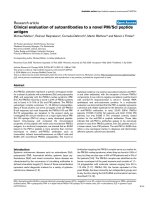
Figure 1 The chemical structures of compounds in water and methanol extracts of Antrodia camphor ata: 1, cytosine; 2, uracil; 3,
guanine; 4, cytidine; 5, uridine; 6, adenine; 7, inosine; 8, guanosine; 9, adenosine; 10,2’-deoxyadenosine; 11, camphorataimide C; 12, 3-isobutyl-
4-[4-(3-methyl-2-butenyloxy)phenyl]-1H-pyrrole-2,5-dione; 13, ergosterol.
Zhao and Leung Chinese Medicine 2010, 5:4
/>Page 3 of 6
phosphoric acid in deionized water, pH 2.19 and metha-
nol was tested. In addition, a neutral aqueous mobile
phase of deionized water and methanol was a lso tested.
The use of neutral aqueous mobile phase showed more
peaks but at the expense of peak shape and symmetry.
Methanol is the best choice of organic components to
facilitate elution of ergosterol, which is only compatible
with solvents of lower polarity.
Method validation
To verify column performance and appropriateness of the
chromatographic conditions, the number of theoretical
plates, selectivity, resolution and peak symmetry va lues
were determined as the indicators of separation efficiency.
Resolution values were all higher than 1.5, which indicates
good separation. Six replicate injections of a sample solu-
tion were performed to assess the precision of the metha-
nol. The relative standard deviation (RSD) of relative
retention time and relative peak area were less than 0.64%
and 4.07%, respectively. Another six independently pre-
pared samples were assessed for the repeatability of the
method. The RSD of relative retention time and relative
peak area were 0.77% and 6.89%, respectively. The sample
stability was determined by three repetitive injections of a
sample solution after three days of storage at room tem-
perature. The RSD of relative retention time and relative
peak area were 0.67% and 7.45%, respectively.
Qualitative chromatographic fingerprint
The full profile of nucleosides and nucleobases was initi-
ally identified by matching the retention times and UV
absorption profiles with respect to standards and was
confirmed using ESI-MS. In total, ten compounds were
identified in the water extract of mycelia AC. However,
adenine, cytosine, and cytidine were not found when
assessed using this new chromatographic co ndition,
likely because their solubilities in the aqueous compo-
nent of the mobile phase render poor column retention.
Due to the bulky structures of these compounds (Figure
1), a specific extraction solvent and mobile phase were
required for their coextraction and elution along with
other compounds in the fingerprint. Our repeated trials
for an optimal extraction showed that 100% methanol is
the only choice capable of coextraction and elution. A
gradient with 100% methanol was therefore adopted. In
this way, different chemical compounds of vari ous pola-
rities are presented within the same chromatographic
window despite the total elution time of all compounds
lasting 120 minutes. Figure 3 shows the chromato-
graphic fingerprint of methanol extract of mycelial AC.
Preliminary application of mycelial AC chromatographic
fingerprint
Two over-the-counter products that claimed consi sted
of mycelial AC were purchased from the Taiwanese
market for our preliminary quality assessment. Figure 4
shows the superimposed chromatograms of methanol
extract of the two commercial products in comparison
to our reference fingerprint. The two commercial myce-
lial products possess very similar fingerprints, but these
fingerprints are distinctively different from our estab-
lished reference fingerprint of mycelial AC. From our
morphological observation and confirmed by micro-
scopic authentication during the species authentication
stage, the powder in capsules are likely dried extracts
rather than crude herbal material. The presence of addi-
tional possible herbal components other than those
declared in the product package may also explain the
difference in their derived fingerprints.
Moreover, the chemical compositions of mycelia and
fruiting bodies have never been compared. The use of
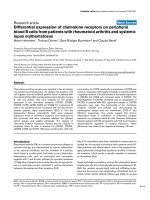
Figure 2 The HPLC-DAD chemical profile of the water extract o f mycelial Antrodia camphorata: 1,cytosine;2, uracil; 3, guanine; 4,
cytidine; 5, uridine; 6, adenine; 7, inosine; 8, guanosine; 9 , adenosine; 10,2’-deoxyadenosine.
Zhao and Leung Chinese Medicine 2010, 5:4
/>Page 4 of 6
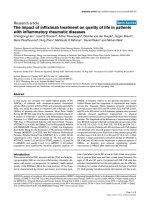
Figure 3 The established HPLC-DAD fingerprint of methanol extract of mycelial Antrodia camphorata: 2, uracil; 3, guanine; 5, uridine; 7,
inosine; 8, guanosine; 9, adenosine; 10,2’-deoxyadenosine; 11, camphorataimide C; 12, 3-isobutyl-4-[4-(3-methyl-2-butenyloxy)phenyl]-1H-pyrrole-
2,5-dione; 13, ergosterol.
Figure 4 The superimposed HPLC-DAD chromatograms of methanol extracts of two commercial mycelial products: crude mycelium
and crude fruiting body of Antrodia camphorata. For the sake of clarity, numbering of compounds is not shown.
Zhao and Leung Chinese Medicine 2010, 5:4
/>Page 5 of 6
our chromatographic fingerprinting tech nique allowed a
comparison of their chemical constituents. The finger-
print of the fruiting body part is distinctively different
from that of the mycelium (Figure 4), suggesting there
are different characteristic chemicals. In literatures, it
suggested that the fruiting body is mainly compose d of
triterpenoids [29]. Therefor e, specific reference chroma-
tographic fingerprints should be used for independent
quality control of the fruiting part of AC.
Conclusions
This study provides the first chromatographic finger-
print to assess the quality of mycelial AC.
Acknowledgements
The authors would like to thank for the financial support of Faculty Research
Grant [FRG/08-09/II-46] of the Hong Kong Baptist University. The generous
donation of crude mycelial and fruiting bodies of Antrodia camphorata for
the present study from GeneFerm Biotechnology Co. Ltd. of Taiwan is
gratefully acknowledged.
Authors’ contributions
Both authors took part in writing this manuscript. SSZ did the literatures
review and all the experimental works. KSYL supervised on the project,
advised and revised the manuscript. All authors read and approved the final
version of the manuscript.
Competing interests
The authors declare that they have no competing interests.
Received: 30 October 2009
Accepted: 29 January 2010 Published: 29 January 2010
References
1. Hu O, Lian ZF, Zhang JY, Lu X: A review of the medicinal and health-care
value: development and utilization of Antrodia camphorata. Subtrop Plant
Sci 2006, 4:77-80.
2. Lee IH, Huang RL, Chen CT, Chen HC, Hsu WC, Lu MK: Antrodia
camphorata polysaccharides exhibit anti-hepatitis B virus effects. FEMS
Microbiol Lett 2002, 209:63-67.
3. Wu SH, Ryvarden L, Chang TT: Antrodia camphorata ("niu-chang-chih”),
new combination of a medicinal fungus in Taiwan. Bot Bull Acad Sin
1997, 38:273-275.
4. Zang M, Su CH: Ganoderma comphoratum, a new taxon in genus
ganoderma from Taiwan, PR China. Acta Bot Yunnanica 1990,
12(4):395-396.
5. Chang TT, Chou WN: Antrodia cinnamomea sp. nov. on Cinnamomum
kanehirai in Taiwan. Mycol Res 1995, 99(6):756-758.
6. Nakamura N, Hirakawa A, Gao JJ, Kakuda H, Shiro M, Komatsu Y, Sheu CC,
Hattori M: Five new maleic and succinic acid derivatives from the
mycelium of Antrodia camphorata and their cytotoxic effects on LLC
tumor cell line. J Nat Prod 2004, 67(1):46-48.
7. Chen CC, Liu YW, Ker YB, Wu YY, Lai EY, Chyau CC, Hseu TH, Peng RY:
Chemical characterization and anti-inflammatory effect of
polysaccharides fractionated from submerge-cultured Antrodia
camphorata mycelia. J Agric Food Chem 2007, 55(13):5007-5012.
8. Mau JL, Huang PN, Huang SJ, Chen CC: Antioxidant properties of
methanolic extracts from two kinds of Antrodia camphorata mycelia.
Food Chem 2004, 86(1):25-31.
9. Huang NK, Cheng JJ, Lai WL, Lu MK: Antrodia camphorata prevents rat
pheochromocytoma cells from serum deprivation-induced apoptosis.
FEMS Microbiol Lett 2005, 244(1):213-219.
10. Lu MK, Cheng JJ, Lai WL, Lin YR, Huang NK: Adenosine as an active
component of Antrodia cinnamomea that prevents rat PC12 cells from
serum deprivation-induced apoptosis through the activation of
adenosine A
2A
receptors. Life Sci 2006, 79(3):252-258.
11. Lu MK, Cheng JJ, Lai WL, Lin YJ, Huang NK: Fermented Antrodia
cinnamomea extract protects rat PC12 cells from serum deprivation-
induced apoptosis: the role of the MAPK family. J Agric Food Chem 2008,
56(3):865-874.
12. Cheng JJ, Huang NK, Chang TT, Wang DL, Lu MK: Study for anti-
angiogenic activities of polysaccharides isolated from Antrodia
cinnamomea in endothelial cells. Life Sci 2005, 76(26):3029-3042.
13. Ao ZH, Xu ZH, Lu ZM, Xu HY, Zhang XM, Dou WF: Niuchangchih (Antrodia
camphorata) and its potential in treating liver diseases. J Ethnopharmacol
2008, 121(2):194-212.
14. Chang HL, Chao GR, Chen CC, Mau JL: Non-volatile taste components of
Agaricus blazei, Antrodia camphorata and Cordyceps militaris mycelia.
Food Chem 2001, 74(2):203-207.
15. Yue YY, Song AR, Tian XM, Wang F, Xu K: Composition analysis of amino
acids in mycelia of Taiwanofungus formosanus. J Fungal Res 2006,
4(2):45-48.
16. Cheng JJ, Yang CJ, Cheng CH, Wang YT, Huang NK, Lu MK:
Characterization and functional study of Antrodia camphorata
lipopolysaccharide. J Agric Food Chem 2005, 53(2):469-474.
17. Wang GJ, Tseng HW, Chou CJ, Tsai TH, Chen CT, Lu MK: The vasorelaxation
of Antrodia camphorata mycelia: involvement of endothelial Ca
2+
-NO-
cGMP pathway. Life Sci 2003, 73(21):2769-2783.
18. Chang CY, Lue MY, Pan TM: Determination of adenosine, cordycepin and
ergosterol contents in cultivated Antrodia camphorata by HPLC method.
J Food Drug Anal 2005, 13(4):338-342.
19. Han HF, Hirakawa A, Zuo F, Nakamura N, Hattori M: Quantitative
determination of maleic and succinic acid derivatives in the mycelium
of Antrodia cinnamomea. J Trad Med 2006, 23(1):19-23.
20. Wu MD, Cheng MJ, Wang BC, Yech YJ, Lai JT, Kuo YH, Yuan GF, Chen IS:
Maleimide and maleic anhydride derivatives from the mycelia of
Antrodia cinnamomea and their nitric oxide inhibitory activities in
macrophages. J Nat Prod 2008, 71(7):1258-1261.
21. Chen JJ, Lin WJ, Liao CH, Shieh PC: Anti-inflammatory benzenoids from
Antrodia camphorata. J Nat Prod 2007, 70(6):989-992.
22. Huang SJ, Mau JL: Antioxidant properties of methanolic extracts from
Antrodia camphorata with various doses of g-irradiation. Food Chem
2007, 105(4):1702-1710.
23. Chen CC, Shiao YJ, Lin RD, Shao YY, Lai MN, Lin CC, Ng LT, Kuo YH:
Neuroprotective diterpenes from the fruiting body of Antrodia
camphorata. J Nat Prod 2006, 69(4):689-691.
24. Hsu FL, Chou CJ, Chang YC, Chang TT, Lu MK: Promotion of hyphal
growth and underlying chemical changes in Antrodia camphorata by
host factors from Cinnamomum camphora. Int J Food Microbiol 2006,
106(1):32-38.
25. Chen SC, Lu MK, Cheng JJ, Wang DL: Antiangiogenic activities of
polysaccharides isolated from medicinal fungi. FEMS Microbiol Lett 2005,
249(2):247-254.
26. Gao JL, Leung KSY, Wang YT, Lai CM, Li SP, Hu LF, Lu GH, Jiang ZH, Yu ZL:
Qualitative and quantitative analyses of nucleosides and nucleobases in
Ganoderma spp. by HPLC-DAD-MS. J Pharm Biomed Anal 2007,
44(3):807-811.
27. Fan H, Li SP, Xiang JJ, Lai CM, Yang FQ, Gao JL, Wang YT: Qualitative and
quantitative determination of nucleosides, bases and their analogues in
natural and cultured Cordyceps by pressurized liquid extraction and high
performance liquid chromatography-electrospray ionization tandem
mass spectrometry (HPLC-ESI-MS/MS). Anal Chim Acta 2006,
567(2):218-228.
28. Xie PS, Leung AY: Understanding the traditional aspect of Chinese
medicine in order to achieve meaningful quality control of Chinese
materia medica. J Chromatogr A 2009,
1216(11):1933-1940.
29. Shen CC, Kuo YC, Huang RL, Lin LC, Don MJ, Chang TT, Chou CJ: New
ergostane and lanostane from Antrodia camphorata. J Chin Med 2003,
14(4):247-258.
doi:10.1186/1749-8546-5-4
Cite this article as: Zhao and Leung: Quality evaluation of mycelial
Antrodia camphorata using high-performance liquid chromatography
(HPLC) coupled with diode array detector and mass spectrometry
(DAD-MS). Chinese Medicine 2010 5:4.
Zhao and Leung Chinese Medicine 2010, 5:4
/>Page 6 of 6