Báo cáo y học: "Expression of aromatase and estrogen receptor alpha in chondrosarcoma, but no beneficial effect of inhibiting estrogen signaling both in vitro and in vivo" ppt
Bạn đang xem bản rút gọn của tài liệu. Xem và tải ngay bản đầy đủ của tài liệu tại đây (2.42 MB, 9 trang )
RESEARC H Open Access
Expression of aromatase and estrogen receptor
alpha in chondrosarcoma, but no beneficial effect
of inhibiting estrogen signaling both in vitro and
in vivo
Danielle Meijer
1
, Hans Gelderblom
2
, Marcel Karperien
3
, Anne-Marie Cleton-Jansen
1
, Pancras CW Hogendoorn
1
and
Judith VMG Bovée
1*
Abstract
Background: Chondrosarcomas are malignant cartilage-forming tumors which are highly resistant to conventional
chemotherapy and radiotherapy. Estrogen signaling is known to play an important role in proliferation and
differentiation of chondrocytes and in growth plate regulation at puberty. Our experiments focus on unraveling the
role of estrogen signaling in the regulation of neoplastic cartilage growth and on interference with estrogen
signaling in chondrosarcomas in vitro and in vivo.
Methods: We investig ated the protein expression of estrogen receptor alpha (ESR1), androgen receptor (AR), and
aromatase in tumor specimens of various chondrosarcoma subtypes, and (primary) chondrosarcoma cultures. Dose-
response curves were generated of conventional central chondrosarcoma cell lines cultured in the presence of
17b-estradiol, dihydrotestosterone, 4-androste ne-3,17 dione, 4-hydroxytamoxifen, fulvestrant and aromatase
inhibitors. In a pilot series, the effect of anastrozole (n = 4) or exemestane (n = 2) treatment in 6 chondrosarcoma
patients with progressive disease was explored.
Results: We showed protein expression of ESR1 and aromatase in a large majority of all subtypes. Only a minority
of the tumors showed few AR positive cells. The dose-resp onse assays showed no effect of any of the compounds
on proliferation of conventional chondrosarcoma in vitro. The me dian progression-free survival of the patients
treated wi th aromatase inhibitors did not significantly deviate from untreated patients.
Conclusions: The presence of ESR1 and aromatase in chondrosarcoma tumors and primary cultures supports a
possible role of estrogen signaling in chondrosarcoma proliferation. However, our in vitro and pilot in vivo studies
have shown no effect of estrogen-signaling inhibition on tumor growth.
Background
Chondrosarcomas of bone are malignant cartilage- form-
ing tumors whic h are hig hly resistant to conventiona l
chemotherapy and radiotherapy [1,2]. However, recently
various promising targets were discovered and the
exploration of suitable therapies continues [3,4]. Con-
ventional chondrosarcomas represent about 90% of all
chondrosarcomas. Most conventional chondrosarcomas
are located in the medullar cavity of the bone and are
called central chondrosarcoma. About 15% of conven-
tional chondrosarcomas arise from the surface of bone
and are designated as peripheral chondrosarcomas. Con-
ventional chondrosarcomas often show local destructive
growth and the high-grade tumors commonly metasta-
size [5].
Besides conventional chondrosarcoma, several rare
chondrosarcoma subtypes are defined, together consti-
tuting 10-15% of all chondrosarcomas. Dedifferentiated
chondrosarcoma (10%) is a tumor containing a high-
grade dedifferentiated non-cartilaginous sarcoma next to
* Correspondence:
1
Department of Pathology, Leiden University Medical Center, Leiden, The
Netherlands
Full list of author information is available at the end of the article
Meijer et al. Clinical Sarcoma Research 2011, 1:5
/>CLINICAL SARCOMA RESEARC
H
© 2011 Meijer et al; licensee BioMed Central Ltd . This is an Open Access article dis tributed under the term s of the Creative Commons
Attribution License ( which permits u nrestricted use, distribu tion, and reproduction in
any medium, provided the original work is properl y cited.
a usually low-grade malignant well-differentiated carti-
lage-forming tumor, with a sharply defined junction
between the two components. It bears a poor prognosis
and no targets for therapy have been reported so far [6].
Mesenchymal chondrosarcoma (2%) is a highly malig-
nant lesion occurring in the bone and soft tissue of rela-
tively young patients. The tumor consists of
differentiated cartilage mixed with undifferentiated small
round cells and usually follows an aggressive course
with a high rate of distant metastases, and a 5-year over-
all survival of 55% [7]. Clear cell chondrosarcoma (2%)
is a low-grade malignant tumor, which rarely metasta-
sizes, but commonly recurs after curettage. About 15%
of the patients die as a result of the dise ase [8]. The
lack of efficacious treatment for all different subtypes of
chondrosarcomas emphasizes the need to identify new
treatment strategies.
One of the potential target s for therapy is the estro-
gen-signaling pathway. Mutations in ESR1 and
CYP19A1, the gene for aromatase, demonstr ated an
important role for estrogen in the proliferation and dif-
ferentiation of cho ndrocytes in the epiphyseal growth
plate [9]. Estrogen induces the pubertal growth spurt,
and at the end of puberty growth plate fusion [10].
Furthermore, osteochondromas, the benign precursors
of peripheral chondrosarcomas, stop growing at the end
of puberty, suggesting an inhibitory effect of estrogens
on these tumors. In addition, ESR1 and ESR2 expression
has been shown to be a common phenomenon in chon-
drosarcomas [11,12]. In a previous study, our group also
demonstrated functional activity of the estrogen-produ-
cing enzyme aromatase in chondrosarcoma cells in vitro
[11]. These results indicated that the ESR signaling
pathway might be a potent ial target for en docrine treat-
ment of metastatic or irresectable chondrosarcoma.
For already three decades endocrine therapy plays a
crucial r ole in the treatment of women with hormone-
responsive breast cancer. Breast cancer and chondrosar-
comas were found to occur relatively frequently in the
same patient. A population-based study by Odink et al.
implicated a 7.62 times increased risk for the same
female patient to have both breast cancer and a cartila-
ginous tumor [13]. The mean age of onset in p atients
with breast ca ncer as the first tumor and chondrosar-
coma as a second tumor is nearly 10 years earlier than
breast cancer in general [13]. The se observations may
suggest a genetic trait. Remarkably, the expression of
ESR1 was significantly higher in breast cancer associated
with chondrosarcoma [14].
The two strategies used for endocrine treatment are
blockade of ESR1 using selective estrogen receptor mod-
ulators/downregulators like tamoxifen and fulvestrant,
and deprivation of estrogen production by inhibiting
aromatase with anastrozole, letrozole, and exemestane.
In our ab ove-mentioned study, we showed that the aro-
matase activity and proliferation of chondrosarcoma
cells slightly decreased after addition of the aromatase
inhibitor exemestane [11]. In our present study, we
focused on further unraveling the role of estrogen in the
regulation of neoplastic cartilage growth in a l arger
cohort of various chondrosarcoma subtypes, including
conventional central and peripheral chondrosarcoma as
well as dedifferentiated, mesenchymal, and clear cell
chondrosarcoma. Moreover, using a larger set of drugs
targeting the estrogen-signaling pathway we investigated
whether interference with estrogen signaling could inhi-
bit chondrosarcoma growth. We aimed to validate and
expand our previous in vitro data by measuring the
effects of estrogens, androgens, tamoxifen, fulvestrant,
and aromatase inhibitors o n the proliferation of various
chondrosarcoma cell cultures. Furthermore, we explored
the ef ficacy of aromatase inhibitors in a set of patients
with metastatic or locally advanced chondrosarcoma.
Methods
Tumor tissue
All specimens in this study were handled according to
the ethical guidelines described in “ Code for Proper
SecondaryUseofHumanTissueinTheNetherlands”
of the Dutch Federation of Medical Scientific Societies.
Conventional central and peripheral chondrosarcoma,
and the rare subtypes dedifferentiated, mesenchymal,
and clear cell chondrosar coma were selected b ased on
accepted clinicopathological and radiological criteria
[15]. In total, formalin-fixed paraffin-embedded (FFPE)
specimens from 175 patients, including the 6 patients
in our pilot study, were collected from the archives of
the D epartment of Pathology, LUMC, The Netherlands
(n = 100), Nuffield Department of Orthopaedic Sur-
gery, University of Oxford, UK (n = 7), Institute of
Orthopaedics and Musculoskeletal Science, UCL, UK
(n = 22), Laboratory of Oncologic Research, ROI, Italy
(n = 30), Department of Pathology, RH, Denmark (n =
9), Department of Pathology, Medizinische Universität
Graz, Austria (n = 7). All were primary tumors except
for three clear cell chondrosarcomas and six mesench-
ymal chondrosarcomas, from which only recurrences
were available. Clinicopathological data are shown in
table 1. Histological grading was perf ormed according
to Evans [5].
Tissue microarray (TMA) construction
Of the rar e chondrosarcoma subtypes we constructed
TMAs using a TMA Master (3DHISTECH Ltd, Buda-
pest, Hungary). TMAs contained 2 mm cores of each
sample, in triplic ate. From the dedifferentiated chondro-
sarcomas we included both the well differentiat ed and
the dedifferentiated components. The clinical detail s are
Meijer et al. Clinical Sarcoma Research 2011, 1:5
/>Page 2 of 9
outlined in Table 1 . Normal non-decalcified liver, kid-
ney, and tonsi l samples were included on the TMAs for
orientation purposes and as internal positive controls.
Immunohistochemistry (IHC)
Details of the primary antibodies used for immunohisto-
chemistry are described in Table 2. As negative controls,
slides were incubated in PBS/BSA 1% without primary
specific antibodies. AR and aromatase immunohisto-
chemical stainings of the patient material were semi-
quantitatively scored for nuclear and cytoplasmic staining
respectively. Scores were given for intensity (0 = absent, 1
= weak, 2 = moderate, 3 = strong) and for the percentage
of positive cells (0 = 0%, 1 = 1-24%, 2 = 25-49%, 3 = 50-
74%, and 4 = 7 5%-100%). To avoid tumors with single
positive cells being regarded as positive, a cut-off level o f
atotalsum≥4 was applied. ESR1 was scored for nuclear
staining, with positivity defined as ≥10% (weakly) positive
cells, according to standard clinical procedures for scor-
ing ESR1-positive breast cancer [16,17]. Scoring wa s per-
formed by two independent observers without knowledge
of the clinicopathological data. For dedifferentiated chon-
drosarcoma, the well differentia ted and the dedifferen-
tiated component were scored separately. Likewise, for
mesenchymal chondrosarcoma both the cartilaginous
areas and the small cell component were evaluated.
Cell cultures and conditions
Chondrosarcoma cell lines SW1353 (ATCC, Manassas,
VA), OUMS27 [18], CH2879 [19], and JJ012 [20], and
breast cancer cell line ZR-75-1 [21] were cultured in
RPMI 1640 supplemented with 10% heat-inactivated fetal
bovine serum (FBS) (Gibco, Invitrogen Life-Technologies,
Scotland, UK) (Table 3). ZR-75-1 cultures wer e addition-
ally supplemented with 1 nmol/L 17b-estradiol (E
2
)
(Sigma). Two chondrosarcoma primary cultures, L835 and
L869, were generated as described previously [11] and
were cultured in collagen I-coated culture flasks in RPMI
1640 supplemented with 20% heat-inac tivated fe tal cal f
serum (Invitrogen), 2% penic illin/strep tomycin (MP Bio-
medicals), and 1% glutamax (Invitrogen) (Table 3). Cells
were grown at 37°C in a humidified incubator with 95%
air and 5% CO
2
. The primary chondros arcoma cultures
expressed mRNA of at least two of the cartilaginous mar-
kers co llagen 2, collagen 10, aggecan or SOX9 [11]. In
addition, karyotyping of L835 and L869 by COBRA-FISH
showed an aberrant number of chromosomes, thereby
confirming their tumorigenic origin.
Protein detection in chondrosarcoma cell cultures
Four T75 culture flasks of 4 chondrosarcoma cell lines
(SW1353, CH2879, OUMS27, JJ012) and 2 primary
chondrosarcoma cell cultures (L869 and L835), and a
positive control (ZR-75-1) were trypsinized and washed
twice with cold PBS. Cells were formalin-fixed over
night and subsequently embedded in paraffin. Using
IHC, we determined ESR1 protein expression, as
described above.
Proliferation assays
To monitor the effects of the estrogen signaling pathway
on chondrosar coma cell proliferation we performed var-
ious experiments. An overview of all different conditions
tested is given in Table 4. For the WST-1 assays with
steroids and inhibitors, SW1353, CH2879, OUMS27,
JJ012, L869, and L835 cells were seeded into collagen I-
Table 1 Clinicopathological data of the 175 formalin-fixed paraffin-embedded cartilaginous tumors
EC CS OC PCS DDCS CCS MCS
Total number of tumors 3 46 10 28 42 23 23
Grade I - 16 - 12 - - -
Grade II - 18 - 13 - - -
Grade III - 12 - 3 - - -
Male 1 22 6 17 21 17 8
Female 2 23 4 10 21 6 15
Enchondromatosis/MO 0 1 5 10 - - -
Median age yrs (range) 38 (37-50) 51 (20-79) 14.5 (6-24) 38 (16-61) 66 (26-85) 43 (20-79) 29.5 (15-70)
Abbreviations: enchondroma (EC), conventional central and peripheral chondrosarcoma (CS and PCS), osteochondroma (OC), dedifferentiated chondrosarcoma
(DDCS), clear cell chondrosarcoma (CCS), and mesenchymal chondrosarcoma (MCS).
Table 2 Procedures and details of the primary antibodies used for immunohistochemistry
Protein Origin Number Dilution Species Antigen retrieval Blocking Positive control
ESR1 Invitrogen/zymed 18-0174Z 1:200 Rabbit Tris-EDTA 30’ 5% ELK milk breast cancer
aromatase Abcam Ab18995 1:300 Rabbit Citrate 30’ 5% ELK milk placenta
AR Dako AR441 1:200 Mouse Tris-EDTA - cervix stroma
Meijer et al. Clinical Sarcoma Research 2011, 1:5
/>Page 3 of 9
coated 96 wells plates (BD Biosciences) at a density of
1500 cells per well for the S W1353 and J J012 cell lines
and 5000 cells per well for the other cultures. The cells
were plated in phenol red-free RPMI 1640 medium
(Invitrogen) supplemented with 10% heat-inactivated
charcoal-stripped FBS (Invitrogen). After 24 hours, serial
dilutions of the st eroids 17b-estradiol (Sigma), 4-andros-
tene-3,17-dione (Sigma) and dihydrotestosterone (F luka
Analytical) (100 pM-1 μM), anti-steroids 4-hydroxyta-
moxifen (Sigma) and fulvestrant (Sigma) (1 nM-10 μM),
aromatase inhibitors anastrozole, letrozole and exemes-
tane (1 nM-10 μM) or combinations were added. The
compounds were solved in ethanol. As vehicle control,
ethanol was added with concentrations never exceeding
0.1%. All concentrations were test ed at least in quadru-
plicate in a total volume of 100 μl. After 3 days, 10 μl
proliferation reagent WST-1 (Roche Diagnostics) were
added t o each well, and the cells wer e returned to the
incubator for three hours. Absorbance was measured at
450 nm with a Victor
3
Multilabel Counter 1420-042
(Perkin Elmer, MA, USA). Values were corrected for
background, averaged and normalized to the vehicle-
control cultures. FBS dependence was tested likewise for
SW1353, CH2879, and OUMS27. For cell counting
experiments, 24000 cells were seeded in a 24 wells plate.
The experimental set up was identical to the prolifera-
tion assays with a total volume o f 1 ml. Cells were
counted after 3 and 7 days of treatme nt according to
experiments previously published by our group [3,11].
Patients
Five patients with grade II or III conventional chondro-
sarcoma a nd one patient wit h dedifferentiated
chondrosarcoma were treated with the aromatase inhibi-
tors anastrozole 1 mg once daily (4 patients, including
the p atient with dedifferentiated chondrosarcoma) and
exemestane 25 mg once daily (2 patients). Median age
was 44 years (range 33-68); 3 patients had metastatic
disease and 3 had locally advanced tumors. Tumor mea-
surements and response evaluations were performed
according to RECIST [22]. From the patients with con-
ventional chondrosarcoma, FFPE tumor specimens were
stained for ESR1 and aromatase protein.
Results
Expression of ESR1, aromatase, and AR in FFPE
chondrosarcoma tumor specimens
Results of ESR1, aromatase, and AR immunohistochem-
ical stainings on 175 FFPE tumor specimens are shown
in Table 5. Expression for ESR1 and aromatase was
detected in the majority of all subtypes (Table 5 and
Figure 1). In conventional central and peripheral chon-
drosarcom a we observed immunoreactivity against ESR1
in 81% (34 out of 42) and 81% (21 out of 26) of the
tumors, respectively. We observed ESR1 in 73% of the
well differentiated and in 84% of the dedifferentiated
component of dedifferentiated cho ndrosarcoma. Positive
stai ning of the two components was strongly correlated.
In mesenchymal chondrosarcomas, 67% of the small cell
components were positive, versus 33 % of the cartilagi-
nous areas. Only a few strongly AR positive cells were
detected in a m inority of the chondrosarcomas of var-
iou s subtypes. Aromatas e protein, the enzyme responsi-
blefortheconversionofandrostenedioneand
androgens to estrogens, was expressed in 86% (38 out of
44) and 93% (25 out of 27) of the central and peripheral
chondrosarcomas respec tively. Almost all well differen-
tiated (97%) and dedifferentiated (89%) components of
dedifferentiated chondrosarco ma were positive for aro-
matase. Of the cartilaginous area of mesenchymal chon-
drosarcoma 77% showed aromatase positivity versus
52% of the small cell component. In central chondrosar-
coma no correlation with histological gr ade was
observed with any of the proteins. In peripheral chon-
drosarcoma only 33% of the grade III tumors s howed
Table 3 Chondrosarcoma cultures
Sample Type Grade Gender Age Passage
1 SW1353 Cell line II F 72 18
2 CH2879 Cell line III F 35 31
3 OUMS27 Cell line III M Na 22
4 JJ012 Cell line II M 39 10
5 L835 Primary culture III M 55 15
6 L869 Primary culture II M 52 18
Table 4 Experimental conditions tested
Test Experimental conditions
Steroids E2 (Fig 2A), ASD, DHT (100 pM-1 μM)
Inhibitors of estrogen signaling OHT, Fulvestrant, Anastrozole, Letrozole, Exemestane (1 nM-10 μM)
Steroids combined with inhibitors E2 (1 nM) with OHT or Fulvestrant (1 nM-10 μM) (Fig. 2B)
FBS 1%, 5%, 10% FBS alone and combined with E2 or ASD (100 pM-1 μM)
Timepoints of measurements 3 days, 7 days
Methods of measurement WST1 viability assay, cell counting
Abbreviations: 17b-estradiol (E2), 4-androstene-3,17-dione, (ASD), dihydrotestosteron (DHT), 4- hydroxytamoxifen (OHT ), Fetal bovine serum (FBS).
Meijer et al. Clinical Sarcoma Research 2011, 1:5
/>Page 4 of 9
ESR1 expression. H owever, only three tumors were
included in this group.
Estrogen responsiveness of central chondrosarcoma in
vitro
SW1353, CH2879, OUMS27, JJ012, L869 and L835 wer e
positive for ESR1 protein staining (Figure 1K, 1L and
data not shown). Therefore, we investigated the effect of
ESR-signaling modulation on the proliferation of chon-
drosarcoma cells in vitro by measuring the effect of ster-
oids and cl inical drugs inhibiting estrogen-signaling. We
evaluated responsiveness of 4 central chondrosarcoma
cell lines and 2 primary cultures to 3 different steroids
(17b-estradiol, the estrogen precursor androstenedione,
and the non-aromatizable androgen dihydroxytestoster-
one). The proliferation of the cells was not significantly
influenced by any of these factors in the chondrosar-
coma cell lines and primary cultures, whereas a clear
response was observed in the proliferation rate of the
ZR-75-1 breast-cancer cell line, which was used as a
positive control (Figure 2Aand d ata not shown). W e
also tested three aromatase inhibitors (anastrozole, letro-
zole, and exemestane), and two estrogen-receptor
antagonists/downregulators (4-hydroxytamoxifen a nd
fulvestrant) and again no effect was shown in the
chondrosarcoma cell lines and cultures under the differ-
ent c onditions described in Table 4 (Figure 2Band data
not shown).
Clinical results
In a pilot series, 6 consecutive patients with locally
advanced or metastatic grade II or I II conventional or
dediffer entiated chondrosarcoma, for whom no st andard
treatment was available, were treated with aromatase
inhibitors after informed consent. All t umors were radi-
ologically progressive in the 6 months before initiat ion
of therapy. All five conventional chondrosarcomas
expressed ESR1 and aromatase protein, supporting the
rationale of the treatm ent. The median prog ression-free
survival was 5 months (range 4-10 months) in the con-
ventional chondro sarcoma patient s and 2 months in the
patient with a metastatic dedifferentiated chondrosar-
coma, which did not significantly deviate from untreated
patients.
Discussion
Chondrosarcoma of bone is a malignant cartilage-form-
ing tumor of which distinct clinical and histological sub-
types are recognized. So far, f or locally advanc ed and
metastatic chondrosarcoma no treatment options are
available. Previous studies have demonstrated the pre-
sence of the ESR1 and activity of aromatase in conven-
tional chondrosarcoma [11,12]. Furthermore, in 2005,
our gro up showed an effect of estrogens and the aroma-
tase inhibitor exemestane on the proliferation of chon-
drosarcoma cells in vitro, indicating that
chondrosarcomas might be susceptible to hormonal
therapy. In that study, ESR1 and CYP19A1 mRNA
expression were demonstrated in a set of 23 conven-
tional chondrosarcomas and 7 (primary) chondrosar-
coma cultures. ESR1 protein expression was
demonstrated in all 23 tumors tested. Addition of 17b-
estradiol, 4-androstene-3,17-dione, and exemestane
showed subtle effects on the proliferation of 2 cell cul-
tures containing ESR1 and aromatase. After addition of
4-androstene-3,17-dione, an increase in proliferation
was demonstrated. Proliferation was 131% of normal
proliferation which decreased to 105% after inhibition
with exemestane. A cell line lacking ESR1 and CYP19A1
did not show any response.
In the current study, we aimed to gain more insight
into the possibility of treating chondrosarcoma patients
with hormonal therapy by further investigating the
expression of the hormone receptors ESR1 and AR, and
of aromatase, the enzyme that mediates the last step in
the biochemical formatio n of estrogen, in a larger set of
conventional chondrosarcomas a s well as three rare
chondrosarcoma subtypes. In conventional chondrosar-
coma, we furthermore monitored the effect of estrogen,
Table 5 Immunohistochemical staining of 175 FFPE
samples of chondrosarcoma patients
ESR1 aromatase AR*
Enchondroma 2/2 100% 3/3 100% 0/2 0%
Central CS 34/42 81% 38/44 86% 6/41 15%
grade I 11/15 73% 13/15 87% 1/13 8%
grade II 14/16 88% 15/17 88% 3/16 19%
grade III 9/11 82% 10/12 83% 2/12 17%
Osteochondroma 5/8 63% 6/8 75% 1/7 14%
Peripheral CS 21/26 81% 25/27 93% 4/28 14%
grade I 9/11 82% 11/12 92% 2/12 17%
grade II 12/13 92% 12/13 92% 1/13 8%
grade III 1/3 33% 3/3 100% 1/3 33%
Dedifferentiated CS
well differentiated component 18/25 72% 31/32 97% 1/27 4%
dedifferentiated component 30/35 86% 34/38 89% 2/37 5%
Clear cell CS 15/22 69% 15/22 69% 0/22 0%
Mesenchymal CS
small cell component 15/23 65% 12/23 52% 1/23 4%
cartilaginous areas 5/15 33% 10/13 77% 0/12 0%
* In positive samples only few strongly positive cells were observed.
Meijer et al. Clinical Sarcoma Research 2011, 1:5
/>Page 5 of 9
the estrogen precurso r androstenedione and the non-
aromatizable androgen dihydrotestosteron, and various
known estrogen signaling-inhibiting drugs on the pro-
gression of chondrosarcoma cells in vitro.
We demonstrated expression of ESR1 in a large pro-
portion of various types of cartilaginous tumors. In con-
ventional central and peripheral chondrosarcoma we
observed immunoreactivity against ESR1 in 81 % (34 out
of 42) and 81% (21 out of 26) of the tumors, respec-
tively. These results confirm and extend 2 previous stu-
dies in which the authors demonstrated nuclear
expression of ESR1 in subsets of 23 [11] and 31 [12]
conventional chondrosarcomas. Grifone et al. [12] sug-
gested a decrease or loss in ESR1 expression in the
higher grade o r dedifferentiated chondrosarcomas. We
observed such a trend in the peripheral chondrosarco-
mas,whereonly33%ofthehighgradetumorsshow
positive staining for ESR1. However, this group included
only three tu mor specimens. In central chondrosarcoma
no correlation with grade was observed. Aromatase pro-
tein, the enzyme responsible for the conversion of
androstenedione and androgens to estrogens, was
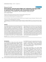
Figure 1 Immunohistochemical staining of ESR1 and aromatase in various chondrosarcoma subtypes and cell lines.Nuclearprotein
expression of ESR1 in well-differentiated (A) and dedifferentiated (B) components of dedifferentiated chondrosarcoma, clear cell chondrosarcoma
(C), and mesenchymal chondrosarcoma (D). Cytoplasmic protein expression of aromatase in well-differentiated (E) and dedifferentiated (F)
components of dedifferentiated chondrosarcoma, clear cell chondrosarcoma (G), and mesenchymal chondrosarcoma (H), and aromatase and
ESR1 protein expression in conventional chondrosarcoma (I and J, respectively). ESR1 protein expression in the JJ012 and CH2879
chondrosarcoma cell lines (K and L). Magnification 200×.
Meijer et al. Clinical Sarcoma Research 2011, 1:5
/>Page 6 of 9
expressed in 86% and 93% of the central and peripheral
chondrosarcomas respectively, suggesting that tumors
are capable of metabolizing estrogens from precursors.
AR is another important target for hormonal therapy in
for example prostate cancer. As androstenedione is a
steroid precursor for estrogens as well as androgens w e
also investigated the possibility of AR involvement in
chondrosarcoma proliferation. However, AR nuclear
protein expression was observed only in a small number
of cases with very few positive cells.
Besides convention al chondro sarcoma, several rare
chondrosarcoma subtypes are defined. Despite aggressive
therapy, approximately 90% of the pa tients with dediffer-
entiated chondrosarcoma die with distant metast asis,
within 2 years after diagnosis of the disease [6,23]. The
low-grade component and the highly malignant compo-
nent display ESR1 protein expression in 72% and 86% of
the samples respectively. Aromatase was observed in 97%
and 89%, suggesting the presence of estrogens.
Mesenchymal chondrosarcomas are usually very
aggressive with a strong tendency of local recurrence
and distant metastases. Patients have a 5-year overall
survival o f 55% [7]. Although mesenchymal chondrosar-
coma of bone is gene rally considered to lack sex predi-
lection [24], Fanburg-Smith et al. [25] suggested a
female predominance and raised the possibility of
hormonal influence in the pathogenesis of this tumor.
However, all their mesenchymal chondrosarcoma cases
were ESR1 negative. In our study, in 65% (15 out of 23)
of the mesenchymal chondrosarcomas the small cell
component was positive for ESR1, while in 33% ( 5 out
of 15) of the tumors also the cartilaginous areas were
positive. Moreover, aromatase expression was observed
in the small cells of 52% (12 out of 23) of the tumors,
whereas the cartilage component demonstrated aroma-
tase expression in 77% (10 out of 13). This might indi-
cate that these tumors do have an active estrogen
signaling pathway, which might be targetable by anties-
trogens or aromatase inhibitors. Discrepant results may
be explained by differences in ESR1 antibody and anti-
gen retrieval protocols.
Clear cell chondrosarcoma is a low-grade variant of
chondrosarcoma, which rarely metastasizes, but has a
recurrence rate of 86% after curettage. About 15% of the
patients die as a result of the disease [8]. We have
observed ESR1 expression and aromatase expression
each in 69% of the clear cell chondrosarcomas, suggest-
ing th at also these chondrosarcoma pa tients potentially
might benef it from a ntiestrogen therapy and/or aroma-
tase inhibition.
In vitro cell models to further study the effect of
estrogen signaling on chondrosarcoma are available for
conventiona l central chondrosarcoma only. No stimul a-
tion of proliferation of central chondrosarcoma cells was
observed after addition of the non-aromatizable andro-
gen dihydrotestosterone. This suggests no significant
role for AR signali ng in chondrosarcoma proliferation,
which i s consistent with the fact that very few tumors
express AR.
In addition, in spite of positive immunohistochemical
staining for ESR1 protein in all in vitro cell cultures, addi-
tion of 17b-estradiol, 4-androstene-3,17-dione or drugs
targeting the estrogen-signaling pathway did not have a
significant effect on the prolifer ation of the conventional
central chondrosarcoma cell cultures. These results con-
trad ict our results published in 2005, where proliferation
was stimulated by 1 7b-estradiol and 4-androstene-3,17-
dione, and inhibited by exemestane [11]. Although we
included an identical experimental set up, cell culture con-
ditions are never 100% identical. For example, each batch
of FBS contains different amounts of growth factors and
other components which might influence experimental
outcome. Also cell characteristics might have changed
over time, resulting in pass ages insensitive to (anti)estro-
gens and aromatase inhibitors, as has been described
before for certain breast cancer cell lines [26-28].
Breast cancer cell line ZR-75-1 is known to be com-
pletely dependent on estrog ens for i ts proliferation, and
proliferation can be fully inhibited by abrogating the
estrogen-signaling pathway [29]. Although we previously
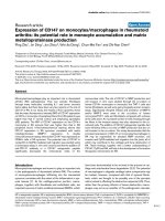
Figure 2 Cell viability assays measuring the effect of estrogen
and antiestrogens in chondrosarcoma cell lines. A) Unlike breast
cancer cell line ZR-75-1, chondrosarcoma cell lines (SW1353,
CH2879, OUMS27, and JJ012) and primary cultures (L869 and L835)
did not respond to E2 with increased proliferation; B) also none of
these cultures responded to 4-hydroxytamoxifen (OHT), and
fulvestrant (Fulv) in the presence of 1 nM E2. Only L835, which is
representative, is shown.
Meijer et al. Clinical Sarcoma Research 2011, 1:5
/>Page 7 of 9
demonstrated an effect of estrogen-signaling on chon-
drosarcoma cell proliferation, as compared to estrogen-
dependent breast cancer cell line ZR-75-1 the effec ts in
chondrosarcoma, if present, were very subtle. As a posi-
tive control, ZR-75-1 showed a 179% increase of prolif-
eration up on addition of 1 nM 17b-estradiol, confirming
a functional e xperimental setup, versus a previously
demonstrated 55% increase in chond rosarcoma prolif-
eration [11] and no significant increase in the current
study. Both studies clearly indicate that, in contrast to
estrogen-depende nt breast cancer, chondrosarcoma pro-
liferation is not fully dependent on estrogens.
Besides investigating estrogen dependence, we tested
aromatase inhibitors which block estrogen production,
and t he effects of tamoxifen and fulvestrant which abro-
gate estrogen receptor function [30,31]. In the estrogen-
dependent ZR-75-1 breast-cancer cell line proliferation
was completely i nhibited upon addition of tamoxifen
and fulvestrant (Figure 2B). However, in the chondrosar-
coma ce ll cultures, estrogen-signaling inhibition c aused
no effects on cell proliferation, suggesting that the
mechanism driving proliferation in chondrosarcoma is
diff erent from the m echanism active in estrogen-depen-
dent breast cancer. In chondro sarcoma, effect s of estro-
gen are much more subtle and likely depend on the
tissue culture conditions u sed, resulting in either mar-
ginal effects (in our previous study) or no effects at all.
In addition, the median time to progression in the
clinical series was five months both before and after
treatment. Therefore, we can conclude that aromatase
inhibition was not effective in five c onventional chon-
drosarcoma patients, nor in a patient with dedifferen-
tiated chondrosarcoma. Although a formal prospective
phase II trial would have bee n more suitable to prove
(in)efficacy of this concept, we were not able to gain
industry support without stronger preclinical data.
Sinceourstudyislimitedtotheeffectsofestrogen
signaling on conventional central chondrosar coma only,
no conclusions can be drawn about the effects of estro-
gen signaling in the other chondrosarcoma subtypes.
However, although we demonstrated the presence of
aromatase and ESR1 in a maj ority of various chondro-
sarcoma subtypes, our in vitro data on conventional
chondrosarcoma and our patient tri al including one
dedifferentiated chondrosarcoma patient suggest that
effects of estrogen-signaling inhibition in other chondro-
sarcoma subtypes, if present at all, will be very small
and that estrogen-signaling inhibition is unlikely to play
a major role in chondrosarcoma management.
Conclusions
In su mmary, we demonstrated the presence of the co m-
ponents involved in estrogen signali ng in a large major-
ity of chondrosarcomas. However, we could not
demonstrate a significant effect of estrogen or inhibitors
of estrogen signaling on cell proliferation and viability in
vitro using central chondrosarcoma cell lines and p ri-
mary cultures. Despite the previously presented and cur-
rently confirmed b iological rationale, our in vitro and
pilo t clinical data suggest that an active estrogen-signal-
ing pathway might just not play a pivotal role in the
development and progression of conventional chondro-
sarcoma an d do not support the further development of
therapeutic strategies including inhibition of estrogen
signaling in chondrosarcoma.
Acknowledgements and funding
We thank B.E. van den Akker, J.J. Baelde, R. Vossen, M.A.J.H. van Ruler, S.
Romeo, I.H. Briaire -de Bruin, and K.G. van der Ham for excellent technical
assistance, J. Oosting for help with data analysis, and T. Krenács for expert
assistance in TMA construction. N. Athanasou, A.M. Flanagan, P. Picci, S.
Daugaard, B. Liegl-Atzwanger, A. Leithner, and the Institute of Orthopaedics,
UCL at the Royal National Orthopaedic Hospital are acknowledged for
providing tumor tissues and clinical data. M. Namba, T. Kalinski, J.A. Block, J.
Trapman, and L.C.J. Dorssers are thanked for providing cell lines OUMS27,
C3842, JJ012, LNCaP, and ZR-75-1, respectively. DM and all experiments were
funded by the Dutch Cancer Society, project no UL 2007-3815 and the
EuroBoNeT consortium [018814], a European Commission granted Network
of Excellence for studying the pathology and genetics of bone tumors. HG,
AMCJ and PCWH were funded by the LUMC, and JVMGB was funded by the
Netherlands Organization for Scientific Research (917-76-315).
Author details
1
Department of Pathology, Leiden University Medical Center, Leiden, The
Netherlands.
2
Department of Clinical Oncology, Leiden University Medical
Center, Leiden, The Netherlands.
3
Department of Tissue Regeneration, MIRA,
Institute for Biomedical Technology and Technical Medicine, University of
Twente, Enschede, The Netherlands.
Authors’ contributions
DM carried out the experiments and drafted the manuscript. HG carried out
the pilot patient study. MK, AMCJ and PCWH participated in the design of
the study, the interpretation of data, and revision of the manuscript. JVMGB
conceived of the study, and participated in its design and coordination, and
helped to draft the manuscript. All authors read and approved the final
manuscript.
Competing interests
The authors declare that they have no competing interests.
Received: 8 February 2011 Accepted: 25 July 2011
Published: 25 July 2011
References
1. Gelderblom H, Hogendoorn PCW, Dijkstra SD, van Rijswijk CS, Krol AD,
Taminiau AH, et al: The clinical approach towards chondrosarcoma.
Oncologist 2008, 13:320-329.
2. Bovee JV, Hogendoorn PC, Wunder JS, Alman BA: Cartilage tumours and
bone development: molecular pathology and possible therapeutic
targets. Nat Rev Cancer 2010, 10:481-488.
3. Schrage YM, Machado I, Meijer D, Briaire-de B, van den Akker BE,
Taminiau AH, et al: COX-2 expression in chondrosarcoma: a role for
celecoxib treatment? Eur J Cancer 2010, 46:616-624.
4. Schrage YM, Briaire-de Bruijn IH, de Miranda NF, van OJ, Taminiau AH,
van WT, et al: Kinome profiling of chondrosarcoma reveals SRC-pathway
activity and dasatinib as option for treatment. Cancer Res 2009,
69:6216-6222.
5. Evans HL, Ayala AG, Romsdahl MM: Prognostic factors in chondrosarcoma
of bone. A clinicopathologic analysis with emphasis on histologic
grading. Cancer 1977, 40:818-831.
Meijer et al. Clinical Sarcoma Research 2011, 1:5
/>Page 8 of 9
6. Milchgrub S, Hogendoorn PCW: Dedifferentiated chondrosarcoma. In
World health organization classification of tumours. Pathology and genetics.
Tumours of soft tissue and bone Edited by: Fletcher C.D.M., Unni KK, Mertens
F 2002, 252-254.
7. Nakashima Y, Park YK, Sugano O: Mesenchymal chondrosarcoma. In World
health organization classification of tumours. In Pathology and genetics.
Tumours of soft tissue and bone Edited by: Fletcher C.D.M., Unni KK, Mertens
F 2002, 255-256.
8. McCarthy EF, Freemont A, Hogendoorn PCW: Clear cell chondrosarcoma.
In World health organization classification of tumours. Pathology and genetics.
Tumours of soft tissue and bone Edited by: Fletcher C.D.M., Unni KK, Mertens
F 2002, 257-258.
9. Grumbach MM: Estrogen, bone, growth and sex: a sea change in
conventional wisdom. J Pediatr Endocrinol Metab 2000, 13(Suppl
6):1439-1455.
10. Van der Eerden BCJ, Karperien M, Wit JM: The estrogen receptor in the
growth plate: implications for pubertal growth. J Pediatr Endocrinol Metab
2001, 14(Suppl 6):1527-1533.
11. Cleton-Jansen AM, van Beerendonk HM, Baelde HJ, Bovée JVMG,
Karperien M, Hogendoorn PCW: Estrogen signaling is active in
cartilaginous tumors: implications for antiestrogen therapy as treatment
option of metastasized or irresectable chondrosarcoma. Clin Cancer Res
2005, 11:8028-8035.
12. Grifone TJ, Haupt HM, Podolski V, Brooks JJ: Immunohistochemical
expression of estrogen receptors in chondrosarcomas and
enchondromas. Int J Surg Pathol 2008, 16:31-37.
13. Odink AE, van Asperen CJ, Vandenbroucke JP, Cleton-Jansen AM,
Hogendoorn PCW: An association between cartilaginous tumours and
breast cancer in the national pathology registration in The Netherlands
points towards a possible genetic trait. J Pathol 2001, 193:190-192.
14. Cleton-Jansen AM, Timmerman MC, Van de Vijver MJ, van Asperen CJ,
Kroon HM, Eilers PH, et al: A distinct phenotype characterizes tumors
from a putative genetic trait involving chondrosarcoma and breast
cancer occurring in the same patient. Lab Invest 2004, 84:191-202.
15. Bertoni F, Bacchini P, Hogendoorn PCW: Chondrosarcoma. In World Health
Organisation classification of tumours. Pathology and genetics of tumours of
soft tissue and bone. Edited by: Fletcher CDM, Unni KK, Mertens F. Lyon:
IARC Press; 2002:247-251.
16. Allred DC, Harvey JM, Berardo M, Clark GM: Prognostic and predictive
factors in breast cancer by immunohistochemical analysis. Mod Pathol
1998, 11:155-168.
17. Harvey JM, Clark GM, Osborne CK, Allred DC: Estrogen receptor status by
immunohistochemistry is superior to the ligand-binding assay for
predicting response to adjuvant endocrine therapy in breast cancer. J
Clin Oncol 1999, 17:1474-1481.
18. Kunisada T, Miyazaki M, Mihara K, Gao C, Kawai A, Inoue H, et al: A new
human chondrosarcoma cell line (OUMS-27) that maintains
chondrocytic differentiation. Int J Cancer 1998, 77
:854-859.
19. Gil-Benso R, Lopez-Gines C, Lopez-Guerrero JA, Carda C, Callaghan RC,
Navarro S, et al: Establishment and characterization of a continuous
human chondrosarcoma cell line, ch-2879: comparative histologic and
genetic studies with its tumor of origin. Lab Invest 2003, 83:877-887.
20. Jagasia AA, Block JA, Qureshi A, Diaz MO, Nobori T, Gitelis S, et al:
Chromosome 9 related aberrations and deletions of the CDKN2 and
MTS2 putative tumor suppressor genes in human chondrosarcomas.
Cancer Lett 1996, 105:91-103.
21. Dorssers LC, van AT, Dekker A, van Agthoven TL, Kok EM: Induction of
antiestrogen resistance in human breast cancer cells by random
insertional mutagenesis using defective retroviruses: identification of
bcar-1, a common integration site. Mol Endocrinol 1993, 7:870-878.
22. Therasse P, Arbuck SG, Eisenhauer EA, Wanders J, Kaplan RS, Rubinstein L,
et al: New guidelines to evaluate the response to treatment in solid
tumors. In J Natl Cancer Inst. Volume 92. European Organization for
Research and Treatment of Cancer, National Cancer Institute of the United
States, National Cancer Institute of Canada; 2000:205-216.
23. Bruns J, Fiedler W, Werner M, Delling G: Dedifferentiated
chondrosarcoma–a fatal disease. J Cancer Res Clin Oncol 2005,
131:333-339.
24. Kulyk WM, Franklin JL, Hoffman LM: Sox9 expression during
chondrogenesis in micromass cultures of embryonic limb mesenchyme.
Exp Cell Res 2000, 255 :327-332.
25. Fanburg-Smith JC, Auerbach A, Marwaha JS, Wang Z, Santi M, Judkins AR,
et al: Immunoprofile of mesenchymal chondrosarcoma: aberrant desmin
and EMA expression, retention of INI1, and negative estrogen receptor
in 22 female-predominant central nervous system and musculoskeletal
cases. Ann Diagn Pathol 2010, 14:8-14.
26. Fasco MJ, Amin A, Pentecost BT, Yang Y, Gierthy JF: Phenotypic changes in
MCF-7 cells during prolonged exposure to tamoxifen. Mol Cell Endocrinol
2003, 206:33-47.
27. Katzenellenbogen BS, Kendra KL, Norman MJ, Berthois Y: Proliferation,
hormonal responsiveness, and estrogen receptor content of MCF-7
human breast cancer cells grown in the short-term and long-term
absence of estrogens. Cancer Res 1987, 47:4355-4360.
28. Reddel RR, Alexander IE, Koga M, Shine J, Sutherland RL: Genetic instability
and the development of steroid hormone insensitivity in cultured T 47D
human breast cancer cells. Cancer Res 1988, 48:4340-4347.
29. van Agthoven T, van Agthoven TL, Portengen H, Foekens JA, Dorssers LC:
Ectopic expression of epidermal growth factor receptors induces
hormone independence in ZR-75-1 human breast cancer cells. Cancer
Res 1992, 52:5082-5088.
30. Howell A: Pure oestrogen antagonists for the treatment of advanced
breast cancer. Endocr Relat Cancer 2006, 13:689-706.
31. Lewis JS, Jordan VC: Selective estrogen receptor modulators (SERMs):
mechanisms of anticarcinogenesis and drug resistance.
Mutat Res 2005,
591:247-263.
doi:10.1186/2045-3329-1-5
Cite this article as: Meijer et al.: Expression of aromatase and estrogen
receptor alpha in chondrosarcoma, but no beneficial effect of inhibiting
estrogen signaling both in vitro and in vivo. Clinical Sarcoma Research
2011 1:5.
Submit your next manuscript to BioMed Central
and take full advantage of:
• Convenient online submission
• Thorough peer review
• No space constraints or color figure charges
• Immediate publication on acceptance
• Inclusion in PubMed, CAS, Scopus and Google Scholar
• Research which is freely available for redistribution
Submit your manuscript at
www.biomedcentral.com/submit
Meijer et al. Clinical Sarcoma Research 2011, 1:5
/>Page 9 of 9