Báo cáo y học: "Local recurrence and assessment of sentinel lymph node biopsy in deep soft tissue leiomyosarcoma of the extremities" pdf
Bạn đang xem bản rút gọn của tài liệu. Xem và tải ngay bản đầy đủ của tài liệu tại đây (623.99 KB, 5 trang )
RESEARCH Open Access
Local recurrence and assessment of sentinel
lymph node biopsy in deep soft tissue
leiomyosarcoma of the extremities
Michael J Lamyman, Henk P Giele, Paul Critchley, Duncan Whitwell, Max Gibbons and Nicholas A Athanasou
*
Abstract
Background: Leiomyosarcoma of deep soft tissues of the extremities is a rare malignant tumour treated primarily
by surgery. The incidence of local recurrence and lymph node metastasis is uncertain and it is not known whether
a sentinel lymph node biopsy is indicated in these tumo urs.
Methods: A retrospective review of pat ients treated for extremity deep soft tissue leiomyosarcoma at our
institution over a 10-year period was conducted. Patients developing local recurrence or lymph node metastasis
were identified. The presence or absence of lymphatics in the primary tumours was assessed by
immunohistochemical expression of LYVE-1 and podoplanin.
Results: 27 patients (mean age 62 years) were included in the study. 15 were female and 12 male. Lymph nod e
metastasis was seen in only two cases (7%); intratumoural lymphatics were identified in the primary tumours of
both these cases. Local recurrence occurred in 25.9% of cases despite complete excision and post-operative
radiotherapy; the mean time to recurrence was 10.1 months.
Conclusion: On the basis of this study, we do not advocate sentinel lymph node biopsy in this group of patients
except in those cases in which intratumoural lymphatics can be demonstrated. Close follow up is important
especially for high grade leiomyosarcomas, particularly in the first year, as these tumours have a high incidence of
local recurrence.
Introduction
Leiomyosarcoma of soft tissues is a malignant tumour
composed of tumour cells that exhibit smooth muscle
differentiation. Leiomyosarcomas are generally thought
to account for 5-10% of soft tissue sarcomas [1-3].
These tumours arise most commonly in the retroperito-
neum but can develop in any location; in one study of
75 soft t issue leiomyosarcomas, 33% were noted to arise
in extremity soft tissues\The behaviour of leiomyosar-
coma of extremity deep soft tissues has not been studied
independently of those arising in other locations.
Regional lymph node meta stasis in patients with soft
tissue sarcomas is an infrequent event occurring in 2.6 -
5% of all patients [4-6]. Sentinel lymph node biopsy
(SLNB) has been employed for staging of soft tissue
sarcomas, particularly epithelial sarcoma [7-9]. The inci-
dence of lymph node metastasis in extremity leiomyo-
sarcomas is cl early important with regard to whether
SLNB should be carried out for this tumour. In previous
retrospective reviews of the literature, pooling data from
published reports on regional lymph node involvement,
Weingrad and R osenberg [10] and Mazeron and Suit
[11] found the incidence in leiomyosarc oma was 10.6%
and 4% respective ly; in the prospective study of Fong et
al [5], the incidence was reported to be 2.7%. These stu-
dies, however, did not distinguish leiomyosarcoma of
extremity deep soft tissues from those arising in other
locations; this is an important factor as leiomyosarcoma
occ urs more commonly in the retroperitoneum, mese n-
tery, abdominal and pelvic viscera than in extremity soft
tissues and lymph node metastasis from sarcomas of
visceral origin occurs less commonly than from sarco-
mas arising in extremity soft tissues [5]. T he recurrence
rate following excision of deep soft tissue extremity
* Correspondence:
Nuffield Department of Orthopaedics, Rheumatology and Musculoskeltal,
Sciences, University of Oxford, Department of Pathology, Nuffield
Orthopaedic Centre, Oxford, OX3 7LD, UK
Lamyman et al. Clinical Sarcoma Research 2011, 1:7
/>CLINICAL SARCOMA RESEARC
H
© 2011 Lamyman et al; licensee BioMed Central Ltd. This is an Open Access article distributed under the terms of the Creative
Commons Attribution License ( which permits unrestricted use, distribution, and
reproduction in any medium, provided the ori ginal work is properly cited.
leiomyosarcomas is also unknown; this has not been
assessed i ndependently of recurrence of superficial
(cutaneous) leiomyosarcomas, which have a favourable
prognosis, or of retroperitoneal tumours, which have a
poor prognosis.
Theaimofthisstudyhasbeentodeterminethe
recurrence rate and i ncidence of lymph node metastasis
of deep soft tissue leiomyosarcomas of the extremities.
As the presence of lymphatic vessels h as been noted in
malignant soft tissue tumours that metastasise to lymph
nodes [12], we determined whether immunohistochem-
ical identification of lymphatics in the primary tumour
coul d provide a guide as to whether lymph node metas-
tasis of extremity leiomyosarcoma occurred and thus,
whether a SLNB might be indicated in such cases.
Patients and methods
A search of the pathology database detected all patients
with a histological diagnosis of deep soft tissue leiomyo-
sarcoma over a 10 year period, between 1998 and 2008.
Only patients diagnosed and treated at the Nuffield
Orthopaedic Centre wit h leiomyosarcoma of the extre-
mities were entered into the study. Patients with superfi-
cial cutaneous soft tissue leiomyosarcomas or
gynaecological, retroperitoneal, intra-abdominal or
intrathoracic primary tumours were excluded. A case
notes review was performed.
Local recurrence and lymph node m etastasis was con-
sidered to have occurred only i f proven though open
biopsy. The histological diagnosis of leiomyosarcoma
was based on morphological and immunohistochemical
criteria detailed in the WHO classification of soft tissue
tumours [1]. Immunohistochemical expression of at
least two smooth muscle antigens (smooth muscle actin,
desmin, h-caldesmon) was seen in all cases. Identifica-
tion of lymphatics was carried out using anti-Lyve -1
and anti-podoplanin antibodies as previously described
[12].
Results
35 patien ts were iden tified as eligible for entry into the
study. Five patients had to be excluded as either the
case notes could not be found or were incomplete. Two
patients were excluded bec ause they died following their
biopsy but before definitive surgery, and one patient was
excluded because metastatic disease was found on pre-
sentation. The case notes of the remaining 27 pa tients
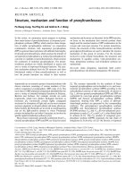
were reviewed. Fifteen were fe male and twelve male
(Figure 1). The mean age at presentation was 62 years.
The mean follow up was 19.9 months, median 15
months (range 4 to 59 months). The sites of the primary
tumour are shown in Figure 2. The size, grade and stage
of the tumours are shown in Table 1. In all cases, local
excision of the tumours was performed aiming for
complete clearance with as wide a margin as possible.
21 of the patients (78%) received adjuvant radiotherapy
following primary excision. Details of patients develop-
ing local recurrence are shown in Table 2 and of those
developing lymph node metastasis in Table 3. Lymph
node metastasis occurred in two patients (7%).
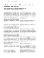
A review of the pathology of the primary tumour in
these two c ases showed that both tumours contained
intratumoural lymphatics, as assessed by endothelial cell
expression of the lymphatic markers, podoplanin and
LYVE-1. (Figure 3) The remaining tumours, which did
not metasta sise to lymph nodes , were negative for lym-
phatic markers. In one patient the nodal recurrence was
extensive, encasing femoral vessels and was not resect-
able. In the second patient an inguinal and iliac lymph
node dissection was performed. In this patient the
lymph node metastasis occurred early, before radiother-
apy was instituted.
Local recurrence occurred in seven patients (25.9%).
The mean time from surgical excision to recurrence was
10.1 months (range 3-24 months). There was no inci-
dence of local recurrence or lymph node metastasis in
patients with low grade leiomyosarcoma. Post-operative
radiotherapy was received by all patients who subse-
quently pre sented with local recurrence. In six of these
seven patients, the tumour had been excised with a
clear margin. In one patient the excision was described
as marg inal. In all but one case the recurrence was trea-
ted by further surgical resection.
Discussion
The role of SLNB in the management of soft tissue sar-
coma has yet to be defined [8,9,13]. In our institutio n it
is current practice to undertake SLNB in patients with
epithelioid sarcoma given the relatively high rate of
lymph node metastasis in these tumours. Previous stu-
dies have reported that the in cidence of lymph node
metastasis in such tumours is between 16.7 and 80%
[5,10,11]. A positive SLNB in these cases is followed b y
a formal lymph node dissection. A number of soft tissue
sarcomas, such as rha bdomyosarcoma, clear cell sar-
coma and sy novial sarcoma, have also been shown to
have a propensit y for regional lymph node metastasis
and some observers have suggested that SLNB may be
of prognostic benefit in these tumours [9]. Previous esti-
mates of the incidence of lymph node metastasis in all
patients with leiomyosarcoma have been between 2 .7
and 10.6%.
[510H]
These studies exam ined the metastatic
rate of leiomyosarcomas arising at several different sites
collectively and not just that of leiomyosarcomas of
deep soft tissues of the extremities. In the present study
we found that the r ate of lymph node metastasis in
extremity deep soft tissue leiomyosarcomas to be 7%.
Lamyman et al. Clinical Sarcoma Research 2011, 1:7
/>Page 2 of 5
In patients with intermediate thickness melanoma,
SLNB has become widely accepted as a minimally inva-
sive method of staging the regional lymph nodes [14,15].
When SLNB is performed in these patients, 20% w ill be
found to have micrometastasis. However when SLNB is
performed in thin melano mas, with a Breslow thickness
less than I mm, the micrometa stasis rate falls to 5%
[16]. Current AJCC guidelines do not recommend rou-
tine use of SLNB in this group [17,18], and on this basis
the comparable rate of lymph node metastasis in deep
soft tissue leiomyosarcomas would not appear to justify
the extra morbidity (eg extra operating time, potential
wound problems) associated with undertaking SLNB.
Recent work at our institution has shown that soft tis-
sue sarcomas with a high propensity to met astasise to
lymph nodes contain intratumoural lymphatics [12];
intratumoural lymphatics were found to be present in
all epithelioid sarcomas and a number of other sarcomas
including leiomyosarcoma. Lymph node metastasis has
been reported in up to 80% of epithelioid sarcomas
[5,10,11]. The lower incidence of lymph node metastasis
in leiomyosarcomas may reflect the fact that intratu-
moural lymphatics are found less commonly in these
tumours. It is none the less signifi cant that in our study
the two leiomyoarcomas which did metastasise to regio-
nal lymph nodes both contained intratumoural lympha-
tics. Immunohistochemical demonstration of lymphatic
vessels in these primary leiomysarcomas was of prognos-
tic significance with regard to the development of lymph
node metastasis, and it could be argued that SLNB is
indicat ed in primary leiomyosarcomas o f the extremities
where intratumoural lymphatics are identified.
We foun d a high rate of local recurrence in extremity
deep soft tissue leiomyosarcoma patients with 25.9%
experiencing recurrence despite adequate resection and
adjuvant radiotherapy. Mankin and Hornicek report a
recurrence rate of 10.8% in 65 patients with leiomyosar-
coma [19]. Again this study did not differentiate
between leiomyosarcoma of the e xtremities and other
Figure 1 Age and sex distribution of cases.
Figure 2 Sites of primary tumour with number and approximate percentage of cases.
Lamyman et al. Clinical Sarcoma Research 2011, 1:7
/>Page 3 of 5
Table 1 Size, Grade and Stage of the primary
leiomyosarcoma
Case Size in maximum Diameter (cm) Grade MSTS Stage
118 2 2b
28 3 2b
35 3 2b
4 Unknown 2 2b
5 7.5 1 lb
6 4.5 2 lb
710 3 2b
S7 3 2b
94 2 2b
10 8 3 2b
11 13 3 2b
12 14 3 2b
13 2 2 2b
14 3.8 1 lb
15 12 2 2b
16 25 2 2b
17 12 2 2b
18 3.5 2 2b
19 8.5 3 2b
20 10 3 2b
21 16 3 2b
22 9 1 lb
23 13 1 lb
24 9 3 3
25 Unknown 2 2b
26 10 3 2b
27 8 3 2b
MSTS - Musculoskeletal Tumour Society
Table 2 Patients with local recurrence following primary excision
Age Sex Site Max Diam eter (cm) Grade MSTS
Stage
Time to first
recurrenc e
(Months)
Number of recurrences Margins Adjuvant Radio therapy
55 F Arm 5 3 2b 3 2 Clear Yes
77 F Calf Unknown 2 2b 24 2 Clear Yes
77 M Buttock 8 3 2b 3 4 Clear Yes
67 F Thigh 13 3 2b 9 2 Clear Yes
67 M Buttock 12 2 2b 4 2 Clear Yes
55 F Calf 9 3 2b 5 1 Marginal Yes
76 M Forearm unknown 2 2b 19 1 Clear Yes
Table 3 Patients with lymph node metastasis
Age Sex Site Max Diameter
(cm)
Grade MSTS Stage Margins Aduvant Radiotherapy Time to detection of lymph node metastasis
(months)
79 F Thigh 8.5 3 2b Clear Yes 21
53 M Thigh 8 3 2b Clear No 3
Figure 3 Intratumoural lymphatic vessels in a primary
leiomyosarcoma, showing podoplanin expression by lymphatic
endothelial cells.
Lamyman et al. Clinical Sarcoma Research 2011, 1:7
/>Page 4 of 5
sites. The findings of the present study indicate that
deep soft tissue leiomyosarcoma of the extremities, in
contrast to leiomyosarcoma arising at other sites has a
greater propensity to local recurrence. Such recurrences
are difficult to treat and surgical resection of an already
irradiated area remains the only option.
Conclusion
This study has shown that patients with leiomyosarcoma
of deep soft tissues of the extremities have a rate of
lymph node metastasis of 7% and a local recurrence rate
of 25.9% despit e adequate excision and post-operat ive
radiotherapy. On the basis of this study, we do not
advocate the use of SLNB to this group of patients
except in cases where lymphatics can be demonstrated
in the primary tumour. Our findings emphasise the
importance of close follow up, especially for high grade
leiomyosarcomas, particularly in the first year post sur-
gery, as there is a high incidence of local recurrence.
Authors’ contributions
HG, PC, MG and DW contributed to the design of the study. HG, MG and PC
conducted the study. NA carried out pathological studies and MJL, HPG, MG
and NA wrote the paper. All authors have read and approved the final
manuscript.
Competing interests
The authors declare that they have no competing interests.
Received: 11 February 2011 Accepted: 1 August 2011
Published: 1 August 2011
References
1. Evans HL, Shipley J: Leiomyosarcoma. In Pathology and genetics of tumours
of soft tissue and bone. Edited by: Fletcher DCM, Unni KK, Mertens F. Lyon,
IARC Press; 2002:131-134.
2. Weiss SW, Goldblum JR: Leiomyosarcoma. Enzinger and Weiss’s Soft Tissue
Tumours. 5 edition. St. Louis Mosby; 2008, 545-564.
3. Hashimoto H, Daimaru Y, Tsuneyoshi M, Enjoji M: Leiomyosarcoma of the
external soft tissue. Cancer 1986, 57:2077-2088.
4. Sondak VK, Chang AE: Clinical evaluation and treatment of soft tissue
sarcomas. In Enzinger and Weiss’s soft tissue tumours Fourth edition. Edited
by: Weiss SW, Goldblum JR. St Louis Mosby; 2001:21-44.
5. Fong Y, Coit DG, Woodruff JM, et al: Lymph node metastasis from soft
tissue sarcoma in Adults: Analysis of data from a prospective database
of 1772 sarcoma patients. Ann Surg 1993, 217:72-77.
6. Behranwala KA, A’Hern R, Al-Muderis O, et al: Prognosis of lymph node
metastasis in soft tissue sarcoma. Ann Surgical Oncol 2004, 11:714-719.
7. Gow KW, Rapkin LB, Olson TA, et al: Sentinel lymph node biopsy in the
pediatric population. J Pediatr Surg 2008, 43:2193-8.
8. Kayton ML, Delgado R, busam K, et al: Experience with 31 sentinel lymph
node biopsies for sarcomas and carcinomas in pediatric patients. Cancer
2008, 1;112:2052-9.
9. Maduekwe UN, Hornicek FJ, Springfield DS, et al: Role of sentinel lymph
node biopsy in the staging of synovial, epithelioid, and clear cell
sarcomas. Ann Surg Oncol 2009, 16:1356063.
10. 10 DN, Weingrad DN, Rosenberg SA: Early lymphatic spread of osteogenic
and soft-tissue sarcomas. Surgery 1978, 84:231-240.
11. Mazeron JJ, Suit HD: Lymph nodes as sites of metastasis from sarcomas
of soft tissue. Cancer 1987, 60:1800-1808.
12. Mahendra G, Kliskey K, et al: Intratumoural lymphatics in benign and
malignant soft tissue tumours. Virchows Archiv Pathol Anat 2008,
453:457-464.
13. Blazer DG, Sabel MS, Sondak VK: Is there a role for sentinel lymph node
biopsy in the management of sarcoma? Surg Oncol 2003, 12:201-206.
14. Morton DL, Cochran AJ, Thompson JF: The rational for sentinel-node
biopsy in melanoma. Nat Clin Pract Oncol 2008, 5:510-511.
15. McMasters KM, Reintgen DS, Ross MI, et al: Sentinel lymph node biopsy
for melanoma: Controversy despite widespread agreement. J Clin Oncol
2001, 19:2851-2855.
16. Puleo CA, Messina JL, Riker Al, et al: Sentinel node biopsy for thin
melanomas: Which patients should be considered. Cancer Control 2005,
12:230-235.
17. Balch CM, Soong SJ, Gershenwald JE, et al: Prognostic factors analysis of
17,6000 melanoma patients: validation of the American Joint Committee
on Cancer melanoma staging system. J Clin Oncol 2001, 19:3622-3634.
18. Balch CM, Buzzaid AC, Atkins MB, et al: A new American Joint Committee
on Cancer staging system for cutaneous melanoma. Cancer 2000,
88:1484-1491.
19. Mankin HJ, Hornicek FJ: Diagnosis, classification, and management of soft
tissue sarcoma. Cancer Control 2005, 12:5-21.
doi:10.1186/2045-3329-1-7
Cite this article as: Lamyman et al.: Local recurrence and assessment of
sentinel lymph node biopsy in deep soft tissue leiomyosarcoma of the
extremities. Clinical Sarcoma Research 2011 1:7.
Submit your next manuscript to BioMed Central
and take full advantage of:
• Convenient online submission
• Thorough peer review
• No space constraints or color figure charges
• Immediate publication on acceptance
• Inclusion in PubMed, CAS, Scopus and Google Scholar
• Research which is freely available for redistribution
Submit your manuscript at
www.biomedcentral.com/submit
Lamyman et al. Clinical Sarcoma Research 2011, 1:7
/>Page 5 of 5