Báo cáo y học: "Low-dose vasopressin infusion results in increased mortality and cardiac dysfunction following ischemia-reperfusion injury in mice" potx
Bạn đang xem bản rút gọn của tài liệu. Xem và tải ngay bản đầy đủ của tài liệu tại đây (278.44 KB, 8 trang )
Open Access
Available online />Page 1 of 8
(page number not for citation purposes)
Vol 13 No 3
Research
Low-dose vasopressin infusion results in increased mortality and
cardiac dysfunction following ischemia-reperfusion injury in mice
Toonchai Indrambarya, John H Boyd, Yingjin Wang, Melissa McConechy and Keith R Walley
Critical Care Research Laboratories, Heart + Lung Institute, University of British Columbia, 166 – 1081 Burrard Street, Vancouver, British Columbia,
V6Z 1Y6, Canada
Corresponding author: John H Boyd,
Received: 5 Mar 2009 Revisions requested: 31 Mar 2009 Revisions received: 2 Jun 2009 Accepted: 23 Jun 2009 Published: 23 Jun 2009
Critical Care 2009, 13:R98 (doi:10.1186/cc7930)
This article is online at: />© 2009 Indrambarya et al.; licensee BioMed Central Ltd.
This is an open access article distributed under the terms of the Creative Commons Attribution License ( />),
which permits unrestricted use, distribution, and reproduction in any medium, provided the original work is properly cited.
Abstract
Introduction Arginine vasopressin is a vasoactive drug
commonly used in distributive shock states including mixed
shock with a cardiac component. However, the direct effect of
arginine vasopressin on the function of the ischemia/reperfusion
injured heart has not been clearly elucidated.
Methods We measured left ventricular ejection fraction using
trans-thoracic echocardiography in C57B6 mice, both in normal
controls and following ischemia/reperfusion injury induced by a
one hour ligation of the left anterior descending coronary artery.
Mice were treated with one of normal saline, dobutamine (8.33
μg/kg/min), or arginine vasopressin (0.00057 Units/kg/min,
equivalent to 0.04 Units/min in a 70 kg human) delivered by an
intraperitoneal micro-osmotic pump. Arterial blood pressure was
measured using a micromanometer catheter. In addition,
mortality was recorded and cardiac tissues processed for RNA
and protein.
Results Baseline left ventricular ejection fraction was 65.6%
(60 to 72). In normal control mice, there was no difference in left
ventricular ejection fraction according to infusion group.
Following ischemia/reperfusion injury, AVP treatment
significantly reduced day 1 left ventricular ejection fraction
46.2% (34.4 to 52.0), both in comparison with baseline and day
1 saline treated controls 56.9% (42.4 to 60.2). There were no
significant differences in preload (left ventricular end diastolic
volume), afterload (blood pressure) or heart rate to account for
the effect of AVP on left ventricular ejection fraction. The seven-
day mortality rate was highest in the arginine vasopressin group.
Following ischemia/reperfusion injury, we found no change in
cardiac V1 Receptor expression but a 40% decrease in
Oxytocin Receptor expression.
Conclusions Arginine vasopressin infusion significantly
depressed the myocardial function in an ischemia/reperfusion
model and increased mortality in comparison with both saline
and dobutamine treated animals. The use of vasopressin may be
contraindicated in non-vasodilatory shock states associated
with significant cardiac injury.
Introduction
With the increasing medical complexity of the critically ill,
shock due to a combination of vasodilation and cardiac dys-
function is increasingly frequent. Two common clinical exam-
ples of this are first, vasodilation following cardiopulmonary
bypass surgery and, second, the cardiac dysfunction during
septic shock. These mixed shock conditions are routinely
treated with intravenous fluids plus inotropes combined with a
vasopressor such as norepinephrine or arginine vasopressin
(AVP). AVP is a vasopressor commonly used in intensive care
units and cardiac surgical units due to its efficacy in restorat-
ing blood pressure [1-6]. The effects of AVP are mediated via
vasopressin 1 receptors (V1R; predominantly vascular), vaso-
pressin 2 receptors (V2R; predominantly renal), vasopressin 3
receptors (V3R; predominantly central), and the oxytocin
receptors (OTR) [7]. In addition, vasopressin blocks K
ATP
channels [8] and potentiates the effect of adrenergic agents
[9]. Vascular V1R appear to mediate the majority of effects of
vasopressin in reversing vasoplegia and catecholamine toler-
ance [4,10].
ANOVA: analysis of variance; AVP: arginine vasopressin; DOB: dobutamine; I/R: ischemia/reperfusion; LAD: left anterior descending coronary artery;
LVEF: left ventricular ejection fraction; LVEDV: left ventricular end diastolic volume; OTR: oxytocin receptors; P2R: purinergic receptors; RT-PCR:
real-time polymerase chain reaction; SL: normal saline solution; V1R: vasopressin 1 receptor; V2R: vasopressin 2 receptor; V3R: vasopressin 3 recep-
tor.
Critical Care Vol 13 No 3 Indrambarya et al.
Page 2 of 8
(page number not for citation purposes)
In healthy individuals, AVP administration at low doses (<0.04
Units/minute) has little effect on blood pressure. However,
there are multiple reports of increased blood pressure respon-
siveness to low-dose AVP in both septic shock and distributive
shock after cardiopulmonary bypass surgery [7,11]. Conse-
quently, low-dose AVP has been increasingly used to treat
these disorders [1-3,12-17].
Despite its widespread use, there remains considerable
uncertainty regarding its cardiac effects. When studied at the
high doses (0.1 to 1 Unit/minute) previously used for
mesenteric vessel constriction in gastrointestinal bleeding
[18], deleterious effects of AVP on myocardial performance
were reported including coronary vasospasm [19-21]. At
these high doses, AVP may also impair indices of ventricular
contraction and relaxation without overt global ischemia [22].
In addition, the baroreflex mediated via V1R might cause
bradycardia and direct cardiac suppression [23,24]. Although
the most highly expressed vasopressin receptor in the heart is
V1R, the other receptors are physiologically active. Gene
transfer of V2R into failing myocardium increases cardiac con-
tractility [25,26], while OTR mediates a calcium-dependent
vasodilatory response via stimulation of the nitric oxide path-
way in endothelial cells [27]. OTR stimulation also results in
release of atrial natriuretic peptide from the heart [28,29].
Clinically, there are conflicting reports on the effect of AVP on
cardiac function. In some series, AVP infusion has been
reported to decrease cardiac output [28,30,31]. Others have
observed a dramatic restoration of blood pressure without a
decrease in stroke volume or other measures of cardiac func-
tion [2,30,32,33]. The clinical observation that AVP increases
mean arterial pressure in patients with shock is uniform across
these studies, so interpreting any direct effect on myocardial
contractility must be done with caution as alterations in after-
load have a significant impact on measures of cardiac perform-
ance.
The uncertainty as to the in vivo action of AVP on the heart
provides the rationale for this study. Further, as the use of AVP
moves into the mainstream [1,12], it is important to understand
its cardiac effects both on the normal heart and in the injured
or ischemic heart. We chose a model of subacute heart failure
without overt shock as the direct in vivo effects of AVP on con-
tractility are extremely difficult to distinguish from indirect
effects due to changes in afterload (blood pressure). In this
study, we used a mouse model of ischemia/reperfusion (I/R)
induced heart failure to compare the effect of continuous infu-
sion of AVP with saline control (SL) or standard inotropic ther-
apy (dobutamine (DOB)) on cardiac function in mice. We
assessed cardiac function using trans-thoracic echocardiog-
raphy, and in parallel experiments used intra-arterial pressure
measurements to determine whether cardiac function was
influenced by changes in systemic blood pressure.
Materials and methods
These experiments were approved by the UBC Animal Care
Committee and conform to Canadian and National Institutes of
Health guidelines regarding animal experimentation. All exper-
iments were conducted in 10- to 14-week-old male C57B6
mice as a control and in mice following I/R injury induced by
one hour ligation of the left anterior descending coronary
artery (LAD; see below). Intraperitoneal pumps (1 μL/hour for
72 hours, Alzet micro-osmotic pump, Alza Corporation, Palo
Alto, CA, USA) delivered normal saline (SL control), DOB at
8.33 μg/kg/minute, or arginine vasopressin at 0.00057 Units/
kg/minute (equivalent to 0.04 Units/minute in a 70 kg human;
AVP treatment). AVP levels in rodents and humans are similar,
while in rodents the intraperitoneal route of administration for
AVP increases plasma AVP levels in a manner very similar to
intravenous dosing in humans [34,35]. At least five mice per
time point in each group were studied.
Ischemia-reperfusion of the LAD
An open-chest model of I/R using ligation and reperfusion of
the LAD was modified from Michael and colleagues [36]. Mice
were anesthetized using ketamine (75 mg/kg) and xylazine (10
mg/kg) in order to facilitate endotracheal intubation using a 22
Gauge catheter. Thereafter, deep anesthesia was maintained
with 1 to 2% isoflurane. Ventilation was controlled using
Mouse Ventilator (Model 687, Harvard Instruments, Holiston,
MA, USA) with a tidal volume of 0.5 mL and a respiratory rate
of 120 breaths/minute. After a left thoracotomy was performed
at the level of the second or third intercostal space, the LAD
was identified and a 6-0 polypropylene suture was placed
around the LAD. Occlusion of the LAD was accomplished by
pulling the suture ends through a small piece of PE-50 tubing
and occlusion was confirmed by discoloration of the anterior
left ventricle wall.
Following one hour of ischemia the ligature was released to
allow reperfusion, which was visualized. Following the thora-
cotomy wound closure, the intraperitoneal pump (see above)
was implanted into the peritoneal cavity. Intra-operatively, 1 mL
of normal saline was injected subcutaneously for volume
resuscitation and subcutaneous buprenorphrine for pain con-
trol were given. After recovery and resumption of spontaneous
ventilation, mice were extubated.
Myocardial function evaluation
Left ventricular ejection fraction (LVEF) was used to measure
cardiac function at baseline, day 1 and day 3 post I/R. Tran-
sthoracic echocardiography using a Vevo 770 cardiac ultra-
sound (Visualsonics, Toronto, Canada) while anesthetized
with 1 to 2% inhaled isofluorane. Left ventricular internal diam-
eter at end-systole and end-diastole from Short Axis 2D views
at the level of the papillary muscles were identified and used
for measurement of LVEF using the manufacturer's software.
All echocardiographs were performed by the same qualified
Available online />Page 3 of 8
(page number not for citation purposes)
investigator (TI), and quality control was ensured by the other
investigator (JB) blinded from treatment group.
Direct blood pressure measurement
As arterial catheterization is a terminal procedure, separate
mice were anesthetized in the same way and a number 2
French micromanometer catheter (Mikro-tip SPR-838, Millar
Instruments Inc., Houston, TX, USA) was advanced via the
carotid artery into the ascending aorta to measure blood pres-
sure. The heart was excised, frozen in liquid nitrogen, and
stored at -80°C for subsequent study.
Quantitative real-time PCR
Total RNA was extracted from frozen heart samples using Tri-
zol (Invitrogen, Carlsbad, CA, USA) as per the manufacturer's
instructions. RNA was obtained from either I/R injured hearts
or control, non-injured heart. RNA 1 μg was treated with
DNAse I Amplification Grade (Invitrogen, Carlsbad, CA, USA)
and the product underwent quantitiative RT-PCR using M-
MLV RT (Invitrogen, Carlsbad, CA, USA) followed by PCR
amplification with Taq DNA Polymerase (Qiagen, Valencia,
CA, USA). PCR was 40 cycles at 94°C for 15 seconds, 58°C
for 30 seconds, and 72°C for 30 seconds. Primers were as fol-
lows, V1R forward; TCGTCCAGATGTGGTCAGTC, V1R
reverse; AGCTGTTCAAGG-AAGCCAGT, V2R forward;
CCTGGTGTCTACCACGTCTG, V2R reverse; GGTCTCG-
GTCATCCAGTAGC. OTR Forward; AGGAGCTGTTCT-
CAACCATC OTR Reverse; QPCR
TGCAAACCAATCAATAGGCAC. SYBER green was used
as the fluorescence indicator, which represented quantity of
amplicon production with PCR cycle (Ct value). All quantita-
tive RT-PCR reactions were run in triplicate and an average Ct
value was calculated for each PCR condition. Fold change of
Ct value of each sample was calculate using glyceraldehyde-
3-phosphate dehydrogenase (GAPDH) as a background con-
trol.
Western blot for OTR
A 20 μg sample of each protein was mixed with equal volumes
of SDS reducing buffer (62.5 mmol Tris l–1, pH 6.8, 2% (w/v)
SDS, 10% (v/v) glycerol, 100 mmol dithiothreitol l–1, 0.05%
(w/v) bromophenol blue) and incubated in a boiling waterbath
for five minutes before loading. Using the discontinuous buffer
system SDS-PAGE, proteins were separated according to
size on 10% polyacrylamide gels and electroblotted on to
nitrocellulose membranes. After blocking non-specific anti-
gens with 5% (w/v) skim milk for one hour, western blots were
probed with rabbit's anti-OTR immunoglobulin (Santa Cruz
Biotechnology, California, USA), dilute 1:2000 in 5% (w/v)
BSA and Tris-Buffered Saline Tween-20 at 4°C overnight.
Using the Enzymatic Chemiluminescence (ECL, Amersham™,
GE Healthcare, Buckinghamshire, UK) assay, anti-rabbit
horseradish peroxidase molecule bound goat immunoglobulin
was used as secondary antibody. The images of ECL reaction
were obtained using Chemigenius2 with CCD camera (Syn-
gene, Cambridge, UK). The densitometry was performed
using imageJ 1.410 (National Institutes of Health, Maryland,
USA).
Data analysis
All graphical values are expressed as means ± standard error
of the mean, and to provide more descriptive data in the results
section we present data as median (inter-quartile range). In the
case of unequal variance, groups were analyzed using Kruskal-
Wallis one-way analysis of variance (ANOVA) on Ranks and
subsequent multiple comparisons were performed using
Dunn's Method. In groups with equal variance one-way
ANOVA determined if differences existed, then pairwise multi-
ple comparison procedures used the Holm-Sidak method. The
analyses were performed using Sigmastat (SPSS, Chicago,
IL, USA), and statistical significance was set at P < 0.05. Kap-
lan Meier survival was used to demonstrate the survival rate of
each treatment group, and log rank test was used to test for
differences between groups.
Results
Vasopressin significantly reduces left ventricular
ejection fraction following I/R but has no effect in intact
mice
The baseline (normal) LVEF obtained from 2D short axis M-
mode left ventricular internal diameter trace was 65.6% (60 to
72; n = 29). In mice (n = 4 per group) who received intraperi-
toneal infusions but were not subjected to I/R of the LAD,
there was no statistically significant difference in LVEF
between SL controls at 62.7% (56.9 to 62.5), DOB 72.94%
(73.9 to 56.0), and AVP treatment 54.73% (52.1 to 57.3). In
mice subjected to I/R injury, AVP treatment significantly
reduced day 1 LVEF to 46.2% (34.4 to 52.0) in comparison
with both baseline and with day 1 SL control 56.9% (42.4 to
60.2), while DOB-treated mice did not demonstrate a signifi-
cant reduction in day 1 LVEF compared with baseline 53.7%
(47.0 to 61.7), as shown in Figure 1. In comparison to day 1,
LVEF measured at day 3 demonstrated improvements in all
groups; however, mice receiving AVP remained significantly
lower than baseline.
The decreased LVEF in vasopressin treated mice is due
to altered contractility rather than through influencing
heart rate, preload or afterload
Baseline heart rate was similar in AVP, SL, and DOB groups
respectively, with no statistically significant differences
between groups. Following I/R of the LAD there was no statis-
tical difference between groups at days 1 and 3 (Table 1). To
assess left ventricular preload, we measured left ventricular
end diastolic volume (LVEDV) using transthoracic echocardi-
ography. Although there was a trend towards decreased
LVEDV at day 1 and day 3 in all groups compared with their
respective baseline values, there was no significant difference
between AVP, SL, and DOB-treated mice at day 1 or day 3
after I/R (Table 1). Afterload was assessed through invasive
Critical Care Vol 13 No 3 Indrambarya et al.
Page 4 of 8
(page number not for citation purposes)
measurement of systolic blood pressure, diastolic blood pres-
sure, and mean arterial pressure are shown in Figure 2.
Although there was no statistically significant differences in
blood pressure, mean arterial pressure trended to lowest in
AVP group with a mean arterial pressure of 91.1 mmHg (88.2
to 98.6) compared with 104.3 mmHg (91.6 to 110.1) in DOB
and 95.9 mmHg (90.8 to 99.8) in SL controls.
Vasopressin infusion results in higher mortality
following I/R of the LAD than saline or dobutamine
When compared with infusions of either saline or DOB, vaso-
pressin results in dramatically increased mortality (Figure 3).
This difference begins as soon as day 1 following I/R and per-
sists throughout our seven-day observation period. The mice
were no different in appearance (grooming, temperature, activ-
ity level) according to infusion group, and in general appeared
healthy during routine monitoring.
Only V1R and OTR are expressed in the heart, I/R of the
LAD results in changes in expression of OTR
Vasopressin has minimal effects on cardiac performance in
intact animals, while I/R injury results in a dramatic suppres-
sion in cardiac ejection fraction when compared with saline
infusion. We therefore verified whether this might be due to
regulation of vasopressin receptor subtype in the heart as a
result of I/R. Expression of V1R, V2R, and OTR in the heart
was assessed in four mice per group at baseline and at day 1
following I/R of the LAD. Using RT-PCR, we found that normal
hearts express only V1R and OTR, while V2R is not detecta-
ble. There was no change in V1R expression as a result of I/R
injury, while OTR expression was reduced by 40% compared
with controls (Figure 4).
Discussion
The major finding of this study is that although continuous infu-
sion of low-dose AVP (equivalent to 0.04 Units/minute in an
average human) had no effect on hemodynamics or cardiac
function in the resting state, following one hour of LAD I/R,
AVP had a negative inotropic effect and seemed to increase
early mortality. As previous studies have noted, AVP may exert
cardiac suppressive effects through a variety of mecha-
nisms[22-24,30,31], therefore, we went on to identify a poten-
tial mechanism behind this ischemia-induced cardiac
sensitization to vasopressin.
Vasopressin is a peptide produced by the hypothalamus. Its
effects are mediated through at least five specific receptors
V1R, V2R, V3R, OTR, and purinergic receptors (P2R) [4,10].
V1R is the receptor thought to be primarily responsible for
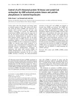
Figure 1
Cardiac function as assessed by 2D ECHO at baseline and following I/R of the LADCardiac function as assessed by 2D ECHO at baseline and following I/
R of the LAD. The baseline (normal) left ventricular ejection fraction
(LVEF) obtained from 2D short axis M-mode left ventricular internal
diameter trace was 67.52 ± 1.8%, 63.48 ± 2.9%, and 65.47 ± 2.4% in
arginine vasopressin (AVP; n = 14), normal saline solution (SL; n = 9),
and dobutamine (DOB; n = 6), resepctively. Following ischemia/reper-
fusion (I/R) injury, AVP treatment significantly reduced day 1 LVEF
(41.1 ± 3.4%) in comparison with SL control (51.6 ± 4.3%), while both
group had significant reductions in LVEF vs baseline. DOB mitigated
the decrease in LVEF (57.7 ± 6.7%) day 1 post I/R. LVEF measured at
day 3 demonstrated improvement in all groups; however, mice receiv-
ing AVP remained significantly lower than baseline. * P < 0.05 vs base-
line, ** P < 0.05 vs SL-treated mice. Results are present as means ±
standard error of the mean. LAD = left anterior descending coronary
artery.
Table 1
Baseline, day 1 and day 3 heart rate and left ventricular end diastolic volume post I/R injury and intraperitoneal pump implantation
Group Parameter Baseline Day 1 post I/R Day 3 post I/R
Vasopressin
n = 14
HR (bpm):
LVEDV (uL):
495 ± 18.1
67 ± 5
517.15 ± 15.67
58 ± 8
485.11 ± 34.94
60 ± 9
Dobutamine
n = 6
HR (bpm):
LVEDV (uL):
439 ± 28.7
64 ± 7
475.17 ± 32.35
55 ± 7
507.1 ± 50.68
55 ± 7
Normal Saline
n = 9
HR (bpm):
LVEDV (uL):
448 ± 15.1
69 ± 4
486.66 ± 16.29
60 ± 6
438 ± 32.10
58 ± 6
Heart rate (HR) was determined using limb lead echocardiography pads during echocardiogram, while left ventricular end diastolic volume
(LVEDV) was determined using echocardiography. No significant differences in HR or LVEDV exist between groups infused with saline (n = 9),
dobutamine (n = 6), or vasopressin (n = 14). Results are present as means ± standard error of the mean. I/R = ischemia/reperfusion.
Available online />Page 5 of 8
(page number not for citation purposes)
increased vascular tone because it mediates vasoconstriction
in vascular smooth muscle. It has also been found to be
expressed on cardiac myocytes and the kidney. V2Rs are
found mainly in the renal collecting duct and are responsible
for the antidiuretic effect of vasopressin. OTRs are found dif-
fusely throughout the body and are thought to mediate vasodi-
lation. Thus vasopressin is able to cause either
vasoconstriction or vasodilation depending on the tissue spe-
cific distribution of V1R vs OTR and is able to enhance the
effect of vasoconstrictor agents such as norepinephrine
through mechanisms yet to be identified [9].
Although its mechanism of action on the vasculature is well
understood, vasopressin has dose-dependent effects on both
cardiac contractility and coronary arterial tone. It appears that
at low doses vasopressin may act mainly through the P2R with
a shifting of physiologic effect from coronary smooth muscle
V1R-mediated vasoconstriction to P2R-mediated vascular
endothelial vasodilation. At higher doses this relation is
reversed with V1R-mediated coronary arterial vasoconstriction
predominating, with a resultant drop in cardiac output. Due to
safety concerns at higher doses, most clinical data relating to
direct cardiac effects are using low doses of vasopressin (≤
0.04 Units/minute), often in conjunction with inotropes.
Patients with vasodilatory shock increase systemic vascular
resistance twofold, while only diminishing cardiac output by
14% in response to vasopressin – indirect evidence of some
positive inotropy [37]. Similarly, when co-infused with the
phosphodiesterase inhibitor milrinone in patients with
advanced heart failure, vasopressin resulted in increased vas-
cular tone and blood pressure with no resultant change in car-
diac output [38]. In hypotensive post-cardiotomy patients who
remain in shock despite catecholamine infusions, the addition
of low-dose vasopressin resulted in a significant increase in
left ventricular work index and a decrease in vasopressor use,
inotrope usage, and heart rate [2]. It is of great interest to the
clinician that the hemodynamic effects of vasopressin are
potentiated by the shock state, because in normal subjects
vasoconstriction only occurs at high doses, while fluid unre-
sponsive shock confers a powerful vasopressor effect at low
doses. This may reflect an acute depletion of circulating vaso-
pressin with subsequent hypersensitivity to its effects [37,39].
Because of these theoretical and practical benefits, vaso-
pressin has come into widespread use for shock states,
including shock in which myocardial injury plays a contributive
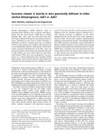
Figure 2
Intra-arterial blood pressure at day 1 following I/R of the LADIntra-arterial blood pressure at day 1 following I/R of the LAD. Systolic
blood pressure (SBP), diastolic blood pressure (DBP), and mean arte-
rial pressure (MAP) are shown. Although there was no statistically sig-
nificant differences in blood pressure, MAP trended to lowest in
arginine vasopressin (AVP) group (n = 5) with a MAP of 89.7 ± 1.7
mmHg compared with 100.1 ± 6.0 in dobutamine (DOB; n = 5) and
94.8 ± 3.4 in normal saline solution (SL) control (n = 5). Results are
present as means ± standard error of the mean. I/R = ischemia/reper-
fusion; LAD = left anterior descending coronary artery.
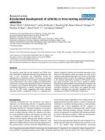
Figure 3
Kaplan Meier survival curve for mice in the three treatment groupsKaplan Meier survival curve for mice in the three treatment groups.
When compared with infusions of either saline (n = 6) or dobutamine (n
= 6), vasopressin (n = 12) results in dramatically increased mortality.
This difference begins as soon as day 1 following ischemia/reperfusion
and persists throughout our seven day observation period.
Figure 4
Western blot of left ventricular OTR levels at baseline and day 1 follow-ing I/R of the LADWestern blot of left ventricular OTR levels at baseline and day 1 follow-
ing I/R of the LAD. Left ventricular tissue was dissected and flash fro-
zen for protein extraction both in control (baseline) animals and at day 1
following ischemia/reperfusion (I/R) of the left anterior descending cor-
onary artery (LAD). We chose this timepoint as the enhanced physio-
logic effect (cardiac suppression) was observed by day 1. In those
animals subjected to I/R of the LAD, oxytocin receptor (OTR) expres-
sion normalized to β-actin was reduced by 40% compared with con-
trols.
Critical Care Vol 13 No 3 Indrambarya et al.
Page 6 of 8
(page number not for citation purposes)
role [1-3,12-17]. However, the cardiovascular effect of vaso-
pressin on the injured myocardium has yet to be elucidated.
In this study we found that low-dose vasopressin did not sig-
nificantly alter arterial blood pressure or cardiac ejection frac-
tion in the uninjured state. This experimental observation
correlates with the clinical finding that normotensive patients
do not exhibit a physiologic response to low-dose vasopressin
[37,39]. In contrast, we found AVP significantly decreased
LVEF in a model of ischemia reperfusion. Our model used one
hour of LAD ischemia and reproducibly depressed day 1 car-
diac ejection fraction by approximately 13% in mice treated
with saline infusions (control animals). We compared these
saline-infused mice with the standard drug used for cardio-
genic shock (DOB) and noted a significant increase in cardiac
ejection fraction. This served as both a positive control to
assure good absorption of the intra-peritoneal medication as
well as a standard of care arm with which to compare cardiac
function and mortality. Vasopressin, on the other hand, demon-
strated a markedly different effect following LAD I/R than in the
intact animal. During the infusion the mean cardiac ejection
fraction dropped by 10% when compared with saline, and by
24% compared with baseline. This decrease in cardiac con-
tractility appeared to be through a direct cardiac effect as
there was no significant change in either preload (LVEDV) or
afterload (arterial blood pressure) due to vasopressin.
Vasopressin had no significant effect on cardiac function in
intact mice, while following I/R injury vasopressin was cardio-
suppressive. We speculated that this may have resulted from
alterations in vasopressin receptor expression as a result of I/
R. We found that V1R and OTR were expressed in the heart,
while V2R was not detectable. V1R expression was not
altered as a result of I/R, while OTR expression was reduced
by 40% (Figure 4). This stable expression of V1R combined
with decreased OTR expression could result in predominant
vasoconstriction in the injured heart, potentially worsening car-
diac ischemia and resulting in dysfunction.
In addition to a decline in cardiac contractility, vasopressin
resulted in a marked increase in early mortality compared with
both saline and DOB-treated mice. The moderate reduction in
cardiac ejection fraction and non-statistically significant trend
towards a 5 mmHg decrease in blood pressure in the vaso-
pressin-infused group essentially excludes cardiogenic shock
as a cause of the excess mortality. Further support for this
comes from routine monitoring of the post-operative appear-
ance (grooming, temperature, activity level), with all groups
appearing healthy with no evidence of general medical deteri-
oration as would be expected with cardiac insufficiency. Vaso-
pressin and its analogue terlipressin have been reported to
induce cardiac arrhythmia (bradycardia and Torsade de
Pointes) not associated with clear evidence of myocardial inf-
arction [40-45]. Given the generally healthy clinical condition
of the mice, we speculate that the majority of deaths may have
related to sudden cardiac events (arrhythmia). How might this
occur? Vasopressin has been found to block K
ATP
channels in
the vascular endothelium, where this reverses vasoplegia in
the systemic circulation [8], but may contribute to sudden
vasospasm in the coronary circulation [46]. K
ATP
channels
expressed on cardiomyocytes are thought to decrease mem-
brane excitability when activated through stress and thus may
be key mediators of ischemic tolerance [46]. Increased mem-
brane excitability as a result of vasopressin acting to close
K
ATP
channels could increase the risk of arrhythmia.
Limitations of this study include a lack of continuous cardiac
and hemodynamic monitoring. Transient changes in afterload
may have influenced the extent of ischemic cardiac damage
but may not have been detected by our single measurement,
while a lack of continuous cardiac rhythm monitoring did not
allow us to determine whether arrhythmia was in fact the major
cause of death. Other limitations were the lack of quantifica-
tion of ischemic myocardium as a result of the I/R, and that we
used whole left ventricular tissue rather than isolated cardio-
myocyte digestion, and were thus not able to assess from
which cell type the vasopressin receptors were derived. There-
fore, the down-regulation of OTR must be regarded as hypoth-
esis generating rather than a proof of mechanism.
In summary, we found that low-dose vasopressin infusion had
no significant cardiovascular effect in normal mice. In contrast,
following ischemic injury to the myocardium vasopressin
exerted a strong negative inotropic effect on the heart, result-
ing in a significant decline in cardiac ejection fraction as meas-
ured by echocardiogram. This decline was not mediated
through changes in left ventricular preload or afterload at the
time point assayed and the possibility of a direct cardiac effect
is raised. We speculate that I/R, by decreasing OTR expres-
sion in the heart, may result in vasopressin-inducing vasocon-
striction and cardiac dysfunction in the injured heart.
Conclusions
AVP infusion significantly depressed the myocardial function
in I/R injured model and increased the mortality rate in compar-
ison with SL and DOB. The use of vasopressin may be asso-
ciated with cardiac suppression in non-vasodilatory shock
states involving significant cardiac injury.
Key messages
• Vasopressin infusion decreases cardiac ejection frac-
tion and increases mortality after I/R injury.
• The decrease in cardiac ejection fraction is not caused
by an increase in afterload, but rather through a
decrease in cardiac contractility.
• Vasopressin should be used with caution in patients
who may have a cardiac component contributing to
their shock.
Available online />Page 7 of 8
(page number not for citation purposes)
Competing interests
The authors declare that they have no competing interests.
Authors' contributions
TI drafted the manuscript, performed echocardiography and
molecular experiments, and assisted with animal experiments.
JB designed the experiments, wrote the manuscript and per-
formed echocardiography. YW performed animal experiments.
MM performed molecular experiments. KW designed the
experiments and wrote the manuscript. All authors read and
approved the final manuscript.
Acknowledgements
This project was funded by Canadian Institutes of Health Research
(CIHR), Heart and Stroke Foundation and Providence Health Care
Research Institute. KW is a Michael Smith Foundation for Health
Research Distinguished Scholar. JB is a Providence Health Care
Research Institute Physician Scholar. TI is a Faculty of Medicine, Chu-
lalongkorn University Scholar.
References
1. Russell JA, Walley KR, Singer J, Gordon AC, Hebert PC, Cooper
DJ, Holmes CL, Mehta S, Granton JT, Storms MM, Cook DJ, Pres-
neill JJ, Ayers D: Vasopressin versus norepinephrine infusion in
patients with septic shock. N Engl J Med 2008, 358:877-887.
2. Dunser MW, Mayr AJ, Stallinger A, Ulmer H, Ritsch N, Knotzer H,
Pajk W, Mutz NJ, Hasibeder WR: Cardiac performance during
vasopressin infusion in postcardiotomy shock. Intensive Care
Med 2002, 28:746-751.
3. Patel BM, Chittock DR, Russell JA, Walley KR: Beneficial effects
of short-term vasopressin infusion during severe septic
shock. Anesthesiology 2002, 96:576-582.
4. Holmes CL, Landry DW, Granton JT: Science review: Vaso-
pressin and the cardiovascular system part 1 – receptor phys-
iology. Crit Care 2003, 7:427-434.
5. Dunser MW, Mayr AJ, Ulmer H, Ritsch N, Knotzer H, Pajk W, Luck-
ner G, Mutz NJ, Hasibeder WR: The effects of vasopressin on
systemic hemodynamics in catecholamine-resistant septic
and postcardiotomy shock: a retrospective analysis. Anesth
Analg 2001, 93:7-13.
6. Gold JA, Cullinane S, Chen J, Oz MC, Oliver JA, Landry DW: Vaso-
pressin as an alternative to norepinephrine in the treatment of
milrinone-induced hypotension. Crit Care Med 2000,
28:249-252.
7. Holmes CL, Patel BM, Russell JA, Walley KR: Physiology of vaso-
pressin relevant to management of septic shock. Chest 2001,
120:989-1002.
8. Landry DW, Oliver JA: The ATP-sensitive K+ channel mediates
hypotension in endotoxemia and hypoxic lactic acidosis in
dog. J Clin Invest 1992, 89:2071-2074.
9. Noguera I, Medina P, Segarra G, Martinez MC, Aldasoro M, Vila
JM, Lluch S: Potentiation by vasopressin of adrenergic vaso-
constriction in the rat isolated mesenteric artery. Br J Pharma-
col 1997, 122:431-438.
10. Holmes CL, Landry DW, Granton JT: Science Review: Vaso-
pressin and the cardiovascular system part 2 – clinical physi-
ology. Crit Care 2004, 8:15-23.
11. Landry DW, Levin HR, Gallant EM, Seo S, D'Alessandro D, Oz
MC, Oliver JA:
Vasopressin pressor hypersensitivity in
vasodilatory septic shock. Crit Care Med 1997, 25:1279-1282.
12. Dellinger RP, Levy MM, Carlet JM, Bion J, Parker MM, Jaeschke R,
Reinhart K, Angus DC, Brun-Buisson C, Beale R, Calandra T, Dhai-
naut JF, Gerlach H, Harvey M, Marini JJ, Marshall J, Ranieri M, Ram-
say G, Sevransky J, Thompson BT, Townsend S, Vender JS,
Zimmerman JL, Vincent JL: Surviving Sepsis Campaign: Interna-
tional guidelines for management of severe sepsis and septic
shock: 2008. Crit Care Med 2008, 36:296-327.
13. Lange M, Ertmer C, Westphal M: Vasopressin vs. terlipressinin
the treatment of cardiovascular failure in sepsis. Intensive
Care Med 2008, 34:821-832.
14. Albanese J, Leone M, Delmas A, Martin C: Terlipressin or nore-
pinephrine in hyperdynamic septic shock: a prospective, rand-
omized study. Crit Care Med 2005, 33:1897-1902.
15. Dubois MJ, De Backer D, Creteur J, Anane S, Vincent JL: Effect of
vasopressin on sublingual microcirculation in a patient with
distributive shock. Intensive Care Med 2003, 29:1020-1023.
16. Dunser MW, Mayr AJ, Ulmer H, Knotzer H, Sumann G, Pajk W,
Friesenecker B, Hasibeder WR: Arginine vasopressin in
advanced vasodilatory shock: a prospective, randomized, con-
trolled study. Circulation 2003, 107:2313-2319.
17. Iijima T, Oguchi T, Kashimoto S: Does vasopressin infusion
improve the outcome of severe septic-shock without any
adverse effects? Anesthesiology 2003, 98:793. author reply
793.
18. Shelly MP, Greatorex R, Calne RY, Park GR: The physiological
effects of vasopressin when used to control intra-abdominal
bleeding. Intensive Care Medicine 1988, 14:526-531.
19. Boyle WASL 3rd: Direct cardiac effects of vasopressin and
their reversal by a vascular antagonist. Am J Physiol 1986,
251:H734-741.
20. Zenteno-Savin T, Sada-Ovalle I, Ceballos G, Rubio R: Effects of
arginine vasopressin in the heart are mediated by specific
intravascular endothelial receptors. Eur J Pharmacol 2000,
410:15-23.
21. Cheng CP, Igarashi Y, Klopfenstein HS, Applegate RJ, Shihabi Z,
Little WC: Effect of vasopressin on left ventricular perform-
ance. Am J Physiol
1993, 264:H53-60.
22. Wilson MF, Brackett DJ, Archer LT, Hinshaw LB: Mechanisms of
impaired cardiac function by vasopressin. Ann Surg 1980,
191:494-500.
23. Oikawa R, Nasa Y, Ishii R, Kuwaki T, Tanoue A, Tsujimoto G, Takeo
S: Vasopressin V1A receptor enhances baroreflex via the cen-
tral component of the reflex arc. Eur J Pharmacol 2007,
558:144-150.
24. Luk J, Ajaelo I, Wong V, Wong J, Chang D, Chou L, Reid IA: Role
of V1 receptors in the action of vasopressin on the baroreflex
control of heart rate. American Journal of Physiology 1993,
265:R524-529.
25. Weig HJ, Laugwitz KL, Moretti A, Kronsbein K, Stadele C, Bruning
S, Seyfarth M, Brill T, Schomig A, Ungerer M: Enhanced cardiac
contractility after gene transfer of V2 vasopressin receptors in
vivo by ultrasound-guided injection or transcoronary delivery.
Circulation 2000, 101:1578-1585.
26. Laugwitz KL, Ungerer M, Schoneberg T, Weig HJ, Kronsbein K,
Moretti A, Hoffmann K, Seyfarth M, Schultz G, Schomig A: Aden-
oviral gene transfer of the human V2 vasopressin receptor
improves contractile force of rat cardiomyocytes. Circulation
1999, 99:925-933.
27. Thibonnier M, Conarty DM, Preston JA, Plesnicher CL, Dweik RA,
Erzurum SC: Human vascular endothelial cells express oxy-
tocin receptors. Endocrinology 1999, 140:1301-1309.
28. D'Souza SP, Davis M, Baxter GF: Autocrine and paracrine
actions of natriuretic peptides in the heart. Pharmacol Ther
2004, 101:113-129.
29. Gutkowska J, Jankowski M, Mukaddam-Daher S, McCann SM:
Oxytocin is a cardiovascular hormone. Braz J Med Biol Res
2000, 33:625-633.
30. Luckner G, Dunser MW, Jochberger S, Mayr VD, Wenzel V, Ulmer
H, Schmid S, Knotzer H, Pajk W, Hasibeder W, Mayr AJ, Friese-
necker B: Arginine vasopressin in 316 patients with advanced
vasodilatory shock. Crit Care Med 2005, 33:2659-2666.
31. Holmes CL, Walley KR, Chittock DR, Lehman T, Russell JA: The
effects of vasopressin on hemodynamics and renal function in
severe septic shock: a case series. Intensive Care Med 2001,
27:1416-1421.
32. Jolly S, Newton G, Horlick E, Seidelin PH, Ross HJ, Husain M, Dza-
vik V: Effect of vasopressin on hemodynamics in patients with
refractory cardiogenic shock complicating acute myocardial
infarction. Am J Cardiol 2005, 96:1617-1620.
33. Holmes CL, Walley KR: Arginine vasopressin in the treatment
of vasodilatory septic shock. Best Pract Res Clin Anaesthesiol
2008, 22:275-286.
34. Yasujima M, Abe K, Tanno M, Omata K, Kasai Y, Sato M, Kimura T,
Yoshinaga K: Decreased urinary active and inactive kallikrein
by chronic infusion of vasopressin in conscious rats. Tohoku J
Exp Med 1985, 145:215-222.
Critical Care Vol 13 No 3 Indrambarya et al.
Page 8 of 8
(page number not for citation purposes)
35. Leclerc F, Walter-Nicolet E, Leteurtre S, Noizet O, Sadik A, Cremer
R, Fourier C: Admission plasma vasopressin levels in children
with meningococcal septic shock. Intensive Care Med 2003,
29:1339-1344.
36. Michael LH, Entman ML, Hartley CJ, Youker KA, Zhu J, Hall SR,
Hawkins HK, Berens K, Ballantyne CM: Myocardial ischemia and
reperfusion: a murine model. Am J Physiol 1995,
269:H2147-2154.
37. Landry DW, Levin HR, Gallant EM, Ashton RC Jr, Seo S, D'Ales-
sandro D, Oz MC, Oliver JA: Vasopressin deficiency contributes
to the vasodilation of septic shock. Circulation 1997,
95:1122-1125.
38. Gold J, Cullinane S, Chen J, Seo S, Oz MC, Oliver JA, Landry DW:
Vasopressin in the treatment of milrinone-induced hypoten-
sion in severe heart failure. Am J Cardiol 2000, 85:506-508.
A511.
39. Chen JM, Cullinane S, Spanier TB, Artrip JH, John R, Edwards NM,
Oz MC, Landry DW: Vasopressin deficiency and pressor hyper-
sensitivity in hemodynamically unstable organ donors. Circu-
lation 1999, 100:II244-246.
40. Kelly KJ, Stang JM, Mekhjian HS: Vasopressin provocation of
ventricular dysrhythmia. Ann Intern Med 1980, 92:205-206.
41. Klein GJ: Vasopressin, "torsades de pointes," and QT syn-
drome. Ann Intern Med 1980, 93:511-512.
42. Fitz JD: Vasopressin induction of ventricular ectopy. Arch Intern
Med 1982, 142:644.
43. Solanki P, Chawla A, Garg R, Gupta R, Jain M, Sarin SK: Benefi-
cial effects of terlipressin in hepatorenal syndrome: a pro-
spective, randomized placebo-controlled clinical trial. J
Gastroenterol Hepatol 2003, 18:152-156.
44. Urge J, Sincl F, Prochazka V, Urbanek K: Terlipressin-induced
ventricular arrhythmia. Scand J Gastroenterol 2008,
43:1145-1148.
45. Hobo R, Netsu S, Koyasu Y, Tsutsumi O: Bradycardia and car-
diac arrest caused by intramyometrial injection of vasopressin
during a laparoscopically assisted myomectomy. Obstet
Gynecol 2009, 113:484-486.
46. Marban E:
The surprising role of vascular K(ATP) channels in
vasospastic angina. J Clin Invest 2002, 110:153-154.