Báo cáo y học: "Prevalence of endotoxemia after surgery and its association with ICU length of stay" potx
Bạn đang xem bản rút gọn của tài liệu. Xem và tải ngay bản đầy đủ của tài liệu tại đây (166.36 KB, 8 trang )
Open Access
Available online />Page 1 of 8
(page number not for citation purposes)
Vol 13 No 3
Research
Prevalence of endotoxemia after surgery and its association with
ICU length of stay
Franco Valenza
1,2
, Lorella Fagnani
2
, Silvia Coppola
2
, Sara Froio
2
, Francesca Sacconi
2
,
Cecilia Tedesco
2
, Micol Maffioletti
2
, Marta Pizzocri
2
, Valentina Salice
2
, Maria Luisa Ranzi
3
,
Cristina Marenghi
1
and Luciano Gattinoni
1,2
1
Dipartimento di Anestesia, Rianimazione (Intensiva e Subintensiva) e Terapia del Dolore, Fondazione IRCCS – "Ospedale Maggiore Policlinico
Mangiagalli Regina Elena", Via Francesco Sforza 35, 20122, Milano, Italy
2
Dipartimento di Anestesiologia Terapia Intensiva e Scienze Dermatologiche, Università degli Studi di Milano, Via Festa del Perdono 7, 20122, Milano,
Italy
3
Laboratorio Centrale di Analisi Chimico Cliniche e Microbiologiche; Fondazione IRCCS – "Ospedale Maggiore Policlinico Mangiagalli Regina Elena",
Via Francesco. Sforza 35, 20122, Milano, Italy
Corresponding author: Franco Valenza,
Received: 7 Dec 2008 Revisions requested: 20 Jan 2009 Revisions received: 28 May 2009 Accepted: 29 Jun 2009 Published: 29 Jun 2009
Critical Care 2009, 13:R102 (doi:10.1186/cc7934)
This article is online at: />© 2009 Valenza et al.; licensee BioMed Central Ltd.
This is an open access article distributed under the terms of the Creative Commons Attribution License ( />),
which permits unrestricted use, distribution, and reproduction in any medium, provided the original work is properly cited.
Abstract
Introduction The aim of this observational study was to
investigate the prevalence of endotoxemia after surgery and its
association with ICU length of stay.
Methods 102 patients admitted to a university ICU after surgery
were recruited. Within four hours of admission, functional data
were collected and APACHE II severity score calculated.
Arterial blood samples were taken and endotoxemia was
measured by chemiluminescence (Endotoxin Activity (EA)).
Patients were stratified according to their endotoxin levels (low,
intermediate and high) and according to their surgical
procedures. Differences between endotoxin levels were
assessed by ANOVA, accepting P < 0.05 as significant. Data
are expressed as mean ± SD.
Results EA levels were low in 68 (66%) patients, intermediate
in 17 (17%) and high in 17 (17%). Age (61 ± 17 years) and
APACHE II score 8.3 ± 3.7 (P = 0.542) were not significantly
different in the three EA groups. Functional parameters on
admission were similar between EA groups: white blood cells
11093 ± 4605 cells/mm
3
(P = 0.385), heart rate 76 ± 16 bpm
(P = 0.898), mean arterial pressure 88.8 ± 13.6 mmHg (P =
0.576), lactate 1.18 ± 0.77 mmol/L (P = 0.370), PaO
2
/FiO
2
383 ± 109 mmHg (P = 0.474). Patients with high levels of EA
were characterized by longer length of stay in the ICU: 1.9 ± 3.0
days in the low EA group, 1.8 ± 1.4 days in intermediate and 5.2
± 7.8 days in high group (P = 0.038).
Conclusions 17% of our patients were characterized by high
levels of endotoxemia as assessed by EA assay, despite their
low level of complexity on admission. High levels of endotoxin
were associated with a longer ICU length of stay.
Introduction
Endotoxin is a constituent of the cell wall of Gram-negative
bacteria capable of inducing potent inflammatory response in
the host [1,2]. Isolated and purified from the wall of several
Gram-negative bacteria, it has been used to investigate many
aspects of the immuno-inflammatory response of sepsis
through inoculation in the laboratory animal [3,4] or in humans
[5-7]. However, endotoxin has also been documented in clini-
cal scenarios such as trauma or burn injury [8-10]. Rather than
a manifestation of exogenous infection, translocation of
lipopolysaccharide across the intestinal membrane when per-
meability is increased is the putative mechanism of these
forms of endotoxemia [11,12].
Patients undergoing surgery may have direct shedding of
lipopolysaccharide into the circulation via manipulation of
APACHE: Acute Physiology and Chronic Health Evaluation; EA: endotoxin activity; ICU: intensive care unit; LAL: Limulus-Amebocyte-Lysate; SIRS:
Systemic Inflammatory Reaction Syndrome.
Critical Care Vol 13 No 3 Valenza et al.
Page 2 of 8
(page number not for citation purposes)
infected surgical sites, violation of natural barriers such as
bowel resection, contamination from the environment, or via
the use of invasive devices. Meanwhile, postural changes,
blood loss, and vasoplegia all cause relative or absolute
hypoperfusion that may favor bacterial translocation. Butten-
schoen and colleagues have in fact shown that major abdom-
inal surgery is associated with transient endotoxemia [13,14].
However, while the likelihood of endotoxemia might be
straightforward in major abdominal surgery, less is known in
other surgical procedures, apart from cardiopulmonary bypass
[15,16].
We studied the prevalence of endotoxemia in a population of
patients admitted to an intensive care unit (ICU) after surgery
and the relation between endotoxemia and their outcome.
Materials and methods
The study was approved by our institution ethics committee,
and informed consent was obtained from all patients. Adult
patients admitted to the ICU of our institution were recruited
for the study unless they were transferred from another ICU,
had no arterial line in place, or were on chronic dialysis.
On admission and the next morning, clinical history and labo-
ratory data were taken. Cardio-respiratory variables were
recorded and Acute Physiology and Chronic Health Evaluation
(APACHE) II score was calculated [17]. Systemic Inflamma-
tory Reaction Syndrome (SIRS) was considered to be present
when at least two of these criteria were met: temperature
above 38°C or below 36°C, heart rate of more than 90 beats/
min, respiratory rate of more than 20 breaths/min or partial
pressure of carbon dioxide of less than 32 mmHg, or white
blood cell count above 12,000 mm
3
or below 4000 mm
3
[18].
Within four hours after admission blood was withdrawn for
Endotoxin Activity (EA) assay. During the course of the ICU
stay blood and other biologic specimens were collected on a
clinical basis and sent to the microbiologic laboratory of the
institution for microorganism detection. Length of stay and
mortality of both ICU and hospital were calculated. Clinicians
were unaware of the results of the EA assay throughout
patient's ICU and hospital stay.
Endotoxin activity assay
The EA assay has been described in detail previously [19].
Briefly, the method allows the measurement of EA as a func-
tion of each patient's neutrophil chemiluminescence's activity
(on a scale from 0 to 1). An EA level of 0.4 is approximately
equivalent to an endotoxin concentration of 25 to 50 pg/mL,
and a level of 0.6 equivalent to 100 to 200 pg/mL. A 2 ml sam-
ple of whole blood was drawn through an indwelling arterial
line into an endotoxin-free blood collection tube (Vacutainer
systems; Becton Dickinson, Franklin Lakes, NJ, USA). Blood
samples were maintained at room temperature and assayed
within 30 minutes of collection. To assay levels of endotoxin, a
10 μl aliquot of whole blood was placed in each of three tubes
containing luminol buffer (300 μl/tube). The control tube con-
tained blood and buffer only, whereas a positive control con-
tained a maximum stimulatory concentration of endotoxin (2
ng/ml); the final tube contained the test sample. All three tubes
were incubated at 37°C for five minutes and assayed in dupli-
cate. Chemiluminescence was initiated by the addition of 20
μl/tube human complement opsonized zymosan. Continuous
measurements were made of light emissions at 30-second
intervals over a total period of 20 minutes in a reciprocating
tube luminometer (Autolumat LB 953; E. G. & G. Berthold,
Wildbad, Germany). Quality control assays were performed
on a 1:1 ratio basis (i.e. one control every sample measured).
Based on the results obtained, samples were considered ade-
quate if the coefficient of variation between duplicates was
lower than 15% if EA level was below 0.2 and 30% if it was
above 0.2.
Statistical analysis
A descriptive analysis was first conducted on the entire popu-
lation. Patients were then stratified according to the EA results
into the following groups: low (EA <0.4), intermediate (EA 0.4
to <0.6) and high (EA ≥ 0.6). To compare continuous variables
on admission such as age, APACHE II score, and functional
parameters based on both surgical or EA stratification, one-
way analysis of variance was used and all pairwise multiple
comparisons were assessed by Student Newman-Keuls test.
If the normality test (Kolmogorov-Smirnov) failed, data were
analysed by Kruskal-Wallis one-way analysis of variance and
for all pairwise multiple comparisons procedures Dunn's
method was used. Functional parameters at entry and on the
next morning were analyzed according to EA stratification by
means of two-way analysis of variance. This was also used to
assess the interaction between EA levels and the type of sur-
gery in determining ICU and hospital length of stay. For statis-
tical purposes, patients were stratified according to the
different kind of surgical procedures they underwent into
major surgery and other procedures. Data are presented as
mean ± standard deviation, unless otherwise specified. Statis-
tical significance was accepted as P < 0.05. The Sigma Stat
for Windows version 3.11 (Systat Software Inc, Pont Rich-
mond, CA, USA) was used.
Results
A total of 122 patients were recruited for the study. All patients
had their EA level measured on admission. However, 20
patients were excluded from analysis: 17 because variation of
coefficients were out of the accepted range, two because the
calculation of EA level was unreliable (value > 1), and one
because quality control failed. Therefore, a total of 102
patients were considered.
Out of the 102 patients included in the study, 27 underwent
thoracic surgical procedures including 25 resective proce-
dures and 2 decortications; 27 patients underwent abdominal
procedures including gastric, intestine, and colon rectum sur-
Available online />Page 3 of 8
(page number not for citation purposes)
gery; 20 obese patients underwent gastric banding; 11
patients underwent hepatectomies; 7 underwent urological
proceures including 3 procedures on the bladder, 3 prostate-
ctomies, and 1 nephroureterectomy. The remaining 10
patients underwent procedures other then those above men-
tioned including three timectomies, three femoral bone frac-
ture repair, two median abdominal wall laparocele synthesis,
and two tiroidectomies. Patients were scheduled for elective
post-operative ICU monitoring. Five patients were admitted
because of intra-operative complications. Patients demo-
graphics were similar between surgical groups except for
obese patients who underwent gastric banding that were
younger (P < 0.05) and had lower APACHE II scores (P <
0.05). Data are shown on Table 1.
Median EA level was 0.282 (25% 0.190, 75% 0.462). When
stratified according to EA levels, 68 (66%) had normal values,
17 (17%) had intermediate, and 17 (17%) had high levels. As
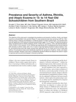
shown in Figure 1, patients with high EA levels (>0.6) on
admission were characterized by a longer ICU length of stay
(P = 0.038); in hospital length of stay was not different
between groups (P = 0.387). In patients with high EA activity
functional parameters were similar on admission to the other
EA stratification groups except for a somewhat higher temper-
ature. However, in contrast to patients without high EA levels,
the number of SIRS criteria, white blood cell count, and lactate
levels did not improve over time from the first day. Data are
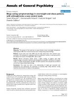
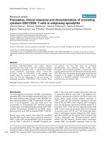
shown on Table 2. Patients who underwent major surgery
(abdominal or thoracic procedures) and presented to the ICU
with higher EA levels were characterized by significantly
longer ICU length of stay (Figure 2). These patients were char-
acterized intraoperatively by a slightly worse oxygenation
despite a more aggressive ventilatory management; the hemo-
dynamic status was similar between EA stratification groups.
Data are shown on Table 3. Complications that resulted in
longer ICU stay included: respiratory insufficiency (n = 11),
septic shock (n = 2), caridac arrhythmia (n = 3), and pneumo-
nia (n = 1). A total of 152 specimens from 41 patients were
sent to the microbiologic laboratory during the ICU stay. Of
these, 46 were positive in a total of 13 patients. The charac-
teristics of these patients are summarised in Table 4.
Discussion
This observational study investigates the prevalence of endo-
toxemia in a population of patients admitted to the ICU after
surgery and evaluates the association between endotoxin lev-
els and outcome. We found that 17% of the patients had lev-
els of endotoxin higher than normal on admission despite the
low level of complexity, and that patients with high endotoxin
levels had longer ICU length of stay.
To detect endotoxemia we chose to use the EA assay as
opposed to the more classic limulus-amebocyte-lysate (LAL)
test [20,21] This method is based on the detection of
enhanced respiratory burst activity in neutrophils following
their priming by complexes of endotoxin and a specific anti-
endotoxin antibody [19]. The method allows the expression of
EA as a function of each patient's neutrophil chemilumines-
cence's activity (on a scale from 0 to 1). The technique has
been validated against the LAL test [22], and has been
recently used in a multicenter trial to assess endotoxin preva-
lence in a mixed surgical/medical ICU cohort of patients
recruited across North America and Europe [23]. According to
this method, most of the patients admitted to the ICU after sur-
gery had normal levels of endotoxin. However, 17 out of 102
had higher than normal levels of endotoxin despite their low
level of complexity (APACHE score) These data confirm previ-
ous observations that circulating endotoxin is a common find-
Table 1
Characteristics of the study population
All Thoracic Abdominal Obesity Hepatic Urological Other
N. of patients 102 27 27 20 11 7 10
Age (years) 62 ± 17 67 ± 8 69 ± 17 40 ± 9 58 ± 18 64 ± 14 69 ± 17
Surgery (min) 178 ± 95 166 ± 64 200 ± 111 121 ± 38 267 ± 106 297 ± 80 146 ± 103
Unscheduled (n) 5 - 2 - - - 3
APACHE II score 8.4 ± 4 9.1 ± 3 9.7 ± 4 4.7 ± 2 8.4 ± 5 9.0 ± 3 9.3 ± 4
EA activity 0,3 ± 0.3 0.4 ± 0.2 0.3 ± 0.2 0.4 ± 0.2 0.3 ± 0.3 0.3 ± 0.3 0.4 ± 0.1
LOS ICU (days) 1 (1 to 2) 1 (1 to 2) 1 (1 to 4.7) 1 (1 to 1) 1 (1 to 1) 1 (1 to 1) 1 (1 to 2)
Mortality ICU (n) 3 - 2 - - - -
LOS hosp (days) 8 (5–11) 8.5 (6–13) 10 (7–16) 4 (4–5) 8.0 (6–10) 12 (9–13) 6.5 (5–10)
Mortality hosp (n) 1 1 - - - - -
Characteristics of the study population are shown. Intensive care unit (ICU) and hospital length of stay (LOS) are presented as median and
interquartile ranges. Other data are presented as mean ± standard deviation.
APACHE = Acute Physiology and Chronic Health Evaluation; EA = endotoxin activity.
Critical Care Vol 13 No 3 Valenza et al.
Page 4 of 8
(page number not for citation purposes)
ing in ICU patients [23], but add to the knowledge on
endotoxin prevalence in post-operative patients. In fact, while
endotoxemia in patients undergoing major abdominal proce-
dures has been previously shown [13,14], our observation
extend to other kind of surgical interventions less likely to be
characterized by endotoxemia.
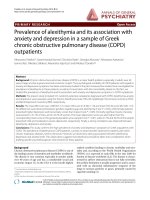
Subjects with high EA levels had a longer ICU length of stay
and a trend towards longer hospital length of stay (Figure 1).
Interestingly, functional parameters on admission were almost
normal and similar between groups of patients stratified by EA
levels (Table 2). Subjects with high EA on admission, despite
being similar to the other groups with respect to functional
data, demonstrated that white blood cell count, SIRS criteria,
and lactate did not significantly decrease on the morning after
admission. Whether this was indicative of an ongoing inflam-
matory process or adequacy of perfusion is difficult to deter-
mine. The role of microbial-derived endotoxin appears to play
a minor role in our study: the clinical suspicion of infection dur-
ing ICU stay was brought on only in a few subjects and even
less had proven infection during the course of their ICU stay
(Table 4). Moreover, the intraoperative hemodynamic variables
were similar between EA stratification groups (Table 3). How-
ever, the prevalence of high endotoxin levels in patients who
underwent thoracic surgery and the trend towards a relative
hypoxemia despite more aggressive ventilatory management
in patients with high EA levels is of interest. Both hypoxemia
[24] and mechanical ventilation [25] are related to endotox-
emia, even if we cannot exclude the potential higher preva-
lence of cigarette smokers in the thoracic group [26]. Except
for obese patients that represent a unique topology, patients
that underwent thoracic and abdominal procedures were sim-
ilar to the others, with respect to age, APACHE score, and
functional data at ICU admission. This suggests that measure-
ment of EA is a potential tool to stratify patients to more
aggressive care or to allocate resources in dynamic ICUs
recruiting post-operative patients for routine monitoring.
Whether EA stratification is useful only for abdominal and tho-
racic procedures cannot be determined from our data: the
number of subjects does not allow for multivariate analysis.
Moreover, we do not have EA data prior to surgery in order to
discriminate between patients who presented to the operating
room with pre-existing endotoxemia that might have persisted
after the surgical procedure itself. These are interesting
aspects that need further attention.
There are limitations to our study. Because of logistical rea-
sons measurements were available only during the week days:
this may have introduced a selection bias. As discussed
above, pre-operative evidence of endotoxemia is lacking: this
would have added to the interpretation of the data. Moreover,
the number of patients recruited is not high enough to gener-
alize our results to a wider ICU population.
Conclusions
In this study we have investigated the prevalence of endotox-
emia in a population of patients admitted to an ICU after sur-
gery. A number of patients were characterized by high levels
of endotoxemia, as assessed by EA assay, despite their low
level of complexity on admission. High levels of endotoxin were
associated with a longer ICU length of stay, particularly in
patients who underwent major surgery.
Figure 1
Intensive care unit and hospital length of stay according to endotoxin activity stratificationIntensive care unit and hospital length of stay according to endotoxin
activity stratification. Intensive care unit (ICU) = black columns; Hospi-
tal = gray columns.
Figure 2
Intensive care unit length of stay according to endotoxin activity stratifi-cation within surgical stratificationIntensive care unit length of stay according to endotoxin activity stratifi-
cation within surgical stratification. White columns represent data from
patients with high endotoxin activity (EA) levels, while dashed columns
refer to patients with intermediate or low EA levels.
Available online />Page 5 of 8
(page number not for citation purposes)
Table 2
Ventilator settings and functional parameters on admission to the intensive care unit
Low Intermediate High
Ventilated patients Admission 62/68 (91%) 16/17 (94%) 17/17 (100%)
Day 1 6/68 (9%)* 1/17 (6%)* 1/17 (6%)*
Tidal volume (l/min) Admission 6.5 ± 1.8 6.4 ± 1.2 7.3 ± 1.9
Day 1 / / /
Respiratory rate (atti/minuto) Admission 11 ± 4 11 ± 2 12 ± 7
Day 1 18 ± 5* 19 ± 5* 16 ± 4*
PaO
2
/FiO
2
(mmHg) Admission 379 ± 111 413 ± 113 372 ± 96
Day 1 329 ± 62* 330 ± 46* 299 ± 83*
PaCO
2
(mmHg) Admission 38.6 ± 4.9 35.1 ± 5.6
#
37.7 ± 5.0
Day 1 39.9 ± 4.7 39.3 ± 5.1* 38.8 ± 4.1
pH arterial blood Admission 7.41 ± 0.05 7.42 ± 0.06 7.39 ± 0.06
Day 1 7.43 ± 0.03 7.43 ± 0.03 7.43 ± 0.04
Lactate (mmol/l) Admission 1.12 ± 0.57 1.23 ± 0.47 1.40 ± 1.39
Day 1 0.79 ± 0.38* 0.81 ± 0.37* 0.89 ± 0.57
Heart rate (bpm) Admission 76 ± 16 75 ± 17 77 ± 18
Day 1 82 ± 15* 80 ± 12 84 ± 15
Mean arterial pressure (mmHg) Admission 88 ± 13 87 ± 13 92 ± 17
Day 1 91 ± 12 94 ± 13 93 ± 16
Central venous pressure (mmHg) Admission 7.5 ± 2.5 6.3 ± 2.9 7.5 ± 2.2
Day 1 6.1 ± 2.6 6.1 ± 2.5 6.1 ± 2.7
Hemoglobin (g/dl) Admission 12.0 ± 3.5 12.4 ± 1.8 12.4 ± 1.6
Day 1 11.3 ± 1.6 11.5 ± 1.7 12.0 ± 1.3
Creatinine (mg/dl) Admission 0.99 ± 0.92 1.06 ± 0.68 0.89 ± 0.37
Day 1 1.04 ± 1.12 1.05 ± 0.47 0.85 ± 0.27
Azotemia (mg/dl) Admission 36 ± 24 40 ± 21 37 ± 21
Day 1 37 ± 27 41 ± 24 36 ± 25
Glycemia (mg/dl) Admission 134 ± 30 134 ± 25 132 ± 35
Day 1 110 ± 29* 111 ± 23* 105 ± 21*
Sodium (Na
+
; mEq/l) Admission 137.5 ± 2.5 137.1 ± 2.2 137.8 ± 2.1
Day 1 137.9 ± 2.7 138.4 ± 3.5 138.1 ± 2.7
Potassium (K
+
;mEq/l) Admission 3.9 ± 0.5 3.9 ± 0.5 3.9 ± 0.4
Day 1 4.1 ± 0.3* 4.0 ± 0.4* 3.9 ± 0.4
Temperature (°C) Admission 35.2 ± 0.9 34.5 ± 0.9 35.4 ± 1.2
#
Day 1 36.6 ± 0.6 36.7 ± 0.5 36.8 ± 0.5
WBC (10
3
/mm
3
) Admission 10.8 ± 4.2 12.5 ± 5.5 10.8 ± 5.1
Day 1 9.4 ± 3.0* 10.4 ± 4.0 10.2 ± 2.7
SIRS criteria Admission 1.4 ± 0.7 1.8 ± 0.8 1.4 ± 0.9
Day 1 0.9 ± 0.9* 1.3 ± 1.1* 0.9 ± 0.8
Ventilatory and functional parameters collected within four hours of admission to the intensive care unit (Admission) and the morning after (Day 1)
are shown in the table. Patients were stratified according to their endotoxin activity levels on admission. * P < 0.05 Admission vs Day 1; # p <
0.05 between EA stratification groups. FiO
2
= fraction of inspired yoghurt; PaCO
2
= partial pressure of arterial carbon dioxide; PaO
2
= partial
pressure of arterial oxygen; SIRS = Systemic Inflammatory Reaction Syndrome; WBC = white blood cell.
Critical Care Vol 13 No 3 Valenza et al.
Page 6 of 8
(page number not for citation purposes)
Table 3
Intra-operative variables of the patients who underwent major surgery
Low Intermediate High
Number of patients (n) 34 10 10
Age (years) 69 ± 15 67 ± 8 67 ± 12
Duration of surgery (min) 174 ± 101 183 ± 73 189 ± 86
Arterial pressure – OR (mmHg) 121 ± 11 118 ± 13 123 ± 11
Arterial pressure – Preop (mmHg) 136 ± 17 140 ± 14 135 ± 11
pH 7.388 ± 0.05 7.420 ± 0.05 7.392 ± 0.04
BE -0.9 ± 2.5 -0.6 ± 1.9 -0.4 ± 2.4
Urine output (ml/h) 239 ± 193 331 ± 346 181 ± 98
Cristalloids (mL) 3045 ± 1590 2895 ± 917 3078 ± 900
Colloids (mL) 580 ± 280 533 ± 57 500 ± 0
Blood transfusion (n) 5 1 1
PaO
2
/FiO
2
(mmHg) 307 ± 141 275 ± 193 243 ± 145
Tidal volume (mL) 679 ± 102 667 ± 129 715 ± 109
Respiratory rate (bpm) 9.9 ± 1.5 9.8 ± 0.6 9.9 ± 1.1
Peak airway pressure (cmH
2
O) 23.8 ± 6.3 20.8 ± 5.9 24.3 ± 6.2
PEEP (cmH
2
O) 2.4 ± 2.3 0.9 ± 2.1 3.5 ± 2.4 *
Invasiveness (n) 5.1 ± 1.1 5.4 ± 0.8 5.4 ± 0.8
Contaminated surgery (n) 2 - -
Intra-operative variables of the patients who underwent abdominal or thoracic surgery are presented. * P < 0.05 analysis of variance. Arterial
pressure – OR = mean systolic arterial pressure recorded during the intervention; Arterial pressure – Preop = systolic arterial pressure taken at
the time of the preoperative evaluation; BE = base excess; FiO
2
= fraction of inspired yoghurt; PaO
2
= partial pressure of arterial oxygen; PEEP =
positive end-expiratory pressure. Invasiveness = sum of invasive procedure including tracheal tube, periferal vein, central vein, arterial line,
nasogastric tube, bladder catheter.
Available online />Page 7 of 8
(page number not for citation purposes)
Competing interests
The authors declare that they have no competing interests.
Authors' contributions
FV conceived the study, collected and analysed the data, and
wrote the manuscript. LF collected and analysed the data, and
wrote the manuscript. SC, SF, FS, CT, MM, MP, and VS col-
lected the data and performed analysis. MLR collected the
microbiologic data. CM collected the data. LG wrote the man-
uscript.
Acknowledgements
The authors would like to thank Spectral Diagnostics Inc. for providing
the instrumentation and the reagents to run the endotoxin activity
assays. This study was funded by Fondazione Ospedale Maggiore,
Mangiagalli e Regina Elena – IRCCS.
References
1. Carswell EA, Old LJ, Kassel RL, Green S, Fiore N, Williamson B:
An endotoxin-induced serum factor that causes necrosis of
tumors. Proc Natl Acad Sci USA 1975, 72:3666-3670.
2. Ulevitch RJ, Tobias PS: Recognition of gram-negative bacteria
and endotoxin by the innate immune system. Curr Opin Immu-
nol 1999, 11:19-22.
3. Emerson TE Jr, Gill CC: Effects of slow intravenous endotoxin
infusion on hemodynamics and survival in dogs. J Appl Physiol
1967, 22:874-877.
4. Natanson C, Eichenholz PW, Danner RL, Eichacker PQ, Hoffman
WD, Kuo GC, Banks SM, MacVittie TJ, Parrillo JE: Endotoxin and
tumor necrosis factor challenges in dogs simulate the cardio-
vascular profile of human septic shock. J Exp Med 1989,
169:823-832.
Key messages
• Endotoxemia is detectable in patients admitted to the
ICU after surgery.
• High levels of endotoxemia are associated with longer
ICU length of stay.
Table 4
Characteristics of the 13 patients who were positive for microbiologic investigations
EA level Age Surgery APACHE SIRS Gram + Gram - Other Time ICU Hosp Alive
0.140 77 A 11 2 Klebsiella Late 4 20 Y
0.446 50 T 6 2 Candida Late 1 6 Y
0.432 73 O 13 2 Contaminants Late 2 6 Y
0.125 78 A 9 2 Pseudomonas Late 24 26 Y
0.333 71 A 9 1 S. epidermidis Early 1 12 Y
0.235 43 A 10 2 S. aureus Early 2 12 Y
0.330 90 O 11 2 Enterobacter Early 1 32 Y
0.191 72 A 10 2 S. aureus Early 8 19 Y
0.558 59 T 4 2 Pseudomonas Early 5 9 Y
0.498 36 O 3 3 Enterococcus Early 5 19 Y
0.750 78 A 7 1 Pseudomonas Early 28 - N
0.825 67 A 13 3 Morganella M Late 22 - N
0.740 76 T 8 1 Klebsiella Late 7 29 Y
Table includes endotoxin activity (EA) level, age, type of surgery (A = abdominal, T = thoracic, O = other), Acute Physiology and Chronic Health
Evaluation (APACHE) score, numbers of Systemic Inflammatory Reaction Syndrome (SIRS) criteria met at the time of intensive care unit (ICU)
admission, strain of microrganisms isolated, whether the microbiological samples were collected early after the admission (E – within three days)
or at a later time (L); the ICU and hospital length of stay, survival.
Critical Care Vol 13 No 3 Valenza et al.
Page 8 of 8
(page number not for citation purposes)
5. Colucci M, Balconi G, Lorenzet R, Pietra A, Locati D, Donati MB,
Semeraro N: Cultured human endothelial cells generate tissue
factor in response to endotoxin. J Clin Invest 1983,
71:1893-1896.
6. Suffredini AF, Fromm RE, Parker MM, Brenner M, Kovacs JA, Wes-
ley RA, Parrillo JE: The cardiovascular response of normal
humans to the administration of endotoxin. N Engl J Med
1989, 321:280-287.
7. Taveira da Silva AM, Kaulbach HC, Chuidian FS, Lambert DR, Suf-
fredini AF, Danner RL: Brief report: shock and multiple-organ
dysfunction after self-administration of Salmonella endotoxin.
N Engl J Med 1993, 328:1457-1460.
8. Munster AM, Winchurch RA, Thupari JN, Ernst CB: Reversal of
postburn immunosuppression with low-dose polymyxin B. J
Trauma 1986, 26:995-998.
9. Winchurch RA, Thupari JN, Munster AM: Endotoxemia in burn
patients: levels of circulating endotoxins are related to burn
size. Surgery 1987, 102:808-812.
10. Kelly JL, O'Sullivan C, O'Riordain M, O'Riordain D, Lyons A,
Doherty J, Mannick JA, Rodrick ML: Is circulating endotoxin the
trigger for the systemic inflammatory response syndrome
seen after injury? Ann Surg 1997, 225:530-541.
11. LeVoyer T, Cioffi WG Jr, Pratt L, Shippee R, McManus WF, Mason
AD Jr, Pruitt BA Jr: Alterations in intestinal permeability after
thermal injury. Arch Surg 1992, 127:26-29.
12. Munster AM, Smith-Meek M, Dickerson C, Winchurch RA: Trans-
location. Incidental phenomenon or true pathology? Ann Surg
1993, 218:321-326.
13. Buttenschoen K, Buttenschoen DC, Berger D, Vasilescu C, Schaf-
heutle S, Goeltenboth B, Seidelmann M, Beger HG: Endotoxemia
and acute-phase proteins in major abdominal surgery. Am J
Surg 2001, 181:36-43.
14. Buttenschoen K, Schneider ME, Utz K, Kornmann M, Beger HG,
Carli BD: Effect of major abdominal surgery on endotoxin
release and expression of Toll-like receptors 2/4. Langen-
becks Arch Surg 2009, 394:293-302.
15. Boelke E, Storck M, Buttenschoen K, Berger D, Hannekum A:
Endotoxemia and mediator release during cardiac surgery.
Angiology 2000,
51:743-749.
16. Jansen PG, Te VH, Oudemans-Van Straaten HM, Bulder ER, Van
Deventer SJ, Sturk A, Eijsman L, Wildevuur CR: Perfusion-related
factors of endotoxin release during cardiopulmonary bypass.
Eur J Cardiothorac Surg 1994, 8:125-129.
17. Knaus WA, Draper EA, Wagner DP, Zimmerman JE: APACHE II: a
severity of disease classification system. Crit Care Med 1985,
13:818-829.
18. Members of the American College of Chest Physicians/Society of
Critical Care Medicine Consensus Conference: American Col-
lege of Chest Physicians/Society of Critical Care Medicine
Consensus Conference: definitions of sepsis and organ failure
and guidelines for the use of innovative therapies in sepsis.
Crit Care Med. 1992, 20:864-874.
19. Romaschin AD, Harris DM, Ribeiro MB, Paice J, Foster DM,
Walker PM, Marshall JC: A rapid assay of endotoxin in whole
blood using autologous neutrophil dependent chemilumines-
cence. J Immunol Methods 1998, 212:169-185.
20. Levin J, Bang FB: Clottable protein in Limulus; its localization
and kinetics of its coagulation by endotoxin. Thromb Diath
Haemorrh 1968, 19:186-197.
21. Tamura H, Tanaka S, Obayashi T, Yoshida M, Kawai T: A new sen-
sitive method for determining endotoxin in whole blood. Clin
Chim Acta 1991, 200:35-42.
22. Marshall JC, Walker PM, Foster DM, Harris D, Ribeiro M, Paice J,
Romaschin AD, Derzko AN: Measurement of endotoxin activity
in critically ill patients using whole blood neutrophil depend-
ent chemiluminescence. Crit Care 2002, 6:342-348.
23. Marshall JC, Foster D, Vincent JL, Cook DJ, Cohen J, Dellinger RP,
Opal S, Abraham E, Brett SJ, Smith T, Mehta S, Derzko A,
Romaschin A: Diagnostic and prognostic implications of endo-
toxemia in critical illness: results of the MEDIC study. J Infect
Dis 2004, 190:527-534.
24. Gaffin SL, Brock-Utne JG, Zanotti A, Wells MT: Hypoxia-induced
endotoxemia in primates: role of reticuloendothelial system
function and anti-lipopolysaccharide plasma. Aviat Space
Environ Med 1986, 57:1044-1049.
25. Nahum A, Hoyt J, Schmitz L, Moody J, Shapiro R, Marini JJ: Effect
of mechanical ventilation strategy on dissemination of intrat-
racheally instilled Escherichia coli in dogs. Crit Care Med
1997, 25:1733-1743.
26. Hasday JD, Bascom R, Costa JJ, Fitzgerald T, Dubin W: Bacterial
endotoxin is an active component of cigarette smoke.
Chest
1999, 115:829-835.