Báo cáo y học: "A general framework for quantifying the effects of DNA repair inhibitors on radiation sensitivity as a function of dose" pptx
Bạn đang xem bản rút gọn của tài liệu. Xem và tải ngay bản đầy đủ của tài liệu tại đây (292.03 KB, 7 trang )
BioMed Central
Page 1 of 7
(page number not for citation purposes)
Theoretical Biology and Medical
Modelling
Open Access
Research
A general framework for quantifying the effects of DNA repair
inhibitors on radiation sensitivity as a function of dose
Anthony J Chalmers*
1
, Soeren M Bentzen
2
and Francesca M Buffa
3
Address:
1
Brighton and Sussex Medical School, University of Sussex, Falmer, Brighton BN1 9RQ, UK,
2
University of Wisconsin Medical School,
Department of Human Oncology, K4/316 Clinical Sciences Center, 600 Highland Avenue, Madison, WI 53792, USA and
3
Cancer Research UK
Molecular Oncology Laboratories, Weatherall Institute of Molecular Medicine, University of Oxford, John RadcliffeHospital, Oxford OX3 9DU,
UK
Email: Anthony J Chalmers* - ; Soeren M Bentzen - ;
Francesca M Buffa -
* Corresponding author
Abstract
Purpose: Current methods for quantifying effects of DNA repair modifiers on radiation sensitivity
assume a constant effect independent of the radiation dose received. The aim of this study was to
develop and evaluate a modelling strategy by which radiation dose dependent effects of DNA repair
inhibitors on clonogenic survival might be identified and their significance assessed.
Methods: An indicator model that allowed quantification of the Sensitiser Effect on Radiation
response as a function of Dose (SERD) was developed. This model was fitted to clonogenic survival
data derived from human tumour and rodent fibroblast cell lines irradiated in the presence and
absence of chemical inhibitors of poly(ADP-ribose) polymerase (PARP) activity.
Results: PARP inhibition affected radiation response in a cell cycle and radiation dose dependent
manner, and was also associated with significant radiation-independent effects on clonogenic
survival. Application of the SERD method enabled identification of components of the radiation
response that were significantly affected by PARP inhibition and indicated the magnitude of the
effects on each component.
Conclusion: The proposed approach improves on current methods of analysing effects of DNA
repair modification on radiation response. Furthermore, it may be generalised to account for other
parameters such as proliferation or dose rate to enable its use in the context of fractionated or
continuous radiation exposures.
Background
Radiotherapy is an effective mode of cancer treatment but
its capacity to cure is limited by toxic effects on healthy tis-
sues. Developing effective treatment schedules requires
detailed knowledge of the cellular effects of radiation in
tumours and normal tissues so that differences may be
exploited and a beneficial therapeutic ratio achieved.
Increasing evidence indicates that DNA repair pathways
are a key determinant of cell survival after radiation, and
that targeting the molecular components of these path-
ways offers therapeutic potential [1-3].
When assessing the impact of modifiers of DNA repair on
cellular responses to ionising radiation, accurate measure-
Published: 19 July 2007
Theoretical Biology and Medical Modelling 2007, 4:25 doi:10.1186/1742-4682-4-25
Received: 26 April 2007
Accepted: 19 July 2007
This article is available from: />© 2007 Chalmers et al; licensee BioMed Central Ltd.
This is an Open Access article distributed under the terms of the Creative Commons Attribution License ( />),
which permits unrestricted use, distribution, and reproduction in any medium, provided the original work is properly cited.
Theoretical Biology and Medical Modelling 2007, 4:25 />Page 2 of 7
(page number not for citation purposes)
ment of effects on clonogenic survival is crucial, since this
is the most clinically relevant radiation response [4]. Data
are generally presented in the form of survival curves,
which illustrate radiation effects over a range of doses and
may be described by parameters that derive primarily
from the Linear Quadratic (LQ) equation [5]. It is well
established, however, that radiation sensitivity may devi-
ate from the LQ model, especially at low doses; mathe-
matical models have been generated to indicate the extent
of such deviation [6]. Assessing the effect of DNA repair
modification on the whole dose-response curve repre-
sents an additional challenge that must be overcome if
accurate assessment of the biological consequences and
therapeutic potential of DNA repair modifiers is to be
achieved.
A conventional approach is to calculate a Sensitiser
Enhancement Ratio (SER) from the radiation dose (D
SF
)
associated with a specified surviving fraction, typically
37% (D
0
)[7], or from the surviving fraction associated
with a specified radiation dose, typically 2 Gray (SF
2
)[8]:
SER values calculated in this way reflect the impact of a
repair modifier at a single dose or survival point [9-11].
D
0
and SF
2
may also be estimated by fitting survival data
as a function of dose [12], thus reflecting the whole data
set, but neither method has the capacity to quantify differ-
ential sensitising effects over different radiation dose
ranges.
Another approach is to calculate the effect of a modifier
on the
α
and
β
parameters of the LQ equation, which
describe respectively the linear and exponential compo-
nents of the survival curve [12,13]. Ratios calculated from
these parameters give an indication of both magnitude
and radiation dose dependency of a sensitising effect, but
the method has limitations.
Firstly, the fitting of these models has always been per-
formed separately on the treated and untreated datasets,
making direct comparison of the parameters difficult. Fur-
thermore, it cannot be applied in situations where the
relationship between survival and dose is more complex
than that predicted by the LQ equation. Secondly, cyto-
toxic effects of sensitising agents that are independent of
radiation are not taken into account. Such effects may be
small, and are often concealed by the method used to cal-
culate surviving fraction, but may be relevant, particularly
if they vary between cell lines or are of similar magnitude
to the radiation modifying effects under investigation. In
such cases, it would be informative to assess the relative
significance of the cytotoxic and radiosensitising effects,
and to ascertain whether the two are interdependent.
The aim of this project was to devise a general approach
that could be applied to complex survival curves and used
to quantify: (1) drug-induced changes in survival at differ-
ent radiation doses, (2) radiation-independent effects on
survival and (3) the relative significance of these changes.
The main features of the method are the inclusion of an
indicator term in the model to indicate the presence of the
drug and a factor
δ
x representing the variation on any
parameter of survival between radiation only and radia-
tion plus drug. This implies that the perturbation on the
parameters introduced by the drug can be approximated
using linear regression and that the linear regression can
be truncated to the first term. The first of these assump-
tions is quite general as a large variety of problems can be
treated within a linear regression framework; the second
holds only if the perturbation is linear or relatively small.
However, the model can easily be extended to include
higher order linear regression terms.
In this study the LQ equation was modified using Joiner's
Induced Repair model [6] and used to express survival at
a given dose, but the approach is general and may be
applied to any other expression of survival. To test the
applicability of the approach, the model was fitted to a
range of clonogenic survival curves that had been previ-
ously derived from rodent and human cell lines irradiated
in the presence and absence of two chemical inhibitors of
the DNA repair enzyme poly(ADP-ribose) polymerase
(PARP).
Results
Development of model: Sensitiser Effect on Radiation
response as a function of Dose (SERD)
Log transformed surviving fraction, SF, was fitted as a
function of dose, d, using the indicator model:
where
δ
z allows for non-null effect of the drug on plating
efficiency;
α
and
β
are the classical linear and quadratic
radiosensitivity parameters; G and d
C
are the low-dose
hyper-sensitivity parameters [14]; i is an indicator which
assumes the value zero for the control case, i.e. radiation
alone, and one for the drug-treated case; and
δ
x – where
"x" is any of the parameters above – is the variation on x
between the control and case under study. General least
square fitting was used and the significance of terms in the
model was tested using the log-likelihood ratio test. This
test considers the ratio of the likelihood of the model with
the parameter to the model without the parameter. Terms
which showed non-significant improvement were
removed from the model; terms which gave a p-value of <
SER
D without sensitiser
D with sensitiser
SER
SF witho
==
0
0
2
or
uut sensitiser
SF with sensitiser
2
SF d z i i d G G i e d
dd di
Cc
( ) ( ) exp ( ) ( ) (
/( )
= + ⋅⋅ − + ⋅⋅− + ⋅⋅ ⋅−
−+⋅
1
δαδαδ
δ
ββδβ
+⋅⋅
{}
id)
2
Theoretical Biology and Medical Modelling 2007, 4:25 />Page 3 of 7
(page number not for citation purposes)
0.05 were considered significant and retained in the final
model (see Table 1). Retention of a
δ
x parameter in the
final model thus indicated a significant drug effect. S-
PLUS 6.1 was used for implementation of the methods
and the analysis [15].
In Joiner's original paper, the low-dose hypersensitivity
parameter g was defined as: g = (
α
S
-
α
R
)/
α
R
where
α
S
is
derived from the very low dose component of the survival
curve and
α
R
from the overall linear component of the
curve. To reduce correlation between the model variables
and to facilitate implementation of the model, we have
used here a re-parameterisation of the model where G = α
S
- α
R
.
In the indicator model (Equation 1), the radiosensitivity
parameter change for drug-treated cells is in the form
x+
δ
x, the underlying hypothesis being that the perturba-
tion introduced by the drug effect on the radiation param-
eters can be approximated to its linear component in the
first instance. For prolonged or fractionated irradiation
regimes, parameters associated with repopulation or
repair effects could also be incorporated into the model.
Although a degree of correlation between the various
parameters in Equation 1 could be expected, this method
allows quantification of linear, quadratic and low dose
survival, and direct comparison of these parameters
between control and drug-treated cells.
Evaluation of SERD model
Figures 1, 2, 3 show clonogenic survival curves generated
by irradiation of two rodent fibroblast and two human
tumour cell lines in the presence and absence of chemical
inhibitors of PARP activity. The variable nature and mag-
nitude of the effects of PARP inhibition on clonogenic sur-
vival among these cell lines offered a useful setting in
which to investigate the utility and applicability of the
SERD model. The radiobiological implications of the
curves have been published elsewhere [16] and will not be
discussed here.
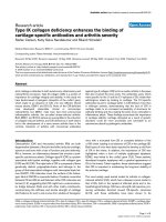
The survival curves shown in figure 1a illustrate radiosen-
sitisation of CHO-K1 fibroblasts by 3-AB, with marked
effect over the dose range 0.05 – 0.3 Gy. Fitting the model
in equation 1 to these data demonstrated that the control
curve is described by the classic linear quadratic equation,
with α and β emerging as the only significant parameters,
Table 1: Significant coefficients generated by fitting the SERD equation to the survival curves shown in Figures 1, 2 and 3.
Cell line Parameter Value (± standard error) p-value*
CHO-K1 (Fig 1a)
α
0.142 (± 0.021) <0.0001
β
0.043 (± 0.005) <0.0001
δ
z -0.133 (± 0.023) <0.0001
δα
0.112 (± 0.015) <0.0001
δ
G 34.649 (± 12.328) 0.005
δ
d
C
0.037 (± 0.008) <0.0001
V79-379A (Fig 1b)
α
0.187 (± 0.019) <0.0001
β
0.016 (± 0.004) 0.0003
G 2.235 (± 0.666) 0.0009
d
C
0.161 (± 0.031) <0.0001
δ
z -0.184 (± 0.017) <0.0001
T98G exponential phase (Fig 2a)
α
0.208 (± 0.006) <0.0001
δ
z -0.101 (± 0.014) <0.0001
δ
G 10.116 (± 10.374) 0.330
δ
G 7.81 0.020
δ
d
C
0.033 (± 0.019) 0.076
δβ
0.013 (± 0.002) <0.0001
T98G growth-arrested (Fig 2b)
α
0.175 (± 0.003) <0.0001
δ
z 0.051 (± 0.007) <0.0001
δα
-0.017 (± 0.005) 0.0005
U373-MG exponential phase (Fig
3a)
α
0.270 (± 0.011) <0.0001
δ
z 0.068 (± 0.021) 0.002
δβ
0.028 (± 0.004) <0.0001
U373-MG growth-arrested (Fig
3b)
α
0.126 (± 0.014) <0.0001
β
0.031 (± 0.003) <0.0001
δ
z -0.044 (± 0.012) 0.0002
* Log-likelihood ratio test (L-ratio) was applied to include or drop parameters from the final equation. p-values shown were derived from a t-test
that the parameter is zero. The L-ratio and associated p-value is shown only when the tests did not agree (i.e. significance in one but not the other).
Theoretical Biology and Medical Modelling 2007, 4:25 />Page 4 of 7
(page number not for citation purposes)
while the high value derived for δG denoted a significant
effect of 3-AB on low-dose hyper-radiosensitivity (Table
1). Addition of the drug also exerted a negative effect on
radiation-independent survival (δz significant and
retained in the reduced model), and enhanced the linear
(δα significant) but not the quadratic component of cell
killing (δβ non-significant).
Similar analysis of the curves in figure 1b indicated that
V79-379A cells exhibited significant low-dose hyper-radi-
osensitivity in the absence of PARP inhibitor, and that
radiation-independent survival was significantly reduced
in its presence. No significant interaction between
NU1025 and any parameter of radiosensitivity was iden-
tified.
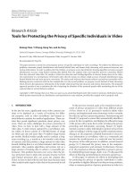
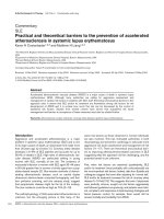
Figure 2a illustrates modification of the low-dose survival
characteristics of exponential phase T98G glioma cells by
PJ34. Fitting the SERD equation to these data indicated
that the effect of the drug on the low-dose hyper-radiosen-
sitivity parameter G was modest and did not reach statis-
tical significance. However, the fit of the model was
significantly superior when the
δ
G parameter was
included than when it was not (see log likelihood ratio);
Clonogenic survival curves derived from (a) exponential and (b) confluence-arrested populations of T98G glioma cells irradiated +/- 3 µM PJ34Figure 2
Clonogenic survival curves derived from (a) exponential and
(b) confluence-arrested populations of T98G glioma cells
irradiated +/- 3 µM PJ34.
0.1
1.0
1.0
0.1
012345
9
2
3
4
5
6
7
8
9
Control
PJ34
Dose (Gy)
Surviving fraction
012345
9
2
3
4
5
6
7
8
9
Control
PJ34
Dose (Gy)
Surviving fraction
(a)
(b)
Clonogenic survival curves derived from asynchronous, irra-diated populations of (a) CHO-K1 hamster fibroblasts +/- 5 mM 3-aminobenzamide and (b) V79-379A hamster fibrob-lasts +/- 100 µM NU1025Figure 1
Clonogenic survival curves derived from asynchronous, irra-
diated populations of (a) CHO-K1 hamster fibroblasts +/- 5
mM 3-aminobenzamide and (b) V79-379A hamster fibrob-
lasts +/- 100 µM NU1025. In all figures, data points represent
means (+/- standard error of the mean) of three independent
experiments.
Surviving fraction
Dose (Gy)
012345
0.1
1.0
9
2
3
4
5
6
7
8
9
Control
NU1025
Dose (Gy)
Surviving fraction
0.1
1.0
012345
4
5
6
7
8
2
3
4
5
6
7
8
Control
3-AB
(a)
(b)
Theoretical Biology and Medical Modelling 2007, 4:25 />Page 5 of 7
(page number not for citation purposes)
thus it was retained in the reduced final model after like-
lihood testing. This supports the interpretation that PJ34
induces low-dose hyper-radiosensitivity in exponential
phase populations of T98G.
By contrast, analysis of figures 2b, 3a and 3b indicated
that PJ34 did not affect low-dose radiation sensitivity of
confluent populations of T98G glioma cells, or of U373-
MG cells. In all cases, the radiation-independent effect of
the drug on survival (
δ
z) was a significant parameter.
The effect of PJ34 on overall radiosensitivity of human gli-
oma cells was dependent on the cell cycle characteristics
of the irradiated population. In exponential phase popu-
lations, addition of the drug increased the quadratic com-
ponent of cell killing (Figs. 2a, 3a), whereas in growth-
arrested populations there was no radio-sensitisation
(Figs. 2b, 3b). The negative effect of PJ34 on the linear
component of cell killing in growth-arrested T98G cells
may reflect a modest radioprotective effect of the drug in
this population.
Discussion
Conventional analysis of the effects of DNA repair modi-
fiers upon clonogenic survival is limited to quantifying
the magnitude of change of a single survival parameter,
typically D
0
or SF
2
. This approach fails to take into
account dose-dependent variations in response modifica-
tion, and is unsuited to the analysis of complex or mul-
tiphasic survival curves. Furthermore, many modifiers
exert a radiation-independent effect on survival that
renders interpretation of their impact on the low dose
region of the survival curve problematic. Finally, as fitting
of the model is usually performed separately on treated
and untreated survival curves, the parameters are not
directly comparable. The SERD method presented here
was generated to enable direct comparison of the param-
eters in the treated and untreated experiments. As a conse-
quence, quantitative assessment of the effect of modifiers
of DNA repair upon four distinct components of the radi-
ation response was achieved: (1) radiation-independent
survival (parameter z, Equation 1), (2) low-dose radiation
sensitivity (parameters G and d
c
), (3) the linear compo-
nent of cell survival (
α
), and (4) the quadratic component
of cell survival (
β
). A data set comprising complex survival
curves and varied responses to DNA repair modification
was used to test the applicability of the SERD equation.
In the absence of an existing method by which survival
parameters can be directly compared between treated and
untreated experiments, the merits of the approach were
evaluated in terms of the capacity of the model to quantify
and indicate the relative significance of the effects of PARP
inhibition on the survival parameters listed above. On a
more subjective level, the ability of the model to enhance
interpretation of complex survival data was considered.
Application of the SERD equation to the data derived
from hamster fibroblast cell lines indicated that, while 3-
AB significantly affected radiosensitivity parameters in
CHO-K1 fibroblasts, any radiosensitising effects of
NU1025 in V79-379A fibroblasts were rendered non-sig-
nificant by the radiation-independent effect of the drug.
Inclusion in the model of the radiation-independent
parameter z thus enabled more robust assessment of drug
effects. The model also indicated that radiosensitising
Clonogenic survival curves derived from (a) exponential and (b) confluence-arrested populations of U373-MG glioma cells irradiated +/- 3 µM PJ34Figure 3
Clonogenic survival curves derived from (a) exponential and
(b) confluence-arrested populations of U373-MG glioma cells
irradiated +/- 3 µM PJ34.
0.1
1.0
012345
7
8
2
3
4
5
6
7
8
Control
PJ34
Dose (Gy)
Surviving fraction
0.1
1.0
Dose (Gy)
Surviving fraction
012345
7
8
2
3
4
5
6
7
8
Control
PJ34
(a)
(b)
Theoretical Biology and Medical Modelling 2007, 4:25 />Page 6 of 7
(page number not for citation purposes)
effects of 3-AB on CHO-K1 cells were restricted to linear
and low dose hypersensitivity parameters.
When applied to data derived from human glioma cell
lines, the method was shown to be sensitive to subtle
changes in shape and gradient of survival curves. An effect
of PARP inhibition on low-dose sensitivity of exponential
phase T98G cells was substantiated by the SERD model,
but the magnitude of the effect was demonstrably smaller
than in CHO-K1 cells. Likewise, diverse effects of PARP
inhibition on exponential phase and growth-arrested
populations of glioma cells were validated by the model.
The observation that
δ
z was a significant parameter in all
cases, and that the magnitude and direction of this effect
varied according to cell line and confluence, suggests that
this variable is an important factor in the measurement of
radiation responses. Including
δ
z in the SERD equation
enabled investigation of its relationship with radiation-
dependent parameters; other methods require correction
for radiation-independent effects prior to analysis.
Conclusion
Measurement of radiation responses over a wide range of
doses is becoming increasingly accurate [17], and exam-
ples of radiation dose-dependent mechanisms are emerg-
ing [18,19]. In its current form, we have shown the SERD
method to be a useful tool in the analysis of survival data
that are not adequately described by the linear quadratic
equation, and in the evaluation of modifiers of the radia-
tion response. Since the framework chosen allows direct
comparison of all new parameters considered, additional
parameters could be incorporated into the model in a
structured way to facilitate its application to scenarios in
which additional radiobiological phenomena such as
repair or repopulation might be important.
Methods
Cell lines and chemical inhibitors
T98G and U373-MG human glioblastoma cells and CHO-
K1 and V79-379A hamster fibroblast cells were routinely
maintained in monolayer culture in Eagle's minimal
essential medium supplemented with 10% fetal calf
serum. For experiments using growth-arrested popula-
tions, cells were allowed to reach confluence and har-
vested 24 h later, after discarding detached cells. For all
other experiments, exponentially growing cells were har-
vested at 50% confluence. 3-aminobenzamide (3-AB)
(Sigma-Aldrich, Dorset), PJ34 (Calbiochem), and
NU1025 (generous gift of Dr. B Durkacz of Newcastle
University) were administered in tissue culture medium
warmed to 37°C at concentrations determined in prelim-
inary cytotoxicity assays: 5 mM 3-AB, 100 µM NU1025
and 3 µM PJ34.
Clonogenic survival assay
Clonogenic survival assays were carried out using the flow
cytometric cell-sorting protocol described previously [16].
Briefly, precise numbers of cells were plated by flow cyto-
metric sorting and incubated for 2 hours for adherence.
Medium was then replaced with prewarmed control or
drug-containing medium. Flasks were irradiated (0.05 – 5
Gy) with 240 kV X-rays after a further 2 hours and drug-
free medium replaced 22 hours later. After an incubation
period of seven cell doubling times, surviving colonies
were stained with crystal violet solution and counted.
Each plot was derived from a minimum of three inde-
pendent experiments, each performed in triplicate. Plat-
ing efficiencies were calculated for all flasks, and surviving
fraction for drug-free flasks was calculated in the usual
way. For drug-treated flasks, surviving fraction was calcu-
lated using the mean, unirradiated, drug-free plating effi-
ciency as the denominator. This method revealed
radiation-independent drug effects and enabled assess-
ment of the relationship of this variable to radiation-
dependent effects.
Competing interests
The author(s) declare that they have no competing inter-
ests.
Authors' contributions
AC participated in the design of the study, executed the
laboratory experiments and drafted the manuscript. SB
participated in the design of the study and advised on sta-
tistical methodology. FB participated in the design of the
study, developed and performed the statistical analysis
and helped to draft the manuscript. All authors read and
approved the final manuscript.
References
1. Belzile JP, Choudhury SA, Cournoyer D, Chow TY: Targeting DNA
repair proteins: a promising avenue for cancer gene therapy.
Curr Gene Ther 2006, 6:111-123.
2. Ding J, Miao ZH, Meng LH, Geng MY: Emerging cancer therapeu-
tic opportunities target DNA-repair systems. Trends Pharmacol
Sci 2006, 27:338-344.
3. Sanchez-Perez I: DNA repair inhibitors in cancer treatment.
Clin Transl Oncol 2006, 8:642-646.
4. Steel GG, Adams K: Stem-cell survival and tumor control in the
Lewis lung carcinoma. Cancer Res 1975, 35:1530-1535.
5. Chadwick KH, Leenhouts HP: A molecular theory of cell sur-
vival. Phys Med Biol 1973, 18:78-87.
6. Joiner MC, Marples B, Johns H: The response of tissues to very
low doses per fraction: a reflection of induced repair? Recent
Results Cancer Res 1993, 130:27-40.
7. McCulloch EA, Till JE: The sensitivity of cells from normal
mouse bone marrow to gamma radiation in vitro and in vivo.
Radiat Res 1962, 16:822-832.
8. Deacon J, Peckham MJ, Steel GG: The radioresponsiveness of
human tumours and the initial slope of the cell survival
curve. Radiother Oncol 1984, 2:317-323.
9. Brock WA, Milas L, Bergh S, Lo R, Szabo C, Mason KA: Radiosensi-
tization of human and rodent cell lines by INO-1001, a novel
inhibitor of poly(ADP-ribose) polymerase. Cancer Lett 2004,
205:155-160.
Publish with Bio Med Central and every
scientist can read your work free of charge
"BioMed Central will be the most significant development for
disseminating the results of biomedical research in our lifetime."
Sir Paul Nurse, Cancer Research UK
Your research papers will be:
available free of charge to the entire biomedical community
peer reviewed and published immediately upon acceptance
cited in PubMed and archived on PubMed Central
yours — you keep the copyright
Submit your manuscript here:
/>BioMedcentral
Theoretical Biology and Medical Modelling 2007, 4:25 />Page 7 of 7
(page number not for citation purposes)
10. Schlicker A, Peschke P, Burkle A, Hahn EW, Kim JH: 4-Amino-1,8-
naphthalimide: a novel inhibitor of poly(ADP-ribose)
polymerase and radiation sensitizer. Int J Radiat Biol 1999,
75:91-100.
11. Boulton S, Kyle S, Durkacz BW: Interactive effects of inhibitors
of poly(ADP-ribose) polymerase and DNA-dependent pro-
tein kinase on cellular responses to DNA damage. Carcinogen-
esis 1999, 20:199-203.
12. Liu XF, Xia YF, Li MZ, Wang HM, He YX, Zheng ML, Yang HL, Huang
WL: The effect of p21 antisense oligodeoxynucleotides on
the radiosensitivity of nasopharyngeal carcinoma cells with
normal p53 function. Cell Biol Int 2006, 30:283-287.
13. Fehlauer F, Barten-Van Rijbroek AD, Stalpers LJ, Leenstra S, Linde-
man J, Tjahja I, Troost D, Wolbers JG, van der Valk P, Sminia P: Addi-
tive cytotoxic effect of cisplatin and X-irradiation on human
glioma cell cultures derived from biopsy-tissue. J Cancer Res
Clin Oncol 2000, 126:711-716.
14. Short S, Mayes C, Woodcock M, Johns H, Joiner MC: Low dose
hypersensitivity in the T98G human glioblastoma cell line.
Int J Radiat Biol 1999, 75:847-855.
15. Insightful: [ />].
16. Chalmers A, Johnston P, Woodcock M, Joiner M, Marples B: PARP-
1, PARP-2, and the cellular response to low doses of ionizing
radiation. Int J Radiat Oncol Biol Phys 2004, 58:410-419.
17. Rothkamm K, Lobrich M: Evidence for a lack of DNA double-
strand break repair in human cells exposed to very low x-ray
doses. Proc Natl Acad Sci U S A 2003, 100:5057-5062.
18. Kuhne M, Riballo E, Rief N, Rothkamm K, Jeggo PA, Lobrich M: A
double-strand break repair defect in ATM-deficient cells
contributes to radiosensitivity. Cancer Res 2004, 64:500-508.
19. Marples B, Wouters BG, Collis SJ, Chalmers AJ, Joiner MC: Low-
Dose Hyper-radiosensitivity: A Consequence of Ineffective
Cell Cycle Arrest of Radiation-Damaged G(2)-Phase Cells.
Radiat Res 2004, 161:247-255.