HANDBOOK OF CARDIAC PACING – PART 6 pdf
Bạn đang xem bản rút gọn của tài liệu. Xem và tải ngay bản đầy đủ của tài liệu tại đây (661.43 KB, 16 trang )
72 Handbook of Cardiac Pacing
8
Atrial fibrillation or flutter with complete heart block or advanced AV-block
and bradycardia unrelated to digitalis or other drugs (unless needed),
with any of the conditions noted for complete AV-block
Persistent advanced 2nd degree AV block (
below the AV-node) with bilateral
bundle branch block or complete AV-block
after acute myocardial infarction
Persistent, symptomatic advanced 2nd or 3rd degree AV block (distal conduction
system) after acute myocardial infarction
Tr ansient advanced AV block and associated bilateral bundle branch block
postmyocardial infarction
Documented symptomatic
sinus
bradycardia, possibly due to long term es-
sential drug therapy for which there is no reasonable alternative
Symptomatic chronotropic incompetence (inability to increase heart rate appro-
priately in response to physiology and/or metabolic demands)
Recurrent syncope with clear spontaneous events provoked by carotid si-
nus stimulation; minimal carotid sinus pressure causing a pause greater
than 3 seconds
Bi- or trifascicular block and intermittent
complete or Mobitz-II AV block with
or without symptoms
Sustained, pause dependent ventricular tachycardia. The efficacy of pacing must
be documented.
CLASS II: DEVICES FREQUENTLY USED, BUT SOME DIVERGENCE OF
OPINION WITH RESPECT TO THE NECESSITY OF THEIR INSERTION
CLASS IIA: WEIGHT OF EVIDENCE/OPINION IS IN FAVOR OF USEFULNESS/
EFFICACY
Asymptomatic complete heart block at any
anatomic
level of the conduc-
tion system and ventricular rates of 40 bpm or faster
Asymptomatic Mobitz II block, permanent or intermittent
Asymptomatic Mobitz I at the intra-His or infra-His levels
First degree AV block with symptoms suggestive of pacemaker syndrome, and docu-
mented correction of symptoms with temporary AV pacing
Heart rate less than 40 bpm without a clear correlation between symptoms and the
bradycardia
Recurrent syncope, but no clear provocative events, and hypersensitive CS response
Syncope of unknown origin with major abnormalities of SA or AV node function
documented or provoked during electrophysiologic study.
Bi- or trifascicular block and syncope without proven AV-block, when other causes
have been excluded
Bi- or trifascicular block and markedly prolonged HV interval (>100ms)
Bi- or trifascicular block and pacing induced infra-His block
High risk patients with congenital long QT syndrome
73Indications for Permanent Pacemaker Implantation
8
CLASS IIB: USEFULNESS/EFFICACY IS LESS WELL ESTABLISHED BY EVIDENCE/
OPINION
First degree AV block in excess of 300 msec in patients with LV dysfunction
and symptoms of CHF, in whom a shorter AV interval results in hemo-
dynamic improvement.
Advanced block at the AV node post myocardial infarction
Minimal symptoms and heart rates less than 30
AV reentrant or AV node reentrant SVT not responsive to medical or abla-
tive therapy
Prevention of symptomatic, drug-refractory, recurrent atrial fibrillation
Recurrent syncope with bradycardia induced by head up tilt testing, the
benefit of pacing proven by temporary pacing
Hypertrophic obstructive cardiomyopathy with a significant gradient at rest
or provoked
CLASS III: GENERAL AGREEMENT THAT DEVICE IS NOT INDICATED
Asymptomatic
1st degree AV block
Asymptomatic Mobitz I AV block
(above the the bundle of His)
Tr ansient AV block that is expected to resolve and not likely to occur again (e.g.
Lyme disease, drug toxicity)
Tr ansient AV block in the absence of an intraventricular conduction delay post myo-
cardial infarction
Transient AV block with isolated left anterior fascicular block post myocardial
infarction
Acquired left anterior fascicular block without AV block post myocardial infarction
Persistent 1st degree AV block and bundle branch block that is old or of uncertain
age postmyocardial infarction
Bifascicular block but no AV block or symptoms
Bifascicular block and first degree AV block without symptoms
Asymptomatic heart rates less than 40 bpm (possibly due to drug therapy), or when
symptoms are clearly not associated with bradycardia
Bradycardia associated with nonessential drug therapy
Hypersensitive carotid sinus response without clinical symptoms
Vague symptoms (dizzy, lightheaded) with hyperactive carotid sinus response
Recurrent syncope, lightheadedness or dizziness in the absence of cardioinhibitory
response to tilt table testing.
Vasovagal syncope that is avoidable by behavioral changes
Long QT syndrome due to reversible causes
Frequent or complex ventricular ectopy without sustained VT in the absence of
long QT
Patients with hypertrophic obstructive cardiomyopathy who are asymptomatic
and/or medically controlled
Hypertrophic cardiomyopathy without evidence of outflow obstruction.
74 Handbook of Cardiac Pacing
8
ADDITIONAL CONSIDERATIONS
Virtually all of the Class I indications refer to symptoms. It is critical that these
be documented in the medical record to show that the pacemaker implant was
indicated. It is also very valuable for medical, legal and reimbursement reasons to
have in the chart an ECG strip that shows the bradycardia or heart block. Ideally,
the correlation between the symptoms and the bradycardia is documented in the
medical record. The symptoms that are looked for as being associated with brady-
cardia are:
Tr ansient dizziness, lightheadedness
Presyncope or syncope
Confusional states
Marked exercise intolerance
Congestive heart failure
Sometimes the indication for pacing may be questionable or fall into a borderline
category. For these patients there are other issues that should be considered:
Overall physical and mental state of the patient
Presence of underlying cardiac disease
Patient’s desire to operate a motor vehicle
Remoteness from medical care
Necessity of rate depressing drugs
Slowing of basic escape rates
Presence of significant cerebrovascular disease
Desires of patient and family
The presence of a life limiting disease or a patient with irreversible brain dam-
age may not be a suitable candidate for pacing. Patients with severe ischemic dis-
ease may require a pacemaker to allow the administration of beta blockers or other
drugs that result in symptomatic bradycardia. If a patient lives a great distance
from medical care, needs to operate a motor vehicle or has the strong urging of
the family, a borderline indication may provide enough of a reason to implant a
pacemaker. Conversely, if a patient or the family has strong feelings against an
implant then a borderline indication might not provide the physician with enough
of a reason to push the issue.
75Follow-Up of Permanent Pacemakers
9
Handbook of Cardiac Pacing, by Charles J. Love. © 1998 Landes Bioscience
Follow-Up of Permanent Pacemakers
Introduction 75
Protocol for Pacemaker Evaluation 76
Frequency of Follow-Up 80
Medicare Guidelines for Pacemaker Follow-Up 80
NASPE Guidelines for Pacemaker Follow-Up 81
INTRODUCTION
Follow-up of implanted pacemakers is an essential and critical part of patient
care. Failure to insure follow-up or to perform it properly may lead to premature
battery wear, failure to provide pacing support when needed, and failure to iden-
tify problems with the pacemaker before they result in serious consequences for
the patient. Ideally, the pacemaker follow-up should be performed by qualified
health care personnel that are familiar with both the patient’s medical status as
well as the device that is implanted. The use of “sales representatives” to perform
this function in an unsupervised setting should not be considered acceptable. It is
highly desirable for persons involved in pacemaker follow-up to be competent,
preferably demonstrated by having taken and passed the NASPE Exam for Com-
petency in Pacing and Defibrillation.
The rationale for regularly scheduled clinic evaluations is as follows:
1. Allow maximum utilization of the pacemaker power source without
endangering the patient. This is accomplished by programming the pace-
maker to the lowest output that still provides an adequate safety margin
allowing for any periodic changes in capture threshold.
2. Detect pacemaker system abnormalities through use of the telemetry
features and pacemaker self diagnostic capabilities before symptoms or
device failure occur.
3. Permit diagnosis of the nature of device abnormalities before re-oper-
ating and allowing correction noninvasively if possible.
4. Allow evaluation and adjustment of sensor-driven pacemakers using
histograms and trending graphs to insure that appropriate device re-
sponse is present between evaluations.
5. Provide an opportunity for continuing patient education regarding their
device.
6. Serve as a periodic contact for the patient with the health care system
for patients that may otherwise not follow-up with a physician.
7. Provide updated information concerning patient’s location and pace-
maker related data should there be a recall or alert for the pacemaker or
pacing lead.
76 Handbook of Cardiac Pacing
9
A simple pacemaker clinic consists of a room with ECG monitoring capability,
the appropriate programming equipment, and a pacemaker magnet. More so-
phisticated centers with dedicated pacemaker services will have a selection of dif-
ferent programmers for many makes and models of devices. They will also have
equipment to measure the pulse duration of the pacemaker output and the ability
to display a magnified view of the pace artifact. Computer based databases for
following the patient and storing ECG data are widely used. This facilitates searches
to find a patient with a specific device, or a group of patients when a recall occurs.
PROTOCOL FOR PACEMAKER EVALUATION
There are many methods for evaluating a pacemaker’s function. The approach
to the patient presenting for a routine evaluation at our institution is as follows:
1. Brief patient history related to heart rhythm symptoms, exertional ca-
pability and general cardiovascular status.
2. Examination of the implant site. Additional directed physical examina-
tion such as blood pressure determination, chest and cardiac ausculta-
tion are performed as indicated.
3. The patient is attached to ECG monitor and the baseline cardiac rhythm
is observed for proper device function. A recording is made to docu-
ment proper or aberrant function. Optionally, a 12 -lead ECG may also
be obtained.
4. A magnet is applied over the pacemaker and another recording is made.
The magnet rate is calculated and noted.
5. The pacemaker is interrogated and the initial programmed parameters,
the measured data, and the diagnostic patient data are printed. These
data are evaluated for proper device function and proper response to
the patient’s needs.
6. While monitored, the patient’s intrinsic heart rhythm and level of pace-
maker dependence is determined. This is done by reducing the lower
pacing rate of the device to see if an intrinsic (nonpaced) rhythm is
present. The sensing threshold is evaluated by making the pacemaker
less sensitive until it is no longer inhibited by the intrinsic events.
7. If the pacemaker is functioning in the unipolar polarity for sensing,
evaluation for myopotential inhibition and/or tracking at the final sen-
sitivity settings is checked for by having the patient do isometric arm
exercises while observing the ECG.
8. The capture threshold is determined by reducing the output until cap-
ture is lost. Many devices have programmer assisted methods for deter-
mining capture. These enhance the safety of the threshold check in pa-
tients who do not have an escape rhythm (pacemaker dependent). This
feature should be used routinely due to the safety of this method.
9. Based on the threshold determination, the final pacemaker parameters
are programmed. For chronic implants in devices without automatic
77Follow-Up of Permanent Pacemakers
9
threshold testing or capture confirmation features, the voltage is pro-
grammed at 1.7 to 2 times the threshold value measured at a pulse width
of .3 to .6 msec. Alternatively, if the threshold was measured by keeping
the voltage stable and reducing the pulse width, the pulse width may be
tripled. The latter method is valid only if the pulse width threshold is
.3 msec or less.
10. The patient is provided with a printout of the final parameters, inform-
ing them of the demand rate and upper rate limit (if applicable). By
allowing the patient to keep a copy of the programmed parameters they
are able to present it to health care personnel in the emergency room or
at other institutions. This can save many phone calls to the pacemaker
clinic or the physician who is on call.
11. A chest X-ray may be taken at routine intervals (e.g., yearly) at the dis-
cretion of the physician.
Adjustments to the device and the frequency of device evaluation should be
made with consideration of the level of risk to the individual patient. Factors to
consider are listed in Table 9.1.
Tr anstelephonic follow-up is a means by which the pacemaker clinic is able to
obtain a rhythm strip over the phone. The capability to reprogram the pacemaker
over the telephone is not currently available. With newer and more advanced trans-
mitters we have just begun to have the capability to receive diagnostic data and
telemetry information from the pacemakers. However, the current standard meth-
odology for telephone evaluation has not changed in two decades. It provides for
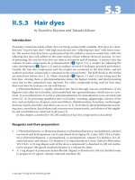
the transmission of a real time rhythm strip by having the patient place a small
device on the chest or by using a set of metal bracelets attached to the transmitter
(Fig 9.1). This device generates a tone that is decoded into a rhythm strip by a
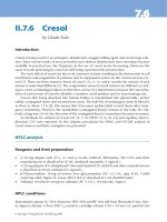
receiving center (Fig 9.2). This is useful in conjunction with magnet application
to determine if the pacemaker is functioning and to get a general idea as to the
condition of the battery. It does not replace the full evaluation that is performed
in the pacemaker clinic. An additional benefit of these transmitters is that the
patient can send a rhythm strip during an episode of palpitations.
The rationale for routine transtelephonic follow-up is as follows:
1. Makes available a method for monitoring the continued safety and lon-
gevity of the pacing system between office visits.
Ta b le 9.1. Risk considerations for programming and follow-up frequency
Degree of pacemaker dependency
Device advisories or recalls on the pacemaker or leads
Changes in underlying heart disease
Severity of underlying heart disease
Epicardial electrodes
Pediatric patients
Exposure to cardioversion, defibrillation, or electrocautery
High stimulation thresholds with high programmed outputs
Undersensing, interference or other sensing problems
Concurrent use of an ICD or other implanted device
78 Handbook of Cardiac Pacing
9
Fig. 9.1a and b. Front and back of basic transmitter. The four metal feet are dampened with water and
applied to the chest. The mouthpiece of the telephone is held over the front of the transmitter to send an
analog signal to the receiving center.
Fig. 9.1c. Cradle type transmitter packaged with
a magnet. The phone is placed in the cradle and
the wrist bands are placed on the patient to ac-
quire the electrocardiogram.
79Follow-Up of Permanent Pacemakers
9
Fig. 9.1d. Cardiophone
TM
Transmitter integrated
into a standard telephone set. The patient may
use the phone for routine calls, and plug in the
wrist bands to transmit to the pacing center
when needed.
Fig. 9.2. Typical
transtelephonic receiv-
ing center. This
Paceart
TM
system is
computer based and
runs on a standard
“PC”. Analysis, report-
ing and storage of the
transmitted rhythm
strip is performed ef-
ficiently as opposed to
the “cut and paste”
method of the older
style heated pen strip
recorders.
2. Provides a method of detecting pacemaker system abnormalities before
symptoms occur.
3. Allows transmission of a rhythm strip into the clinic office when pa-
tient is symptomatic.
4. For patients that are not able to come to the pacemaker clinic, this pro-
vides at least a minimal level of follow-up.
The standard procedure for routine transtelephonic evaluation at most cen-
ters is as follows:
1. The patient is questioned as to their general health status as well as any
symptoms that relate to cardiac rhythm.
2. Transmission of the rhythm for 30 seconds without a magnet.
3. Transmission of the rhythm for 30 seconds with a magnet over the pace-
maker.
4. The magnet is removed and another 30 seconds of rhythm is recorded.
80 Handbook of Cardiac Pacing
9
5. The patient is assured that the pacemaker function is normal. If a prob-
lem is found the patient has the situation explained. Arrangement is
made for a more thorough evaluation in the clinic or for corrective ac-
tion to be taken as indicated.
There is some concern that pacing a patient asynchronously could provoke a
ventricular or atrial arrhythmia. This could occur by delivering a pace output
during the vulnerable period of the cardiac cycle (R on T). While this is theoreti-
cally possible, it occurs extremely infrequently in clinical practice. For any patient
that has demonstrated a predisposition towards significant arrhythmia from mag-
net application, this type of testing should be avoided unless done in a proper
medically supervised environment.
FREQUENCY OF FOLLOW-UP
There are two approaches for routine evaluation in the clinic and by telephone;
Medicare guidelines and the NASPE guidelines. The former were developed for
pacemakers that are no longer in general use. They are antiquated and are used by
those who, in general, wish to maximize clinic revenue. The latter are more ratio-
nal and were developed with regard to the modern pacemakers. We use the NASPE
guidelines and strongly encourage others to do so as well. Follow-up frequency
should be adjusted based on the patient’s needs. These may be more frequent if
medically justified. The Medicare and NASPE guidelines are presented below.
MEDICARE GUIDELINES FOR PACEMAKER FOLLOW-UP
Pacemaker Clinic Monitoring:
Single chamber pacemakers
Tw ice the first 6 months following implant
Once every 12 months.
Dual chamber pacemakers:
Tw ice the first 6 months following implant
Once every 6 months.
Tr anstelephonic Monitoring (TTM):Guideline 1**
Single chamber pacemakers
1st month q 2 weeks
2nd–36th months q 8 weeks
37th and later q 4 weeks
Dual chamber pacemakers
1st month q 2 weeks
2nd–6th months q 4 weeks
7th–36th months q 8 weeks
36th month and later q 4 weeks
81Follow-Up of Permanent Pacemakers
9
Tr anstelephonic Monitoring (TTM):Guideline 2**
Single chamber pacemakers
1st month q 2 weeks
2nd–48th months q 12 weeks
49th–72nd month q 8 weeks
73rd month and later q 4 weeks
Dual chamber pacemakers
1st month q 2 weeks
2nd–30th months q 12 weeks
31st–48th month q 8 weeks
49th month and later q 4 weeks
**Medicare guideline 2 is for pacemakers that have demonstrated better than
90% longevity at 5 years, whose output voltage decreases less than 50% over at
least 3 months and whose magnet rate decreases less than 20% or 5 pulses per
minute over the same period. Virtually all modern pacemakers would fall under
this guideline.
Guideline 1 is for devices that do not meet the above criteria.
NASPE GUIDELINES FOR PACEMAKER FOLLOW-UP
Predischarge:
Full clinic evaluation + PA & Lateral CXR and 12-lead ECG,
Provide TTM transmitter and training in its use.
1st Outpatient Follow-up (6-8 weeks postimplant)
Full clinic evaluation
Programming changes to chronic values
Review patient education and retention of concepts
TTM only as required for symptoms prior to this visit
Early Surveillance Period (through 5th month)
One clinic or one TTM contact
Maintenance Period (beginning at 6 months)
Full Clinic evaluation yearly.
TTM with patient interview q3 months, unless clinic evaluation is performed
near scheduled TTM.
Intensified Period (Latest interval in the Medicare schedule or when battery shows
significant wear)
Full Clinic evaluation yearly
TTM with patient interview q1 month, unless clinic evaluation is performed
near a scheduled TTM.
For older pacemakers that are not showing significant signs of battery wear
there is really no need to perform monthly TTM evaluations unless indicated for
other reasons such as device reliability or recalls.
82 Handbook of Cardiac Pacing
10
Handbook of Cardiac Pacing, by Charles J. Love. © 1998 Landes Bioscience
Preoperative, Operative
and Postoperative Considerations
Preoperative Preparation of the Patient 82
Lead Insertion 83
Lead Positioning 83
Complications 84
Pacemaker Lead Extraction 84
Postoperative Management of the Pacemaker Patient 87
PREOPERATIVE PREPARATION OF THE PATIENT
Education of the patient is critical to ensure that there are appropriate expec-
tations regarding both the operative procedure as well as the outcome. It is impor-
tant that the patient understand not only what the pacemaker will do, but what it
will not do. Patients expecting that their gout will be cured or that their aortic
valve stenosis will resolve with a pacemaker implant will be quite disappointed.
Patients with appropriate expectations will be very satisfied. It is important to
give an adequate explanation to reduce the apprehension and anxiety that is nor-
mal. Important points to cover during the explanation include:
1. The indication for pacing
2. An explanation of the type of pacemaker chosen
3. A description of the basic function of the pacemaker
4. Determination of the site of the incision. This should be done with con-
sideration as to dominant hand, prior chest or breast surgery, chest or
clavicular injury, known vascular anomaly and special sporting or other
needs.
5. The type of sedation, analgesia and anesthesia to be used
6. A general description of the surgical technique
7. The risks of the operation
Prior to the operation, routine orders may include some or all of the following:
1. NPO for 6-8 hours
2. Basic laboratory studies (CBC, differential, electrolytes, BUN, creati-
nine, PT, PTT, platelet count)
3. Discontinuation of anticoagulants and aspirin if possible
4. 18 ga or larger intravenous access
5. Shave insertion area
6. Antiseptic soap bath or shower to area where incision is to be made
7. Void bladder on call to the operating room
83Preoperative, Operative and Postoperative Considerations
10
8. Prophylactic antibiotic (e.g., vancomycin, cefazolin, etc.)
9. Analgesia/sedation (e.g., midazolam, meperidine, etc.)
P
ACEMAKER POCKET LOCATION
As noted above, the location for the site of the pacemaker implant in the body
should be based on the patient needs and not on physician convenience. It is pre-
ferred that the nondominant side be used to minimize the exposure to flexion of
the implanted lead as it passes under the clavicle. Most patients find that having
the pacemaker on the dominant side is less comfortable and that it seems to “get
in the way.” If the patient has had a mastectomy then the opposite side should be
used as more tissue will be present to cover the pacemaker. Patients that like to
hunt may use either shoulder to brace the weapon. They should be asked which
shoulder they use for shooting so the device may be placed on the opposite side. It
is not considered good medical practice to have a gun lover angry at the physician.
A history of clavicular fracture or edema of an upper extremity should alert one
to the possibility of anatomic or vascular abnormalities that could make the im-
plant a challenge. Golfers present a true challenge as no matter which side one
places the device they will claim you ruined their game. In some cases the upper
chest is not appropriate for implant, or the transvenous route is not available from
above. In this case the pacemaker may be placed in the abdominal wall. Epicardial
leads are placed on the heart or alternate venous approaches may be considered.
For most pacemakers, the pocket is made in the prepectoral region, one or two
centimeters below and parallel to the clavicle. If the cephalic vein is used the inci-
sion may be in the deltopectoral groove.
LEAD INSERTION
The venous system is usually accessed for lead placement using a subclavian
vein cannulation or cephalic vein cutdown. The cutdown technique virtually elimi-
nates the risk of pneumothorax or hemothorax. It does take a bit more time to
perform, and occasionally only a small vein is found. Both approaches are com-
mon, however when diagnosing postoperative complications it is useful to know
which was used. On rare occasions the internal jugular vein may be used and the
lead tunneled over or under the clavicle to the pocket area.
LEAD POSITIONING
Tr ansvenous ventricular leads have been typically placed in the right ventricu-
lar apex, and transvenous atrial leads in the right atrial appendage. However, leads
may be placed in other positions as well. The use of active fixation leads with a
helix to screw them into the myocardium allows stable positioning in virtually
any position. New data suggests that ventricular leads placed high on the intra-
ventricular septum or in the right ventricular outflow tract will improve stroke
84 Handbook of Cardiac Pacing
10
volume as compared to lead placement in the apex. This may be due to a more
synchronized depolarization with the left ventricle. Atrial leads may be placed
anywhere in the right atrium as long as the thresholds for sensing and pacing are
good. If a lateral position for the atrial lead is used it is essential to be sure that the
phrenic nerve does not get stimulated with atrial pacing. Epicardial leads may be
screwed in, stabbed in, or sutured on. The approach to placing them on the heart
may be via sternotomy, thoracotomy (limited or otherwise) or subxyphoid.
Congenital anomalies may make transvenous lead placement difficult or im-
possible. The most common anomaly is the “persistent left superior vena cava”. In
this situation the left inominate vein does not cross over to meet the right inominate
to form the superior vena cava. Instead, the left inominate stays on the left side of
the chest and empties into the coronary sinus via the great cardiac vein. This
anomaly occurs in approximately 1% of the population. Though more difficult to
place, both single and dual chamber pacing systems may be inserted via this route.
Obviously, if the presence of this anomaly is known ahead of time the implant
would be best done on the right side. Other less common anomalies are related to
repairs of congenital defects such as transposition of the great vessels. Issues re-
garding these problems are beyond the scope of this publication. One must be
aware that they exist as the X-ray appearance may be unusual.
COMPLICATIONS OF PACEMAKER INSERTION
As with any surgical procedure there are many potential complications. Many
of these may be avoided by careful planning and performance of the procedure.
However, even in the best and most experienced hands problems may arise. It is
vital that a problem be identified as soon as possible so that corrective action may
be initiated. I have divided the types of complications into three categories as
shown in Table 10.1. Most of the listed problems are self explanatory. The
“Twiddler” syndrome is caused by a patient (often with Alzheimer’s Disease) flip-
ping the pacemaker over and over in the pocket. This causes the lead to wind up
much like a telephone cord that twists over on itself. The lead may be pulled out of
the heart by tension or the lead may be damaged by the severe stress caused by
torsion.
PACEMAKER LEAD EXTRACTION
When a pacing lead becomes infected or when certain other complications
arise it may be necessary to remove a chronically implanted lead or group of leads.
If the lead has been implanted less than one year, it will likely (but not always) pull
out of the heart with minimal traction. Leads that have been implanted for more
than a year begin to aggressively fibrose to the vascular and myocardial tissues.
Over time they become encased in scar tissue and even calcified. This makes them
difficult and potentially dangerous to remove. The indications based on a scheme
85Preoperative, Operative and Postoperative Considerations
10
published by Charles Byrd for removal of chronically implanted pacing leads are
listed in Table 10.2. The indications are currently under review and will be pub-
lished by NASPE in the near future. Mandatory reasons for removing a pacing
lead are the result of a condition that is or could be life threatening, while necessary
reasons are to correct a problem. Some problems do not cause an immediate medi-
cal threat, but one might wish to prevent future problems. In some cases it is
simply to the patient’s advantage to have the leads removed. These latter two situ-
ations fall under the discretionary category.
Leads, pacemakers and defibrillators that are infected present a difficult thera-
peutic problem. An infection involving only the incision and not the implanted
hardware may be curable with antibiotics. If the pocket itself is infected or if a
portion of the lead, pacemaker or defibrillator has eroded, antibiotics are unlikely
to result in a cure. At best, suppression of the infection will be possible until the
antibiotics are stopped. All of the prosthetic material must be removed from the
Ta b le 10.1. Complications of pacemaker insertion
Lead Insertion Related:
Pneumothorax
Hemothorax
Subclavian artery injury
Air embolus
Thoracic duct injury
Brachial plexus injury
Chordae or valve entanglement/rupture
Tr icuspid insufficiency by the lead holding the valve open
Cardiac perforation
Pericardial tamponade
Lead Related (other):
Diaphragm pacing (chest wall or phrenic nerve)
Endocarditis
Insulation failure/damage
Lead conductor failure
Lead connector failure
Lead dislodgment
Exit block (high-capture threshold)
Loss of sensing
Ve nous thrombosis
Poor connection at generator
Malposition in the coronary sinus
Malposition across a patent foramen ovale or atrial septal defect
Pocket Related:
Hematoma
Erosion
Infection
Chronic pain
Migration of pacemaker
“Twiddler” syndrome
Pectoralis muscle stimulation
Pocket stimulation
86 Handbook of Cardiac Pacing
10
patient to insure a cure. Infected pockets must be excised or left open to heal by
secondary intention. Patients with sepsis or endocarditis caused by the pacing
system must be treated aggressively with antibiotics and removal of all hardware.
Removal of pacing leads used to require an open heart procedure when they
would not come out with traction. Eventually, the “weight and pulley” method
was used. This technique was performed by exposing the pacing lead and tying a
piece of suture to it. The suture was then run over a pulley and a small weight was
tied to it. The patient was placed on a nursing unit until the weight crashed to the
floor. It was at this time that all would know that the lead had pulled free from the
heart. While this was a somewhat effective (not to mention dramatic) method of
removing leads, it was time consuming and the same implant site could not be
reused as it had been exposed for too long a period of time. Over the past decade
the use of locking stylets and counter traction sheaths have been developed to
provide a safe and rapid method to consistently remove leads. The locking stylets
stabalize the lead and provide traction at the tip of the lead. The sheaths are passed
over the lead to tear away adhesive scar tissue, and to provide a localized force at
the lead myocardial interface to prevent an avulsion. More recently the Excimer
laser has been approved for use in the removal of pacing leads. It is used to vapor-
ize the scar tissue instead of tearing it away. The laser energy is delivered by a
fiberoptic sheath that is passed over the lead. When the sheath encounters a bind-
ing site energy is applied and the sheath vaporizes the tissue. Though highly effec-
Ta b le 10.2. Indications for extraction of pacing leads
Mandatory
Endocarditis
Sepsis due to the pacing system
Obliteration of all usable veins
Mishap during attempted extraction
Tr aumatic injury to the lead / vein site
Necessary
Pocket infection
Occult infection
Erosion of the pacemaker or lead
Chronic draining sinus
Multiple leads in a single vessel
Discretionary
Malignancy
Pocket pain
Deteriorating polyurethane insulation
Accufix and Encor “J” leads*
*The need for removal of these leads will depend on the individual patient and the risk
benefit ratio as determined by the physician. The actual classification for extraction of
these leads will depend on the clinical situation. Elderly patients with normal leads need
not have them removed. Younger patients ought to have the leads extracted as a
precautionary measure. Patients with failed leads and protruding wires should have the
leads removed under most circumstances. Consultation with a lead extraction expert is
recommended in situations involving this lead.
87Preoperative, Operative and Postoperative Considerations
10
tive, this is an expensive methodology. Lead extraction is a difficult and poten-
tially dangerous procedure. It should only be performed by physicians well trained
and experienced in the technique, and at centers that are capable of dealing with
the potential life threatening problems that can occur.
POSTOPERATIVE MANAGEMENT OF THE PACEMAKER PATIENT
Postoperative management of the patient is directed at insuring that the de-
vice is functioning properly and at minimizing pain and the risk of wound prob-
lems. Common postoperative orders are listed in Table 10.3.
Many physicians obtain not only the PA and lateral chest X-ray on the follow-
ing day, but also a STAT portable film to rule out a pneumothorax. We obtain this
extra film only if there is a suspicion that the pleura was penetrated or if the pa-
tient becomes symptomatic. The ice pack to the site helps to reduce edema and
pain. The use of IV antibiotics is always controversial. Most surgical literature
supports the use of a single dose preoperatively as prophylaxis against infection.
The use of additional antibiotic postoperatively is not proven to reduce the inci-
dence of infection. However, most implanting physicians give at least one addi-
tional dose. Maintaining the head of the bed slightly elevated will help to keep the
venous pressures in the upper body lower and reduce the amount of bleeding into
the pacemaker pocket. Some physicians restrict the movement of the patient’s
arm with a sling to reduce the risk of lead dislodgment in the early postoperative
period. We have found that if the leads are properly secured and are placed with
the right amount of slack that dislodgment is very unlikely with or without the
sling. Indeed, if a lead is going to dislodge it tends to do so when the PA X-ray is
taken. It is during this time that the patient is standing with arms raised over the
X-ray plate during a deep inspiration. I have found this to be the ultimate test of
lead stability.
The pacemaker should be thoroughly evaluated prior to patient discharge. This
includes threshold checks, sensor evaluation and adjustment, activation of special
features and counters, and a final interrogation with printing of all programmed
parameters. The patient will have many questions that will require answers. In
addition, final instructions regarding wound care and follow-up must be discussed.
A summary of the instructions that we give our patients is presented in Table 10.4.
Most of the issues listed in Table 10.4 are straightforward. However, issues in-
volving electromagnetic interference (EMI) are of concern to both patients and
physicians. If the pacemaker does sense EMI, it will usually recognize this as ab-
normal and nonphysiologic. As the EMI will “blind” the pacemaker to the patient’s
heart rhythm, it will begin to pace asychronously at an “interference rate.” This is
usually around 60 or 70 bpm. It does this to be sure that the patient is not without
pacing support during this time. The patient may notice a change in the heart
rhythm due to loss of AV synchrony or competition with the native rhythm. The
pacemaker will resume normal function when the source is turned off or if the
patient moves away from it.