Báo cáo y học: "Thoracic epidural anesthesia reverses sepsis-induced hepatic hyperperfusion and reduces leukocyte adhesion in septic rats" potx
Bạn đang xem bản rút gọn của tài liệu. Xem và tải ngay bản đầy đủ của tài liệu tại đây (325.37 KB, 8 trang )
Open Access
Available online />Page 1 of 8
(page number not for citation purposes)
Vol 13 No 4
Research
Thoracic epidural anesthesia reverses sepsis-induced hepatic
hyperperfusion and reduces leukocyte adhesion in septic rats
Hendrik Freise
1
, Fritz Daudel
2
, Christina Grosserichter
1
, Stefan Lauer
1
, Juergen Hinkelmann
1
,
Hugo K Van Aken
1
, Andreas W Sielenkaemper
3
, Martin Westphal
1
and Lars G Fischer
1
1
Department of Anesthesiology and Intensive Care, University Hospital of Muenster, Albert-Schweitzer-Strasse 33, 48149 Muenster, Germany
2
Department of Intensive Care Medicine, Inselspital, University of Bern, Freiburgstrasse, 3010 Bern, Switzerland
3
Department of Anesthesiology and Intensive Care Medicine, St. Theresien-Hospital Saarbrücken, Rheinstraße 2, 66113 Saarbrücken, Germany
Corresponding author: Hendrik Freise,
Received: 13 Mar 2009 Revisions requested: 5 May 2009 Revisions received: 26 May 2009 Accepted: 13 Jul 2009 Published: 13 Jul 2009
Critical Care 2009, 13:R116 (doi:10.1186/cc7965)
This article is online at: />© 2009 Freise et al.; licensee BioMed Central Ltd.
This is an open access article distributed under the terms of the Creative Commons Attribution License ( />),
which permits unrestricted use, distribution, and reproduction in any medium, provided the original work is properly cited.
Abstract
Introduction Liver dysfunction is a common feature of severe
sepsis and is associated with a poor outcome. Both liver
perfusion and hepatic inflammatory response in sepsis might be
affected by sympathetic nerve activity. However, the effects of
thoracic epidural anesthesia (TEA), which is associated with
regional sympathetic block, on septic liver injury are unknown.
Therefore, we investigated hepatic microcirculation and
inflammatory response during TEA in septic rats.
Methods Forty-five male Sprague-Dawley-rats were
instrumented with thoracic epidural catheters and randomized
to receive a sham procedure (Sham), cecal ligation and
puncture (CLP) without epidural anesthesia (Sepsis) and CLP
with epidural infusion of 15 ul/h bupivacaine 0.5% (Sepsis +
TEA). All animals received 2 ml/100 g/h NaCl 0.9%. In 24 (n =
8 in each group) rats, sinusoidal diameter, loss of sinusoidal
perfusion and sinusoidal blood flow as well as temporary and
permanent leukocyte adhesion to sinusoidal and venolar
endothelium were recorded by intravital microscopy after 24
hours. In 21 (n = 7 in each group) separate rats, cardiac output
was measured by thermodilution. Blood pressure, heart rate,
serum transaminase activity, serum TNF-alpha concentration
and histologic signs of tissue injury were recorded.
Results Whereas cardiac output remained constant in all
groups, sinusoidal blood flow increased in the Sepsis group and
was normalized in rats subjected to sepsis and TEA. Sepsis-
induced sinusoidal vasoconstriction was not ameliorated by
TEA. In the Sepsis + TEA group, the increase in temporary
venolar leukocyte adherence was blunted. In contrast to this,
sinusoidal leukocyte adherence was not ameliorated in the
Sepsis + TEA group. Sepsis-related release of TNF-alpha and
liver tissue injury were not affected by Sepsis + TEA.
Conclusions This study demonstrates that TEA reverses
sepsis-induced alterations in hepatic perfusion and ameliorates
hepatic leukocyte recruitment in sepsis.
Introduction
The liver is critically involved in a multitude of vital physiological
processes and contributes to the host's immune reaction in
systemic inflammatory response and sepsis [1-3]. Impaired
microcirculation and intrahepatic inflammatory reaction are
hallmarks in primary and secondary hepatic injuries [4-6]. In
severe sepsis and trauma, liver injury is associated with
increased mortality and length of hospital stay [7-10]. The
hepatic immune response determines pathogen clearance
and the systemic immune reaction [1,5,11]. After prolonged
inflammation, hepatic immune dysfunction contributes to mor-
tality [12]. Protection of liver function is therefore crucial to the
maintenance of homeostasis in perioperative and critical care
medicine.
Sympathetic nerve activity plays a crucial role in hepatic injury
and immune response. Increased sympathetic tone alone
induces intrahepatic inflammation and liver injury in healthy
ANOVA: analysis of variance; CLP: cecal ligation and puncture; ELISA: enzyme-linked immunosorbent assay; HABR: hepatic arterial buffer response;
H&E: hematoxylin and eosin; NaCl: sodium chloride; TEA: thoracic epidural anesthesia; TNF: tumor necrosis factor.
Critical Care Vol 13 No 4 Freise et al.
Page 2 of 8
(page number not for citation purposes)
mice, whereas sympathetic denervation reduced perioperative
hepatic injury [13-15]. In sepsis, both α- and β-adrenorecep-
tors impair hepatic function and immune response [16-18].
Thoracic epidural anesthesia (TEA) promotes postoperative
intestinal recovery and reduces cardiovascular mortality, most
probably mediated by regional sympathetic block [19-28].
Recently, TEA has also been shown to ameliorate organ injury
and improve outcome in sepsis and necrotizing pancreatitis
[28-32]. The hepatic effects of TEA in sepsis, however, have
never been subject to investigation.
Therefore, we conducted a randomized, blinded experimental
study to test the hypothesis that TEA: improves hepatic micro-
vascular perfusion and attenuates leukocyte activation in sep-
sis; and influences systemic inflammatory response and liver
tissue injury induced by cecal ligation and puncture (CLP) in
rats.
Materials and methods
The study was approved by the animal care committee of the
District Government of Muenster. Animals received standard
chow and were kept in a 12 hour light-dark-cycle. Food was
withheld 12 hours prior to surgery. The animals had free
access to water.
Male Sprague-Dawley rats (weighing 275 to 300 g; Harlan-
Winkelmann, Borchen, Germany) were anesthetized by isoflu-
ran in 50% oxygen. Central venous and arterial lines (0.96 mm
once daily; Liquidscan, Ueberlingen, Germany) were intro-
duced. Epidural catheters (0.61 mm once daily) were inserted
at L3/L4 and advanced to Th6 [26]. All catheters were exteri-
orized at the neck of the animal and protected by a swivel
device. The cecum was ligated below the ileocecal valve to
maintain intestinal continuity and then punctured at two loca-
tions with an 18-gauge needle. Subsequently anesthesia was
terminated and volume resuscitation was performed using 2
mL/100 g/hour isotonic sodium chloride (NaCl) solution intra-
venously. The animals were housed individually for the follow-
ing 24 hours. The correct position of the epidural catheter was
confirmed by autopsy after completion of the experiment.
After instrumentation, animals were randomly allocated to one
of the three groups by closed envelopes: Sham = sham oper-
ation, 15 μl/hour NaCl 0.9% epidural; Sepsis = CLP 24 hours,
15 μl/hour NaCl 0.9% epidural; Sepsis + TEA = CLP 24
hours, 15 μl/h bupivacaine 0.5% epidural. The investigators
were not aware of the group assignment.
Twenty-four hours after CLP and sham-laparotomy, mean arte-
rial blood pressure was recorded using a standard transducer
(PMSET 1 DT, Becton Dickinson, Germany) and a monitor
(Siemens Sirecust 404, Siemens, Germany). Heart rate was
derived from the arterial pressure curve. For blood gas analy-
ses, 80 μl blood was withdrawn. Motoric block was quantified
using an established motor score derived from the Bromage
score and adapted to rats [33].
Intravital microscopy
Twenty-four hours after sepsis induction, 24 animals (n = 8 per
group) were then re-anesthetized and tracheotomized [29].
Intravital microscopy of the left liver lobe was performed as fol-
lows: median laparotomy was extended by a left subcostal
incision and the hepatic ligaments of the left liver lobe were
carefully dissected. The animal was placed in a 110° position
on its left side onto the microscope (Eclipse 300, Nikon, Düs-
seldorf, Germany). The left liver lobe was exteriorized and the
lower surface was placed on a microscope slide in a tension
free position. Intravenously, 2 μmol/kg sodium fluorescein and
0.2 μmol/kg rhodamine 6 G (Sigma, Deisenhofen, Germany)
were used for contrast enhancement.
In each experiment, 10 randomly chosen acini and 10 postsi-
nusoidal venoles were recorded for 30 seconds both with
sodium-fluorescein and rhodamine contrast enhancement.
Offline image analysis was performed by a blinded investigator
(CG) using a computer-assisted image analysis system (Anal-
ySIS, OSIS, Muenster, Germany). Hepatic microcirculation
was assessed by the periportal sinusoidal diameter of 10 sinu-
soids per acinus and the loss of sinusoidal perfusion, defined
as the number of non-perfused sinusoids divided by all visible
sinusoids of the acinus.
The leukocyte adhesion was evaluated separately in sinusoids
and postsinusoidal venoles. Temporary adherent, that is,
slowly moving or adhering at the sinusoidal wall for less than
20 seconds, and permanently adherent, that is, adherent for
more than 20 seconds, leukocytes were counted in each aci-
nus and expressed as cells/μm
2
. Accordingly, temporarily and
permanently adherent leukocytes in the venoles were counted
as cells/μm
2
venolar endothelium.
Cardiac output and liver injury
In another set of 21 animals, cardiac output was determined
applying the thermodilution technique 24 hours after sepsis. In
these animals, a thermocouple catheter (IT21, Physitemp,
Clifton, NJ, USA) was introduced into the aortic arch via the
left carotid artery during instrumentation. For measurement of
cardiac output, the area under temperature curve after injec-
tion of 0.3 ml cold saline solution (8°C) was recorded (Cardiac
Output pod and Powerlab 4/20, ADInstruments, Spechbach,
Germany). The results of three measurements were averaged
in each animal. To reduce bias of emotional stress the proce-
dure was mimicked every hour for four hours before measure-
ment.
Liver cell injury was assessed 24 hours after induction of sep-
sis by measuring serum activities of aspartate aminotrans-
ferase and alanine aminotransferase. Blood was withdrawn via
aortic puncture and plasma enzyme activity was determined by
Available online />Page 3 of 8
(page number not for citation purposes)
means of standard enzymatic techniques (Ektachem, Kodak,
Stuttgart, Germany).
Specimens of the left liver lobe were collected immediately
after death and fixed by immersion in 4% formaldehyde solu-
tion. Subsequently, they were dehydrated and embedded in
paraffin wax to cut sections at a thickness of 5 μm. Slides were
stained with H&E and assessed by an experienced patholo-
gist.
Serum TNF-α
Twenty-four hours after CLP and sham-procedure respec-
tively, serum concentration of TNF-α was measured by a com-
mercially available anti-rat TNF-α ELISA (BD OptEIA, Cat No.
550734, Becton Dickinson, Heidelberg, Germany) according
to the manufacturer's instructions and read-out by a fluoromet-
ric plate reader (EL808, BioTek, Bad Friedrichshall, Germany).
Statistics
Sigmastat 3.0 (Systat Software, Richmond, CA, USA) was
used for statistical analysis. Normal distribution and equal var-
iance tests were performed. Sepsis-induced and TEA-related
effects were evaluated by one-way analysis of variance
(ANOVA) with post-hoc Student Newman Keuls test or
ANOVA on Ranks with post-hoc Dunn's test as appropriate. A
P < 0.05 was defined as the level of significance. Data are pre-
sented as mean ± 95% confidence interval or as median
(25%/75% percentiles) as appropriate.
Results
All animals allocated to the sepsis groups showed signs of
lethargy, piloerection, and exudation around the eyes and nose
24 hours after induction of sepsis. Peritoneal inflammation and
purulent ascites was present when the abdomen was reo-
pened for intravital microscopy.
Compared with the Sham-group, mean arterial blood pressure
and heart rate were not affected in the untreated Sepsis or
Sepsis + TEA groups. Cardiac output also remained constant
both in the Sepsis and Sepsis + TEA groups. Arterial oxygen
tension and pH were not altered by sepsis or treatment with
TEA (Table 1). Serum-lactate concentrations were increased
to 1.25 (1.00/1.50) mmol/l in the Sepsis group compared with
0.8 (0.7/0.9) mmol/l in the Sham group (P < 0.05). Leukocyte
count dropped from 6430 ± 3099 cells/μL in Sham animals to
2228 ± 1129 cells/μl in the CLP group (P < 0.05). In addition,
the Sepsis group was characterized by a drop in platelet count
compared with the Sham-group (197,000 ± 102,000 cells/μl
vs. 338,000 ± 64,000/μl; P < 0.05). These parameters were
not altered in animals subjected to Sepsis + TEA as compared
with the untreated Sepsis group. Serum TNF-α concentration
was elevated after 24 hours in the Sepsis-group (P < 0.05 vs.
Sham). This increase was not ameliorated in the Sepsis+TEA
group (Figure 1).
Hepatic microcirculation and leukocyte adherence
Sinusoidal blood flow increased in the Sepsis group, whereas
in the Sepsis + TEA group flow returned to Sham levels (Fig-
ure 2). The numbers of perfused sinusoids did not differ
between groups. However, in the Sepsis group sinusoidal
constriction was induced, which was not influenced in the
Sepsis + TEA group (Figure 3).
Temporary leukocyte adhesion increased in sepsis both in the
sinusoids and in the postsinusoidal venules. TEA reduced the
temporary venolar leukocyte adhesion significantly, whereas it
did not affect the increased sinusoidal adherence (Figure 4).
The permanent sinusoidal and venolar leukocyte adherence
was neither affected in the Sepsis group, nor in animals sub-
jected to Sepsis + TEA.
Liver Injury
In the untreated Sepsis group, serum activity of aspartate ami-
notransferase rose from 275 ± 101 U/l to 454 ± 108 U/l and
alanine aminotransferase activity increased from 97 ± 81 U/l
to 185 ± 58 U/l (P < 0.05 vs. Sham). These increases were
not significantly affected by TEA. Similarly, histopathologic
examination revealed only mild edema formation and patchy
pericentral necrosis in sepsis.
Discussion
The knowledge about the hepatic effects of TEA is just the
beginning. Recent investigation in the pre- and intraoperative
period in human and animals revealed conflicting results with
respect to hepatic perfusion [34-38]. All these studies were
performed in healthy subjects after a single bolus of epidural
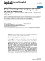
Figure 1
Serum TNF-αSerum TNF-α. Serum TNF-α 24 hours after induction of sepsis by
cecal ligation and puncture and sham procedure respectively. In Sep-
sis, serum TNF-α was increased compared with Sham (* P < 0.05 vs.
Sham). Thoracic epidural anesthesia (TEA) did not ameliorate this sign
of systemic inflammation. Data (n = 7 in each group) are displayed as
mean ± 95% confidence interval.
Critical Care Vol 13 No 4 Freise et al.
Page 4 of 8
(page number not for citation purposes)
local anesthetics. The impact of continuous TEA on liver injury
in severe sepsis were not investigated.
Hepatic dysfunction in critical illness is still not completely
understood. In the current concept of septic liver injury, two
phases of dysfunction are distinguished [1]. The early phase is
related to hypoperfusion in the presence of hypovolemia and
inadequate cardiac output and resolves fast under supportive
therapy. The late and persistent dysfunction is characterized
by (supra-) normal tissue perfusion.
In this study, the microvascular liver blood flow was signifi-
cantly increased in the untreated Sepsis group. In the Sepsis
+ TEA group, sinusoidal blood flow was normalized compared
with the untreated Sepsis group. These changes in hepatic
perfusion were not correlated to changes in cardiac output,
which remained stable both in the Sepsis group and in the
Sepsis + TEA group. Furthermore, the effects of TEA on
hepatic tissue blood flow were also not associated with
altered sinusoidal vasoregulation or increased sinusoidal
recruitment.
Effects of sepsis and TEA on hepatic perfusion
Hepatic macrovascular inflow, although not directly measured
in this study, most likely remained constant because cardiac
output was not altered. This assumption is supported by
numerous studies showing a consistent correlation of cardiac
output and macrovascular hepatosplanchnic inflow in sepsis.
In human sepsis, macrovascular hepatic inflow rose with car-
diac output after therapeutic interventions [39-41]. Both in
early and late CLP-sepsis macrovascular hepatic inflow was
reduced in parallel with cardiac output [42,43]. Furthermore,
in the presence of unchanged or increased cardiac output,
hepatic macrovascular inflow also paralleled these changes in
cardiac output [42,44,45]. Consequently, the sepsis-induced
increase in sinusoidal blood flow and its reversal by TEA are
most probably not caused by changes of macrovascular
hepatic inflow.
Microvascular tissue perfusion in sepsis, however, is often
uncoupled from the systemic circulation. In the clinical therapy
of critical illness this dissociation might contribute to the per-
sistent organ failure after hemodynamic stabilization [46]. Ear-
lier studies demonstrated unchanged or even decreased
microvascular blood flow in the presence of two to three-fold
increased regional blood flow [42,45]. In our study sinusoidal
blood flow was increased in the Sepsis group whereas car-
diac output was not altered. Intravital microscopy and cardiac
output, however, needed to be performed in different sets of
animals to minimize interaction between both techniques.
The increase in hepatic microvascular blood flow occurred
despite sinusoidal vasoconstriction and consequently was
Table 1
Cardiorespiratory parameters
MAP
(mmHg)
HR
(bpm)
CO
(ml/min)
pH PaO
2
(mmHg)
Sham 136 ± 10 432
(404/444)
420 ± 74 7.42 ± 0.02 87 ± 10
Sepsis 121 ± 11 468
(447/477)
402 ± 68 7.40 ± 0.09 91 ± 20
Sepsis + TEA 129 ± 16 420
(297/480)
391 ± 152 7.39 ± 0.09 90 ± 11
Twenty-four hours after induction of sepsis by cecal ligation and puncture and sham procedure, respectively. None of these parameters were
significantly altered by sepsis or sepsis + thoracic epidural anesthesia (TEA). Data (n = 7) each group are displayed as mean ± 95% confidence
interval (CI) or median (25%/75% percentile).
CO = cardiac output; HR = heart rate; MAP = mean arterial pressure; PaO
2
= partial pressure of arterial oxygen.
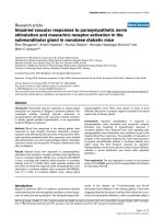
Figure 2
Hepatic microvascular blood flowHepatic microvascular blood flow. Sinusoidal blood flow 24 hour after
induction of sepsis by cecal ligation and puncture and sham procedure,
respectively. In Sepsis, blood flow was increased compared with Sham
(* P < 0.05 vs. Sham). Thoracic epidural anesthesia (TEA) reduced
blood flow (# P < 0.05 vs. Sepsis). Data (n = 8 in each group) are dis-
played as mean ± 95% confidence interval.
Available online />Page 5 of 8
(page number not for citation purposes)
related to increased sinusoidal blood flow velocity. These find-
ings are consistent with an increased arteriolar inflow and are
thus the first hint to an impaired hepatic arterial buffer
response (HABR) in late polymicrobial sepsis. In HABR, liver
arterial blood flow is adapted in response to changes in portal
blood flow. Intrahepatic vasodilatation occurs at the level of
the preterminal branches of the hepatic artery and is regulated
by hydrogen sulfide and adenosine washout by portal blood
flow [47,48]. Our results are supported by earlier findings
demonstrating impaired HABR and a selective increase in
hepatic arterial blood flow in endotoxemia [49,50]. In the Sep-
sis + TEA group, the sepsis-related increase in liver microvas-
cular blood flow was blunted. It is therefore most likely that the
continuous TEA restored HABR. However, this line of interpre-
tation of the presented data is limited by the fact that we did
not measure hepatic and portal flow, pressure and resistance
separately in this study. Further investigations on the influence
of TEA on hepatic blood flow regulation in sepsis and other
clinically relevant conditions such as major liver resections are
warranted.
In this study, loss of sinusoidal perfusion was not present in
the Sepsis group whereas in earlier studies intravital micros-
copy revealed sinusoidal vasoconstriction and up to 30%
reduction in sinusoidal perfusion both in early and late rodent
CLP-sepsis [51-53]. Differences in volume resuscitation might
partly explain the differing results. In the previous studies an
initial bolus of 20 to 60 ml/kg saline solution was administered
during 20 to 24 hour CLP-sepsis [53,54]. In our study, volume
was infused continuously to a total dose of 144 ml/kg/24
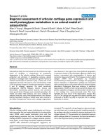
Figure 3
Hepatic microcirculationHepatic microcirculation. (a) Percentage of non-perfused sinusoids
and (b) sinusoidal width 24 hours after induction of sepsis by cecal
ligation and puncture and sham procedure, respectively. In Sepsis sinu-
soidal vasoconstriction occurred (* P < 0.05 vs. Sham), which was not
influenced in Sepsis + thoracic epidural anesthesia (TEA). Sinusoidal
perfusion was neither influenced in Sepsis nor in Sepsis + TEA. Data (n
= 8 in each group) are displayed as mean ± 95% confidence interval.
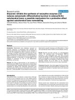
Figure 4
Temporary leukocyte adhesionTemporary leukocyte adhesion. Numbers of leukocytes adhering tem-
porarily to the (a) sinusoidal and (b) postsinusoidal venolar endothe-
lium 24 hours after induction of sepsis by cecal ligation and puncture
and sham procedure respectively. In Sepsis, temporary adherence
increased both to the sinusoidal and to the venolar endothelium (* P <
0.05 vs. Sham). The venolar leukocyte adherence was prevented in
Sepsis + TEA (# P < 0.05 vs. Sepsis). Data (n = 8 in each group) are
displayed as mean ± 95% confidence interval.
Critical Care Vol 13 No 4 Freise et al.
Page 6 of 8
(page number not for citation purposes)
hours. Detection of hypovolemia is often difficult in critical
care. In experimental rat sepsis, volume depletion is even
harder to exclude. In the present study, mean arterial pressure
and heart rate were not significantly affected. Cardiac output
remained stable both in untreated rats and in the sepsis + TEA
group. The stable systemic hemodynamic parameters com-
bined with stable acid-base-balance and a well-maintained
microvascular perfusion in the liver suggests a sufficient resus-
citation in this model. This clinically relevant infusion regimen
might have prevented loss of perfused sinusoids and contrib-
uted to increased tissue blood flow in our study.
Effects of sepsis and TEA on leukocyte adhesion
In the Sepsis + TEA group, sepsis-induced temporary leuko-
cyte adhesion to the venolar endothelium decreased, whereas
temporary sinusoidal leukocyte adhesion was not prevented.
In contrast, the permanent leukocyte adhesion after 24 hour of
sepsis was neither affected in the Sepsis group nor in the Sep-
sis + TEA group. This observation is in line with prior findings
of time-dependent pattern of leukocyte recruitment in rat CLP-
sepsis with increased leukocyte adhesion after seven hours
and normalized values after 20 hours [55]. In a recent study
hepatic neutrophil recruitment declined after eight hours [56].
In contrast to sinusoidal temporary adhesion, hepatic venolar
temporary leukocyte adhesion is initiated by selectins [4,57-
59].
Reduced venolar rolling in the Sepsis + TEA group might have
been related to immune regulatory consequences of splanch-
nic sympathetic block. The technique of continuous TEA used
in this study induced a sympathetic block including hepatic
and intestinal sympathetic nerve roots as demonstrated by
thermography [26]. This block can be induced as long as 72
hours after catheter placement. There is some evidence of
hepatic sympathetic immune regulation supporting this inter-
pretation. Increased sympathetic activity in acute urinary reten-
tion results in hepatic inter-cellular adhesion molecule-1
expression [60]. Increased portal norepinephrine in sepsis
trigger hepatic release of TNF-α [17,18], which is in turn a pre-
requisite for venolar temporary leukocyte adhesion [61,62].
Furthermore, TEA has already been shown to reduce tempo-
rary adhesion in mesenteric venoles in hemorrhagic shock
[63]. Therefore, the abdominal sympathetic block associated
with TEA also might have reduced the venolar temporary leu-
kocyte adhesion in sepsis.
Finally, intestinal injury and portal inflow of inflammatory medi-
ators induce secondary hepatic inflammation [1,64,65]. Con-
sequently, the decreased hepatic leukocyte adhesion may be
related to the intestinal protection provided by TEA
[29,30,66]. The systemic inflammatory response as measured
by systemic release of TNF-alpha was not influenced in the
Sepsis + TEA group.
Thoracic epidural infusion of local anesthetics is related to a
segmental sensoric block, analgesia, and sympathetic block.
Each of these aspects, as well as systemically resorbed bupi-
vacaine might contribute to the observed effects of TEA on
hepatic microvascular perfusion and leukocyte adhesion.
However, the present study does not allow to distinguish or
weight these potential mechanisms or to separate primarily
hepatic effects from those secondary to intestinal effects of
TEA.
Conclusions
In this study, TEA ameliorated the sepsis-induced increase in
microvascular liver blood flow and attenuated leukocyte
recruitment. These results suggest an altered regulation of
liver blood flow and a modified intrahepatic immune response
during continuous TEA in sepsis. The consequences of TEA
with respect to liver injury, remote organ dysfunction and out-
come needs to be further explored.
Competing interests
The authors declare that they have no competing interests.
Authors' contributions
HF contributed to design, funding, data acquisition, statistical
analysis and drafted the manuscript. FD contributed to the
design of the study. CG participated in data acquisition and
analysis. SL participated in study planning, data acquisition
and statistical analysis. JH contributed to data analysis and to
the manuscript. HVA participated in design, funding analysis
and manuscript drafting. AWS contributed to funding of the
study and participated to planning and statistical analysis. MW
contributed to data analysis and drafting of the manuscript.
LGF took part in data acquisition and drafting of the manu-
script.
Acknowledgements
This work was supported by the German Research Society (DFG, Grant
Si 629/2-1, Le 625/8-1).
References
1. Dhainaut JF, Marin N, Mignon A, Vinsonneau C: Hepatic response
to sepsis: interaction between coagulation and inflammatory
processes. Crit Care Med 2001, 29:S42-47.
2. Folch-Puy E: Importance of the liver in systemic complications
associated with acute pancreatitis: the role of Kupffer cells. J
Pathol 2007, 211:383-388.
3. Fong YM, Marano MA, Moldawer LL, Wei H, Calvano SE, Kenney
JS, Allison AC, Cerami A, Shires GT, Lowry SF: The acute
Key messages
• TEA does not affect cardiac output in late sepsis.
• TEA reverses hepatic hyperperfusion in late sepsis,
probably by restoring hepatic arterial buffer response.
• TEA ameliorates intrahepatic temporary leukocyte adhe-
sion in late sepsis.
Available online />Page 7 of 8
(page number not for citation purposes)
splanchnic and peripheral tissue metabolic response to endo-
toxin in humans. J Clin Invest 1990, 85:1896-1904.
4. McDonald B, McAvoy EF, Lam F, Gill V, de la Motte C, Savani RC,
Kubes P: Interaction of CD44 and hyaluronan is the dominant
mechanism for neutrophil sequestration in inflamed liver sinu-
soids. J Exp Med 2008, 205:915-927.
5. Gregory SH, Wing EJ: Neutrophil-Kupffer cell interaction: a crit-
ical component of host defenses to systemic bacterial infec-
tions. J Leukoc Biol 2002, 72:239-248.
6. Pannen BH: New insights into the regulation of hepatic blood
flow after ischemia and reperfusion. Anesth Analg 2002,
94:1448-1457.
7. Derikx JP, Poeze M, van Bijnen AA, Buurman WA, Heineman E:
Evidence for intestinal and liver epithelial cell injury in the early
phase of sepsis. Shock 2007, 28:544-548.
8. Harbrecht BG, Doyle HR, Clancy KD, Townsend RN, Billiar TR,
Peitzman AB: The impact of liver dysfunction on outcome in
patients with multiple injuries. Am Surg 2001, 67:122-126.
9. Harbrecht BG, Zenati MS, Doyle HR, McMichael J, Townsend RN,
Clancy KD, Peitzman AB: Hepatic dysfunction increases length
of stay and risk of death after injury. J Trauma 2002,
53:517-523.
10. te Boekhorst T, Urlus M, Doesburg W, Yap SH, Goris RJ: Etiologic
factors of jaundice in severely ill patients. A retrospective
study in patients admitted to an intensive care unit with severe
trauma or with septic intra-abdominal complications following
surgery and without evidence of bile duct obstruction. J Hepa-
tol 1988, 7:111-117.
11. Hildebrand F, Hubbard WJ, Choudhry MA, Frink M, Pape HC, Kun-
kel SL, Chaudry IH: Kupffer cells and their mediators: the cul-
prits in producing distant organ damage after trauma-
hemorrhage. Am J Pathol 2006, 169:784-794.
12. Xiao H, Siddiqui J, Remick DG: Mechanisms of mortality in early
and late sepsis. Infect Immun 2006, 74:5227-5235.
13. Sanchez O, Viladrich M, Ramirez I, Soley M: Liver injury after an
aggressive encounter in male mice. Am J Physiol Regul Integr
Comp Physiol 2007, 293:R1908-1916.
14. Schemmer P, Schoonhoven R, Swenberg JA, Bunzendahl H, Thur-
man RG:
Gentle in situ liver manipulation during organ harvest
decreases survival after rat liver transplantation: role of
Kupffer cells. Transplantation 1998, 65:1015-1020.
15. Schemmer P, Bunzendahl H, Raleigh JA, Thurman RG: Graft sur-
vival is improved by hepatic denervation before organ harvest-
ing. Transplantation 1999, 67:1301-1307.
16. Zhou M, Yang S, Koo DJ, Ornan DA, Chaudry IH, Wang P: The
role of Kupffer cell alpha(2)-adrenoceptors in norepinephrine-
induced TNF-alpha production. Biochim Biophys Acta 2001,
1537:49-57.
17. Zhou M, Das P, Simms HH, Wang P: Gut-derived norepine-
phrine plays an important role in up-regulating IL-1beta and
IL-10. Biochim Biophys Acta 2005, 1740:446-452.
18. Yang S, Zhou M, Chaudry IH, Wang P: Norepinephrine-induced
hepatocellular dysfunction in early sepsis is mediated by acti-
vation of alpha2-adrenoceptors. Am J Physiol Gastrointest
Liver Physiol 2001, 281:G1014-1021.
19. Kehlet H: Manipulation of the metabolic response in clinical
practice. World J Surg 2000, 24:690-695.
20. Rodgers A, Walker N, Schug S, McKee A, Kehlet H, van Zundert
A, Sage D, Futter M, Saville G, Clark T, MacMahon S: Reduction
of postoperative mortality and morbidity with epidural or spi-
nal anaesthesia: results from overview of randomised trials.
BMJ 2000, 321:1493.
21. Tziavrangos E, Schug SA: Regional anaesthesia and periopera-
tive outcome. Curr Opin Anaesthesiol 2006, 19:521-525.
22. Wu CL, Hurley RW, Anderson GF, Herbert R, Rowlingson AJ,
Fleisher LA: Effect of postoperative epidural analgesia on mor-
bidity and mortality following surgery in medicare patients.
Reg Anesth Pain Med 2004, 29:525-533. discussion 515–529.
23. Brodner G, Van Aken H, Hertle L, Fobker M, Von Eckardstein A,
Goeters C, Buerkle H, Harks A, Kehlet H: Multimodal periopera-
tive management – combining thoracic epidural analgesia,
forced mobilization, and oral nutrition – reduces hormonal and
metabolic stress and improves convalescence after major uro-
logic surgery. Anesth Analg 2001, 92:1594-1600.
24. Berendes E, Schmidt C, Van Aken H, Hartlage MG, Wirtz S, Rei-
necke H, Rothenburger M, Scheld HH, Schluter B, Brodner G,
Walter M:
Reversible cardiac sympathectomy by high thoracic
epidural anesthesia improves regional left ventricular function
in patients undergoing coronary artery bypass grafting: a ran-
domized trial. Arch Surg 2003, 138:1283-1290. discussion
1291.
25. Bakhtiary F, Therapidis P, Dzemali O, Ak K, Ackermann H, Meinin-
ger D, Kessler P, Kleine P, Moritz A, Aybek T, Dogan S: Impact of
high thoracic epidural anesthesia on incidence of periopera-
tive atrial fibrillation in off-pump coronary bypass grafting: a
prospective randomized study. J Thorac Cardiovasc Surg
2007, 134:460-464.
26. Freise H, Anthonsen S, Fischer LG, Van Aken HK, Sielenkamper
AW: Continuous thoracic epidural anesthesia induces seg-
mental sympathetic block in the awake rat. Anesth Analg 2005,
100:255-262.
27. Hogan QH, Stekiel TA, Stadnicka A, Bosnjak ZJ, Kampine JP:
Region of epidural blockade determines sympathetic and
mesenteric capacitance effects in rabbits. Anesthesiology
1995, 83:604-610.
28. Scott AM, Starling JR, Ruscher AE, DeLessio ST, Harms BA: Tho-
racic versus lumbar epidural anesthesia's effect on pain con-
trol and ileus resolution after restorative proctocolectomy.
Surgery 1996, 120:688-695. discussion 695-687.
29. Freise H, Lauer S, Anthonsen S, Hlouschek V, Minin E, Fischer LG,
Lerch MM, Van Aken HK, Sielenkamper AW: Thoracic epidural
analgesia augments ileal mucosal capillary perfusion and
improves survival in severe acute pancreatitis in rats. Anesthe-
siology 2006, 105:354-359.
30. Daudel F, Freise H, Westphal M, Stubbe HD, Lauer S, Bone HG,
Van Aken H, Sielenkamper AW: Continuous thoracic epidural
anesthesia improves gut mucosal microcirculation in rats with
sepsis. Shock 2007, 28:610-614.
31. Lauer S, Freise H, Fischer LG, Singbartl K, Aken HV, Lerch MM,
Sielenkamper AW: The role of thoracic epidural analgesia in
receptor-dependent and receptor-independent pulmonary
vasoconstriction in experimental pancreatitis. Anesth Analg
2007, 105:453-459.
32. Demirag A, Pastor CM, Morel P, Jean-Christophe C, Sielenkamper
AW, Guvener N, Mai G, Berney T, Frossard JL, Buhler LH: Epi-
dural anaesthesia restores pancreatic microcirculation and
decreases the severity of acute pancreatitis. World J Gastro-
enterol 2006, 12:
915-920.
33. Sielenkamper AW, Eicker K, Van Aken H: Thoracic epidural
anesthesia increases mucosal perfusion in ileum of rats.
Anesthesiology 2000, 93:844-851.
34. Greitz T, Andreen M, Irestedt L: Haemodynamics and oxygen
consumption in the dog during high epidural block with spe-
cial reference to the splanchnic region. Acta Anaesthesiol
Scand 1983, 27:211-217.
35. Kennedy WF Jr, Everett GB, Cobb LA, Allen GD: Simultaneous
systemic and hepatic hemodynamic measurements during
high peridural anesthesia in normal man. Anesth Analg 1971,
50:1069-1077.
36. Meierhenrich R, Wagner F, Schutz W, Rockemann M, Steffen P,
Senftleben U, Gauss A: The effects of thoracic epidural
anesthesia on hepatic blood flow in patients under general
anesthesia. Anesth Analg 2009, 108:1331-1337.
37. Vagts DA, Iber T, Puccini M, Szabo B, Haberstroh J, Villinger F,
Geiger K, Noldge-Schomburg GF: The effects of thoracic epi-
dural anesthesia on hepatic perfusion and oxygenation in
healthy pigs during general anesthesia and surgical stress.
Anesth Analg 2003, 97:1824-1832.
38. Sivarajan M, Amory DW, Lindbloom LE: Systemic and regional
blood flow during epidural anesthesia without epinephrine in
the rhesus monkey. Anesthesiology 1976, 45:300-310.
39. Jakob SM, Ruokonen E, Rosenberg PH, Takala J: Effect of
dopamine-induced changes in splanchnic blood flow on
MEGX production from lidocaine in septic and cardiac surgery
patients. Shock 2002, 18:1-7.
40. Jakob SM, Ruokonen E, Takala J: Effects of dopamine on sys-
temic and regional blood flow and metabolism in septic and
cardiac surgery patients. Shock 2002, 18:8-13.
41. Rank N, Michel C, Haertel C, Lenhart A, Welte M, Meier-Hellmann
A, Spies C: N-acetylcysteine increases liver blood flow and
improves liver function in septic shock patients: results of a
prospective, randomized, double-blind study. Crit Care Med
2000, 28:3799-3807.
Critical Care Vol 13 No 4 Freise et al.
Page 8 of 8
(page number not for citation purposes)
42. Hiltebrand LB, Krejci V, Banic A, Erni D, Wheatley AM, Sigurdsson
GH: Dynamic study of the distribution of microcirculatory
blood flow in multiple splanchnic organs in septic shock. Crit
Care Med 2000, 28:3233-3241.
43. Chung CS, Yang S, Song GY, Lomas J, Wang P, Simms HH,
Chaudry IH, Ayala A: Inhibition of Fas signaling prevents
hepatic injury and improves organ blood flow during sepsis.
Surgery 2001, 130:339-345.
44. Wang P, Tait SM, Chaudry IH: Sustained elevation of norepine-
phrine depresses hepatocellular function. Biochim Biophys
Acta 2000, 1535:36-44.
45. Albuszies G, Radermacher P, Vogt J, Wachter U, Weber S, Scho-
aff M, Georgieff M, Barth E: Effect of increased cardiac output
on hepatic and intestinal microcirculatory blood flow, oxygen-
ation, and metabolism in hyperdynamic murine septic shock.
Crit Care Med 2005, 33:2332-2338.
46. Ince C: The microcirculation is the motor of sepsis. Crit Care
2005, 9(Suppl 4):S13-19.
47. Siebert N, Cantre D, Eipel C, Vollmar B: H2S contributes to the
hepatic arterial buffer response and mediates vasorelaxation
of the hepatic artery via activation of K(ATP) channels. Am J
Physiol Gastrointest Liver Physiol 2008, 295:G1266-1273.
48. Lautt WW: The 1995 Ciba-Geigy Award Lecture. Intrinsic reg-
ulation of hepatic blood flow. Can J Physiol Pharmacol 1996,
74:223-233.
49. Ayuse T, Brienza N, Revelly JP, O'Donnell CP, Boitnott JK,
Robotham JL: Alternations in liver hemodynamics in an intact
porcine model of endotoxin shock. Am J Physiol 1995,
268:H1106-1114.
50. Schiffer ER, Mentha G, Schwieger IM, Morel DR: Sequential
changes in the splanchnic circulation during continuous endo-
toxin infusion in sedated sheep: evidence for a selective
increase of hepatic artery blood flow and loss of the hepatic
arterial buffer response. Acta Physiol Scand 1993,
147:251-261.
51. Caldwell CC, Martignoni A, Leonis MA, Ondiveeran HK, Fox-
Robichaud AE, Waltz SE: Ron receptor tyrosine kinase-
dependent hepatic neutrophil recruitment and survival benefit
in a murine model of bacterial peritonitis. Crit Care Med 2008,
36:1585-1593.
52. Singer G, Urakami H, Specian RD, Stokes KY, Granger DN: Plate-
let recruitment in the murine hepatic microvasculature during
experimental sepsis: role of neutrophils.
Microcirculation
2006, 13:89-97.
53. Keller SA, Paxian M, Ashburn JH, Clemens MG, Huynh T: Kupffer
cell ablation improves hepatic microcirculation after trauma
and sepsis. J Trauma 2005, 58:740-749. discussion 749–751.
54. Wang P, Ba ZF, Ayala A, Chaudry IH: Hepatocellular dysfunction
persists during early sepsis despite increased volume of crys-
talloid resuscitation. J Trauma 1992, 32:389-396. discussion
396-387.
55. Zhang P, Xie M, Spitzer JA: Hepatic neutrophil sequestration in
early sepsis: enhanced expression of adhesion molecules and
phagocytic activity. Shock 1994, 2:133-140.
56. Zhang H, Zhi L, Moochhala SM, Moore PK, Bhatia M: Endog-
enous hydrogen sulfide regulates leukocyte trafficking in
cecal ligation and puncture-induced sepsis. J Leukoc Biol
2007, 82:894-905.
57. Vollmar B, Glasz J, Menger MD, Messmer K: Leukocytes contrib-
ute to hepatic ischemia/reperfusion injury via intercellular
adhesion molecule-1-mediated venular adherence. Surgery
1995, 117:195-200.
58. Klintman D, Li X, Thorlacius H: Important role of P-selectin for
leukocyte recruitment, hepatocellular injury, and apoptosis in
endotoxemic mice. Clin Diagn Lab Immunol 2004, 11:56-62.
59. Essani NA, Fisher MA, Simmons CA, Hoover JL, Farhood A, Jae-
schke H: Increased P-selectin gene expression in the liver vas-
culature and its role in the pathophysiology of neutrophil-
induced liver injury in murine endotoxin shock. J Leukoc Biol
1998, 63:288-296.
60. Yu HJ, Lin BR, Lee HS, Shun CT, Yang CC, Lai TY, Chien CT, Hsu
SM: Sympathetic vesicovascular reflex induced by acute uri-
nary retention evokes proinflammatory and proapoptotic
injury in rat liver. Am J Physiol Renal Physiol 2005,
288:F1005-1014.
61. Tian Y, Jochum W, Georgiev P, Moritz W, Graf R, Clavien PA:
Kupffer cell-dependent TNF-alpha signaling mediates injury in
the arterialized small-for-size liver transplantation in the
mouse. Proc Natl Acad Sci USA 2006, 103:4598-4603.
62. Patrick AL, Rullo J, Beaudin S, Liaw P, Fox-Robichaud AE: Hepatic
leukocyte recruitment in response to time-limited expression
of TNF-alpha and IL-1beta. Am J Physiol Gastrointest Liver
Physiol 2007, 293:G663-672.
63. Adolphs J, Schmidt DK, Korsukewitz I, Kamin B, Habazettl H,
Schafer M, Welte M: Effects of thoracic epidural anaesthesia
on intestinal microvascular perfusion in a rodent model of nor-
motensive endotoxaemia. Intensive Care Med 2004,
30:2094-2101.
64. Horie Y, Wolf R, Anderson DC, Granger DN: Nitric oxide modu-
lates gut ischemia-reperfusion-induced P-selectin expression
in murine liver. Am J Physiol 1998, 275:H520-526.
65. Iwao T, Toyonaga A, Shigemori H, Oho K, Sakai T, Tayama C, Mas-
umoto H, Sato M, Tanikawa K: Hepatic artery hemodynamic
responsiveness to altered portal blood flow in normal and cir-
rhotic livers. Radiology 1996, 200:793-798.
66. Ai K, Kotake Y, Satoh T, Serita R, Takeda J, Morisaki H: Epidural
anesthesia retards intestinal acidosis and reduces portal vein
endotoxin concentrations during progressive hypoxia in rab-
bits. Anesthesiology 2001, 94:263-269.