Báo cáo y học: " Bench-to-bedside review: Diaphragm muscle function in disuse and acute high-dose corticosteroid treatment" ppsx
Bạn đang xem bản rút gọn của tài liệu. Xem và tải ngay bản đầy đủ của tài liệu tại đây (1.7 MB, 9 trang )
Available online />Page 1 of 9
(page number not for citation purposes)
Abstract
Critically ill patients may require mechanical ventilatory support and
short-term high-dose corticosteroid to treat some specific under-
lying disease processes. Diaphragm muscle inactivity induced by
controlled mechanical ventilation produces dramatic alterations in
diaphragm muscle structure and significant losses in function.
Although the exact mechanisms responsible for losses in dia-
phragm muscle function are still unknown, recent studies have
highlighted the importance of proteolysis and oxidative stress. In
experimental animals, short-term strategies that maintain partial
diaphragm muscle neuromechanical activation mitigate diaphrag-
matic force loss. In animal models, studies on the influence of
combined controlled mechanical ventilation and short-term high-
dose methylprednisolone have given inconsistent results in regard
to the effects on diaphragm muscle function. In the critically ill
patient, further research is needed to establish the prevalence and
mechanisms of ventilator-induced diaphragm muscle dysfunction,
and the possible interaction between mechanical ventilation and
the administration of high-dose corticosteroid. Until then, in caring
for these patients, it is imperative to allow partial activation of the
diaphragm, and to administer the lowest dose of corticosteroid for
the shortest duration possible.
Introduction
Through a complex integration of feedback signals, the
respiratory center generates signal output to the diaphragm
muscle leading to its rhythmic contractions. Some critically ill
patients, including those with acute insults to the respiratory
center, upper spinal cord, bilateral phrenic nerves or neuro-
muscular junction or those receiving neuromuscular paralysis –
for instance, patients with acute respiratory distress
syndrome [1] – must be supported with the application of
controlled mechanical ventilation (CMV), where the ventilator
takes full control of the act of breathing and the respiratory
muscles do not contract. In addition to mechanical ventilation,
some critically ill patients – such as victims of acute spinal
cord injury [2], of lung transplant rejection [3], of hematologic
malignancy [4] and of status asthmaticus [5] – may require
administration of high doses of corticosteroids.
The extent to which CMV [6] or (short-term) high-dose
corticosteroid administration [7] negatively impacts diaphragm
muscle function has been demonstrated in experimental
animals. In critically ill patients, however, the presence of
confounding factors (for example, sepsis, malnutrition,
hyperglycemia) makes it difficult to determine the extent to
which diaphragm muscle dysfunction is attributable to disuse
or high-dose corticosteroid alone, or in combination. The
reported studies suggest that deleterious effects in the
diaphragm occurred with both diaphragm muscle disuse [8]
and possibly with the administration of high-dose cortico-
steroid [9], leading to difficulty weaning from mechanical
ventilation.
The goal of the present article is to address two key issues:
to identify the underlying mechanisms responsible for the loss
of diaphragmatic function that occur as a result of CMV and
acute high-doses of corticosteroids; and to determine the
evidence of diaphragm muscle impairment in humans, and
the potential approaches for protecting the diaphragm
muscle.
Mechanisms of diaphragm muscle
dysfunction with disuse
Several animal studies have demonstrated that CMV reduces
the contractile function of previously healthy diaphragm
Review
Bench-to-bedside review: Diaphragm muscle function in disuse
and acute high-dose corticosteroid treatment
Catherine SH Sassoon
1,2
and Vincent J Caiozzo
3
1
Department of Medicine, University of California, Irvine, California, USA
2
Department of Medicine, Pulmonary and Critical Care Section, VA Long Beach Healthcare System (11/111P), 5901 East 7th Street, Long Beach,
CA 90822, USA
3
Department of Orthopedic Surgery, Physiology and Biophysics, University of California, Irvine, California, USA
Corresponding author: Catherine SH Sassoon,
Published: 8 September 2009 Critical Care 2009, 13:221 (doi:10.1186/cc7971)
This article is online at />© 2009 BioMed Central Ltd
Akt = protein kinase-B serine/threonine kinase; AMV = assist-control mechanical ventilation; CMV = controlled mechanical ventilation; IGF-1 =
insulin-like growth factor-1; IMT = inspiratory muscle training; MAFbox = muscle atrophy F-box; MP = methylprednisolone; MuRF1 = muscle ring
finger-1; NADPH = nicotinamide adenine dinucleotide phosphate; PI3K = phosphotidylinositol-3-kinase; PI
max
= maximal inspiratory pressure; ROS =
reactive oxygen species; VIDD = ventilator-induced diaphragmatic dysfunction.
Critical Care Vol 13 No 5 Sassoon and Caiozzo
Page 2 of 9
(page number not for citation purposes)
muscle with intact neural outflow tract and neurotrophic influ-
ences, a condition referred to as ventilator-induced diaphrag-
matic dysfunction (VIDD) [6,10,11]. The impairment occurs
fairly rapidly and is progressive. In rabbits, compared with a
control, the diaphragmatic force-generating capacity declined
by 25% after 24 hours of CMV, and by 44% after 72 hours of
CMV [11]. In rats, the rate of diaphragmatic force loss was
more profound than that in rabbits (46% after only 24 hours
of CMV) [10]. The deleterious effects of CMV-induced
diaphragmatic dysfunction are not exclusive to rodents
[6,12,13]. It is plausible that the diaphragm’s lack of constant
rhythmic contractions makes it susceptible to functional
derangement with inactivity, even when the inactivity is of
short duration.
CMV induces diaphragm muscle inactivity via phrenic inhi-
bition. Superimposed to the already inactive diaphragm from
CMV application, the administration of cisatracurium – a
benzylisoquinolinium nondepolarizing paralytic – does not
exacerbate the force loss [14]. In contrast, rocuronium – an
aminosteroid nondepolarizing paralytic – worsens diaphrag-
matic force loss [15]. Testelmans and colleagues postulated
that this difference is related to rocuronium’s corticosteroid
molecular structure [15].
Studies assessing the mechanisms of CMV-induced dia-
phragm muscle dysfunction have attributed the dysfunction
predominantly to increased proteolysis [16-18] with and
without the requirement of oxidative stress [19,20]. Proteo-
lysis is conducive to myofibrilar disruption and/or atrophy
(reduced cross-sectional area) [21]. It should be noted that
impairment in excitation–contraction coupling has not been
investigated systematically. Impaired excitation–contraction
coupling (that is, a decrease in sarcolemma resting membrane
action potential and/or sarcoplasmic reticulum Ca
2+
release
capacity) leads to reduced force development [22].
Oxidative stress
Excessive oxidative stress results from a decrease in anti-
oxidant buffering capacity and/or the overproduction of
reactive oxygen species (ROS) [23]. CMV compromises anti-
oxidant defenses [24,25]. CMV decreases the total anti-
oxidant capacity and glutathione (a nonenzymatic antioxidant)
concentrations [24,25]. The effects of CMV on enzymatic
antioxidant (for example, glutathione peroxidase) are variable.
For instance, in rats the glutathione-peroxidase activity
decreases after 12 hours of CMV [25], while in piglets the
activity remains unchanged after 3 days of CMV [24].
Overproduction of ROS can occur even following short
periods of CMV. For instance, Zergeroglu and colleagues
observed significant elevations in ROS levels after only
6 hours of CMV [26]. Importantly, the elevated ROS levels
were associated with atrophy of all fiber types and diaphrag-
matic force loss after 12 to 18 hours of CMV [16,27]. The
trigger for the increased oxidative stress remains unknown.
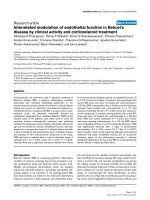
Oxidative stress pathways capable of producing ROS in
skeletal muscle inactivity include nitric oxide synthase-genera-
ting, xanthine oxidase-generating, nicotinamide adenine
dinucleotide phosphate (NADPH) oxidase-generating, and
mitochondrial oxidant-generating pathways (Figure 1) [21].
The nitric oxide synthase pathway does not seem to be
involved in VIDD [28]. Conversely, Whidden and coworkers
recently reported that the xanthine oxidase pathway
contributes to the oxidative damage of diaphragm muscle
[29]. This hypothesis was supported by the observation that
administration of oxypurinol, a xanthine-oxidase inhibitor,
partially attenuates diaphragmatic dysfunction after 12 hours
and 18 hours of CMV [29]. Markers of protein and lipid per-
oxidation, protein carbonyls and 4-hydroxynoneal, respec-
tively, are also suppressed with the administration of
oxypurinol. While xanthine oxidase contributes to diaphragm
muscle force loss, xanthine-oxidase inhibition does not
attenuate CMV-induced diaphragm muscle atrophy [29],
suggesting that other oxidative stress pathways may be
involved in the atrophic process.
In addition to xanthine oxidase, McClung and colleagues
demonstrated the role of the NADPH oxidase pathway in
producing oxidative damage in the diaphragm [30]. In rats
receiving 18 hours of CMV, apocynin (an inhibitor of NADPH
oxidase) attenuated diaphragm muscle dysfunction, prevent-
ed atrophy of all myofiber types, and prevented CMV-induced
reduction in glutathione. Furthermore, apocynin not only
suppressed calpain-1 and caspase-3 activation, but in fact
increased calpastatin, an endogenous calpain inhibitor.
Among the oxidative stress pathways, however, the mitochon-
drial oxidant-generating pathway is key in the development of
oxidative stress damage of the diaphragm with CMV [31].
Recently, Kavazis and colleagues demonstrated that
mitochondriae are a major source of ROS production
associated with mitochondrial oxidative damage and with
mitochondrial respiratory dysfunction [31].
Consistent with the mitochondrial oxidant-generating pathway,
an earlier study from the same laboratory demonstrated
elevated intracellular oxidant production with CMV [25]. The
latter was estimated from the intracellular increased emission
of dichlorodihydrofluorescein dye, a chemical that fluoresces
upon reaction with oxidative species [25]. The enhanced
production of lipid and protein oxidation markers underscores
the elevated oxidative stress [16]. Lipid oxidation may result
in cellular membrane dysfunction (that is, decreased Ca
2+
ATPase activity) and may delay Ca
2+
removal from the
cytosol, causing its accumulation within the cytosol itself
[23]. The elevated Ca
2+
concentration in the cytosol can
activate calpain, the Ca
2+
-dependent proteases [21]. In fact,
calpain-1 is an absolute requirement for oxidative stress-
induced myofiber atrophy [32]. This contention was
supported in experiments with hydrogen-peroxide-incubated
myotube cell cultures. Hydrogen peroxide induced myotube
atrophy. In contrast, calpain-1 RNA interference (gene
knocked out) completely prevents the atrophy [32].
Protein oxidation preferentially targets myofibrillar proteins
including myosin and actin [26]. The contractile proteins
damaged by oxidation become susceptible to degradation by
proteases [23]. Such protein degradation results in both
decreased diaphragmatic force-generating capacity and dia-
phragm muscle atrophy. In mechanically ventilated animals,
pretreatment with the antioxidant Trolox, a soluble vitamin E
analog, preserves diaphragmatic force-generating capacity
and prevents atrophy [27]. Trolox reduces production of
protein carbonyls – oxidative byproducts of proteins [19] –
but does not alter the suppressed antioxidant glutathione
concentrations. The protective effect of the diaphragm by
Trolox is achieved through a reduction in myofilament protein
availability to degradation by the proteasome [19,23]. In
addition to its antioxidant activity, Trolox has direct suppres-
sive effects on calpain, caspase proteases, and 20S protea-
some activity [19,21,27]. The 20S proteasome is the core
structure of the 26S proteasome complex in the ubiquitin–
proteasome pathway [33], and the unbound form can
independently degrade oxidized proteins without requiring
ubiquitin conjugation (Figure 2).
Proteolytic systems
All of the major proteolytic systems are responsible for CMV-
induced proteolysis; these include lysosomal proteases [18],
calcium-dependent proteolysis or calpains [14], caspase-3
[34], and the ATP-dependent ubiquitin proteasome [17].
Lysosomal proteases are primarily responsible for proteolysis
of extracellular proteins and cell surface receptors [35].
Calpain proteases are involved in the cleavage of cytoskeletal
proteins (for example, titin, nebulin, desmin) that anchor
contractile elements of myosin to actin [36]. Caspases
(endoproteases responsible for the final execution of cell
death) including caspase-3 proteases induce DNA
fragmentation, induce myonuclear apoptosis, and cleave
actomyosin complexes [34,37], whereas the ubiquitin–
proteasome pathway degrades the myofilament actin and
myosin [20].
Lysosomal proteases and calpains play a significant role in
diaphragmatic dysfunction with inactivity [18]. Pretreatment
with leupeptin – an inhibitor of lysosomal thiol proteases and
calcium-activated proteases – completely prevents the CMV-
induced reduction in diaphragmatic force and atrophy [18].
Likewise, caspase-3 vitally contributes in the detrimental
effects of CMV on the diaphragm [34]. Treatment of animals
with caspase inhibitor prevents myonuclei loss, DNA frag-
mentation, and myofiber atrophy [34].
The ubiquitin–proteasome pathway is responsible for most
muscle protein degradation [33]. The ubiquitin–proteasome
system, however, does not break down complexes of proteins
contained in myofibrils. One or more other proteases are
required in the initial process to release myofilament contrac-
tile proteins (that is, actin and myosin) for the ubiquitin–
proteasome system to degrade those proteins [23]. The
binding of ubiquitin to protein substrates requires ubiquitin-
activating enzyme (E1), ubiquitin-carrier enzyme (E2), and
ubiquitin ligases (E3) [33] (Figure 2). Two of the E3 ligases –
the muscle atrophy F-box (MAFbox; atrogin-1, atrogenes) and
muscle ring finger-1 (MuRF1) genes – are overexpressed in
various models of skeletal muscle atrophy [38]. Similarly,
MAFbox and MuRF1 are upregulated during CMV-induced
diaphragm muscle inactivity [11,19].
An important upstream signaling pathway of atrogene expres-
sion is the insulin-like growth factor-1–phosphotidylinositol-3-
Available online />Page 3 of 9
(page number not for citation purposes)
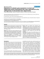
Figure 1
Oxidative stress pathways capable of producing reactive oxidant species. These pathways include nitric oxide synthase pathway, xanthine oxidase
pathway, nicotinamide adenine dinucleotide phosphate (NADPH) oxidase pathway, and mitochondrial oxidant-generating pathway. The
mitochondrial oxidant-generating pathway is key to oxidative damage of diaphragm muscle inactivity. O
2
•
, superoxide; NO
•
, nitric oxide. Adapted
with permission from [21].
kinase–protein kinase-B serine/threonine kinase (IGF-1/PI3K/
Akt) pathway [39] (Figure 3). IGF-1/PI3K/Akt suppresses
MAFbox by inactivating the expression of forkhead box-O and
preventing its nuclear translocation. After 6 hours and 18 hours
of CMV, diaphragmatic Akt activation decreased while both
forkhead box-O nuclear translocation and MAFbox and
MuRF1 expression increased [19]. IGF-1/PI3K/Akt signaling
therefore seems to play an important role in regulating the E3
ligase in the ubiquitin–proteasome pathway.
Influence of neuromechanical activation on
diaphragmatic function
Maintaining diaphragm muscle activation with assist-control
mechanical ventilation (AMV) represents an important
strategy for maintaining diaphragm muscle function [40]. For
instance, we have shown that 3 days of CMV produces a
dramatic loss (~45%) in diaphragmatic function, as defined
by the maximal isometric tension. In contrast, 3 days of AMV
produces much smaller losses in diaphragmatic force-
generating capacity, an approximately 15% loss in maximal
isometric tension [40]. The minimal extent of diaphragmatic
activation sufficient to preserve function is unclear. From our
previous data [40], however, it appears that activation levels
of 30% and above are associated with relatively small losses
in diaphragm muscle function (Figure 4). The influence of
minimal diaphragm muscle activation between 0% and 30%
on maximal isometric tension remains unknown. It is also
unclear whether AMV can preserve diaphragmatic force under
prolonged mechanical ventilation >3 days. The decline in force
with CMV was associated with an approximately threefold
increase in MAFbox mRNA expression, while with AMV the
expression did not differ significantly from controls [40].
In another study, when spontaneous breathing for 5 minutes
or 60 minutes was interposed during 24 hours of CMV four
times a day, the diaphragmatic force-generating capacity
decreased by an average of 19% and decreased by 28%
with continuous CMV, respectively [41]. Although the
protective effects of a brief duration of diaphragm muscle
activation on functional loss were modest (~9%), the
activation prevents diaphragm muscle atrophy. Futier and
colleagues recently demonstrated that maintaining diaphrag-
matic activation with pressure support ventilation for 18 hours
did not augment proteolysis [42]. Protein carbonyls (markers
of oxidative stress), however, were elevated to the same
extent as with CMV. Unfortunately, measures of diaphrag-
matic function were not performed, and whether pressure
support ventilation preserves diaphragmatic force therefore
remains unknown [42].
Evidence of CMV-induced diaphragmatic
dysfunction in humans and a potential
approach to prevention
In critically ill patients it is extremely difficult to establish
whether CMV is responsible for diaphragm muscle dys-
function and weaning failure, because multiple confounding
factors (for example, sepsis, malnutrition, hyperglycemia)
contribute to diaphragm muscle weakness and atrophy.
Consistent with studies in animals, Levine and colleagues
reported that diaphragm muscle atrophy also occurred fairly
Critical Care Vol 13 No 5 Sassoon and Caiozzo
Page 4 of 9
(page number not for citation purposes)
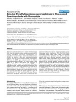
Figure 2
The ubiquitin–proteasome pathway. The substrate proteins are designated for degradation by conjugation to ubiquitin in an ATP-dependent
reaction. The ubiquitin-activating enzyme (E1) uses ATP to create a highly reactive thiolester form of ubiquitin, and then transfers it to a ubiquitin-
carrier protein (E2). The subsequent transfer of the activated ubiquitin to the protein substrate requires a ubiquitin-protein ligase (E3). The E3
ligases muscle atrophy F-box (MAFbox) and muscle ring finger-1 (MuRF-1) have important roles in skeletal muscle atrophy. Once the ubiquitin
conjugates are formed, they are transported to a proteolytic complex known as the 26S proteasome, consisting of two 19S regulators and the 20S
core proteasome. The 19S regulators recognize and bind the ubiquitinated protein. Energy from ATP hydrolysis releases the ubiquitin chain and
unfolds the substrate protein. The unfolded protein is fed into the 20S proteasome for degradation into small peptides and amino acids. The 20S
proteasome can degrade oxidized protein without ubiquitination. Adapted with permission from [33].
rapidly in brain-dead organ donors with CMV application of
18 to 69 hours, compared with control subjects who under-
went lung surgery and received mechanical ventilation for 2
to 3 hours [8]. Diaphragm muscle atrophy involved both slow
and fast fiber types, decreasing cross-sectional areas by
57% and 53%, respectively (Figure 5). The atrophy was
associated with decreased antioxidant glutathione concen-
tration (by 23%), increased active caspase-3 protease (by
twofold), and elevated mRNA levels of MAFbox (by threefold)
and MuRF1 (by sevenfold). Interestingly, a biopsy of the
pectoralis major muscle did not show any fiber atrophy [8].
The study of Levine and colleagues lacked measurement of
diaphragm muscle function, and was confined to brain-dead
organ donors whose neural activation and possibly neuro-
trophic factors to the diaphragm were completely absent and
thus were not typical of critically ill patients in the intensive
care unit [8]. Nevertheless, the negative impact of diaphragm
muscle disuse in critically ill patients cannot be ignored [43].
In humans, the degree of diaphragm muscle activation that
will preserve force remains unknown. In a prospective trial,
critically ill patients who were predicted to receive mecha-
nical ventilation for longer than 72 hours were randomized
into controls (n = 13) and those receiving inspiratory muscle
training (IMT) (n = 12) from the onset of mechanical
ventilation [44]. A threshold load was used for the IMT by
setting the ventilator pressure-triggering sensitivity at 10% or
20% of the initial maximum inspiratory pressure (PI
max
),
whichever was tolerated, and was applied twice daily for
5 minutes. When the patient tolerated the initial load, the next
training duration was increased by 5 minutes up to a
maximum of 30 minutes. Afterwards, the load was increased
by 10% increments until 40% of the initial PI
max
value was
attained. Sedation and analgesia with intravenous midazolam
and fentanyl, respectively, were administered. The IMT
session was aborted according to specified criteria. Weaning
with decreasing pressure support was initiated once the
patient met the weaning criteria. The initial and final PI
max
values in the training group were similar to those of the
control group (initial, –51 cmH
2
O vs. –48 cmH
2
O; final,
–56 cmH
2
O vs. –55 cmH
2
O, respectively). The duration of
mechanical ventilation or of the weaning trial was similar for
both groups, with a trend toward a shorter duration for the
IMT group compared with the control group (mean duration
of mechanical ventilation, 8.6 days vs. 9.8 days; mean
duration of weaning trial, 23 hours vs. 31 hours, respectively).
Available online />Page 5 of 9
(page number not for citation purposes)
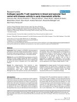
Figure 3
The insulin-like growth factor-1–phosphotidylinositol-3-kinase–protein kinase-B serine/threonine kinase–forkhead box-O pathway. (a) Increased
insulin-like growth factor-1 (IGF-1) activates phosphotidylinositol-3-kinase (PI3K), leading to phosphorylation of protein kinase-B serine/threonine
kinase (Akt) and forkhead box-O (Foxo). Phosphorylated Foxo is sequestered within the cytoplasm and prevents its nuclear translocation and
atrogin-1 (muscle atrophy F-box (MAFbox)) activation. Phosphorylated Akt also activates mammalian target of rapamycin (mTOR) and p70Sk,
resulting in increased protein synthesis. (b) Suppression of IGF-1 with controlled mechanical ventilation-induced diaphragm muscle inactivity
deactivates Akt, leading to nuclear translocation of Foxo, which then activates atrogin-1 and other atrogenes resulting in increased proteolysis.
Reprinted from Cell, 117, Sandri M, Sandri C, Gilbert A, Skurk C, Calabria E, Picard A, Walsh K, Schiaffino S, Lecker SH, Goldberg AL, Foxo
Transcription Factors Induce the Atrophy-Related Ubiquitin Ligase Atrogin-1 and Cause Skeletal Muscle Atrophy, 14 Pages, Copyright (2004),
with permission from Elsevier [39].
The lack of IMT benefits may be due to the small sample size
[44]. It is also conceivable that the magnitude of the stimulus
for IMT, as a percentage of the PI
max
, in the critically ill
patients (that is, the threshold load applied, and/or session
frequency and duration) was inadequate to elicit a physio-
logical training effect. Measurements of PI
max
in the critically ill
patients are challenging and highly dependent on patient
volitional effort and the methods of measurement. Alter-
natively, in view of the complexity of the underlying mecha-
nisms of CMV-induced diaphragmatic dysfunction, diaphragm
muscle conditioning alone is inadequate, and pharmaco-
logical intervention may be required to mitigate diaphragm
muscle weakness.
Mechanisms of the interactive effects of
mechanical ventilation and short-term
high-dose corticosteroid on diaphragm
muscle dysfunction
Short-term high-dose corticosteroid has been administered
for its anti-inflammatory and immunosuppressive effects in
critically ill patients [9,45]; the treatment might be
responsible for the development of acquired paresis in the
critically ill patient, referred to as critical-illness myopathy
[46]. Patients may receive both mechanical ventilation and
short-term high-dose corticosteroid, yet the effects of acute
high-dose corticosteroid alone or its interaction with
mechanical ventilation is not well understood.
We recently studied the temporal relationship (1 to 3 days of
80 mg/kg/day intramuscularly) and dose–response effects
(3 days of 80 mg/kg/day vs. 10 mg/kg/day intramuscularly) of
methylprednisolone (MP) treatment in rabbits [7]. MP induced
a progressive decline in diaphragmatic force by 19%, 24%,
and 34% after 1 day, 2 days, and 3 days, respectively. The
decline in diaphragmatic force correlated with the degree of
abnormal myofibril volume density. Low-dose MP (10 mg/kg/day,
but a high dose by clinical standards) decreased diaphrag-
matic force modestly, by 12%. The suppression of IGF-1 and
upregulation of MAFbox mRNA were independent of the MP
dose [7]. Both high-dose and low-dose MP decreased IGF-1
by 35%, and increased MAFbox mRNA by threefold [7].
Clearly, short-term high doses of MP in spontaneously
breathing animals produced detrimental effects on the dia-
phragm. The combination of both CMV and high-dose MP is
therefore expected to aggravate the decline in diaphragmatic
force compared with either CMV or MP alone.
Interestingly, Maes and colleagues demonstrated in rats that
24 hours of combined CMV and high-dose MP
(80 mg/kg/day intramuscularly) preserved the diaphragmatic
force compared with CMV alone [47]. The mechanism by
which MP prevented diaphragmatic force loss was via
inhibition of calpain activity. Our preliminary data [48] in
rabbits contrast with those of Maes and colleagues. After
2 days of combined MP (60 mg/kg/day intravenously) plus
CMV, MP plus AMV, or MP plus continuous positive airway
pressure, the diaphragmatic force decreased from that
without MP by 10%, 16% and 18% from the average values
of 16.1 Newton/cm
2
, 22.6 Newton/cm
2
, and 23.3 Newton/cm
2
with CMV, AMV, and continuous positive airway pressure
alone, respectively [48]. The diaphragmatic force with the
combined CMV and MP approach was not significantly
different from that with CMV alone. This suggests that both
CMV and MP share common mechanisms for the decrease in
diaphragmatic force. It is unclear whether the discrepancy
between our preliminary results [48] and those of Maes and
colleagues [47] is related to species differences or to the
duration of MP treatment.
Evidence of methylprednisolone-induced
diaphragmatic dysfunction in humans and a
potential approach to prevention
As with CMV, the extent to which acute, high-dose MP could
contribute to diaphragm muscle weakness in critically ill
patients is difficult to determine. This difficulty stems from the
many confounding factors in these patients, and from the lack
of data on functional or structural alterations in humans. In-
direct data, however, suggest that such interaction may occur
in critically ill patients [46].
Critical Care Vol 13 No 5 Sassoon and Caiozzo
Page 6 of 9
(page number not for citation purposes)
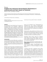
Figure 4
Monoexponential relationships between diaphragm muscle maximal
tetanic force and its electrical activity. The maximal isometric tension
(P
o
) is normalized for muscle cross-sectional area. The diaphragm
muscle electrical activity (EMG
d
) during assist-control mechanical
ventilation (AMV) was estimated by measuring the area subtended by
the moving average EMG
d
curve and its baseline, and is expressed as
a percentage of spontaneous breathing. P
o
is maintained almost
identically to that of the control after 3 days of AMV with diaphragm
muscle activation between 30% and 80% of spontaneous breathing.
Whether diaphragm muscle activation between 0% and 30% is
effective to maintain P
o
remains unknown. Data obtained from [41]: n
= 6 for the control and controlled mechanical ventilation (CMV)
groups; n = 5 for the AMV group.
First, in a prospective study of critically ill patients receiving
mechanical ventilation for longer than 7 days, De Jonghe and
colleagues reported a strong association between the
occurrence of neuromyopathy and the administration of
corticosteroids [46]. Second, in patients with acute spinal
cord injury, the administration of recommended high-dose MP
for 48 hours resulted in paraspinal muscle necrosis and type
II fiber atrophy in four out of five patients [9]. Three of the
patients remained ventilator dependent at discharge from the
Spinal Cord Injury Center despite the relatively low level of
injury [9]. Finally, among 26 patients with chronic obstructive
pulmonary disease who received mechanical ventilation and
MP (240 mg/day), nine (35%) patients developed myopathy
of the extremities – a condition associated with higher total
doses of MP treatment (1,649 mg vs. 979 mg), with
prolonged mechanical ventilation, and with prolonged
hospital length of stay [45].
Whether a dose and duration of corticosteroids that confers
beneficial anti-inflammatory effects and yet preserves
diaphragm muscle integrity/function does exist remains
unknown. More research is necessary to dissect the
underlying mechanisms of the effects of corticosteroid on the
diaphragm, particularly its interaction with mechanical
ventilation. Because of the corticosteroid dose–response
effects in both animal studies [7] and human studies [45],
clinicians must carefully weigh the risks and benefits ratio,
and must use the lowest corticosteroid dose for the shortest
duration possible.
Future research
In laboratory animals the mechanisms responsible for VIDD
have been the focus of intense investigation. Unfortunately,
the triggering factor(s) for enhanced proteolysis in VIDD
remain unknown. Similarly, the contribution of excitation–
contraction coupling and the degree or duration of neuro-
mechanical activation for preventing diaphragmatic force loss
are unknown. Whether the benefits of AMV depend on the
level of diaphragmatic activity or whether the benefits cease
with time remains unclear. Diaphragm muscle conditioning
Available online />Page 7 of 9
(page number not for citation purposes)
Figure 5
Cross-sectional areas of diaphragm muscle. Cross-sections of diaphragm muscle from biopsy specimens of a representative organ donor subject
((a), (c), (e)) and from a control ((b), (d), (f)). (a) and (b) Muscle fibers in the organ donor subject are in general smaller than those in the control
diaphragm. No inflammatory infiltrate or necrosis is seen. Stained with hematoxylin and eosin. (c) and (d) Stained with antibody specific for slow
myosin, heavy chain. (e) and (f) Stained with antibody specific for fast myosin, heavy chain. In (c) to (f), fibers reacting with the antibody appear
orange–red, whereas fibers not reacting with the antibody appear black; open circle, slow-twitch fibers; open square, fast-twitch fibers. In addition,
all fibers in each section are outlined by an antibody reactive to laminin. Reproduced with permission from [8]. Copyright © 2008 Massachusetts
Medical Society. All rights reserved.
using noninvasive phrenic nerve stimulation is a potential
strategy for preventing VIDD that remains to be explored. In
animal studies, treatment with specific inhibitors to the
signaling cascade involved in proteolysis completely
preserves diaphragm muscle function. Whether a similar
strategy should be attempted in patients remains to be
determined.
Competing interests
The authors declare that they have no competing interests.
Acknowledgements
The present work was supported by grants from the Department of
Veterans Affairs Medical Research Service (to CSHS) and the National
Institute of Arthritis and Musculoskeletal and Skin Diseases AR-46856
(to VJC). We thank Ercheng Zhu, Ph.D. for generating the data pre-
sented in Figure 4.
References
1. Gainnier M, Roch A, Forel JM, Thirion X, Arnal JM, Donati S,
Papazian L: Effect of neuromuscular blocking agents on gas
exchange in patients presenting with acute respiratory dis-
tress syndrome. Crit Care Med 2004, 32:113-119.
2. Bracken MB, Shepard MJ, Holford TR, Leo-Summers L, Aldrich
EF, Fazl M, Fehlings M, Herr DL, Hitchon PW, Marshall LF,
Nockels RP, Pascale V, Perot PL Jr, Piepmeier J, Sonntag VK,
Wagner F, Wilberger JE, Winn HR, Young W: Administration of
methylprednisolone for 24 or 48 hours or tirilazad mesylate
for 48 hours in the treatment of acute spinal cord injury.
Results of the Third National Acute Spinal Cord Injury Ran-
domized Controlled Trial. National Acute Spinal Cord Injury
Study. JAMA 1997, 277:1597-1604.
3. Nava S, Fracchia C, Callegari G, Ambrosino N, Barbarito N,
Felicetti G: Weakness of respiratory and skeletal muscles
after a short course of steroids in patients with acute lung
rejection. Eur Respir J 2002, 20:497-499.
4. Jagannath S: Treatment of myeloma in patients not eligible for
transplantation. Curr Treat Options Oncol 2005, 6:241-253.
5. Kaplan PW, Rocha W, Sanders DB, D’Souza B, Spock A: Acute
steroid-induced tetraplegia following status asthmaticus.
Pediatrics 1986, 78:121-123.
6. Sassoon CSH, Caiozzo VJ, Manka A, Sieck GC: Altered
diaphragm contractile properties with controlled mechanical
ventilation. J Appl Physiol 2002, 92:2585-2595.
7. Sassoon CS, Zhu E, Pham HT, Nelson RS, Fang L, Baker MJ,
Caiozzo VJ: Acute effects of high-dose methylprednisolone on
diaphragm muscle function. Muscle Nerve 2008, 38:1161-
1172.
8. Levine S, Nguyen T, Taylor N, Friscia ME, Budak MT, Rothenberg
P, Zhu J, Sachdeva R, Sonnad S, Kaiser LR, Rubinstein NA,
Powers SK, Shrager JB: Rapid disuse atrophy of diaphragm
fibers in mechanically ventilated humans. N Engl J Med 2008,
358:1327-1335.
9. Qian T, Guo X, Levi AD, Vanni S, Shebert RT, Sipski ML: High-
dose methylprednisolone may cause myopathy in acute
spinal cord injury patients. Spinal Cord 2005, 43:199-203.
10. Powers SK, Shanely RA, Coombes JS, Koesterer TJ, McKenzie M,
Van Gammeren D, Cicale M, Dodd SL: Mechanical ventilation
results in progressive contractile dysfunction in the diaphragm.
J Appl Physiol 2002, 92:1851-1858.
11. Zhu E, Sassoon CS, Nelson R, Pham HT, Zhu L, Baker MJ,
Caiozzo VJ: Early effects of mechanical ventilation on isotonic
contractile properties and MAF-box gene expression in the
diaphragm. J Appl Physiol 2005, 99:747-756.
12. Radell PJ, Remahl S, Nichols DG, Eriksson LI: Effects of pro-
longed mechanical ventilation and inactivity on piglet
diaphragm function. Intensive Care Med 2002, 28:358-364.
13. Anzueto A, Peters JI, Tobin MJ, de los Santos R, Seidenfeld JJ,
Moore G, Cox WJ, Coalson JJ: Effects of prolonged controlled
mechanical ventilation on diaphragmatic function in healthy
adult baboons. Crit Care Med 1997, 25:1187-1190.
14. Testelmans D, Maes K, Wouters P, Powers SK, Decramer M,
Gayan-Ramirez G: Infusions of rocuronium and cisatracurium
exert different effects on rat diaphragm function. Intensive
Care Med 2007, 33:872-879.
15. Testelmans D, Maes K, Wouters P, Gosselin N, Deruisseau K,
Powers S, Sciot R, Decramer M, Gayan-Ramirez G: Rocuronium
exacerbates mechanical ventilation induced diaphragm dys-
function in rats. Crit Care Med 2006, 34:3018-3023.
16. Shanely RA, Zergeroglu MA, Lennon SL, Sugiura T, Yimlamai T,
Enns D, Belcastro A, Powers SK: Mechanical ventilation-
induced diaphragmatic atrophy is associated with oxidative
injury and increased proteolytic activity. Am J Respir Crit Care
Med 2002, 166:1369-1374.
17. DeRuisseau KC, Kavazis AN, Deering MA, Falk DJ, Van Gam-
meren D, Yimlamai T, Ordway GA, Powers SK: Mechanical venti-
lation induces alterations of the ubiquitin–proteasome
pathway in the diaphragm. J Appl Physiol 2005, 98:1314-1321.
18. Maes K, Testelmans D, Powers S, Decramer M, Gayan-Ramirez
G: Leupeptin inhibits ventilator-induced diaphragm dysfunc-
tion in rats. Am J Respir Crit Care Med 2007, 175:1134-1138.
19. McClung JM, Kavazis AN, Whidden MA, DeRuisseau KC, Falk DJ,
Criswell DS, Powers SK: Antioxidant administration attenuates
mechanical ventilation-induced rat diaphragm muscle atrophy
independent of protein kinase B (PKB Akt) signalling.
J Physiol 2007, 585:203-215.
20. McClung JM, Whidden MA, Kavazis AN, Falk DJ, Deruisseau KC,
Powers SK: Redox regulation of diaphragm proteolysis during
mechanical ventilation. Am J Physiol Regul Integr Comp Physiol
2008, 294:R1608-R1617.
21. Powers SK, Kavazis AN, McClung JM: Oxidative stress and
disuse muscle atrophy. J Appl Physiol 2007, 102:2389-2397.
22. Clark BC, Fernhall B, Ploutz-Snyder LL: Adaptations in human
neuromuscular function following prolonged unweighting: I.
Skeletal muscle contractile properties and applied ischemia
efficacy. J Appl Physiol 2006, 101:256-263.
23. Powers SK, Kavazis AN, DeRuisseau KC: Mechanisms of disuse
muscle atrophy: role of oxidative stress. Am J Physiol Regul
Integr Comp Physiol 2005, 288:R337-R344.
24. Jaber S, Sebbane M, Koechlin C, Hayot M, Capdevila X, Eledjam
JJ, Prefaut C, Ramonatxo M, Matecki S: Effects of short vs. pro-
longed mechanical ventilation on antioxidant systems in
piglet diaphragm. Intensive Care Med 2005, 31:1427-1433.
25. Falk DJ, Deruisseau KC, Van Gammeren DL, Deering MA, Kavazis
AN, Powers SK: Mechanical ventilation promotes redox status
alterations in the diaphragm. J Appl Physiol 2006, 101:1017-
1024.
26. Zergeroglu MA, McKenzie MJ, Shanely RA, Van Gammeren D,
DeRuisseau KC, Powers SK: Mechanical ventilation-induced
oxidative stress in the diaphragm. J Appl Physiol 2003, 95:
1116-1124.
27. Betters JL, Criswell DS, Shanely RA, Van Gammeren D, Falk D,
Deruisseau KC, Deering M, Yimlamai T, Powers SK: Trolox atten-
uates mechanical ventilation-induced diaphragmatic dysfunc-
tion and proteolysis. Am J Respir Crit Care Med 2004, 170:
1179-1184.
28. Van Gammeren D, Falk DJ, Deering MA, Deruisseau KC, Powers
SK: Diaphragmatic nitric oxide synthase is not induced during
mechanical ventilation. J Appl Physiol 2007, 102:157-162.
29. Whidden MA, McClung JM, Falk DJ, Hudson MB, Smuder AJ,
Nelson WB, Powers SK: Xanthine oxidase contributes to
mechanical ventilation-induced diaphragmatic oxidative
stress and contractile dysfunction. J Appl Physiol 2009, 106:
385-394.
30. McClung JM, Van Gammeren D, Whidden MA, Falk DJ, Kavazis
AN, Hudson MB, Gayan-Ramirez G, Decramer M, DeRuisseau
KC, Powers SK: Apocynin attenuates diaphragm oxidative
stress and protease activation during prolonged mechanical
ventilation. Crit Care Med 2009, 37:1373-1379.
31. Kavazis AN, Talbert EE, Smuder AJ, Hudson MB, Nelson WB,
Powers SK: Mechanical ventilation induces diaphragmatic
mitochondrial dysfunction and increased oxidant production.
Free Radic Biol Med 2009, 46:842-850.
32. McClung JM, Judge AR, Talbert EE, Powers SK: Calpain-1 is
required for hydrogen peroxide-induced myotube atrophy. Am
J Physiol Cell Physiol 2009, 296:C363-C371.
33. Goldberg AL, Elledge SJ, Harper JW: The cellular chamber of
doom. Sci Am 2001, 284:68-73.
Critical Care Vol 13 No 5 Sassoon and Caiozzo
Page 8 of 9
(page number not for citation purposes)
34. McClung JM, Kavazis AN, DeRuisseau KC, Falk DJ, Deering MA,
Lee Y, Sugiura T, Powers SK: Caspase-3 regulation of
diaphragm myonuclear domain during mechanical ventilation-
induced atrophy. Am J Respir Crit Care Med 2007, 175:150-
159.
35. Lecker SH, Solomon V, Mitch WE, Goldberg AL: Muscle protein
breakdown and the critical role of the ubiquitin–proteasome
pathway in normal and disease states. J Nutr 1999, 129:227S-
237S.
36. Koh TJ, Tidball JG: Nitric oxide inhibits calpain-mediated prote-
olysis of talin in skeletal muscle cells. Am J Physiol Cell
Physiol 2000, 279:C806-C812.
37. Du J, Wang X, Miereles C, Bailey JL, Debigare R, Zheng B, Price
SR, Mitch WE: Activation of caspase-3 is an initial step trig-
gering accelerated muscle proteolysis in catabolic conditions.
J Clin Invest 2004, 113:115-123.
38. Bodine SC, Latres E, Baumhueter S, Lai VK, Nunez L, Clarke BA,
Poueymirou WT, Panaro FJ, Na E, Dharmarajan K, Pan ZQ, Valen-
zuela DM, DeChiara TM, Stitt TN, Yancopoulos GD, Glass DJ:
Identification of ubiquitin ligases required for skeletal muscle
atrophy. Science 2001, 294:1704-1708.
39. Sandri M, Sandri C, Gilbert A, Skurk C, Calabria E, Picard A,
Walsh K, Schiaffino S, Lecker SH, Goldberg AL: Foxo transcrip-
tion factors induce the atrophy-related ubiquitin ligase
atrogin-1 and cause skeletal muscle atrophy. Cell 2004, 117:
399-412.
40. Sassoon CS, Zhu E, Caiozzo VJ: Assist-control mechanical ven-
tilation attenuates ventilator-induced diaphragmatic dysfunc-
tion. Am J Respir Crit Care Med 2004, 170:626-632.
41. Gayan-Ramirez G, Testelmans D, Maes K, Rácz GZ, Cadot P,
Zádor E, Wuytack F, Decramer M: Intermittent spontaneous
breathing protects the rat diaphragm from mechanical venti-
lation effects. Crit Care Med 2005, 33:2804-2809.
42. Futier E, Constantin JM, Combaret L, Mosoni L, Roszyk L, Sapin
V, Attaix D, Jung B, Jaber S, Bazin JE: Pressure support ventila-
tion attenuates ventilator-induced protein modifications in the
diaphragm. Crit Care 2008 12:R116.
43. Sieck GC, Mantilla CB: Effects of mechanical ventilation on the
diaphragm. N Engl J Med 2008, 358:1392-1393.
44. Caruso P, Denari SD, Ruiz SA, Bernal KG, Manfrin GM, Friedrich
C, Deheinzelin D: Inspiratory muscle training is ineffective in
mechanically ventilated critically ill patients. Clinics 2005, 60:
479-484.
45. Amaya-Villar R, Garnacho-Montero J, Garcia-Garmendia JL,
Madrazo-Osuna J, Garnacho-Montero MC, Luque R, Ortiz-Leyba
C: Steroid-induced myopathy in patients intubated due to
exacerbation of chronic obstructive pulmonary disease. Inten-
sive Care Med 2005, 31:157-161.
46. De Jonghe B, Sharshar T, Lefaucheur JP, Authier FJ, Durand-
Zaleski I, Boussarsar M, Cerf C, Renaud E, Mesrati F, Carlet J,
Raphaël JC, Outin H, Bastuji-Garin S, Groupe de Réflexion et
d’Etude des Neuromyopathies en Réanimation: Paresis acquired
in the intensive care unit: a prospective multicenter study.
JAMA 2002, 288:2859-2867.
47. Maes K, Testelmans D, Cadot P, Deruisseau K, Powers SK,
Decramer M, Gayan-Ramirez G: Effects of acute administration
of corticosteroids during mechanical ventilation on rat
diaphragm. Am J Respir Crit Care Med 2008, 178:1219-1226.
48. Tom L, Zhu E, Pham TH, Jiao G, Caiozzo VJ, Sassoon CSH:
Effects of methylprednisolone on diaphragmatic contractile
properties during mechanical ventilation. Proc Am Thoracic
Soc 2006, 3:A137.
Available online />Page 9 of 9
(page number not for citation purposes)