Báo cáo y học: " Comparison of two non-bronchoscopic methods for evaluating inflammation in patients with acute hypoxaemic respiratory failure" doc
Bạn đang xem bản rút gọn của tài liệu. Xem và tải ngay bản đầy đủ của tài liệu tại đây (487.86 KB, 9 trang )
Open Access
Available online />Page 1 of 9
(page number not for citation purposes)
Vol 13 No 4
Research
Comparison of two non-bronchoscopic methods for evaluating
inflammation in patients with acute hypoxaemic respiratory
failure
Giuseppe Colucci
1
*, Guido Domenighetti
1
*, Roberto Della Bruna
2
, Josè Bonilla
3
,
Costanzo Limoni
4
, Michael A Matthay
5
and Thomas R Martin
6
1
Multidisciplinary Intensive Care Unit, Regional Hospital EOC, Via Ospedale 14, Locarno 6600, Switzerland
2
EOLAB, Ente Ospedaliero Cantonale, Viale Officina 3, Bellinzona 6500, Switzerland
3
Cantonal Pathological Institute, Lab for Clinical Cytology, Via A. Franzoni 45, Locarno 6600, Switzerland
4
Department for Social Sciences, University of Applied Sciences and Arts of Southern Switzerland, Le Gerre, Manno 6928, Switzerland
5
Departments of Medicine and Anaesthesia, Cardiovascular Research Institute, University of California, San Francisco, 505 Parnassus Ave, M917,
Box 0624, San Francisco, CA 94143, USA
6
Medical Research Service of the VA Puget Sound Medical Center and the Division of Pulmonary and Critical Care Medicine, Department of Medicine,
University of Washington, Seattle, 1660 S. Columbian Way, Seattle, WA 98108, USA
* Contributed equally
Corresponding author: Guido Domenighetti,
Received: 19 Apr 2009 Revisions requested: 4 Jun 2009 Revisions received: 28 Jul 2009 Accepted: 11 Aug 2009 Published: 11 Aug 2009
Critical Care 2009, 13:R134 (doi:10.1186/cc7995)
This article is online at: />© 2009 Colucci et al.; licensee BioMed Central Ltd.
This is an open access article distributed under the terms of the Creative Commons Attribution License ( />),
which permits unrestricted use, distribution, and reproduction in any medium, provided the original work is properly cited.
Abstract
Introduction The simple bedside method for sampling undiluted
distal pulmonary edema fluid through a normal suction catheter
(s-Cath) has been experimentally and clinically validated.
However, there are no data comparing non-bronchoscopic
bronchoalveolar lavage (mini-BAL) and s-Cath for assessing
lung inflammation in acute hypoxaemic respiratory failure. We
designed a prospective study in two groups of patients, those
with acute lung injury (ALI)/acute respiratory distress syndrome
(ARDS) and those with acute cardiogenic lung edema (ACLE),
designed to investigate the clinical feasibility of these
techniques and to evaluate inflammation in both groups using
undiluted sampling obtained by s-Cath. To test the
interchangeability of the two methods in the same patient for
studying the inflammation response, we further compared mini-
BAL and s-Cath for agreement of protein concentration and
percentage of polymorphonuclear cells (PMNs).
Methods Mini-BAL and s-Cath sampling was assessed in 30
mechanically ventilated patients, 21 with ALI/ARDS and 9 with
ACLE. To analyse agreement between the two sampling
techniques, we considered only simultaneously collected mini-
BAL and s-Cath paired samples. The protein concentration and
polymorphonuclear cell (PMN) count comparisons were
performed using undiluted sampling. Bland-Altman plots were
used for assessing the mean bias and the limits of agreement
between the two sampling techniques; comparison between
groups was performed by using the non-parametric Mann-
Whitney-U test; continuous variables were compared by using
the Student t-test, Wilcoxon signed rank test, analysis of
variance or Student-Newman-Keuls test; and categorical
variables were compared by using chi-square analysis or Fisher
exact test.
Results Using protein content and PMN percentage as
parameters, we identified substantial variations between the two
sampling techniques. When the protein concentration in the
lung was high, the s-Cath was a more sensitive method; by
contrast, as inflammation increased, both methods provided
similar estimates of neutrophil percentages in the lung. The
patients with ACLE showed an increased PMN count,
suggesting that hydrostatic lung edema can be associated with
a concomitant inflammatory process.
ACLE: acute cardiogenic lung oedema; ALI: acute lung injury; ARDS: acute respiratory distress syndrome; bBAL: bronchoscopic bronchoalveolar
lavage; CI: confidence interval; FiO
2
: fraction of inspired oxygen; Fr: French; HR: heart rate; ICU: intensive care unit; IL: interleukin; LIS: Lung Injury
Score; LOS: length of stay; mini-BAL: non-bronchoscopic bronchoalveolar lavage; PaO
2
: partial pressure of oxygen in arterial blood; PEEP: positive
end-expiratory pressure; PMN: polymorphonuclear cell; P
peak
: peak pressure; P
plat
: plateau pressure; RBC: red blood cell; SAP: systemic arterial pres-
sure; SAPS II: Simplified Acute Physiology Score II; s-Cath: suction catheter; SpO
2
: pulsed oxygen saturation; V
E
: minute ventilation; V
t
: expiratory
tidal volume; WBC: white blood cell.
Critical Care Vol 13 No 4 Colucci et al.
Page 2 of 9
(page number not for citation purposes)
Conclusions There are significant differences between the s-
Cath and mini-BAL sampling techniques, indicating that these
procedures cannot be used interchangeably for studying the
lung inflammatory response in patients with acute hypoxaemic
lung injury.
Introduction
In patients with acute hypoxaemic respiratory failure, acute
respiratory distress syndrome (ARDS) represents the more
severe form of acute lung injury (ALI) [1]. Although a wide
spectrum of clinical disorders may be associated with the
development of ALI/ARDS, aetiologies can be divided into dis-
eases associated with direct lung injury (i.e., pneumonia, aspi-
ration, inhalation injury; primary ARDS) and indirect lung injury
in the setting of a systemic process (i.e., sepsis, severe trauma
with shock, pancreatitis; secondary ARDS) [2]. The inflamma-
tory response of the lung is intense in the alveolar space, and
the hallmark of ALI/ARDS in the early phase is severe damage
of the alveolocapillary barrier, leading to increased permeabil-
ity, development of protein-rich and biomarker-rich oedema
fluid, and impaired clearance of the oedema [3-5]. The study
of the composition and resolution of oedema fluid is of primary
importance because it may lead to new insights into the patho-
genesis of ALI/ARDS. Sequential sampling of oedema fluid is
required for this purpose.
Another common cause of acute respiratory failure is acute
cardiogenic lung oedema (ACLE). Although the mechanism of
cardiogenic oedema is different from that of ALI/ARDS, recent
studies have found that endothelial-derived and epithelial-
derived inflammatory mediators are released into the blood
even during this form of hydrostatic oedema [6].
Sampling of pulmonary oedema fluid from the distal air spaces
is an important procedure that allows the study of the lung
inflammatory response. The gold standard technique for this
purpose is bronchoscopic bronchoalveolar lavage (bBAL).
However, bBAL performed with the standard adult broncho-
scope may be poorly tolerated in some critically ill ARDS
patients, because it can lead to a worsening of hypoxaemia
and hypercapnia, haemodynamic instability, temporary loss of
recruited lung areas and development of positive end-expira-
tory pressure (PEEP) of unknown magnitude [7].
Less invasive bedside techniques have been developed that
overcome these difficulties and simplify the procedure, provid-
ing alternatives for the rapid study of alveolar fluid in patients
with ALI/ARDS. Non-bronchoscopic bronchoalveolar lavage
(mini-BAL) and the distal collection of oedema fluid through a
simple suction catheter (s-Cath) are examples of these less
invasive techniques [4,8,9].
The simple bedside method for sampling distal pulmonary
oedema fluid through an s-Cath has been experimentally vali-
dated and used in many studies [10]. However, an assess-
ment of inflammation using undiluted sampling obtained by s-
Cath in patients with ALI/ARDS and ACLE or a comparison of
mini-BAL with s-Cath have not been performed. We therefore
designed a prospective study in two groups of patients with
acute hypoxaemic respiratory failure, those with ALI/ARDS
and those with ACLE, in order to investigate the clinical feasi-
bility of these techniques. To determine whether the two meth-
ods can be used interchangeably for sampling the distal air
spaces of the lung, we compared mini-BAL and s-Cath for
agreement of protein concentration and percentage of poly-
morphonuclear cells (PMNs), as surrogate markers of acute
lung inflammation.
Materials and methods
All patients admitted to the multidisciplinary intensive care unit
(ICU) of the EOC Regional Hospital "La Carità" in Locarno,
Switzerland, between 2002 and 2004 were screened for eli-
gibility. Patients with ALI or ARDS of different causes and with
ACLE requiring immediate intubation and mechanical ventila-
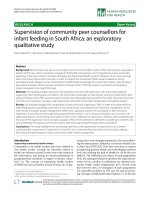
tory support were enrolled (n = 54; Figure 1). Patients with
ALI/ARDS were identified by the American-European Consen-
sus Conference definitions [1]. The clinical diagnosis of ACLE
was confirmed by reviewing patient records and chest radio-
graphs, recent medical history, and echocardiography or pul-
monary artery catheter if the diagnosis was not clear. ACLE
was classified as acute exacerbation of congestive heart fail-
ure, acute coronary syndrome or exacerbation of diastolic left
ventricular dysfunction. Patients were excluded if they had
known HIV infection, immunodeficiency necessitating granulo-
cyte colony stimulating factor, ALI/ARDS after thoracic sur-
gery, and mixed causes of pulmonary oedema with elements
of both ALI/ARDS and elevated hydrostatic pressure (n = 24).
Finally, 30 mechanically ventilated patients met the eligibility
criteria (Figure 1). Patients were intubated with oral endotra-
cheal tubes with an internal diameter of 8 mm or more.
This study was approved by the Committee of Human
Research of the Canton of Ticino, Switzerland, and informed
written consent was obtained from each patient's next of kin.
Clinical data
Clinical, physiological and biological data at the time of fluid
sampling and throughout the hospital course were recorded
using a standardised data collection form. The Simplified
Acute Physiology Score II (SAPS II) [11] and the Lung Injury
Score (LIS) [12] were calculated. Outcome variables included
ICU and hospital mortality and length of stay (LOS) in the ICU.
Available online />Page 3 of 9
(page number not for citation purposes)
Stable patients were sedated at the time of fluid sampling with
midazolam and/or propofol. Ventilatory support in patients
with ALI/ARDS was carried out in accordance with the ARDS
Network criteria for a protective lung strategy [13]. Patients
with ACLE were ventilated with a plateau pressure (P
plat
) limit
of 30 cmH
2
O and a pressure-controlled or volume-controlled
mode. During sampling with the mini-BAL catheter, arterial
oxygen saturation (SpO
2
), haemodynamics (heart rate (HR),
systemic arterial pressure (SAP)) and ventilatory variables
(expiratory tidal volume (V
t
), minute volume (V
E
), auto PEEP,
peak pressure (P
peak
), and P
plat
) were recorded. At the time of
fluid collection, patients were treated with vasoactive agents,
diuretics, antiarrhythmic agents, antibiotics and fluids (mainly
sodium chloride 0.9%). None of the patients were treated with
inhaled beta-adrenergic agonists before the sampling proce-
dures.
Collection of samples
In order to enhance the comparison of the two methods, lung
samples with s-Cath and mini-BAL were obtained early in the
course of ALI/ARDS and ACLE (within one hour of intubation
for the s-Cath and four hours for the mini-BAL).
s-Cath
Samples of distal pulmonary oedema fluid were collected with-
out saline instillation by two of the authors (GC, GD) or by
trained ICU nurses, following the method previously described
by Matthay and co-workers [4,5].
A 14-French (Fr) gauge tracheal s-Cath was blindly advanced
through the silicone rubber diaphragm of the swivel adapter of
the endotracheal tube into a wedge position in a distal bron-
chus. Undiluted fluid was then aspirated into a suction trap by
gentle suction and stored for less than four hours at 4°C
before processing. If the sample was sticky from airway
mucus, a small amount (0.2 ml) of sodium citrate was added.
The resulting new dilution factor was taken into consideration
for the protein content measurements. The collection proce-
dure lasted less than two minutes and was performed without
complications in all patients. No modification of ventilatory set-
tings was necessary during the s-Cath procedure.
mini-BAL
Mini-BAL was performed by means of a 16-Fr 5 mm outer
diameter catheter introduced through a swivel adapter to allow
maintenance of PEEP and to set V
E
(BAL Cath, Ballard Medi-
cal Products, Draper, UT, USA). By means of the external oxy-
gen port, which allows the catheter to be directed, the 12-Fr
inner catheter was advanced until a slight resistance was felt,
indicating a wedged position. In three patients, the correct
peripheral position of the tip was confirmed by fluoroscopy.
Lavage was performed with 30 ml aliquots of sterile saline,
with the goal of instilling a total of 150 ml in five separate aliq-
uots. After each aliquot, a gentle manual suction was applied
to recover the instilled fluid. Fluid was kept in specimen traps
and immediately processed in the laboratory. Dwell time was
Figure 1
Flowchart of compared subgroups of patientsFlowchart of compared subgroups of patients. ACLE, acute cardiogenic lung oedema; ALI = acute lung injury; ARDS = acute respiratory distress
syndrome; PMN = polymorphonuclear cell; s-Cath = suction catheter.
Critical Care Vol 13 No 4 Colucci et al.
Page 4 of 9
(page number not for citation purposes)
as short as possible and the whole procedure lasted less than
15 minutes after the instillation of the first aliquot. The patient's
stability was monitored during this procedure by recording
SpO
2
, HR, SAP, V
t
, V
E
, auto-PEEP, P
peak
and P
plat
. Arterial
blood gas analysis was performed before and 30 minutes after
the mini-BAL procedure.
Patients were pre-oxygenated with 100% fraction of inspired
oxygen (FiO
2
) 15 minutes prior to sampling. This oxygen con-
centration was maintained during the sampling collection and
for up to 30 minutes after removing the catheter. Then, if SpO
2
was stable, the pre-BAL FiO
2
was progressively restored over
30 to 60 minutes. The small 5 mm outer mini-BAL catheter
diameter made it possible to maintain the pre-procedure ven-
tilatory settings in most patients during the entire sampling col-
lection [7]; the maintenance of the settings enabled analysis of
ventilatory variables (pressures, blood gas) during and after
the procedure. A peripheral blood specimen was collected
from each patient at the time of the mini-BAL procedure. The
mini-BAL procedure was not performed in eight patients
because of haemoptysis, major cardiovascular instability or
extreme hypoxaemia (partial pressure of oxygen in arterial
blood (PaO
2
)/FiO
2
< 100 with 100% oxygen). During the mini-
BAL procedure, 5 of 22 patients experienced minor bronchial
bleeding and the procedure was stopped prematurely.
Measurements
Oedema fluid obtained by means of the s-Cath was filtered
through a 100 μm nylon cell strainer (Falcon 2360, Becton
Dickinson, Frankling Lakes, NJ, USA). One aliquot (200 μl)
was used for cell count (white blood cells (WBCs) and red
blood cells (RBCs) respectively, including cell differential),
with a Sysmex NE 1500 and the Sysmex K 1000 hematocy-
tometer (Sysmex Europe GmbH, Norderstedt, Germany).
Total protein concentration was measured after centrifugation
by the Biuret technique. After recording the total volume of
mini-BAL fluid, we filtered it through a 100 μm nylon cell
strainer; at least 15 ml of the filtered solution was used for
measurement of total and differential leukocyte counts. Cell
count (WBC, RBC) was performed with a Sysmex NE 1500
and a Sysmex K 1000 hematocytometer. A centrifuged portion
of mini-BAL fluid was used for measurement of total protein
(Biuret method). The protein content was computed, after cen-
trifugation, by taking into account the total BAL fluid volume for
a given patient. The same strategy was used for all patients.
The plasma total protein concentration was measured in dupli-
cate by the Biuret method. A protein concentration ratio of
oedema fluid:plasma was calculated.
Statistical analysis
Data are reported as means ± standard deviation or as medi-
ans and ranges. Comparison between groups was performed
using the non-parametric Mann-Whitney-U test; normally dis-
tributed variables were compared by using the unpaired Stu-
dent t-test. Continuous variables (variations of respiratory and
haemodynamic variables during mini-BAL) were compared by
using Student t-test, Wilcoxon signed rank test, analysis of var-
iance or Student-Newman-Keuls test. Categorical variables
were compared by using chi-squared analysis or Fisher's exact
test. Finally, Bland-Altman plots [14] were used for assessing
the mean bias and the limits of agreement between the two
sampling techniques, using protein content and neutrophil
percentage.
Results
Patient characteristics
There were 30 mechanically ventilated patients; 21 with ALI/
ARDS (5 with ALI and 16 with ARDS) and 9 with ACLE were
studied. The clinical disorders associated with the develop-
ment of primary ALI/ARDS (n = 14) were pneumonia (n = 11),
carmustine-induced lung injury (n = 1), methotrexate-induced
lung injury (n = 1) and cryptogenic organising pneumonia (n =
1). Secondary (indirect pulmonary) ALI/ARDS (n = 7) was
caused by sepsis (n = 6) and necrotising pancreatitis (n = 1).
ACLE was associated with acute coronary syndrome (n = 6),
exacerbation of congestive heart failure (n = 2) or left ventricu-
lar diastolic dysfunction (n = 1). Patients with ACLE were
older than patients with ALI/ARDS and had similarly high
SAPS II and LIS. Both groups (ALI/ARDS and ACLE) had a
similar impairment in oxygenation (PaO
2
/FiO
2
) at admission
and at inclusion in the study. LOS in the ICU was significantly
shorter for patients with ACLE. The ICU and hospital mortality
rates were lower than expected for patients with ALI/ARDS
(19% and 24%, respectively) [15]. In patients with ACLE, the
Table 1
Characteristics of patients with ALI/ARDS and ACLE
Variable ALI/ARDS ACLE P
Number 21 9
Age, years 58 ± 18 77 ± 9 0.01
Men/women 16/5 4/5
PaO
2
/FiO
2
at intubation 135 ± 69 133 ± 55 0.96
PaO
2
/FiO
2
at inclusion 160 ± 62 153 ± 49 0.73
CRP at inclusion, mg/L 183 ± 142 79 ± 72 0.05
LIS 2.4 ± 0.5 2.25 ± 0.5 0.42
SAPS II 51 ± 19 66 ± 21 0.06
LOS in ICU, days 14 (2 to 42)
a
7 (1 to 14)
a
0.001
ICU mortality (%) 19 22 1.0
Hospital mortality (%) 24 44 0.4
Data shown as mean ± standard deviation.
a
Data as median (range)
ACLE = acute cardiogenic lung oedema; ALI = acute lung injury;
ARDS = acute respiratory distress syndrome; CRP = C-reactive
protein; FiO
2
= fraction of inspired oxygen; ICU = intensive care unit;
LIS = Lung Injury Score; LOS = length of stay; PaO
2
= partial
pressure of oxygen in arterial blood; SAPS II = Simplified Acute
Physiology Score II.
Available online />Page 5 of 9
(page number not for citation purposes)
ICU mortality rate was 22%. A summary of demographic and
clinical data is shown in Tables 1 and 2.
Variations of haemodynamic and respiratory variables
during s-Cath and mini-BAL
s-Cath was performed in all included patients (n = 30) and did
not induce changes in haemodynamics or ventilation during or
after the procedure.
Mini-BAL was performed in 22 patients (8 patients with ACLE
and 14 with ALI/ARDS). The mean value of injected volume
was 120 ± 18 ml (range 100 to 150 ml) and the mean recov-
ered volume was 41 ± 15 ml (range 20 to 65 ml). Common
haemodynamic variables (HR, SAP) recorded during and 30
minutes after mini-BAL sampling collection were not signifi-
cantly different from baseline (pre-procedure) in the whole
group. By contrast, with an FiO
2
of 1.0, the SpO
2
decreased
in the whole group from 95 ± 3% at baseline to 93 ± 4% at
the end of the procedure (P < 0.01) and the PaO
2
/FiO
2
decreased from 206 ± 68 to 185 ± 51 (P = 0.04). The
recorded ventilator P
peak
was 28 ± 5 cmH
2
O before and 32 ±
9 cmH
2
O during the procedure (P < 0.05); at the end of sam-
pling collection, this pressure returned to the pre-procedure
values (28 ± 6 cmH
2
O; P < 0.05). The mean V
t
(measured on
three consecutive breathing cycles) was 433 ± 41 ml before
and 389 ± 43 ml (P = 0.50) during sampling.
Protein concentration ratio, C-reactive protein and PMN
count in patients with ALI/ARDS and ACLE
The protein concentration in undiluted oedema fluid sampling
obtained by s-Cath was measured in 18 patients with ALI/
ARDS (11 primary and 7 secondary ALI/ARDS forms). Three
patients with ALI/ARDS were excluded from this analysis
because of the presence of thick secretions. The s-Cath pro-
cedure allowed us to obtain oedema fluid in all patients with
ACLE (n = 9). Comparisons of the protein concentration ratio
of oedema fluid:plasma were performed between these
groups. The PMN count comparison was performed in 10
patients with ALI/ARDS without pneumonia and in 8 patients
with ACLE by using non-contaminated (by airways secretion)
undiluted sampling obtained by s-Cath. The PMN count was
not possible because of thick secretions in eight patients with
ALI/ARDS and because of an insufficient quantity of oedema
fluid in one patient with ACLE. For the Bland-Altman analysis
of agreement between the two sampling techniques, with pro-
tein content and neutrophil percentage as parameters, we
used only simultaneously collected mini-BAL and s-Cath
paired samples. Paired collection was possible in 14 patients
for the protein content (8 patients with ALI/ARDS without thick
secretions and 6 patients with ACLE) and in 15 patients for
PMN percentage determination (9 patients with ALI/ARDS
without thick secretions and 6 patients with ACLE; Figure 1).
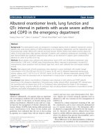
As shown in Figure 2, the mean ratio of oedema fluid (obtained
by s-Cath) to plasma protein in patients with ACLE (n = 9) at
the time of intubation was 0.20 ± 0.19, a value significantly dif-
ferent from that found in patients with ALI/ARDS with a sec-
ondary (indirect) origin (n = 7; 0.81 ± 0.33; P = 0.002).
Patients with primary ALI/ARDS (direct pulmonary, mainly
pneumonia; n = 11) had a mean ratio value of 0.32 ± 0.42 (P
Table 2
Causes for ALI/ARDS and ACLE
Definition N
➢ Primary (direct pulmonary) ALI/ARDS 14
• Pneumonia/aspiration 11
• Carmustine-induced lung injury 1
• Methotrexate-induced lung injury 1
• COP 1
➢ Secondary (indirect pulmonary) ALI/ARDS 7
• Sepsis 6
• Necrotising pancreatitis 1
➢ ACLE 9
• Acute exacerbation of CHF 2
• Acute coronary syndrome
- AMI 5
- Unstable angina 1
• Acute exacerbation of LV diastolic dysfunction 1
ACLE = acute cardiogenic lung edema; ALI = acute lung injury; AMI
= acute myocardial infarction; ARDS = acute respiratory distress
syndrome; CHF = congestive heart failure; COP = cryptogenic
organising pneumonia; LV = left ventricular.
Figure 2
Protein concentration ratio in patients with ACLE (n = 9), primary (n = 11) and secondary (n = 7) ALI/ARDSProtein concentration ratio in patients with ACLE (n = 9), primary (n =
11) and secondary (n = 7) ALI/ARDS. Sampling obtained by s-Cath.
ACLE = acute cardiogenic lung oedema; ALI = acute lung injury;
ARDS = acute respiratory distress syndrome; s-Cath = suction cathe-
ter.
Critical Care Vol 13 No 4 Colucci et al.
Page 6 of 9
(page number not for citation purposes)
= 0.03 vs. secondary ALI/ARDS protein concentration ratio).
The mean plasma C-reactive protein level at inclusion was 183
± 142 mg/L in the whole ALI/ARDS group (n = 21) and 79 ±
72 mg/L in patients with ACLE (n = 9; P = 0.05; Table 1). Fig-
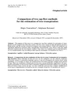
ure 3 shows the median value of the absolute PMN count for
all but one of the patients with ACLE (n = 8) compared with
the PMN count for patients with ALI/ARDS without pneumonia
(n = 10), obtained by s-Cath. There was no statistically signif-
icant difference between groups. The patients with ACLE also
showed an increased PMN count, but this was not as great as
that observed in the patients with ALI/ARDS.
Evaluation of agreement between s-Cath and mini-BAL
sampling methods
Bland-Altman plots evaluating agreement between the two
sampling techniques using protein content and PMN percent-
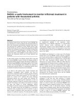
age as efficacy parameters are shown in Figure 4 and 5. The
average difference in protein content was 12.1 g/L (n = 14
paired collections, 6 patients with ACLE and 8 patients with
ALI/ARDS without thick secretions; P = 0.025; 95% confi-
dence interval (CI) 1.73 to 22.4), indicating that the protein
content detected in the same patient was significantly higher
when sampled by s-Cath. The differences increase as the
average protein content increases in the two methods (Figure
4). Specifically, as the average total protein concentration in
the lung increases, the s-Cath method returns more protein
than does the mini-BAL method. The average difference in the
PMN percentage was 14.0% (n = 15 paired collections, 6
patients with ACLE and 9 patients with ALI/ARDS without
thick secretions; P = 0.16; 95% CI -6.12 to 34.05), indicating
that the PMN percentage detected by the two techniques in
the same patient was not significantly different. The power of
this test was nevertheless only 65% with our paired sample
size of 15 patients. The difference between the two tech-
niques tended to decrease as the average PMN percentage
increased (Figure 5). Finally, we did not find any association
related to the underlying disease process.
Discussion
The sampling of alveolar fluid from patients with acute respira-
tory failure allows the study of lung inflammatory response to
various injuries. As demonstrated by Matthay and co-workers
[4,5,10], direct sampling of undiluted lung oedema fluid may
provide fundamental insights into the onset and evolution of
acute inflammatory changes in permeability lung oedema as
well as help to determine whether the pulmonary oedema is
primarily from a hydrostatic or increased permeability mecha-
nism. bBAL is generally well tolerated even in severely hypox-
aemic patients with ALI/ARDS [15]. Yet, the sampling
collections with this gold standard technique may sometimes
be restricted by persistent severe hypoxaemia or cardiovascu-
lar instability, the presence of small endotracheal tubes or the
unavailability of a bronchoscopist. Mini-BAL has been suc-
cessfully evaluated in comparison with bBAL for the diagnosis
of ventilator-associated pneumonia [8], but it showed disap-
pointing results in a recent study where both techniques were
compared for assessing alveolar permeability and inflamma-
tion in patients at risk for ARDS or with ARDS [9]. However,
considering that the s-Cath sampling might not perform ade-
quately after 24 hours because the ability to obtain oedema
fluid may decline over the course of ALI/ARDS, we decided to
use the mini-BAL as a comparison methodology, because it is
easily performed at the bedside and may be completed with-
out a bronchoscopist.
Mini-BAL was generally a safe procedure. However, when per-
formed with a 16-Fr 5 mm outer diameter catheter, the mini-
BAL procedure on rare occasions induced significant gas
exchange abnormalities lasting up to 30 minutes after the end
of collection, as shown by SpO
2
and PaO
2
/FiO
2
abnormalities.
Hypoxaemia was probably induced by the lavage itself and by
reduced tidal volumes delivered while the catheter was in
place [7,16]. We measured the P
peak
on the ventilator (back
pressure) before, during and after the procedure. This pres-
sure significantly increased during mini-BAL sampling, repre-
senting an indirect sign of unstable tidal volumes during the
ventilatory cycle [7,17]. Moreover, mini-BAL caused minor
bronchial haemorrhage in five patients, leading us to stop the
investigation prematurely. Another potential side effect of mini-
BAL is sepsis-like systemic effects, which may emerge pre-
dominantly in patients with pneumonia [18]. With s-Cath, sam-
ples were collected by physicians and trained ICU nurses; the
procedure was rapid and no complications occurred. This
result indicates an advantage of the s-Cath procedure
because collection can be performed shortly after intubation
and at the onset of ALI or hydrostatic oedema. Another advan-
Figure 3
Absolute PMN count in patients with ACLE (n = 8) and ALI/ARDS with-out pneumonia (n = 10)Absolute PMN count in patients with ACLE (n = 8) and ALI/ARDS with-
out pneumonia (n = 10). The horizontal line represents the median. The
box encompasses the 25
th
to 75
th
percentiles and the error bars show
the 10
th
to 90
th
percentiles. Filled circles: outliers. The difference is non-
significant. Sampling obtained by s-Cath. ACLE = acute cardiogenic
lung oedema; ALI = acute lung injury; ARDS = acute respiratory dis-
tress syndrome; PMN = polymorphonuclear cell; s-Cath = suction cath-
eter.
Available online />Page 7 of 9
(page number not for citation purposes)
tage is that fluid is suctioned undiluted without saline, and,
therefore, the measurement of protein or potential mediators
of lung injury can be made without dilution. For this reason, the
protein concentration ratio of the oedema fluid:plasma was
calculated in our different groups using samples obtained by
the s-Cath. The main disadvantage of the s-Cath oedema fluid
sampling method is that it seldom yields lung oedema fluid
after the first 24 hours of intubation. Therefore, this sampling
technique is preferred for studying lung fluid at the onset of
lung injury shortly after endotracheal intubation.
In the patient population in this study, the mean value of the
oedema fluid protein/plasma ratio in patients with primary ALI/
ARDS was significantly lower compared with the value in the
group of patients with a secondary form of ALI/ARDS. We
speculate that during secondary ARDS, there is a more severe
capillary leak that may flood the alveoli [19], possibly explain-
ing the higher protein concentration ratio in the early disease
phase of indirect ALI/ARDS while the early direct insult of the
alveoli in pneumonia-associated ALI/ARDS may exude less
protein resulting in a lower oedema fluid/plasma ratio.
Figure 4
Bland-Altman analysis of agreement showing the differences between protein content (g/L) measurements plotted against the average between methodsBland-Altman analysis of agreement showing the differences between protein content (g/L) measurements plotted against the average between
methods. Squares correspond to patients. The middle horizontal line indicates the average difference between the two methods (12.1 g/L), whereas
the outer lines represent the upper and lower limits of agreement. The black squares represents patients with acute cardiogenic lung oedema.
Figure 5
Bland-Altman analysis of agreement showing the differences between measurements of percentage of neutrophils plotted against the average between methodsBland-Altman analysis of agreement showing the differences between measurements of percentage of neutrophils plotted against the average
between methods. Squares correspond to patients. The middle horizontal line indicates the average difference between the two methods (14%),
whereas the outer lines represent the upper and lower limits of agreement. The black squares represents patients with acute cardiogenic lung
oedema.
Critical Care Vol 13 No 4 Colucci et al.
Page 8 of 9
(page number not for citation purposes)
Our results did not show a good agreement between the s-
Cath and mini-BAL sampling techniques. Using protein con-
tent and PMN percentage as efficacy parameters, we found, in
applying Bland-Altman plots, a significant bias with wide limits
of agreement between the two methods. When the protein
concentration in the lung was high, the s-Cath method is a bet-
ter method for estimating protein concentration (Figure 4); in
contrast, as inflammation increases, both methods provide
similar estimates of the percentage of neutrophils in the air
spaces of the lung (Figure 5). The analysis of our plots indi-
cates that, compared with the results for mini-BAL, the protein
content was significantly higher in the same patient when
measured by s-Cath. In other words, the s-Cath sampling tech-
nique 'detected more' protein content, meaning that this
method could be more sensitive than mini-BAL itself for this
purpose. These results suggest that the s-Cath and mini-BAL
procedures cannot be used interchangeably for studying lung
fluid composition during lung injury and that collection of lung
oedema fluid should be performed with the same method.
Interestingly, our results show an increased absolute PMN
count recorded in patients with ACLE. Recent laboratory and
clinical studies have provided evidence that cardiogenic pul-
monary oedema may be associated with a mild increase in per-
meability of the alveolocapillary barrier and that ongoing
pulmonary injury and inflammation may characterise this disor-
der, particularly when the hydrostatic pressure elevations are
severe [20-24]. For example, Pugin and colleagues [25] found
that inflammatory cytokines and IL-8 increased rapidly after
intubation and positive pressure ventilation in patients with
ACLE, although these levels were lower than in patients with
ALI. Considering that our samples were obtained shortly after
intubation through the s-Cath procedure, the increased abso-
lute PMN count in patients with ACLE was probably not
related to ventilator-induced lung injury. We speculate that this
finding may indicate an inflammatory process during the hydro-
static form of pulmonary oedema. Although the mean plasma
C-reactive protein level in patients with ACLE was significantly
lower than the level recorded in the group of patients with ALI/
ARDS, the raised C-reactive protein concentration in patients
with the hydrostatic form of lung oedema, devoid of any treat-
ment with corticosteroids or clinical and bacteriological evi-
dence of infection, is notable. Dysregulation of C-reactive
protein in the setting of acute hydrostatic lung oedema seems
to be a common finding that could be associated with a con-
comitant inflammatory process, therefore perhaps playing a
role in the evolution of this form of oedema [26,27].
Our study has some limitations. A lack of agreement between
s-Cath and mini-BAL may occur for several reasons: the varia-
bility of instilled volume of the mini-BAL may have influenced
the results; the techniques have two distinct dilution features,
the region of the lung where oedema is sampled is achieved
blindly and the lung injury is heterogeneous; and the difficulty
in wedging the mini-BAL catheter properly in a distal airway
may further represent a barrier in achieving comparable
results. Another limitation for using s-Cath is the presence of
sticky airways secretions, typically found in primary ALI/ARDS
following bilateral pneumonia, making it impossible to obtain
free-flowing oedema fluid. This problem was the main reason
for excluding few patients from our paired analysis.
Studies assessing the impact of pulmonary heterogeneity in
patients with ALI, ARDS or ACLE would therefore be helpful
in the future for evaluating sampling agreement of different
techniques. Finally, although we tried to study our patients as
early as possible after the clinical recognition of injury, some
patients were not investigated with the mini-BAL procedure at
exactly the same time as the s-Cath sampling but all the pro-
cedures were completed within a four-hour time window. Nev-
ertheless, we consider this frame of time as likely to be
representative of the functional status of lung neutrophils and
protein concentration because lung PMN and total protein
does not change significantly over the first three days after the
onset of ARDS when measured by the traditional bBAL proce-
dure [28-30].
Conclusions
This study in patients with ALI/ARDS and cardiogenic lung
oedema compared two minimally invasive methods for sam-
pling oedema fluid from distal lung air spaces. The results
show significant differences between the s-Cath and mini-BAL
techniques, suggesting that these procedures cannot be used
interchangeably for sequentially studying the lung inflamma-
tory response in the distal air spaces. Except for use in
patients with purulent airway secretions, the s-Cath method
has more advantages than the mini-BAL technique, because
the s-Cath procedure is rapid, non-invasive, inexpensive and,
above all, can be performed shortly after intubation at the
onset of ALI or hydrostatic oedema. Moreover, the oedema
fluid is undiluted with saline, allowing the accurate measure-
ment of protein and potential mediators of lung injury. The
oedema fluid sampling technique remains a preferred method
for studying lung fluid at the onset of ALI in intubated patients.
Nevertheless, both techniques are minimally invasive and pro-
vide a method to quantify the inflammatory response and the
degree of protein exudation in the distal airspaces of the lung
in patients with bilateral pulmonary infiltrates and acute respi-
ratory failure that requires mechanical ventilation.
Competing interests
The authors declare that they have no competing interests.
Available online />Page 9 of 9
(page number not for citation purposes)
Authors' contributions
GD collected the samples and wrote the initial draft and the
final manuscript. GC collected the samples and data and par-
ticipated in writing and revising the final manuscript. RDB and
JB performed the data analysis. CL directed the statistical
analysis and interpretation and participated in drafting the ini-
tial manuscript. MAM and TRM conceived the premise and
participated in writing, interpretation and analysis. All authors
have read and approved the final manuscript.
Acknowledgements
The authors are grateful to the nurses for their invaluable and precious
help during the collection of samples in the ICU.
References
1. Bernard GR, Artigas A, Brigham KL, Carlet J, Falke K, Hudson L,
Lamy M, LeGall JR, Morris A, Spragg R: The American-European
Consensus Conference on ARDS. Definitions, mechanisms,
relevant outcomes and clinical trial coordination. Am J Respir
Crit Care Med 1994, 149:818-824.
2. Ware LB, Matthay MA: The acute respiratory distress syn-
drome. N Engl J Med 2000, 342:1334-1349.
3. Pugin J, Verghese G, Widmer MC, Matthay MA: The alveolar
space is the site of intense inflammatory and profibrotic reac-
tions in the early phase of acute respiratory distress syn-
drome. Crit Care Med 1999, 27:304-312.
4. Matthay MA, Wiener-Kronish J: Intact epithelial barrier function
is critical for the resolution of alveolar edema in humans. Am
Rev Respir Dis 1990, 142:1250-1257.
5. Ware LB, Matthay MA: Alveolar fluid clearance is impaired in
the majority of patients with acute lung injury and the acute
respiratory distress syndrome. Am J Respir Crit Care Med
2001, 163:1376-1383.
6. De Pasquale CG, Arnolda LF, Doyle IR, Grant RL, Aylward PE, Ber-
sten AD: Prolonged alveolocapillary barrier damage after acute
cardiogenic pulmonary edema. Crit Care Med 2003,
31:1060-1067.
7. Lindholm CE, Ollman B, Snyder JV, Millen EG, Grenvik A: Cardi-
orespiratory effects of flexible fiberoptic broncoscopy in criti-
cally ill patients. Chest 1978, 74:362-368.
8. Chastre J, Fagon JY: Ventilator-associated pneumonia. Am J
Respir Crit Care Med 2002, 165:867-903.
9. Perkins GD, Chatterjie S, McAuley DF, Gao F, Thickett DR: Role
of nonbronchoscopic lavage for investigating alveolar inflam-
mation and permeability in acute respiratory distress syn-
drome. Crit Care Med 2006, 34:57-64.
10. Verghese GM, Ware LB, Matthay BA, Matthay MA: Alveolar epi-
thelial fluid transport and the resolution of clinically severe
hydrostatic pulmonary edema. J Appl Physiol 1999,
87:1301-1312.
11. LeGall JR, Lemeshow St, Saulnier F: A new simplified acute
physiology score (SAPS II) based on a European/North Amer-
ican multicenter study. JAMA 1993, 270:2957-2963.
12. Murray JF, Matthay MA, Luce JM, Flick MR: An expanded defini-
tion of the adult respiratory distress syndrome. Am Rev Respir
Dis 1988, 138:720-723.
13. The Acute Respiratory Distress Syndrome Network: Ventilation
with lower tidal volumes as compared with traditional tidal vol-
umes for acute lung injury and the acute respiratory distress
syndrome. N Engl J Med 2000, 342:1301-1308.
14. Bland JM, Altman DG: Statistical methods for assessing agree-
ment between two methods of clinical measurement. Lancet
1986, i:567-569.
15. Steinberg KP, Mitchell DR, Maunder RJ, Milberg JA, Whitcomb
ME, Hudson LD: Safety of bronchoalveolar lavage in patients
with adult respiratory distress syndrome. Am Rev Respir Dis
1993, 148:556-561.
16. Jolliet Ph, Chévrolet JC: Bronchoscopy in the intensive care unit.
Intensive Care Med 1992, 18:160-169.
17. Fein A, Grossman RF, Jones JC, Overland E, Pitts L, Murray JF,
Staub NC: The value of edema protein measurements in
patients with pulmonary edema. Am J Med 1979, 67:32-39.
18. Pugin J, Suter PM: Diagnostic bronchoalveolar lavage in
patients with pneumonia produces sepsis-like systemic
effects. Intensive Care Med 1992, 18:6-10.
19. Gattinoni L, Pelosi P, Suter PM, Pedoto A, Vercesi P, Lissoni A:
Acute respiratory distress syndrome caused by pulmonary
and extrapulmonary disease. Different syndromes? Am J
Respir Crit Care Med 1998, 158:3-11.
20. Steinberg KP, Milberg JA, Martin TR, Maunder RJ, Cockrill BA,
Hudson LD: Evolution of bronchoalveolar cell populations in
the adult respiratory distress syndrome. Am J Respir Crit Care
Med 1994, 150:113-122.
21. Cohen AB, Stevens MD, Miller EJ, Atkinson MA, Mullenbach G,
Maunder RJ, Martin TR, Wiener-Kronish JP, Matthay MA: Neu-
trophil-activating peptide-2 in patients with pulmonary edema
from congestive heart failure or ARDS. Am J Physiol
1993,
264:L490-L495.
22. Doyle IR, Nicholas TE, Bersten AD: Serum surfactant protein-A
levels in patients with acute cardiogenic pulmonary edema
and adult respiratory distress syndrome. Am J Respir Crit Care
Med 1995, 152:307-317.
23. West JB, Mathieu-Costello O: Vulnerability of pulmonary capil-
laries in heart disease. Circulation 1995, 92:622-631.
24. De Pasquale CG, Arnolda LF, Doyle IR, Grant RL, Aylward PE, Ber-
sten AD: Prolonged alveolocapillary barrier damage after acute
cardiogenic pulmonary edema. Crit Care Med 2003,
31:1060-1067.
25. Pugin J, Verghese G, Widmer M-C, Matthay MA: The alveolar
space is the site of intense inflammatory and profibrotic reac-
tions in the early phase of acute respiratory distress syn-
drome. Crit Care Med 1999, 27:304-312.
26. Yeh ETH, Willerson JT: Coming of age of C-reactive protein.
Using inflammation markers in cardiology. Circulation 2003,
107:370-372.
27. Labarrere CA, Zaloga GP: C-reactive protein: from innocent
bystander to pivotal mediator of atherosclerosis. Am J Med
2004, 117:499-507.
28. Martin TR, Pistorese BP, Hudson LD, Maunder RJ: The function
of lung and blood neutrophils in patients with the adult respi-
ratory distress syndrome. Implications for the pathogenesis of
lung infections. Am Rev Respir Dis 1991, 144:254-262.
29. Pittet JF, Mackersie RC, Martin TR, Matthay MA: Biological mark-
ers of acute lung injury: prognostic and pathogenetic signifi-
cance. Am J Respir Crit Care Med 1997, 155:1187-1205.
30. Park WY, Goodman RB, Steinberg KP, Ruzinski JT, Radella F 2nd,
Park DR, Pugin J, Skerrett SJ, Hudson LD, Martin TR: Cytokine
balance in the lungs of patients with acute respiratory distress
syndrome. Am J Respir Crit Care Med 2001, 164:1896-1903.
Key messages
• No data exist comparing mini-BAL and s-Cath for
assessing lung inflammation in acute hypoxaemic respi-
ratory failure.
• Using protein content and PMN percentage as parame-
ters, we identified substantial variations between the
two sampling techniques.
• When the protein concentration in the lung was high,
the s-Cath was a more sensitive method.
• As inflammation increased, both methods provided sim-
ilar estimates of neutrophil percentages in the lung.
• Both procedures cannot be used interchangeably for
sequentially studying the lung inflammatory response in
the distal air spaces.