Báo cáo y học: " Cardiac effects of induction agents in the septic rat heart" potx
Bạn đang xem bản rút gọn của tài liệu. Xem và tải ngay bản đầy đủ của tài liệu tại đây (391.62 KB, 8 trang )
Open Access
Available online />Page 1 of 8
(page number not for citation purposes)
Vol 13 No 5
Research
Cardiac effects of induction agents in the septic rat heart
York A Zausig, Hendrik Busse, Dirk Lunz, Barbara Sinner, Wolfgang Zink and Bernhard M Graf
Department of Anaesthesiology and Critical Care, University of Regensburg, Franz-Josef-Strauss-Allee 11, Regensburg, 93053, Germany
Corresponding author: York A Zausig,
Received: 3 Aug 2009 Revisions requested: 28 Aug 2009 Revisions received: 2 Sep 2009 Accepted: 8 Sep 2009 Published: 8 Sep 2009
Critical Care 2009, 13:R144 (doi:10.1186/cc8038)
This article is online at: />© 2009 Zausig et al.; licensee BioMed Central Ltd.
This is an open access article distributed under the terms of the Creative Commons Attribution License ( />),
which permits unrestricted use, distribution, and reproduction in any medium, provided the original work is properly cited.
Abstract
Introduction The current debate about the side effects of
induction agents, e.g. possible adrenal suppression through
etomidate, emphasizes the relevance of choosing the correct
induction agent in septic patients. However, cardiovascular
depression is still the most prominent adverse effect of these
agents, and might be especially hazardous in septic patients
presenting with a biventricular cardiac dysfunction - or so-called
septic cardiomyopathy. Therefore, we tested the dose-response
direct cardiac effects of clinically available induction agents in
an isolated septic rat heart model.
Methods A polymicrobial sepsis was induced via cecal ligation
and single puncture. Hearts (n = 50) were isolated and
randomly assigned to five groups, each receiving etomidate,
s(+)-ketamine, midazolam, propofol, or methohexitone at
concentrations of 1 × 10
-8
to 1 × 10
-4
M. Left ventricular
pressure, contractility and lusitropy, and coronary flow were
measured. Cardiac work, myocardial oxygen delivery, oxygen
consumption, and percentage of oxygen extraction were
calculated.
Results All of the induction agents tested showed a dose-
dependent depression of cardiac work. Maximal cardiac work
dysfunction occurred in the rank order of s(+)-ketamine (-6%)
<etomidate (-17%) <methohexitone (-31%) <midazolam (-
38%) <propofol (-50%). In addition, propofol showed a
maximum decrease in contractility of -38%, a reduction in
lusitropy of -44%, and a direct vasodilator effect by increasing
coronary flow by +29%.
Conclusions Overall, this study demonstrates that these tested
drugs indeed have differential direct cardiac effects in the
isolated septic heart. Propofol showed the most pronounced
adverse direct cardiac effects. In contrast, S(+)ketamine
showed cardiovascular stability over a wide range of
concentrations, and might therefore be a beneficial alternative to
etomidate.
Introduction
Although the ideal induction agent for critically ill patients has
not yet been found, there is general agreement that in those
patients an induction agent that provides cardiovascular sta-
bility upon induction of anesthesia would be first choice. Nev-
ertheless, current guidelines do not recommend one induction
agent over another [1,2]. However, there are concerns that
non-cardiovascular side effects, such as possible adrenal sup-
pression by etomidate, could compromise critically ill patients
and last at least 24 hours [3]. At present the clinical conse-
quences are not clear [4].
However, the most significant adverse effect of induction
agents is cardiovascular depression, which has already been
well described in healthy animal models and humans after
intravenous administration. The degree of negative cardiovas-
cular effects depends on dose and speed of administration
and appears to vary greatly among the commonly used drugs
[5,6]. In non-septic patients or experimental settings, clinically
available induction agents, such as etomidate, propofol, keta-
mine, methohexitone or midazolam, show dose-dependent
effects [5,6]. These effects result from their variable impact on
peripheral arteriolar and venous dilation, from direct cardiac
depression or both. Surprisingly, direct cardiac effects of
induction agents in isolated septic hearts have so far not been
systematically evaluated.
The cardiovascular dysfunction in sepsis derives from a
reduced systemic vascular resistance typically complicated by
decreased cardiac function [1,2]. This cardiac dysfunction -
the so-called septic cardiomyopathy - is a major contributor to
sepsis-related morbidity and mortality [7,8]. It affects both
+dLVP/dt: left ventricular contractility; -dLVP/dt: left ventricular relaxation; DO
2
: myocardial oxygen supply; LVP: Left ventricular pressure; MVO
2
: myo-
cardial oxygen consumption; pCO
2
: partial pressure of carbon dioxide; PO
2
: and partial pressure of oxygen.
Critical Care Vol 13 No 5 Zausig et al.
Page 2 of 8
(page number not for citation purposes)
ventricles in the phases of contraction and relaxation [7-11].
Almost one-fifth of all septic patients with refractory hypoten-
sion die because of a low cardiac output deriving from this
severe myocardial dysfunction. It is, therefore, the everyday
clinical challenge of each intensive care unit physician to suffi-
ciently treat septic patients without further compromising the
already reduced function of the septic heart [9]. This mechan-
ical impairment is accompanied by disturbed myocardial
metabolism and coronary flow, which influences a balanced
myocardial oxygen supply-demand ratio [10].
However, global cardiac mechanical and metabolic effects of
these induction agents in septic cardiomyopathy have thus far
not been systematically compared in a dose-dependent fash-
ion. There is very little evidence on the direct in vitro effects of
these agents on cardiac contractile function in sepsis, and the
isolated, dose-dependent effects of these induction agents on
myocardial excitability, contractility, coronary flow, and oxygen
utilization in a septic heart are still unknown. We used the iso-
lated ex vivo heart model to study the direct cardiac effects in
the absence of confounding neurohormonal, metabolic, or sys-
temic factors.
Therefore, the aim of this study was to directly compare elec-
trical, mechanical, and metabolic effects of etomidate, s(+)-
ketamine, midazolam, propofol, and methohexitone at equimo-
lar concentrations, with special emphasis on their impact on
cardiac work.
Materials and methods
Approval from the Institutional Animal Care Committee of the
University of Goettingen was obtained before initiation of this
study. All experimental procedures conformed with German
animal safety regulations. Fifty male Wistar rats (weighing 245
± 3 g) were injected intraperitoneally with 100 mg/kg keta-
mine and 2.5 to 5 mg/kg xylazine hydrochloride. A polymicro-
bial sepsis was induced via cecal ligation and a single
puncture as reported previously in detail [12]. After 20 hours
of incubation, hearts were isolated and prepared as has been
described in recent reports [13]. All hearts were perfused at a
perfusion pressure of 55 mmHg with a modified Krebs-
Ringer's salt solution, which was filtered in-line (5 μm pore-
size filter disk, Sigma-Aldrich
®
, Munich, Germany) and had the
following composition: Na
+
140 mM; K
+
4.5 mM; Mg
2+
1.2
mM; Ca
2+
2.5 mM; Cl
-
134 mM; HCO
3
-
15.5 mM; H
2
PO
4
-
1.2
mM; EDTA 0.05 mM; glucose 11.5 mM; pyruvate 2 mM; man-
nitol 10 mM; and insulin 5 U/L. Mean aortic inflow pH, partial
pressure of carbon dioxide (pCO
2
), and partial pressure of
oxygen (PO
2
) were 7.39 ± 0.01, 36 ± 1 mmHg, and 580 ± 25
mmHg, respectively. Perfusate and heart temperature was
maintained at 36.9 ± 0.3°C throughout the experiment.
Spontaneous atrial rate, atrio-ventricular conduction time, and
systolic left ventricular pressure (LVP) and its derivative were
measured as detailed previously [13]. Coronary inflow was
measured at constant temperature and under constant pres-
sure of 55 mmHg by a transit-time in-line ultrasound flow meter
(Research Flowmeter T106, Transonic Systems, Ithaca, USA).
Coronary inflow and outflow (coronary sinus) oxygen tensions
(mmHg) were measured discontinuously using a self-calibrat-
ing gas analyzer (AVL OMNI 9
®
, Roche Diagnostic, Man-
nheim, Germany). Oxygen delivery, percent oxygen extraction,
and myocardial oxygen consumption were calculated as noted
previously [6,13]. Cardiac work ((left ventricular systolic pres-
sure - left ventricular diastolic pressure) × heart rate) was cal-
culated [14]. All measurements were taken during the last
minute of each 15-minute experimental period for statistical
analysis.
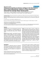
The experimental protocol is shown in Figure 1. After steady
state, the hearts were randomly assigned by lottery to five
groups (10 hearts each) and received propofol (Disoprivan
®
,
AstraZeneca, Wedel, Germany), midazolam (Midazolam-Rati-
opharm
®
, Ratiopharm, Ulm, Germany), s(+)-ketamine (S(+)-
Ketamine
®
, Pfizer Pharma, Berlin, Germany), methohexitone
(Brevimytal
®
, Hikma Pharma, Graefeling, Germany), or etomi-
date (Etomidat-Lipuro
®
, B. Braun, Melsungen, Germany).
Each heart was perfused, in randomized order, at concentra-
tions of 10
-8
to 10
-4
M with one of these drugs for a period of
15 minutes. There was a 20-minute drug-free washout period.
Prior to this study we tested higher concentrations for each of
these drugs. However, at concentrations higher than 10
-3
M
some hearts showed cardiac arrest. Therefore, concentrations
of 10
-3
M or more were not included in the present study.
The concentrations tested in our study (10
-8
to 10
-4
M), which
are equivalent to 0.002 to 18 μg/mL propofol (molecular
weight: 178.3 mM), 0.003 to 33 μg/mL midazolam (325.8
mM), 0.003 to 27 μg/mL s(+)-ketamine (274.2 mM), 0.003 to
26 μg/mL methohexitone (262.3 mM), and 0.002 to 24 μg/mL
etomidate (244.3 mM), correspond to approximate therapeu-
tic plasma-free values (corrected for plasma protein binding, in
%) of 5.1 to 11 × 10
-7
(97 to 98%) propofol, 3.7 to 37 × 10
-9
M (94 to 95%) midazolam, 3.2 to 19 × 10
-6
M (12 to 30%)
Figure 1
Study protocolStudy protocol. CLP-OP = cecal ligation and puncture operating
procedure.
Available online />Page 3 of 8
(page number not for citation purposes)
s(+)-ketamine, 4.6 to 9.1 × 10
-6
M (70 to 73%) methohexi-
tone, and 0.9 to 4.7 × 10
-7
M (77 to 94%) etomidate
[6,15,16]. However, even higher concentrations up to 10 fold
can easily be achieved by bolus injection [17].
Statistical analysis
All data in the text, tables and figures are displayed as means
± standard error of the mean. Raw data from each functional
and metabolic variable were compared by analysis of variance
with repeated measures. If F tests were significant, Bonferoni
tests were used to compare absolute group means for each
variable measured at the same concentration and individual
drug concentrations (and washout, WASH) against the initial
control (CTRL). P < 0.05 was considered to be statistically
significant.
Results
Control values of sham-operated hearts (heart rate 309 ± 4
beats/min, LVP contractility (+dLVP/dt) 3275 ± 84 mmHg/
sec, LVP relaxation (-dLVP/dt) 2629 ± 74 mmHg/sec, cardiac
work 36036 ± 639 mmHg/beats, and myocardial oxygen sup-
ply (DO
2
)/myocardial oxygen consumption (MVO
2
) ratio 1.5 ±
0.0 were statistically different from control values of septic
hearts. Sham-operated hearts showed control values of etomi-
date, s(+)-ketamine, midazolam, propofol, and methohexitone
in septic hearts were not statistically different between the
groups. After a washout period, each parameter returned to
baseline level.
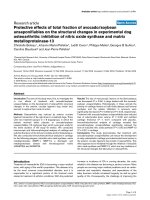
The comparative effects of etomidate, s(+)-ketamine, mida-
zolam, propofol, and methohexitone on heart rate are shown in
Figure 2. No effects on heart rate were observed at 1 × 10
-8
to 1 × 10
-6
M for any induction agent. At higher concentra-
tions, heart rate was significantly suppressed at 1 × 10
-4
M for
propofol (maximum decrease: -29 ± 4%) and at 1 × 10
-5
to 1
× 10
-4
M for midazolam (maximum decrease: -47 ± 5%).
Reduction of heart rate by midazolam at 1 × 10
-4
M was signif-
icantly more pronounced compared with all other tested
induction agents. Maximum decreases in heart rate were -12
± 3% for s(+)-ketamine (1 × 10
-4
M) and -11 ± 4% for meth-
ohexitone (1 × 10
-4
M). Only etomidate showed no chrono-
tropic effect at any tested concentration in this study.
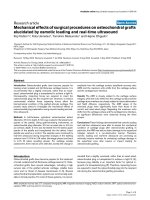
All tested induction agents showed a dose-dependent
decrease in cardiac contractility except for midazolam and
s(+)-ketamine (Figure 3). The maximum decrease in +dLVP/dt
of -38 ± 5% at 1 × 10
-4
M and -19 ± 5% at 1 × 10
-4
M was
significant for propofol and methohexitone, respectively. The
effects of propofol were significantly more pronounced com-
pared with all other agents tested at equimolar concentrations.
Other induction agents showed a maximum decrease in con-
tractility of -5 ± 6% for etomidate at 1 × 10
-5
M, and a maxi-
mum increase in contractility of +7 ± 5% for s(+)-ketamine at
1 × 10
-4
M, and +9 ± 6% for midazolam at 1 × 10
-6
M. As
shown in Figure 4, etomidate, midazolam, methohexitone, and
propofol showed negative lusitropic effects with maximal
decreases in -dLVP/dt of -7 ± 6% (at 1 × 10
-5
M, not signifi-
cant), -21 ± 5% (at 1 × 10
-4
M, significant), -21 ± 6% (at 1 ×
10
-4
M, significant), and -44 ± 5% (at 1 × 10
-4
M, significant),
respectively. At 1 × 10
-4
M the negative reduction of lusitropy
by propofol was significantly different compared with all other
tested induction agents. In contrast, at 1 × 10
-4
M s(+)-keta-
mine demonstrated an increase in lusitropy of +14 ± 6%.
There was a significant difference compared with propofol and
midazolam at equimolar concentration.
Cardiac work (Figure 5) - the product of LVP and heart rate -
was reduced at 1 × 10
-4
M by etomidate (maximum decrease:
Figure 2
Comparative effects of etomidate, s(+)-ketamine, midazolam, propofol, and methohexitone on heart rate in rat isolated septic heartsComparative effects of etomidate, s(+)-ketamine, midazolam, propofol, and methohexitone on heart rate in rat isolated septic hearts. All drugs except
etomidate decreased chronotropic effects. For control values, only the first (CTRL) and the washout (WASH) periods are displayed. After the wash-
out period, the heart rate returned to baseline level. *P < 0.05 midazolam (10
-5
to 10
-4
M) and propofol (10
-4
M) versus control;
#
P < 0.05 midazolam
versus etomidate, s(+)-ketamine, propofol, and methohexitone. Data are the means ± standard error of the mean.
Critical Care Vol 13 No 5 Zausig et al.
Page 4 of 8
(page number not for citation purposes)
-17 ± 6%), s(+)-ketamine (-6 ± 6%), midazolam (-38 ± 7%),
propofol (-50 ± 6%), and methohexitone (-31 ± 4%) in a dose-
dependent fashion. At this concentration, the reduction of car-
diac performance was significantly different for propofol, mida-
zolam and methohexitone compared with s(+)-ketamine.
Additionally, propofol significantly decreased cardiac work at
1 × 10
-5
M by -17 ± 4%.
Etomidate, s(+)-ketamine, midazolam, and methohexitone
showed no direct effects on coronary flow, myocardial oxygen
supply and demand (all not shown). Therefore, DO
2
/MVO
2
ratio (Figure 6) was not affected by these agents. These
effects were similar for propofol at 1 × 10
-8
to 1 × 10
-5
M.
However, at 1 × 10
-4
M propofol significantly increased coro-
nary flow of +29 ± 4%. Additionally, there was a considerable
cardiac-work induced decrease in MVO
2
and oxygen extrac-
tion, accompanied by a coronary flow dependent rise of DO
2
Figure 3
Comparative effects of etomidate, s(+)-ketamine, midazolam, propofol, and methohexitone on left ventricular contractility in rat isolated septic heartsComparative effects of etomidate, s(+)-ketamine, midazolam, propofol, and methohexitone on left ventricular contractility in rat isolated septic hearts.
All drugs except for s(+)ketamine and midazolam decreased contractility. For control values, only the first (CTRL) and the washout (WASH) periods
are displayed. After the washout period, left ventricular contractility (+ dLVP/dt
-1
) returned to baseline level. *P < 0.05 for methohexitone and propo-
fol vs. control;
#
P < 0.05 propofol vs. etomidate, s(+)-ketamine, midazolam, and methohexitone. Data are the means ± standard error of the mean.
Figure 4
Comparative effects of etomidate, s(+)-ketamine, midazolam, propofol, and methohexitone on left ventricular relaxation in rat isolated septic heartsComparative effects of etomidate, s(+)-ketamine, midazolam, propofol, and methohexitone on left ventricular relaxation in rat isolated septic hearts.
All drugs except for s(+)-ketamine decreased lusitropy. For control values, only the first (CTRL) and the washout (WASH) periods are displayed.
After the washout period left ventricular relaxation (-dLVP/dt
-1
) returned to baseline level. *P < 0.05 midazolam, propofol, and methohexitone vs. con-
trol;
#
P < 0.05 midazolam, propofol, and methohexitone vs. etomidate and s(+)-ketamine;
$
P < 0.05 propofol vs. midazolam and methohexitone.
Data are the means ± standard error of the mean.
Available online />Page 5 of 8
(page number not for citation purposes)
leading to increase of DO
2
/MVO
2
ratio (Figure 6) of +58 ±
4%. This was significantly different compared with etomidate,
s(+)-ketamine, midazolam, and methohexitone at equimolar
concentration.
Discussion
The study was designed to compare the direct effects of five
commonly used intravenous induction agents by analyzing car-
diac responses at equimolar concentrations in septic hearts.
The tested drugs demonstrate differential direct effects on
electrical properties, myocardial function, andoxygen supply-
to-demand ratio. Propofol showed the most pronounced
adverse direct cardiac effects, whereas s(+)ketamine was
most beneficial, as it showed cardiac functionality over a wide
range of concentration.
There are concerns regarding the application of etomidate in
critically ill patients, especially in septic patients due to possi-
Figure 5
Comparative effects of etomidate, s(+)-ketamine, midazolam, propofol, and methohexitone on cardiac work in rat isolated septic heartsComparative effects of etomidate, s(+)-ketamine, midazolam, propofol, and methohexitone on cardiac work in rat isolated septic hearts. Each drug
decreased cardiac work (CW). For control values, only the first (CTRL) and the washout (WASH) periods are displayed. After washout period CW
returned to baseline level. *P < 0.05 midazolam (10
-4
M), propofol (10
-5
to 10
-4
M), and methohexitone (10
-4
M) vs. control;
#
P < 0.05 midazolam and
propofol vs. etomidate and methohexitone.
$
P < 0.05 s(+)-ketamine vs. propofol, midazolam, and methohexitone. Data are the means ± standard
error of the mean.
Figure 6
Comparative effects of etomidate, s(+)-ketamine, midazolam, propofol, and methohexitone on myocardial oxygen supply/myocardial oxygen con-sumption in rat isolated septic heartsComparative effects of etomidate, s(+)-ketamine, midazolam, propofol, and methohexitone on myocardial oxygen supply/myocardial oxygen con-
sumption in rat isolated septic hearts. All drugs decreased myocardial oxygen supply/myocardial oxygen consumption (DO
2
/MVO
2
-1
) ratio. For con-
trol values, only the first (CTRL) and the washout (WASH) periods are displayed. After the washout period DO
2
/MVO
2
-1
returned to baseline level.
*P < 0.05 propofol vs. control, etomidate, s(+)-ketamine, midazolam, and methohexitone. Data are the means ± standard error of the mean.
Critical Care Vol 13 No 5 Zausig et al.
Page 6 of 8
(page number not for citation purposes)
ble adrenal suppression [18]. The incidence of this adrenal
suppression in sepsis ranges from 9 to 67%, and cortisol
response to corticotrophin is more frequently impaired after
administration of etomidate as compared with alternative
induction agents [19]. However, in septic patients, cardiovas-
cular instability is the main focus of clinicians because it is the
major cause of morbidity and mortality in sepsis. The presence
of cardiac dysfunction - demonstrated as septic cardiomyopa-
thy - additionally decreases survival rate in septic patients [10].
Therefore, an induction agent that provides cardiovascular sta-
bility such as etomidate is frequently used in healthy subjects
as it is intended to show minimal cardiovascular effects [4,5].
In the present study, we show that etomidate is safe with
regard to cardiac function at concentrations of 10
-8
to 10
-5
M
in septic hearts. However, at higher concentrations it markedly
depresses cardiac work. These concentrations can easily be
achieved either by bolus administration or by long-term infu-
sion in patients with severe sepsis or septic shock, especially
with multiple-organ failure accompanied by a decreased
hepatic and renal metabolism [20]. Therefore, these effects
must be kept in mind, especially because other studies under-
line that a higher induction dose of etomidate is also associ-
ated with a decrease in systolic arterial blood pressure in
animal models and patients with advanced age and heart dis-
ease [21].
In contrast, s(+)ketamine showed cardiac functionality over a
wide range of concentrations. S(+)ketamine is an optical iso-
mer of ketamine and exhibits stereoselective bindings to differ-
ent receptors, accounting for its three to four times higher
anesthetic potency compared with the R(-)-isomer [16,22].
The racemic ketamine and both ketamine stereoisomers show
negative chronotrope, dromotrope, and inotrope effects in the
isolated healthy heart [22]. In septic hearts, s(+)ketamine has
no significant negative effect on LVP, contractility or lusitropy.
This discrepancy might be explained by the fact that both the
R(-)-isomer and racemic ketamine in general show significantly
more cardio-depressant effects as compared with the S(+)-
isomer [16,22]. The mechanism behind this is a stereoselec-
tive suppression of the trans-sarcolemmal Ca
2+
current
(ICa
2+
), which play an important role in the force of contraction
and spontaneous firing of sinoatrial node cells, as demon-
strated in electrophysiological experiments [23].
Midazolam and methohexitone, together with propofol,
showed the most adverse effects on cardiac stability. Propo-
fol, midazolam and methohexitone decreased cardiac work in
a dose-dependent fashion. At very similar concentrations,
Stowe and colleagues showed a decrease in contractility in
guinea pig hearts from midazolam, propofol, and thiopental [6].
However, the degree of contractility reduction was more pro-
nounced in healthy hearts as compared with septic ones.
These surprisingly different results might be model or protocol
dependent. Otherwise, as the mechanisms of the cardiac
depressant effects of these induction agents is likely to involve
attenuation of trans-sarcolemmal Ca
2+
flux [6], the dysfunction
of sarcoplasmic reticulum Ca
2+
handling or altered calcium
transient properties described in septic hearts might be attrib-
utable to these differing results [24,25]. The most striking find-
ing on coronary flow was a direct vasodilating effect by
propofol at 1 × 10
-4
M. This effect suggests that coronary
autoregulation was inhibited at this concentration, and propo-
fol may cause a substantial coronary vasodilation when used
as an anesthetic induction agent [6,13]. In contrast, no other
tested induction agent showed a direct vasodilating effect at
any concentration. However, care has to be taken because
depression of heart function is not always an expression of
hazardous effects. For example, vasodilatation of the coronary
arteries induced by propofol might have led to improving myo-
cardial blood and oxygen supply as shown in Figure 6. Addi-
tionally, the slow down of the heart rate by midazolam might
reduce myocardial energy demands, and may additionally
improve diastolic filling of the heart.
We recognise the limitations of this study. Although the
applied sepsis method has the advantages of inducing a 'nat-
ural' course of infection, it has limitations with regard to note-
worthy outcome variability [26,27]. In contrast, other sepsis
models, such as the bolus injection-type method, offer a sim-
ple and highly standardized method. However, failure of trans-
mission of therapeutic results from bolus shock experiments
into clinical use has emphasized that these models do not
reflect all aspects of the sepsis syndrome [26]. In contrast, the
cecal ligation and single puncture method is generally recog-
nized as closely mimicing human disease by activating pro-
and anti-inflammatory pathways. Another limitation of this
study is that in addition to cardiac depression, induction
agents also induce a systemic vascular dilatation that leads to
hypotension. This is associated with an increased risk of death
in critically ill patients [28]. However, the diagnosis of hypoten-
sion is easy, whereas the diagnosis of septic cardiomyopathy
is more sophisticated and requires a more complex analysis.
Therefore, at the moment of induction, this diagnosis may not
be available, and septic patients would be at an increased risk
in terms of choosing the wrong induction agent. On this
account, we used an ex vivo approach and isolated hearts and
focused on the direct cardiac effects of the applied induction
agents. The advantages of this method are to measure
mechanical and metabolic properties in the absence of the
confounding effects of other organs, systemic circulation, and
a host of peripheral complications such as circulating neuro-
hormonal factors [29]. One potential limitation of an isolated
heart preparation study is the possible influence of a force-fre-
quency relationship. Although there are significant changes in
heart rate for midazolam, which are not accompanied by a sig-
nificant change in +dLVP/dt (Figure 3) at 10
-5
M, the possible
influence of a force-frequency relationship has to be kept in
mind when interpretating the presented results.
Available online />Page 7 of 8
(page number not for citation purposes)
Conclusions
In conclusion, this study showed that the tested drugs - etomi-
date, s(+)-ketamine, midazolam, propofol, and methohexitone
- indeed have differential direct cardiac effects, even in the iso-
lated septic heart. Propofol showed the most pronounced
adverse direct cardiac effects, while S(+)ketamine demon-
strated cardiac stability over a wide range of concentrations.
Thus, if our data can be extrapolated to apply to humans, it
seems that there are alternatives to etomidate such as s(+)ket-
amine, which demonstrates similar cardiac stability, but with
less non-cardiovascular side effects affecting the outcome of
septic patients.
Competing interests
On behalf of my co-authors I attest that the work has not been
funded by any source(s) other than described in the statement.
No author or participant has any financial interest in the sub-
ject matter, materials or equipment discussed or in competing
materials. The laboratory in which the research was performed
has not been funded by, or has any participant in the planning,
conduct, or reporting of the research been funded by or have
financial interests in any source with a real or potential interest
in the subject matter, materials, equipment or devices dis-
cussed or in any competing product or subject. And the labo-
ratory in which the work was performed or any of the authors
or participants have not been funded by any foundation or
other non-governmental source that has received funding from
any organization with a real or potential interest in the subject
matter, materials, equipment or devices discussed, or in any
competing product or subject.
Authors' contributions
YZ and BG originated the idea and performed preliminary
experiments. HB continued to perform the experiments. BS
coordinated to the laboratory support. YZ and WZ were
responsible for writing the paper. DL, BS and BG supported
the editing of the manuscript and added important comments
to the paper. All authors read and approved the final
manuscript.
Authors' information
The data was presented in part at the 3rd International Con-
gress of the German Sepsis Society in Weimar from 5 to 8
September, 2007.
Acknowledgements
This study was supported with institutional funding from the Department
of Anaesthesiology, University of Regensburg.
References
1. Dellinger RP, Levy MM, Carlet JM, Bion J, Parker MM, Jaeschke R,
Reinhart K, Angus DC, Brun-Buisson C, Beale R, Calandra T, Dhai-
naut JF, Gerlach H, Harvey M, Marini JJ, Marshall J, Ranieri M, Ram-
say G, Sevransky J, Thompson BT, Townsend S, Vender JS,
Zimmerman JL, Vincent JL: Surviving Sepsis Campaign: interna-
tional guidelines for management of severe sepsis and septic
shock: 2008. Crit Care Med 2008, 36:296-327.
2. Reinhart K, Brunkhorst F, Bone H, Gerlach H, Grundling M, Krey-
mann G, Kujath P, Marggraf G, Mayer K, Meier-Hellmann A, Peck-
elsen C, Putensen C, Quintel M, Ragaller M, Rossaint R, Stuber F,
Weiler N, Welte T, Werdan K: [Diagnosis and therapy of sepsis:
guidelines of the German Sepsis Society Inc. and the German
Interdisciplinary Society for Intensive and Emergency
Medicine]. Anaesthesist 2006, 55(Suppl 1):43-56.
3. Malerba G, Romano-Girard F, Cravoisy A, Dousset B, Nace L, Levy
B, Bollaert PE: Risk factors of relative adrenocortical deficiency
in intensive care patients needing mechanical ventilation.
Intensive Care Med 2005, 31:388-392.
4. Ray DC, McKeown DW: Effect of induction agent on vasopres-
sor and steroid use, and outcome in patients with septic
shock. Crit Care 2007, 11:R56.
5. McCollum JS, Dundee JW: Comparison of induction character-
istics of four intravenous anaesthetic agents. Anaesthesia
1986, 41:995-1000.
6. Stowe DF, Bosnjak ZJ, Kampine JP: Comparison of etomidate,
ketamine, midazolam, propofol, and thiopental on function and
metabolism of isolated hearts. Anesth Analg 1992,
74:547-558.
7. Parrillo JE: Myocardial depression during septic shock in
humans. Crit Care Med 1990, 18:1183-1184.
8. Muller-Werdan U, Buerke M, Ebelt H, Heinroth KM, Herklotz A,
Loppnow H, Russ M, Schlegel F, Schlitt A, Schmidt HB, Schmidt
HB, Soffker G, Werdan K: Septic cardiomyopathy - A not yet
discovered cardiomyopathy? Exp Clin Cardiol 2006,
11:226-236.
9. Fernandes CJ Jr, Akamine N, Knobel E: Myocardial depression in
sepsis. Shock 2008, 30(Suppl 1):14-17.
10. Merx MW, Weber C: Sepsis and the heart. Circulation 2007,
116:793-802.
11. Krishnagopalan S, Kumar A, Parrillo JE: Myocardial dysfunction in
the patient with sepsis.
Curr Opin Crit Care 2002, 8:376-388.
12. Zink W, Kaess M, Hofer S, Plachky J, Zausig YA, Sinner B, Wei-
gand MA, Fink RH, Graf BM: Alterations in intracellular Ca2+-
homeostasis of skeletal muscle fibers during sepsis. Crit Care
Med 2008, 36:1559-1563.
13. Zausig YA, Stowe DF, Zink W, Grube C, Martin E, Graf BM: A
comparison of three phosphodiesterase type III inhibitors on
mechanical and metabolic function in guinea pig isolated
hearts. Anesth Analg 2006, 102:1646-1652.
14. Stowe DF, Graf BM, Fujita S, Gross GJ: One-day cold perfusion
of bimakalim and butanedione monoxime restores ex situ car-
diac function. Am J Physiol 1996, 271:H1884-1892.
15. Heck M, Fresenius M: Repetitorium Anaesthesiologie. 5th edi-
tion. Heidelberg: Springer; 2007.
16. Sinner B, Graf BM: Ketamine. Handb Exp Pharmacol
2008:313-333.
17. Johnson KB, Egan TD, Kern SE, McJames SW, Cluff ML, Pace NL:
Influence of hemorrhagic shock followed by crystalloid resus-
citation on propofol: a pharmacokinetic and pharmacody-
namic analysis. Anesthesiology 2004, 101:647-659.
18. Annane D: ICU physicians should abandon the use of
etomidate! Intensive Care Med 2005, 31:325-326.
19. Absalom A, Pledger D, Kong A: Adrenocortical function in criti-
cally ill patients 24 h after a single dose of etomidate. Anaes-
thesia 1999, 54:861-867.
20. Johnson KB, Egan TD, Kern SE, White JL, McJames SW, Syroid N,
Whiddon D, Church T: The influence of hemorrhagic shock on
propofol: a pharmacokinetic and pharmacodynamic analysis.
Anesthesiology 2003, 99:409-420.
Key messages
• Induction agents show differential direct cardiac effects
in septic cardiomyopathy.
• propofol show most pronounced adverse effects.
• S(+)ketamine demonstrates cardiac stability over a
wide range of concentrations.
Critical Care Vol 13 No 5 Zausig et al.
Page 8 of 8
(page number not for citation purposes)
21. Shirozu K, Akata T, Yoshino J, Setoguchi H, Morikawa K, Hoka S:
The mechanisms of the direct action of etomidate on vascular
reactivity in rat mesenteric resistance arteries. Anesth Analg
2009, 108:496-507.
22. Graf BM, Vicenzi MN, Martin E, Bosnjak ZJ, Stowe DF: Ketamine
has stereospecific effects in the isolated perfused guinea pig
heart. Anesthesiology 1995, 82:1426-1437. discussion 1425A
23. Sekino N, Endou M, Hajiri E, Okumura F: Nonstereospecific
actions of ketamine isomers on the force of contraction, spon-
taneous beating rate, and Ca2+ current in the guinea pig heart.
Anesth Analg 1996, 83:75-80.
24. Hassoun SM, Marechal X, Montaigne D, Bouazza Y, Decoster B,
Lancel S, Neviere R: Prevention of endotoxin-induced sarco-
plasmic reticulum calcium leak improves mitochondrial and
myocardial dysfunction. Crit Care Med 2008, 36:2590-2596.
25. Ren J, Ren BH, Sharma AC: Sepsis-induced depressed con-
tractile function of isolated ventricular myocytes is due to
altered calcium transient properties. Shock 2002, 18:285-288.
26. Freise H, Bruckner UB, Spiegel HU: Animal models of sepsis. J
Invest Surg 2001, 14:195-212.
27. Hubbard WJ, Choudhry M, Schwacha MG, Kerby JD, Rue LW 3rd,
Bland KI, Chaudry IH: Cecal ligation and puncture. Shock 2005,
24(Suppl 1):52-57.
28. Leibowitz AB: Tracheal intubation in the intensive care unit:
extremely hazardous even in the best of hands. Crit Care Med
2006, 34:2497-2498.
29. Graf BM, Abraham I, Eberbach N, Kunst G, Stowe DF, Martin E:
Differences in cardiotoxicity of bupivacaine and ropivacaine
are the result of physicochemical and stereoselective
properties. Anesthesiology 2002, 96:1427-1434.