Báo cáo y học: "Role of CD14 in lung inflammation and infection" pptx
Bạn đang xem bản rút gọn của tài liệu. Xem và tải ngay bản đầy đủ của tài liệu tại đây (245.27 KB, 8 trang )
Introduction
Toll-like receptors (TLR) on the surface of cells of the
respiratory tract play an essential role in sensing the
presence of microorganisms in the airways and lungs.
ese receptors trigger infl ammatory responses, activate
innate immune responses, and prime adaptive immune
responses to eradicate invading microbes [1]. TLR are
members of a family of pattern-recognition receptors,
which recognize molecular structures of bacteria, viruses,
fungi and protozoa (pathogen-associated molecular
patterns or PAMPs), as well as endogenous structures
and proteins released during infl ammation (damage/
danger-associated molecular patterns or DAMPs). To
date, ten diff erent TLR have been identifi ed in humans
and twelve in mice. TLR are expressed on all cells of the
immune system, but also on parenchymal cells of many
organs and tissues. e binding of a PAMP to a TLR
results in cellular activation and initiates a variety of
eff ector functions, including cytokine secretion, proli-
fera tion, co-stimulation or phagocyte maturation. To
facilitate microbial recognition and to amplify cellular
responses, certain TLR require additional proteins, such
as lipopolysaccharide (LPS) binding protein (LBP), CD14,
CD36 and high mobility group box-1 protein (HMGB-1).
In this chapter, the role of CD14 as an accessory receptor
for TLR in lung infl ammation and infection is discussed.
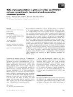
e central role of CD14 in the recognition of various
PAMPs and amplifi cation of immune and infl ammatory
responses in the lung is depicted in Figure 1.
CD14 was characterized as a receptor for bacterial
endotoxin (LPS) in 1990, almost a decade before the dis-
covery and characterization of TLR, and can be regarded
as the fi rst described pattern-recognition receptor [2].
e protein was fi rst identifi ed as a diff erentiation marker
on the surface of monocytes and macrophages and was
designated CD14 at the fi rst leukocyte typing workshop
in Paris in 1982. e genomic DNA of human CD14 was
cloned in 1988 and the gene was later mapped to
chromo some 5q23–31. Several polymorphisms have
been found in the CD14 gene, of which nucleotide poly-
morphisms at position –159 and –1619 correlated with
decreased lung function in endotoxin-exposed farmers [3].
e CD14 gene consists of two exons which code for a
single mRNA that is translated into a protein of 375 amino
acids. e CD14 protein is composed of eleven leucin-rich
repeats, which are also found in TLR and which are
important in PAMP binding. Moreover, the crystal
structure of CD14 revealed that the protein has a `horse-
shoe’ shape, similar to TLR4, and that LPS is bound within
the pocket [4]. In contrast to TLR, however, CD14 lacks a
transmembrane domain, and thus cannot initiate
intracellular signal transduction by itself. e CD14
protein is processed in the endoplasmatic reticu lum and
expressed as a 55 kDa glycoprotein on the cell surface via a
glycosylphosphatidyl (GPI) anchor [5]. Like other GPI-
anchored proteins, CD14 accumulates on the cell surface
in microdomains known as lipid rafts, which are fairly rich
in cholesterol and accumulate several kinases at the
intracellular site. CD14 is expressed pre dominantly on the
surface of `myeloid’ cells, such as mono cytes, macrophages
and neutrophils, but at lower levels also on epithelial cells,
endothelial cells and fi broblasts.
In addition to being expressed as a GPI-anchored
membrane protein, CD14 is also expressed in a soluble
form (sCD14) [2]. sCD14 may result from secretion of
the protein before coupling to the GPI anchor or from
shedding or cleavage from the surface of monocytes.
sCD14 is present in the circulation and other body fl uids
and levels of sCD14 in plasma increase during infl am-
mation and infection. Since interleukin (IL)-6 induces
sCD14 expression in liver cells it is regarded as an acute
© 2010 BioMed Central Ltd
Role of CD14 in lung in ammation and infection
Adam Anas, Tom van der Poll, and Alex F de Vos*
This article is one of ten reviews selected from the Yearbook of Intensive Care and Emergency Medicine 2010 (Springer Verlag) and co-published
as a series in Critical Care. Other articles in the series can be found online at http://ccforum/series/yearbook. Further information about the
Yearbook of Intensive Care and Emergency Medicine is available from />REVIEW
*Correspondence:
Center for Experimental and Molecular Medicine, Center of Infection and
Immunity, Academic Medical Center, Meibergdreef 9, G2-130, 1105AZ Amsterdam,
Netherlands
Anas et al. Critical Care 2010, 14:209
/>© Springer-Verlag Berlin Heidelberg 2010. This work is subject to copyright. All rights are reserved, whether the whole or part of the
material is concerned, speci cally the rights of translation, reprinting, reuse of illustrations, recitation, broadcasting, reproduction on
micro lm or in any other way, and storage in data banks. Duplication of this publication or parts thereof is permitted only under the
provisions of the German Copyright Law of September9, 1965, in its current version, and permission for use must always be obtained
from Springer-Verlag. Violations are liable for prosecution under the German Copyright Law.
phase protein. In bronchoalveolar lavage (BAL) fl uid
from patients with acute respiratory distress syndrome
(ARDS), sCD14 levels were strongly increased and
correlated with total protein levels and neutrophil
numbers in the BAL fl uid [6], suggesting that sCD14
contributes to the infl ammatory process in the lung.
CD14 is a molecule with a wide range of functions. In
addition to functioning as a pattern recognition receptor
for a variety of microbial ligands, CD14 also acts as a
receptor for endogenous molecules like intercellular
adhesion molecule (ICAM)-3 on the surface of apoptotic
cells, amyloid peptid, ceramide, and urate crystals.
Ligation of CD14 by these ligands, except for apoptotic
cells, mediates activation of infl ammatory responses.
CD14 and the LPS receptor complex
LPS is the major constituent of the outer membrane of
Gram-negative bacteria and is one of the most potent
TLR ligands. CD14 together with LBP plays an essential
role in binding of LPS to the TLR4/MD-2 complex [7].
LBP, which, among others, is present in the bloodstream
and BAL fl uid [8], binds to LPS aggregates and transfers
LPS monomers to CD14. CD14 associates with TLR4/
MD-2 and transfers the LPS monomer to this complex
[7]. Likewise, sCD14 is able to mediate LPS-activation
of cells with low membrane CD14 expression, such as
epithelial and endothelial cells [9]. However, at high
con cen trations, LBP and sCD14 are also able to
downregulate LPS-induced responses by transfer of LPS
to lipoproteins for subsequent removal [10]. Recent data
indicate that LPS is bound by MD-2 within the TLR4/
MD-2 complex [11] and that subsequent conformational
changes in TLR4 lead to reorganization of its cyto-
plasmic domain, enabling the recruitment of the adaptor
proteins, myeloid diff erentiation primary-response
protein 88 (MyD88) and TIR-domain-containing-adaptor-
protein-inducing-inter feron (IFN)-β (TRIF) [12]. ese
adaptors initiate signal transduction to the nucleus by
activation of nuclear factor (NF)-κB and IFN regulatory
transcription factor (IRF)-3, leading to the production
of cytokines that regulate infl ammatory cells [12]. In
macrophages, TRIF-dependent signaling is essential for
the expression of the majority of LPS-induced genes,
including IFN-α/β.
Figure 1. Central role of CD14 in pathogen- and pathogen-associated molecular pattern (PAMP)-induced responses in the lung.
CD14,which lacks an intracellular domain for signal transduction, is expressed on the surface of alveolar macrophages, in ltrating monocytes and
neutrophils, and at lower levels also on epithelial and endothelial cells in the lung. CD14 recognizes and binds various structures from invading
microbes, such as lipopolysaccharide (LPS) from Gram-negative bacteria, lipoteichoic acid (LTA) from Gram-positive bacteria, lipoarabinomannan
(LAM) from mycobacteria, viral double stranded (ds) RNA and F glycoprotein (F-gp) from respiratory syncytial virus (RSV). CD14 subsequently
transfers these bound components to Toll-like receptors (TLR) which than trigger cell activation. Binding of LPS to CD14 is regulated by additional
accessory receptors in the lung, including LPS-binding protein (LBP) and a number of surfactant proteins (SP). Furthermore, soluble CD14 (sCD14)
enhances LPS-induced activation of cells with low CD14 expression. Depending on the microbe and the PAMPs it expresses, CD14-ampli ed
responses can either be bene cial to the host by induction of an adequate in ammatory and immune response to eradicate the invading microbe,
or detrimental to the host by excessive in ammation and/or dissemination of the pathogen.
(myco)bacteria
viruses
inammation
clearance
overstimulation
dissemination
sCD14
LTA
LPS
TLR CD14
LAM
dsRNA
RSV
F-gp
SP
LBP
SP
Anas et al. Critical Care 2010, 14:209
/>Page 2 of 8
Recently, it was reported that, in the absence of CD14,
the TLR4/MD-2 complex can distinguish between diff er-
ent chemotypes of LPS [13]. Smooth LPS is synthesized
by most Gram-negative bacteria and consists of three
modules: e lipid A moiety, a core poly saccharide, and
an O-polysaccharide of variable length (made up of 1 to
over 50 monosaccharide units) [7]. Gram-negative bacteria
that fail to add the core polysaccharide or the O-poly-
saccharide chain to the lipid A moiety produce `rough’
LPS, named after the rough morphology of the colonies
these bacteria form. Lipid A, the bioactive part of both
smooth and rough LPS, is responsible for most of the
pathogenic eff ects in Gram-negative bacterial infections
[7, 12]. Murine macrophages lacking CD14 secreted equal
amounts of tumor necrosis factor-α (TNF) to macro-
phages expressing CD14 upon stimulation with rough
LPS, but failed to secrete TNF in response to smooth
LPS, an eff ect which was reversed by addition of sCD14
[13]. Moreover, macrophages lacking CD14 failed to
secrete IFN-α/β in response to either rough or smooth
LPS. ese fi ndings indicate that CD14 is required for
activation of the TLR4/TRIF pathway by either smooth
or rough LPS, and required for the activation of TLR4/
MyD88 pathway by smooth but not by rough LPS [13]. In
addition to LPS, CD14 also facilitates TLR4 activation by
other PAMPs including certain viral components [13, 14].
In the lung, binding of LPS to TLR4 is infl uenced by a
number of surfactant proteins (SP), including SP-A, SP-C
and SP-D [15]. ese surfactants are able to infl uence the
interaction between TLR4 and LPS by direct binding to
LPS; i.e., SP-A binds to rough LPS and lipid A, but not to
smooth LPS, SP-C also binds to rough LPS, and SP-D
binds to both rough and smooth LPS. SP-A and SP-C
binding to LPS inhibits TNF secretion by alveolar macro-
phages, whereas SP-D binding to LPS moderately
enhances TNF secretion by alveolar macrophages. In
addition, SP-A, SP-C and SP-D also bind to CD14 at the
site which recognizes LPS. Strikingly, binding of SP-A to
CD14 enhanced the binding of rough LPS and binding of
SP-C to CD14 augmented binding of smooth LPS [15],
whereas binding of SP-A to CD14 reduced binding of
smooth LPS and binding of SP-D to CD14 decreased
binding of both smooth and rough LPS. Furthermore,
SP-D infl uences LPS-induced TNF secretion by alveolar
macrophages by regulating matrix metalloproteinase-
mediated cleavage of CD14 from the surface of these cells
[16].
Together, these fi ndings suggest that LPS recognition in
the lung and subsequent induction of infl ammatory
immune response is a complexly regulated process.
CD14 and other pattern recognition receptors
In addition to LPS-induced activation of TLR4, CD14
also amplifi es a number of TLR-dependent responses
triggered by other bacterial PAMPs, including peptido-
glycan, lipoteichoic acid (LTA) and lipoarabinomannan
(LAM) [17–19].
Peptidoglycan is an essential cell wall component of
virtually all bacteria. Peptidoglycan is a polymer of N-
acetylglucosamine and N-acetylmuramic acid, cross-
linked by short peptides. Breakdown products of
peptido glycan are recognized by diff erent classes of
pattern-recognition receptors [19]. Polymeric soluble
peptidoglycan is recognized by TLR2 on the surface of
cells, and the interaction of peptidoglycan with TLR2
triggers MyD88-dependent activation and nuclear trans-
location of NF-κB, and subsequently the transcription
and secretion of cytokines. Muramyl dipeptide and γ-D-
glutamyl-meso-diaminopimelic acid, which are low-
molecular weight breakdown fragments of peptidoglycan,
are recognized by intracellular pathogen recognition
receptors, nucleotide-binding oligomerization domain
containing (Nod)2 and Nod1, respectively [19]. Ligand
binding to these receptors triggers interaction with the
receptor-interacting protein kinase, RIP2, which activates
NF-κB. Of these peptidoglycan breakdown products,
only polymeric peptidoglycan binds to CD14, and CD14
enhances polymeric peptidoglycan-induced TLR2 activa-
tion. e low molecular weight fragments of peptido-
glycan, like muramyl dipeptide, do not bind to CD14, do
not induce cell activation through CD14 and also do not
interfere with the binding of polymeric peptidoglycan to
CD14 [19]. Furthermore, unlike LPS, peptidoglycan
bound to sCD14 is not able to activate epithelial and
endothelial cells with low membrane CD14 expression.
LTA is a constituent of the cell wall of Gram-positive
bacteria, anchored on the outer face of the cytoplasmic
membrane and commonly released during growth and
antibiotic therapy. Like polymeric peptidoglycan, LTA
induces NF-κB activation and cytokine secretion in a
TLR2-dependent manner. LTA is recognized by LBP and
CD14, and these accessory receptors both enhance LTA-
induced cell activation [18]. Presumably in a similar
manner, CD14 also enhances TLR2-dependent cellular
activation by LAM derived from the cell-wall of
mycobacteria. LAM derived from slowly growing virulent
mycobacteria like Mycobacterium tuberculosis and
M.leprae is capped with mannose (ManLAM), whereas
LAM from avirulent and fast growing mycobacterial
species is uncapped (AraLAM). Strikingly, AraLAM from
avirulent mycobacteria is much more potent in inducing
TNF secretion by macrophages than ManLAM from
virulent mycobacterial strains [12]. AraLAM-, but not
ManLAM-induced TNF secretion by monocytes and
macrophages was largely CD14-, TLR2- and MyD88-
dependent [17].
Recently CD14 was also found to enhance the innate
immune response triggered by the TLR3 ligand poly(I:C),
Anas et al. Critical Care 2010, 14:209
/>Page 3 of 8
a synthetic mimic of double stranded RNA [20]. TLR3
together with TLR7 and TLR8 are regarded as sensors for
viral infection, since these receptors recognize viral
nucleic acids, like single and double stranded RNA. e
potentiating eff ect of CD14 on TLR3 activation resulted
from increased uptake of poly(I:C) and intracellular
delivery to the compartment where TLR3 resides [20].
Taken together, these fi ndings suggest that CD14 plays an
important role in the induction and amplifi cation of
infl ammatory responses evoked by a wide variety of
pathogens.
Role of CD14 in LPS- and LTA-induced lung
in ammation
e contribution of CD14 to TLR ligand-induced lung
infl ammation has been investigated in several animal
studies (Table1). Intratracheal administration of LPS did
not signifi cantly induce TNF release and neutrophil
accumulation in the lungs of rabbits, unless LPS was
complexed with LBP [21] or the animals were subjected
to mechanical ventilation [22]. Intratracheal instillation
of anti-CD14 antibodies together with LPS/LBP or
intravenous pretreatment with anti-CD14 or anti-TLR4
antibodies before mechanical ventilation markedly
reduced these infl ammatory responses [21, 22]. Despite a
reduction in lung neutrophil number, intravenous anti-
CD14 treatment of rabbits exposed to LPS and subjected
to ventilation did not cause a decrease in lung
chemokines, including CXCL8 (IL-8), growth related
oncogene (GRO) and monocyte chemoattractant protein
(MCP)-1, whereas anti-TLR4 treatment did lower the
level of GRO moderately and of CXCL8 signifi cantly [22].
ese fi ndings reveal that LPS alone does not cause
signifi cant lung infl ammation in rabbits and suggest that
additional accessory signals are required. Whether
mechanical ventilation induces increased release of LBP
or release of (endogenous) DAMPs which potentiate the
LPS-induced response remains to be determined.
In contrast to rabbits, administration of LPS alone to
lungs of naive mice induced severe pneumonitis, irres-
pective of the manner of LPS delivery (inhalation or
intra tracheal or intranasal instillation) or the source of
LPS (Escherichia coli or Acinetobacter baumannii). Using
antibody-treated and gene-defi cient mice, CD14 was
found to be critically involved in the development of
LPS-induced lung infl ammation [23–26]. A study with
CD14-defi cient mice and TLR4 mutant mice (lacking a
functional TLR4) showed that LPS-induced vascular
leakage, neutrophil infi ltration, nuclear translocation of
NF-κB. e release of cytokines (TNF and IL-6) and
chemo kines (CXCL1 and CXCL2) in the lung was
completely dependent on these pattern recognition
receptors [24]. Similar observations were made by others
using mice treated intravenously with anti-CD14
Table 1. E ect of CD14 `neutralization’ in lung in ammation and lung infection
Inciting ligand/pathogen Animal model* E ect of CD14 `neutralization’ in the lung** Ref.
LPS (E. coli +LBP) rabbit αCD14 neutrophil in ux, cytokines 21
LPS (E. coli +ventilation) neutrophil in ux, ~chemokines 22
LPS (E. coli) mouse αCD14 neutrophil in ux, vascular leakage, NF-κB activation 23
LPS (E. coli) mouse CD14
-/-
neutrophil in ux (reversed by sCD14), cytokines (restored by sCD14), 24, 26
chemokines, vascular leakage
LPS (A. baumannii) neutrophil in ux, cytokines 25
LTA (S. aureus) mouse CD14
-/-
~neutrophil in ux, cytokines, chemokines 28
LTA (S. pneumoniae) neutrophil in ux, ~cytokines, ~chemokines 29
nontypeable H. in uenza mouse CD14
-/-
clearance, (early) (late) neutrophil in ux, (early) (late) cytokines 30
A. baumannii mouse CD14
-/-
clearance, ~neutrophil in ux, ~cytokines (dissemination) 25
E. coli rabbit αCD14 clearance, ~neutrophil in ux, ~cytokines, 32
~chemokines (systemic responses)
B. pseudomallei mouse CD14
-/-
clearance (reversed by sCD14), neutrophil in ux (reversed by sCD14), 40
~cytokines (systemic clearance (reversed by sCD14)) (mortality)
S. pneumoniae mouse CD14
-/-
clearance (reversed by sCD14), neutrophil in ux, cytokines, 41
chemokines ( dissemination (reversed by sCD14))
(mortality (reversed by sCD14))
M. tuberculosis mouse CD14
-/-
~clearance, cellular in ltration, ~/cytokines (mortality) 44
In uenza A mouse CD14
-/-
/~clearance, ~lymphocyte recruitment and activation, ~neutrophil in ux, 50
~cytokines
* αCD14: anti-CD14 antibody treatment; CD14
-/-
: CD14-gene de cient. **
(
): (strongly) reduced; ~: unaltered;
(
): (strongly) increased. LPS = lipopolysaccharide;
LTA = lipoteichoic acid.
Anas et al. Critical Care 2010, 14:209
/>Page 4 of 8
antibodies [23] and by our group using CD14-defi cient
and TLR4-defi cient mice [25]. Furthermore, intratracheal
treatment of CD14-defi cient mice with sCD14 restored
the infl ammatory response to the level present in wild-
type mice, whereas treatment with wild-type alveolar
macrophages restored the neutrophil infi ltration of the
lung but not pulmonary TNF release [26]. Moreover,
treatment with wild-type alveolar macrophages also
restored neutrophil infi ltration in the lung of LPS-
exposed TLR4-defi cient mice [27]. ese fi ndings
indicate that sCD14, and CD14 and TLR4 on the surface
of alveolar macrophages contribute to the development
of LPS-induced lung infl ammation. However, when a
high dose of LPS was administered to the lungs of mice,
acute lung infl ammation was absent in mice lacking
functional TLR4, but only partially reduced in CD14
defi cient mice [24]. us, LPS-induced lung infl am ma-
tion is entirely dependent on TLR4 and, depending on the
dose of LPS, also on the presence of CD14 in the lung.
Our group determined whether CD14 also contributes
to the development of lung infl ammation induced by
LTA, a TLR2 ligand from the cell wall of Gram-positive
bacteria [28, 29]. Lung infl ammation induced by
Staphylo coccus aureus LTA was completely dependent on
TLR2, but independent of LBP and only moderately
dependent on CD14 expression. As compared to wild-
type mice, S. aureus LTA-induced neutrophil infl ux was
unchanged in CD14-defi cient mice, whereas TNF and
CXCL2 release in the lung were partially reduced [28].
Strikingly, however, pulmonary infl ammation was also
greatly diminished in TLR4-defi cient mice, as well as in
mice defi cient for platelet activating factor receptor
(PAFR), a known receptor for LTA on epithelial cells.
Similarly, lung infl ammation induced by Streptococcus
pneumoniae LTA, which is less potent compared
S.aureus LTA, was also completely dependent on TLR2
expression. However, in contrast to S. aureus LTA ,
neutrophil infi ltration of the lung was moderately
reduced in CD14-defi cent mice treated with pneumo-
coccal LTA, whereas TNF and CXCL2 release in the lung
was unchanged [29]. Moreover, pneumococcal LTA-
induced lung infl ammation was moderately diminished
in TLR4-defi cient mice. us, despite the amplifying
eff ect on LTA-induced TLR2-mediated responses in
vitro, CD14 contributes minimally to lung infl ammation
induced by LTA. e unexpected contribution of TLR4
to LTA-induced lung infl ammation may result from
DAMPs generated during the infl ammatory process in
the respiratory tract.
Role of CD14 in lung infection
In line with the fi ndings that CD14 contributes to LPS-
induced lung infl ammation in mice, a number of studies
have shown that CD14 is essential for the host defense
response in the lung against Gram-negative bacteria, such
as nontypeable Haemophilus infl uenzae, a possible cause
of community acquired pneumonia, and A. baumannii
and E. coli, which are frequent inducers of nosocomial
pneumonia (Table 1). Nontypeable H.infl uenzae expresses
the TLR4 ligands LPS and lipooligosaccharide on its cell
wall, as well as several TLR2 ligands, including lipo-
proteins and porins. Previously, we found that activa tion
of alveolar macrophages by nontypeable H. infl uenzae
depended on expression of TLR4, TLR2, and CD14 [30].
Moreover, bacterial clearance after intranasal infection
with nontypeable H. infl uenzae was markedly reduced in
CD14-defi cient and TLR4-defi cient mice, as well as in
TLR2-defi cient mice at later stages of the disease [30].
Interestingly, despite impaired bacterial clearance in
CD14-defi cient and TLR4-defi cient mice, the infl amma-
tory response in the lung was strongly reduced in TLR4
defi cient mice, but elevated in CD14 defi cient mice.
Similar observations were made with encapsulated
H. infl uenzae in TLR4-mutant mice [31]. Furthermore,
clearance of nontypeable H. infl uenzae was also signifi -
cantly impaired in MyD88-defi cient mice, but not in mice
lacking functional TRIF [30]. In a similar manner, CD14
was involved in the host defense response against
A. baumanii [25]. CD14-defi cient mice, like TLR4-
defi cient mice, suff ered from impaired bacterial clearance
in the lungs and enhanced bacterial dissemination after
intranasal infection with A. baumannii. However, unlike
TLR4-defi cient mice, CD14-defi cient mice developed
similar infl ammatory responses compared to wild-type
mice. ese fi ndings suggest a role for CD14 in anti-
bacterial responses against nontypeable H. infl uenzae
and A. baumannii. Although the role of TLR4 (and TLR2)
in phagocytic killing is controversial, it is unknown
whether CD14 is involved in such processes. e role of
CD14 in E. coli-induced pneumonia was determined in
anti-CD14 antibody treated rabbits. Intravenous anti-
CD14 antibody treatment of rabbits inoculated with
E. coli by bronchial instillation, resulted in decreased
bacterial clearance from the lungs, but had no eff ect on
neutrophil infi ltration or cytokine release in the lungs
[32]. However, anti-CD14 treatment protected against
sustained hypotension and reduced the levels of nitrate
and nitrite in the blood. e contribution of CD14 to
E. coli-induced pneumonia has not been investigated in
mice, whereas the role of the other components of the
LPS receptor complex (TLR4, MD-2, MyD88, TRIF) has
been determined using gene-defi cient or mutant mice.
Although analysis of bacterial clearance after intranasal
infection of TLR4-mutant mice with E. coli produced
inconsistent results [33], lack of MD-2 or TRIF resulted
in impaired bacterial clearance after E. coli instillation in
the lungs [34, 35]. Moreover, E. coli-induced neutrophil
accumulation and cytokine release was signifi
cantly
Anas et al. Critical Care 2010, 14:209
/>Page 5 of 8
reduced in mice devoid of functional TLR4, MD-2, MyD88
or TRIF [33–35]. ese fi ndings indicate that signaling
through the TLR4 receptor complex is essential in the host
defense response against E. coli, and suggests that CD14
may contribute to these E. coli-induced responses.
To our knowledge, it is unclear whether CD14
contributes to host defense against Pseudomonas
aeruginosa, a frequent cause of nosocomial pneumonia,
and Burkholderia cepacia, a prevalent Gram-negative
bacterium, together with P. aeruginosa, in patients with
cystic fi brosis. Recently, it was found that both TLR4 and
TLR5 are critical in the host response to P. aeruginosa
and that TLR4-defi cient mice were not susceptible to
intratracheal P. aeruginosa infection unless a bacterial
mutant devoid of fl agellin production was used [36]. A
similar approach is required to determine a role for CD14
in Pseudomonas-induced pneumonia. It is plausible that
CD14 also contributes to the host response against
B.cepacia, since LPS from this bacterium signals through
TLR4 and anti-CD14 antibodies dramatically inhibited
B.cepacia-induced chemokine secretion by lung epithelial
cells [37]. Whether CD14 contributes to host defense
response against Klebsiella pneumoniae, a known cause
of nosocomial pneumonia, also remains to be deter-
mined, but data from our study with TLR4-mutant mice
indicate that signaling through TLR4 is essential for
successful clearance of this bacterium [38].
In contrast to the essential role of pulmonary TLR4 and
CD14 in the host defense response against most Gram-
negative bacteria, we found that TLR4 was not involved
and CD14 played a remarkable detrimental role in the
host response to B. pseudomallei, the causative organism
of melioidosis (the most common cause of community-
acquired sepsis in Southeast Asia) [39, 40]. CD14-
defi cient mice infected intranasally with B. pseudomallei
were protected from mortality, accompanied by
enhanced bacterial clearance in the lung, blood and liver,
and reduced cellular infi ltration in the lung [39], whereas
the course of disease in TLR4-defi cient mice was indis-
tinguishable from wild-type mice [40]. Moreover, intranasal
administration of sCD14 to CD14-defi cient mice partially
reversed the phenotype into that of wild-type mice [40].
Interestingly, these fi ndings in B. pseudo mallei-infected
CD14-defi cient mice strongly resemble our previous results
found with TLR2-defi cient mice, and are in line with the
observation that B. pseudomallei expresses an atypical LPS
which signals through TLR2 [39]. Whether CD14 interacts
with TLR2 in B. pseudo mallei-induced responses, and by
which mechanism these receptors facilitate the growth and
dissemination of B. pseudomallei after intranasal infection
remains to be determined.
In the model for S. pneumoniae-induced pneumonia,
we observed an unexpected detrimental role for CD14 in
the innate host defense response. S. pneumoniae, a
Gram-positive bacterium and the single most frequent
pathogen causing community-acquired pneumonia,
induces severe lung infl ammation and sepsis in wild-type
mice after intranasal instillation. Strikingly, CD14-
defi cient mice were protected against pneumococcal
pneumonia, presumably as a result of reduced bacterial
spread to the circulation and reduced lung infl ammation
[41]. In contrast, TLR2-defi cient and TLR4-mutant mice
were not protected against pneumococcal pneumonia
[38, 42], but in fact TLR2 seemed redundant for effi cient
bacterial clearance and TLR4-mutant mice were more
susceptible to pneumonia, accompanied by impaired
bacterial clearance. However, as in CD14-defi cient mice,
lung infl ammation was also reduced in pneumococci-
infected TLR2-defi cient mice [42]. Since intrapulmonary
treatment with sCD14 rendered CD14-defi cient mice
equally susceptible to S. pneumoniae as wild-type mice
[41], these results suggest that S. pneumoniae abuses (s)
CD14 in the lung to cause invasive respiratory tract
infection. Interestingly, the phenotype of CD14 defi cient
mice strongly resembled the phenotype of mice defi cient
for PAFR [43], a receptor for phosphoryl choline from the
pneumococcal cell wall which facilitates pneumococcal
invasion of cells. Further studies are required to
determine whether CD14 serves as a chaperone in the
presentation of S. pneumoniae to the PAFR so that the
phosphoryl–PAFR-mediated invasion is facilitated.
Since M. tuberculosis expresses a number of molecules,
such as lipoproteins, which activate immune cells in a
CD14-dependent manner, we and others investigated
whether CD14 also contributed to the host immune
response in mice with lung tuberculosis [44]. Although
initially after intranasal infection of wild-type and CD14-
defi cient mice no diff erences in bacterial loads, cell
infi ltration and release of most cytokines in the lung were
found [44, 45], at later time points (> 20 weeks after
infection) CD14-defi cient mice were protected from
mortality presumably as a result of a reduced infl am-
matory response in the lungs [44]. ese fi ndings are
completely opposite to the results from M. tuberculosis-
infected TLR2-defi cient and TLR4-mutant mice, which
suff ered from reduced bacterial clearance, chronic
infl ammation, increased cellular infi ltration of the lungs
and reduced survival [46–48]. e mechanism underlying
the detrimental eff ect of CD14 in the host response
against M. tuberculosis remains to be established.
In addition to its role in (myco)bacterial infections,
CD14 may also play a role in the pulmonary host
response against respiratory syncytial virus (RSV), the
most common cause of lower respiratory tract disease in
infants and young children worldwide, and infl uenza A
virus, a cause of pneumonia in very young children, the
elderly and immunocompromised patients. e envelop
F glycoprotein from RSV and certain infl uenza A virus
Anas et al. Critical Care 2010, 14:209
/>Page 6 of 8
components activate macrophages in a CD14-dependent
manner [14, 20]. Experiments with wild-type and TLR4-
mutant mice infected intranasally with RSV showed that
viral clearance was reduced in the absence of functional
TLR4 [14], due to impaired natural killer (NK) cell
migration and function and impaired cytokine secretion.
Recently, it was found that TLR2 and TLR6 are also
involved in recognition of RSV [49]. Whether CD14
contributes to these TLR-mediated immune responses
against RSV remains to be determined. Using CD14-
defi cient mice, we demonstrated that CD14 played a
minimal role in infl uenza A virus-induced pneumonia
[50]. During the entire course of disease, viral loads were
slightly reduced in CD14-defi cient mice, but this did not
result from improved lymphocyte recruitment or
lympho cyte activation, or consistent changes in pulmo-
nary cytokines [50]. us, despite the fact that infl uenza
A expresses ligands that require CD14 for immune cell
activation [20], CD14 seems redundant in the host
defense response against infl uenza A virus.
Conclusion
CD14 plays a central role in the lung in the recognition
and binding of a variety of (myco)bacterial and viral
components, and in the amplifi cation of subsequent host
responses. e studies discussed in this chapter indicate
that the contribution of CD14 to the pulmonary host
defense responses may range from benefi cial to detri-
mental, depending on the microbe and the PAMPs it
expresses. Interfering with CD14-LPS or CD14-LTA
inter actions reduced lung infl ammation. Interference
with CD14-pathogen interactions, however, did not have
a signifi cant eff ect on M. tuberculosis or infl uenza A virus
infection, resulted in reduced clearance of nontypeable
H. infl uenzae, E. coli or A. baumannii in the lung, but
enhanced clearance (and reduced dissemination) of B.
pseudomallei or S. pneumoniae. e latter observation
indicates that certain pathogens may abuse CD14 in the
lung to cause invasive disease. Whether CD14 is a
suitable target for intervention in these latter infectious
diseases and/or in aberrant infl ammatory responses
during pneumonia requires further study.
Abbreviations
ARDS = acute respiratory distress syndrome, BAL – broncoalveolar lavage,
DAMP = damage/danger-associated molecular pattern, F-gp = F glycoprotein,
GPI = glycosylphosphatidyl, GRO = growth related oncogene, HMGB-1 =
high mobility group box-1 protein, ICAM = intracellular adhesion molecule,
IFN = interferon, IL = interleukin, IRF = IFN regulatory transcription factor,
LAM = lipoarabinomannan, LBP = lipopolysaccharide binding protein,
LPS = lipopolysaccharide, LTA = lipoteichoic acid, MCP = monocyte
chemoattractant protein, MyD88 = myeloid di erentiation primary-response
protein 88, NF = nuclear factor, NK = natural killer, Nod = nucleotide-binding
oligomerization domain containing, PAFR = platelet activating factor
resceptor, PAMP = pathogen-associated molecular pattern, RIP = receptor-
interacting protein kinase, RSV = respiratory syncytial virus, SP = surfactant
protein, TLR = Toll-like receptors, TNF = tumour necrosis factor, TRIF =
TIR-domain-containing-adaptor-protein-inducing-interferon-β
Competing interests
The authors declare that they have no competing interests.
Published: 9 March 2010
References
1. Basu S, Fenton MJ: Toll-like receptors: function and roles in lung disease.
Am J Physiol Lung Cell Mol Physiol 2004, 286:L887-L892.
2. Wright SD: CD14 and innate recognition of bacteria. J Immunol 1995,
155:6–8.
3. LeVan TD, Von Essen S, Romberger DJ, et al.: Polymorphisms in the CD14
gene associated with pulmonary function in farmers. Am J Respir Crit Care
Med 2005, 171:773–779.
4. Kim JI, Lee CJ, Jin MS, et al.: Crystal structure of CD14 and its implications
for lipopolysaccharide signaling. J Biol Chem 2005, 280:11347–11351.
5. Wright SD, Ramos RA, Tobias PS, Ulevitch RJ, Mathison JC: CD14, a receptor
for complexes of lipopolysaccharide (LPS) and LPS binding protein.
Science 1990, 249:1431–1433.
6. Martin TR, Rubenfeld GD, Ruzinski JT, et al.: Relationship between soluble
CD14, lipopolysaccharide binding protein, and the alveolar in ammatory
response in patients with acute respiratory distress syndrome. Am J Respir
Crit Care Med 1997, 155:937–944.
7. Beutler B, Rietschel ET: Innate immune sensing and its roots: the story of
endotoxin. Nat Rev Immunol 2003, 3:169–176.
8. Knapp S, Florquin S, Golenbock DT, Van der Poll T: Pulmonary
lipopolysaccharide (LPS)-binding protein inhibits the LPS-induced lung
in ammation in vivo. J Immunol 2006, 176:3189–3195.
9. Pugin J, Schurer-Maly CC, Leturcq D, et al.: Lipopolysaccharide activation of
human endothelial and epithelial cells is mediated by lipopolysaccharide-
binding protein and soluble CD14. Proc Natl Acad Sci USA 1993,
90:2744–2748.
10. Kitchens RL, Thompson PA: Modulatory e ects of sCD14 and LBP on
LPS-host cell interactions. J Endotoxin Res 2005, 11:225–229.
11. Park BS, Song DH, Kim HM, et al.: The structural basis of lipopolysaccharide
recognition by the TLR4-MD-2 complex. Nature 2009, 458:1191–1195.
12. Akira S, Uematsu S, Takeuchi O:
Pathogen recognition and innate immunity.
Cell 2006, 124:783–801.
13. Jiang Z, Georgel P, Du X, et al.: CD14 is required for MyD88-independent
LPS signaling. Nat Immunol 2005, 6:565–570.
14. Kurt-Jones EA, Popova L, Kwinn L, et al.: Pattern recognition receptors TLR4
and CD14 mediate response to respiratory syncytial virus. Nat Immunol
2000, 1:398–401.
15. Chaby R, Garcia-Verdugo I, Espinassous Q, Augusto: LA Interactions between
LPS and lung surfactant proteins. J Endotoxin Res 2005, 11:181–185.
16. Senft AP, Korfhagen TR, Whitsett JA, Shapiro SD, LeVine AM: Surfactant
protein-D regulates soluble CD14 through matrix metalloproteinase-12.
JImmunol 2005, 174:4953–4959.
17. Pugin J, Heumann ID, Tomasz A, et al.: CD14 is a pattern recognition
receptor. Immunity 1994, 1:509–516.
18. Schroder NW, Morath S, Alexander C, et al.: Lipoteichoic acid (LTA) of
Streptococcus pneumoniae and Staphylococcus aureus activates immune
cells via Toll-like receptor (TLR)-2, lipopolysaccharide-binding protein
(LBP), and CD14, whereas TLR-4 and MD-2 are not involved. J Biol Chem
2003, 278:15587–15594.
19. Dziarski R, Gupta D: Peptidoglycan recognition in innate immunity.
JEndotoxin Res 2005, 11:304–310.
20. Lee HK, Dunzendorfer S, Soldau K, Tobias PS: Double-stranded
RNA-mediated TLR3 activation is enhanced by CD14. Immunity 2006,
24:153–163.
21. Ishii Y, Wang Y, Haziot A, et al.: Lipopolysaccharide binding protein and
CD14 interaction induces tumor necrosis factor-alpha generation and
neutrophil sequestration in lungs after intratracheal endotoxin. Circ Res
1993, 73:15–23.
22. Smith LS, Kajikawa O, Elson G, et al.: E ect of Toll-like receptor 4 blockade on
pulmonary in ammation caused by mechanical ventilation and bacterial
endotoxin. Exp Lung Res
2008, 34:225–243.
23. Tasaka S, Ishizaka A, Yamada W, et al.: E ect of CD14 blockade on
endotoxin-induced acute lung injury in mice. Am J Respir Cell Mol Biol 2003,
29:252–258.
24. Jeyaseelan S, Chu HW, Young SK, Freeman MW, Worthen GS: Distinct roles of
pattern recognition receptors CD14 and Toll-like receptor 4 in acute lung
Anas et al. Critical Care 2010, 14:209
/>Page 7 of 8
injury. Infect Immun 2005, 73:1754–1763.
25. Knapp S, Wieland CW, Florquin S, et al.: Di erential roles of CD14 and toll-
like receptors 4 and 2 in murine Acinetobacter pneumonia. Am J Respir Crit
Care Med 2006, 173:122–129.
26. Brass DM, Hollingsworth JW, McElvania-Tekippe E, et al.: CD14 is an essential
mediator of LPS induced airway disease. Am J Physiol Lung Cell Mol Physiol
2007, 293:L77–L83.
27. Hollingsworth JW 2nd, Cook DN, Brass DM, et al.: The role of Toll-like
receptor 4 in environmental airway injury in mice. Am J Respir Crit Care Med
2004, 170:126–132.
28. Knapp S, von Aulock S, Leendertse M, et al.: Lipoteichoic acid-induced lung
in ammation depends on TLR2 and the concerted action of TLR4 and the
platelet-activating factor receptor. J Immunol 2008, 180:3478–3484.
29. Dessing MC, Schouten M, Draing C, et al.: Role played by Toll-like receptors 2
and 4 in lipoteichoic acid-induced lung in ammation and coagulation.
JInfect Dis 2008, 197:245–252.
30. Wieland CW, Florquin S, Maris NA, et al.: The MyD88-dependent, but not the
MyD88-independent, pathway of TLR4 signaling is important in clearing
nontypeable haemophilus in uenzae from the mouse lung. J Immunol
2005, 175:6042–6049.
31. Wang X, Moser C, Louboutin JP, et al.: Toll-like receptor 4 mediates innate
immune responses to Haemophilus in uenzae infection in mouse lung.
JImmunol 2002, 168:810–815.
32. Frevert CW, Matute-Bello G, Skerrett SJ, et al.: E ect of CD14 blockade in
rabbits with Escherichia coli pneumonia and sepsis. J Immunol 2000,
164:5439–5445.
33. Lee JS, Frevert CW, Matute-Bello G, et al.: TLR-4 pathway mediates the
in
ammatory response but not bacterial elimination in E. coli pneumonia.
Am J Physiol Lung Cell Mol Physiol 2005, 289:L731-L738.
34. Jeyaseelan S, Young SK, Fessler MB, et al.: Toll/IL-1 receptor domain-
containing adaptor inducing IFN-beta (TRIF)-mediated signaling
contributes to innate immune responses in the lung during Escherichia
coli pneumonia. J Immunol 2007, 178:3153–3160.
35. Cai S, Zemans RL, Young SK, Worthen GS, Jeyaseelan S: Myeloid
di erentiation protein-2-dependent and -independent neutrophil
accumulation during Escherichia coli pneumonia. Am J Respir Cell Mol Biol
2009, 40:701–709.
36. Ramphal R, Balloy V, Jyot J, et al.: Control of Pseudomonas aeruginosa in the
lung requires the recognition of either lipopolysaccharide or agellin.
JImmunol 2008, 181:586–592.
37. Reddi K, Phagoo SB, Anderson KD, Warburton D: Burkholderia cepacia-
induced IL-8 gene expression in an alveolar epithelial cell line: signaling
through CD14 and mitogen-activated protein kinase. Pediatr Res 2003,
54:297–305.
38. Branger J, Knapp S, Weijer S, et al.: Role of Toll-like receptor 4 in
gram-positive and gram-negative pneumonia in mice. Infect Immun 2004,
72:788–794.
39. Wiersinga WJ, Wieland CW, Dessing MC, et al.: Toll-like receptor 2 impairs
host defense in gram-negative sepsis caused by Burkholderia
pseudomallei (Melioidosis). PLoS Med 2007, 4:e248.
40. Wiersinga WJ, de Vos AF, Wieland CW, et al.: CD14 impairs host defense
against gram-negative sepsis caused by Burkholderia pseudomallei in
mice. J Infect Dis 2008, 198:1388 –1397.
41. Dessing MC, Knapp S, Florquin S, De Vos AF, Van der Poll T: CD14 facilitates
invasive respiratory tract infection by Streptococcus pneumoniae. Am J
Respir Crit Care Med 2007, 175:604–611.
42. Knapp S, Wieland CW, Murawskian ‘t Veer C, et al.: Toll-like receptor 2 plays a
role in the early in ammatory response to murine pneumococcal
pneumonia but does not contribute to antibacterial defense. J Immunol
2004, 172:3132–3138.
43. Rijneveld AW, Weijer S, Florquin S, et al
.: Improved host defense against
pneumococcal pneumonia in platelet-activating factor receptor-de cient
mice. J Infect Dis 2004, 189:711–716.
44. Wieland CW, Van der Windt GJ, Wiersinga WJ, Florquin S, Van der Poll T: CD14
contributes to pulmonary in ammation and mortality during murine
tuberculosis. Immunology 2008, 125:272–279.
45. Reiling N, Holscher C, Fehrenbach A, et al.: Toll-like receptor (TLR)2- and
TLR4-mediated pathogen recognition in resistance to airborne infection
with Mycobacterium tuberculosis. J Immunol 2002, 169:3480–3484.
46. Abel B, Thieblemont N, Quesniaux VJ, et al.: Toll-like receptor 4 expression is
required to control chronic Mycobacterium tuberculosis infection in mice.
J Immunol 2002, 169:3155–3162.
47. Drennan MB, Nicolle D, Quesniaux VJ, et al.: Toll-like receptor 2-de cient
mice succumb to Mycobacterium tuberculosis infection. Am J Pathol 2004,
164:49–57.
48. Branger J, Leemans JC, Florquin S, et al.: Toll-like receptor 4 plays a
protective role in pulmonary tuberculosis in mice. Int Immunol 2004,
16:509–516.
49. Murawski MR, Bowen GN, Cerny AM, et al.: Respiratory syncytial virus
activates innate immunity through Toll-like receptor 2. J Virol 2009,
83:1492–1500.
50. Dessing MC, Van der Sluijs KF, Florquin S, Van der Poll T: CD14 plays a limited
role during in uenza A virus infection in vivo. Immunol Lett 2007,
113:47–51.
Anas et al. Critical Care 2010, 14:209
/>doi:10.1186/cc8850
Cite this article as: Anas A, et al.: Role of CD14 in lung in ammation and
infection. Critical Care 2010, 14:209.
Page 8 of 8