Báo cáo y học: " Near-infrared spectroscopy during stagnant ischemia estimates central venous oxygen saturation and mixed venous oxygen saturation discrepancy in patients with severe left heart failure and additional sepsis/septic shock" potx
Bạn đang xem bản rút gọn của tài liệu. Xem và tải ngay bản đầy đủ của tài liệu tại đây (350.67 KB, 10 trang )
RESEARC H Open Access
Near-infrared spectroscopy during stagnant
ischemia estimates central venous oxygen
saturation and mixed venous oxygen saturation
discrepancy in patients with severe left heart
failure and additional sepsis/septic shock
Hugo Možina, Matej Podbregar
*
Abstract
Introduction: Discrepancies of 5-24% between supe rior vena cava oxygen saturation (ScvO
2
) and mixed venous
oxygen saturation (SvO
2
) have been reported in patients with severe heart failure. Thenar muscle tissue
oxygenation (StO
2
) measured with near-infrared spectroscopy (NIRS) during arterial occlusion testing decreases
slower in sepsis/septic shock patients (lower StO
2
deoxygenation rate). The StO
2
deoxygenation rate is influenced
by dobutamine. The aim of this study was to determine the relationship between the StO
2
deoxygenation rate and
the ScvO
2
-SvO
2
discrepancy in patients with severe left heart failure and additional sepsis/septic shock treated with
or without dobutamine.
Methods: Fifty-two patients with severe left heart failure due to primary heart disease with additional severe
sepsis/septic shock were included. SvO
2
and ScvO
2
were compared to the thenar muscle StO
2
before and during
arterial occlusion.
Results: SvO
2
correlated significantly with ScvO
2
(Pearson correlation 0.659, P = 0.001), however, Bland Altman
analysis showed a clinically important difference between both variables (ScvO
2
-SvO
2
mean 72 ± 8%, ScvO
2
-SvO
2
difference 9.4 ± 7.5%). The ScvO
2
-SvO
2
difference correlated with plasma lactate (Pearson correlation 0.400, P =
0.003) and the StO
2
deoxygenation rate (Pearson correlation 0.651, P = 0.001). In the group of patients treated with
dobutamine, the ScvO
2
-SvO
2
difference correlated with plasma lactate (Pearson correlation 0.389, P = 0.011) and
the StO
2
deoxygenation rate (Pearson correlation 0.777, P = 0.0001).
Conclusions: In pa tients with severe heart failure with additional severe sepsis/septic shock the ScvO
2
-SvO
2
discrepancy presents a clinical problem. In these patients the skeletal muscle StO
2
deoxygenation rate is inversely
proportional to the difference between ScvO
2
and SvO
2
; dobutam ine does not influence this relationship. When
using ScvO
2
as a treatment goal, the NIRS measurement may prove to be a useful non-invasive diagnostic test to
uncover patients with a normal ScvO
2
but potentially an abnormally low SvO
2
.
Trial Registration: NCT0038 4644 ClinicalTrials.Gov.
* Correspondence:
Clinical Department of Intensive Care Medicine, University Clinical Centre
Ljubljana, Zaloska cesta 7, SI-1000 Ljubljana, Slovenia
Možina and Podbregar Critical Care 2010, 14:R42
/>©2010Možina et al.; licensee BioMed Central Ltd. This is an open access article distributed under the terms of the Creative Commons
Attribution License (http: //creativecomm ons.org/licenses/by/2.0), which permits unrestricted use, distribution, and reproduction in
any me dium, provided the original work is properly cited.
Introduction
Maintenanceofadequateoxygendelivery(DO
2
)is
essential to preserve organ function, because a sustained
low D O
2
leads to organ failure and death [1]. Low car-
diac output states (cardiogenic, hypovolemic and
obstructive types of shock), anemic and hypoxic hypoxe-
mia are characterized by a decreased DO
2
but a pre-
served oxygen extraction ratio. In distributive shock, the
oxygen extraction capability is altered so that the critical
oxygen extraction ratio is typically decreased [2]. Mea-
surement of mixed venous oxygen saturation (SvO
2
)
from the pulmonary artery is used for calculations of
oxygen consumption and has been advocated as an
indirect index of tissue oxygenation and a prognostic
predictor in critically ill patients [ 3-6]. However, cathe-
terization of the pulmonary artery is costly, has inherent
risks and its usefulness remains under debate [7,8].
Not surprisingly the monitoring of central venous oxy-
gensaturation(ScvO
2
) was suggested as a simpler and
cheaper assessment of global DO
2
to oxy gen consump-
tion ratio [1,2].
AconcernwithScvO
2
compared with mixed venous
oxygen saturation (SvO
2
) is that it may not accurately
reflect global hypoxia, because organs with capillary
beds that drain into the inferior vena cava or coronary
sinus will not be involved in this measurement. H ealthy
resting individuals have a ScvO
2
that is slightly lower
than the SvO
2
[3]. In heart failure and shock, however,
this situation is reversed. Most authors attribute this
pattern to changes in the distribution of cardiac output
that occur in periods of haemodynamic instability. In
shock states, blood flow to the splanchni c and renal cir-
culations fall, while flow to the heart and brain is main-
tained due to redistribution of blood away from the
mesenteric and renal vascular beds and additional right
heart dysfunction [4]. Discrepancies of 5 to 24% have
been reported [5-7,9].
Near infrared spectroscopy (NIRS) is a technique used
for cont inuous, non-invasive, bedside monitoring of tis-
sue oxygen saturation (StO
2
) [8,10].
We have previously shown t hat skeletal muscle StO
2
does not estimate SvO
2
in patients with severe left
heart failure and additional severe sepsis or septic
shock. However, in patients with severe left heart fail-
ure without additional severe sepsis or septic sh ock,
StO
2
values could be used for fast noninvasive SvO
2
estimation; the trend of StO
2
may be substituted for
the trend of SvO
2
[8].
We have also shown that thenar skeletal muscle StO
2
during stagnant ischemia (deoxygenation rate during
arterial occlusion test) decreases slower in septic shock
patients compared with p atients with severe sep sis or
localized infection or healthy volunteers [10].
Impaired skeletal muscle microcirculation, especially
impaired deoxygenation rate during arterial occlusion
test, was recently detected in patients with chronic heart
failure. Dobutamine, but not levosimendan, partiall y
reversed this impairment [11].
The aim of current study was to combine our previous
findings. We t ested the hypothesis that in patients with
severe left heart failure and additional sepsis/septic
shock the skeletal muscle deoxygenation rate during an
arterial occlusion test could predict a ScvO
2
-SvO
2
dis-
crepancy. The second aim was to explore the effect of
dobutamine treatment on any ScvO
2
-SvO
2
discrepancy.
Materials and methods
Patients
The study protocol wa s approved by the National Ethics
Committee of Slovenia; informed consent was obtained
from all patients or their relatives. The study was per-
formed between October 2004 and June 2007.
After initial hemodynamic resuscitation according to
early goal-directed therapy [12] and S urviving Sepsis
Campaign guidelines [13], transthoracic echocardiogra-
phy for the assessment of left ventricular volume, ejec-
tion fraction (Simpson’s rule) and valvular function w as
performed in all patients admitted to our ICU (Hewlett-
Packard HD 5000, Hewlett Packard, Andover, MA,
USA) by experienced ICU doctors (HM and MP) trained
in echocardiography.
In patients with primary heart disease, low cardiac
output, and no signs of hypovolemia, a right heart
catheterization with a pu lmonary artery floating catheter
(Swan-Ganz CCOmboV CCO/SvO
2
/CEDV, Edwards
Lifesciences, Irvine, CA, USA) was performed following
a decision of the treating physician. The site of insertion
was confirmed by the transducer w aveform, the length
of catheter insertion, and chest radiography. Systemic
arterial pressure was measured invasively using radial or
femoral arterial catheterization. Consecutive patients
with severe left heart failure due to primary heart dis-
ease (left ventricular systolic ejection fraction below
40%, pulmo nary artery o cclusion pressure ab ove 18
mmHg) and additional severe sepsis/septic shock were
included in our study. Severe sepsis and septic shock
were defined according to the 1992 American College of
Chest Physicians/Society of Critical Care Medicine
(ACCP/SCCM) consensus c onference definitions [14].
Patients with heart failure confirmed by echocardiogra-
phy without sepsis/septic shock were excluded. Patients
with cachexia were not included.
Patients were divided into t wo groups depending on
treatment with dobutamine or not.
All patients received standard treatment of localized
infection, severe sepsis and septic or cardiogenic shock
Možina and Podbregar Critical Care 2010, 14:R42
/>Page 2 of 10
including: source control, fluid infusion, catecholamine
infusion, organ failure re placement and/or support ther-
apy, intensive control of blood glucose and corticoster-
oid substitution therapy according to current Surviving
Sepsis Campaign G uidelines [13]. Mechanically venti-
lated patients were sedated with midazolam and/or pro-
pofol infusion. Paralytic agents were not used.
Measurements
Skeletal muscle oxygenation
Thenar muscle StO
2
was measured non-invasivel y by
NIRS (25 mm Probe, InSpectra™, H utchinson Technol-
ogy Inc., West Highland Park Drive NE, MN, USA)
[8,10,15]. Maximal thenar muscle StO
2
was located by
moving the probe over the thenar prominence. StO
2
was continuously monitored and stored onto a compu-
ter using InSpectra™ software. The average of StO
2
changing over a 15 s econd span was used. The arterial
occlusion test was performed as previously reported
[10]: StO
2
was monitored before and during (StO
2
deox-
ygenation rate) upper limb ischemia until StO
2
decreased to 40%. Upper limb ischemia was induced by
rapid automatic pneumatic cuff inflation (to
260 mmHg) placed above the elbow.
Severity of disease
Sepsis-related Organ Failure Assessment (SOFA) score
was calculated at the time of each measurement to
assess the level of organ d ysfunction [16]. Dobutamine
and norepinephrine requirement represented the dose of
drug during the StO
2
measurement. Use of an intra-
aortic balloon pump during the ICU stay is reported.
Plasma lactate concentration was measured using an
enzymatic colorimetric method (Lactate, Roche Diagnos-
tics, Hoffman-La Roche, Basel, Switzerland) at the time
of each StO
2
measurement.
Laboratory analysis
Blood was withdrawn from the superior vena cava
approximately 2 cm above the right atrium and from
the pulmonary artery at the time of each StO
2
measur e-
ment to determine ScvO
2
(%) and SvO
2
(%), respec-
tively. In view of known problems arising during
sampling from the pulmonary artery, including the pos-
sibility o f contaminating arterial blood with pulmonary
capillary blood, all samples from this site were with-
drawn over 30 seconds, using a low-n egative pressure
technique, without inflating the balloon. A standard
volume of 1 mL of blood was obtained f rom each side
after withdrawal of dead-space blood and flushing fluid.
All measurements were made using a cooximeter (Rapi-
dLab 1265, Bayer HealthCare, Leverkusen, Germany).
Data analysis
A sample size of 41 patients was estimated for a correla-
tion coefficient of 0.6 with a desired power o f0.95 and
alpha of 0.01 (SigmaPlot 2004 for Windows, version
9.01 SyStat Software, Inc., Chicago, IL, USA).
Data was expressed as mean ± s tandard deviation
(SD). The Mann Whitney non-param etric test was used
to compare groups. A P value of less than 0.05 was con-
sidered statistically significant. The Pearson correlation
test was applied to determi ne corr elation (SPSS 10.0 for
Windows™ , SPSS Inc., Chicago, IL, USA). In order to
compare ScvO
2
and SvO
2
we calculated bias, systemic
disagreement between measurements (mean difference
between two measurements), precision and the random
error in measuring (SD of bias) [17]. The 95% limits of
agreement were arbitrarily set following Bland a nd Alt-
man as the bias ± two SD.
Results
During the study period (20 months), 2,121 patients
were admitted to the 15-b ed university center internal
medicine ICU. In that period 151 right heart catheteri-
zati ons were perfor med. The final sample of 52 patients
was reached after exclusion of 65 patients with heart
failure without sepsis/septic shock, 24 patients who did
not have heart failure, 2 patien ts for whom consent was
not given and 8 patients for whom NIRS measurements
were not performed. The detailed description of our
select ed population is given in Tab le 1. Patients were all
mechanically ventilated.
Intra-aortic balloon pumps were inserted in patients
who were treated with percutaneous coronary interven-
tion and stent implantat ion after primary cardiac arrest
due t o ST-elevation myocardial infarction (STEMI; n =
42) and cardiogenic shock. Patients with STEMI after
cardiac arrest were treated with medically induced
hypothermia for 24 hours. During the ICU stay and
before study inclusion they all developed pneumonia.
All other patie nts were admitted to the ICU primarily
because of sepsis or septic shock.
Forty-three patients were treated with dobutamine.
There was no difference between patients treated with
or without dobutamine in additional hemodynamic sup-
port (Table 2). Patients treated with dob utamine had a
lower cardiac index (Table 3) and a high er procalcitonin
value (Table 4).
Thenar StO
2
before (basal StO
2
) and during the v as-
cular occlusion test is presented in Table 5. There was
no difference between patients treated with and without
dobutamine in NIRS data.
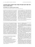
SvO
2
correlated significantly with ScvO
2
(Pearson cor-
relation 0.659, P = 0.001; Figure 1); however, Bland Alt-
man analysis showed a clinically important difference
between both variables (ScvO
2
-SvO
2
mean 72 ± 8%,
ScvO
2
-SvO
2
difference 9.4 ± 7.5%; Figure 2).
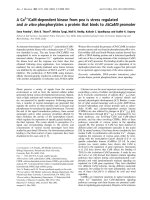
The ScvO
2
-SvO
2
difference correlated with plasma
lactate (Pearson correlation 0.400, P = 0.003; Figure 3)
Možina and Podbregar Critical Care 2010, 14:R42
/>Page 3 of 10
and StO
2
deoxygenation rate (Pearson correlation 0.651,
P = 0.001; Figure 4).
In the group of patients treated with dobutamine the
ScvO
2
-SvO
2
difference correlated with plasma lactate
(Pearson correlation 0.389, P = 0.011 ) and StO
2
deoxy-
genation rate (Pearson correlation 0.777, P = 0.0001).
In a small group of patients (n = 9) treated witho ut
dobutamine the ScvO
2
-SvO
2
difference correlated with
the StO
2
deoxygenation rate (Pearson correlation 0.673,
P = 0.033); however, there was no correlation between
the ScvO
2
-SvO
2
difference and plasma l actate (Pearson
correlation 0.503, P = 0.139).
Discussion
Our study confirmed the hypothesis that the skeletal
muscle StO
2
deoxygenation rate correlates (or is
inversely proportional) to the ScvO
2
-SvO
2
difference in
patients with severe heart failure with additional sepsis/
septic shock. This relation between the StO
2
deoxygena-
tion rate and the ScvO
2
-SvO
2
difference was also pre-
sent in patients treated with or without dobutamine. We
also showed that these patients have a clinically consid-
erable ScvO
2
-SvO
2
discrepancy. Monitoring of ScvO
2
is
a simpler and cheaper assessment of global DO
2
to oxy-
gen consumption ratio, but its use as a treatment goal
in patients with severe heart failure with additional sep-
sis/septic shock is questionable.
The high StO
2
/low SvO
2
seen in patients with severe
sepsis and septic shock suggests blood flow redistribu-
tion. Thenar muscle StO
2
correla tes with central venous
oxygen saturation that is measured in a mixture of
blood from the head and both arms [18]. In healthy
Table 1 Description of patients
Parameter All
(n = 52)
Treatment
with dobutemine
(n = 43)
Treatment
without dobutamine
(n = 9)
P value
Age (years) 68 ± 13 68 ± 14 69 ± 8 0.8
Female (n) 7 5 2 0.6
Heart disease
Ischemic heart disease (n) 42 36 6 0.4
Aortic stenosis (n) 6 4 2 0.6
Dilated cardiomyopathy (n) 1 1 0 0.9
Myocarditis (n) 3 2 1 0.6
Echocardiography
LVEF (%) 28 ± 5 25 ± 8 29 ± 9 0.1
LVEDD (cm) 5.8 ± 0.9 5.8 ± 0.7 6.0 ± 0.9 0.2
Severe mitral regurgitation (n) 26 22 4 0.8
Cause of infection
Pneumonia (n) 45 38 7 0.6
Urosepsis (n) 5 4 1 0.9
Other (n) 2 1 1 0.7
SOFA score 12.2 ± 2.5 12. ± 2.2 12.6 ± 2.6 0.8
ICU stay (days) 9 ± 4 9 ± 6 9 ± 5 0.9
ICU survival (%) 48 47 55 0.8
LVEF, left ventricular ejection fraction; LVEDD, left ventricular end-diastolic diameter; SOFA, Sequential Organ Failure Assessment.
Table 2 Treatment of patients
Treatment All
(n = 52)
Treatment
with dobutemine
(n = 43)
Treatment
without dobutamine
(n = 9)
P value
Norepinephrine (mg/h, n) 0.09 ± 0.10 (43) 0.08 ± 0.11
(37)
0.04 ± 0.06
(9)
0.1
Dobutamine (μg/kg/min) - 0.47 ± 0.25 - -
Levosimendan (n) 23 17 6 0.2
IAPB (n) 20 15 5 0.3
Mechanical ventilation(n) 52 43 9 1.0
FiO
2
0.72 ± 0.22 0.73 ± 0.23 0.71 ± 0.23 0.8
FiO
2
, fractional inspired oxy gen; IAPB, intra- aortic balloon pump.
Možina and Podbregar Critical Care 2010, 14:R42
/>Page 4 of 10
resting individuals the ScvO
2
is slightly lower than the
SvO
2
[3]. Blood in the inferior vena cava has a high oxy-
gen content because the kidneys do not utilise much
oxygen but receive a high proportion of the cardiac out-
put [19]. Blood in the inferior vena cava blood has a
higher oxygen content than b lood from the u pper body
and the SvO
2
is thus greater than the ScvO
2
.
This relation changes in periods of cardiovascular
instability. Scheinman and colleagues performed the ear-
liest comparison of ScvO
2
and SvO
2
in both hemodyna-
mically stable and sho ckedpatients[5].Instable
patients, ScvO
2
was similar to SvO
2
. In patients wit h a
failing heart, ScvO
2
was slightly higher than SvO
2
and in
patients with shock the difference between SvO
2
and
ScvO
2
was even more expressed (47.5% ± 15.11% vs.
58.0% ± 13.05%, respectively, P < 0.001). Lee and collea-
gues described similar findings [20]. Other more
detailed studies in mixed groups of cri tically ill patients
designed to test if the ScvO
2
measurements could sub-
stitute the SvO
2
showed problematically large confi-
dence limits [6] and poor correlation between the two
values [7].
Table 3 Hemodynamic data in patients with heart failure and additional sepsis treated with and without dobutamine
Hemodynamic data All
(n = 52)
Treatment
with dobutemine
(n = 43)
Treatment
without dobutamine
(n = 9)
P value
HR (bpm) 113 ± 20 113 ± 20 114 ± 21 0.8
SAP (mmHg) 118 ± 21 117 ± 20 124 ± 27 0.9
DAP (mmHg) 74 ± 22 76 ± 22 66 ± 21 0.4
PAP
s
(mmHg) 57 ± 14 56 ± 13 57 ± 16 0.9
PAP
d
(mmHg) 28 ± 8 27 ± 8 29 ± 7 0.4
CVP (mmHg) 16 ± 5 16 ± 5 15 ± 5 0.8
DO
2
(ml/kg/min) 406 ± 128 391 ± 134 470 ± 121 0.1
VO
2
(ml/kg/min) 118 ± 42 116 ± 43 126 ± 38 0.5
PAOP (mmHg) 23 ± 7 24 ± 7 22 ± 8 0.7
CI (L/min/m
2
) 2.5 ± 0.7 2.4 ± 0.7 2.9 ± 0.6 0.03
SvO
2
(%) 67 ± 10% 66 ± 10 71 ± 7 0.2
ScvO
2
(%) 77 ± 8% 77 ± 7 78 ± 10 0.6
Bold: statistically significant difference, P < 0.05.
CI, cardiac index; CVP, central venous pressure; DAP, diastolic arterial pressure; DO
2
, delivery of oxygen; HR, heart rate; PAOP, pulmonary artery occlusion
pressure; PAP
d
, diastolic pulmonary arterial pressure; PAP
s
, systolic pulmonary arterial pressure; SAP, systolic arterial pressure; SvO
2
, mixed venous hemoglobin
saturation; ScvO
2
, central venous oxygen saturation; VO
2
, oxygen consu mption.
Table 4 Laboratory data
Laboratory data All
(n = 52)
Treatment
with dobutemine
(n = 43)
Treatment
without dobutamine
(n = 9)
P value
Core temperature (°C) 38.0 ± 0.9 37.9 ± 0.87 38.2 ± 0.92 0.5
Lactate (mmol/l) 3.5 ± 3.0 3.6 ± 3.3 3.0 ± 1.7 0.4
CRP (mg/l) 127 ± 78 124 ± 65 154 ± 120 0.6
PCT (mg/l) 6.2 ± 6.1 7.2 ± 6.3 2.5 ± 4.2 0.01
Leucocytes (*10
9
/l) 14.0 ± 5.4 13.8 ± 5.3 15.4 ± 6.3 0.5
Hemoglobin (g/L) 11.6 ± 1.5 11.6 ± 1.6 11.6 ± 1.0 0.9
Creatinine 198 ± 160 162 ± 142 231 ± 182 0.1
Sodium (mmol/L) 144 ± 12 144 ± 11 147 ± 14 0.8
Arterial blood gal analysis
pH 7.35 ± 0.09 7.35 ± 0.08 7.33 ± 0.09 0.6
pCO
2
(kPa) 4.7 ± 1.0 4.6 ± 1.0 5.3 ± 0.8 0.06
pO
2
(kPa) 15.3 ± 5.4 14.6 ± 4.8 18.5 ± 7.4 0.1
HCO
3
(mmol/L) 20.6 ± 5.6 20.4 ± 6.1 21.5 ± 3.9 0.5
BE(mEq/l) -5.1 ± 6.4 -5.4 ± 6.9 -4.2 ± 4.8 0.5
SatHbO
2
(%) 97 ± 3% 97 ± 2 98 ± 3 0.4
Bold: statistically significant difference, P < 0.05.
BE, base excess; CRP, C-reactive protein; HCO
3
, bicarbonate; PCT, procalcitonin; pCO
2
, partial pressure of carbon dioxide; pO
2
, partial pressure of oxygen; SatHbO
2
,
hemoglobin oxygen saturation.
Možina and Podbregar Critical Care 2010, 14:R42
/>Page 5 of 10
Most authors attribute this pattern to changes in the dis-
tribution of cardiac output that occur in periods of hemo-
dynamic instability. In shock states, b lood flow to the
splanchnic and renal circulations falls, while flow to the
heart and brain is maintained [21]. This results in a fall in
the oxygen content of blood in the inferior vena cava. As a
consequence, in shock states the normal relation is
reversed and ScvO
2
is greater than SvO
2
[5]. Theref ore,
when using ScvO
2
or StO
2
as a treatment goal, the relative
oxygen consumption of the superior vena cava system
may remain stable, while the oxidative metabolism of vital
organs, such as t he splan chnic region, may reach a level
where a flow-limited oxygen consumption is achieved,
together with a marked decrease in oxygen saturation. In
this situation skeletal muscle StO
2
provides a false favor-
able impression of an adequate body perfusion, because of
the inability to detect organ ischemia in the lower part of
the body.
In our study, three patients with septic shock had ske-
letal muscle StO
2
of 75% or less (under the lower
boundary of 95% confidence interval for the mean of
StO
2
in contr ols); they were all in septic shock (lactate
value above 2.5 mmol/L) with a low cardiac index below
2.0 L/min/m
2
. These patients were probab ly in an early
under-resuscitated phase of septic shock. The low quan-
tity of septic patients with low StO
2
did not allow statis-
tical comparison of StO
2
and SvO
2
/SvO
2
in these types
of patients. Additional research is necessary to study
muscle skeletal StO
2
in under resuscitated septic
patients.
OurdataaresupportedbypreviousworkbyBoekste-
gers and colleagues who measured the oxygen partial
Table 5 NIRS data of skeletal muscle tissue oxygenation (StO
2
) during vascular occlusion test in patients with heart
failure and additional sepsis
NIRS data All
(n = 52)
Treatment
with dobutemine
(n = 43)
Treatment
without dobutamine
(n = 9)
P value
Basal StO
2
(%) 89 ± 8 88 ± 8 92 ± 6 0.1
StO
2
deoxygenation
rate (%/min)
-12.6 ± 4.9 -12.7 ± 5.2 -12.6 ± 4.6 0.9
NIRS, near-infrared spectroscopy; StO
2,
skeletal muscle tissue oxygenation.
90.0080.0070.0060.0050.0040.0030.00
SvO
2
(%)
100.00
90.00
80.00
70.00
60.00
50.00
ScvO
2
(%)
Figure 1 Correlation between mixed venous (SvO
2
) and central venous saturation (ScvO
2
) in patients with heart failure and additional
sepsis/septic shock. Pearson correlation 0.659, P = 0.001.
Možina and Podbregar Critical Care 2010, 14:R42
/>Page 6 of 10
40.0030.0020.0010.000.00
ScvO
2
-SvO
2
difference (%)
90.00
80.00
70.00
60.00
50.00
ScvO
2
- SVO
2
mean (%)
bias
bias+2SD
bias-2SD
Figure 2 Bland Altman analysis of clinically important difference between mixed venous (SvO
2
) and central venous saturation (ScvO
2
)
in patients with heart failure and additional sepsis/septic shock. ScvO
2
-SvO
2
mean 72 ± 8%, Scv-Svo2 difference 9.4 ± 7.5%.
15.0010.005.000.00
Lactate (mmol/L)
40.00
30.00
20.00
10.00
0.00
ScvO
2
-SvO
2
difference (%)
Figure 3 Correlation of mixed venous (SvO
2
) and central venous saturation (ScvO
2
) difference with plasma lactate (mmol/L).Pearson
correlation 0.400, P = 0.003.
Možina and Podbregar Critical Care 2010, 14:R42
/>Page 7 of 10
pressure distribution in bicep muscle [22]. They f ound
low peripheral oxygen availability in cardiogenic shock
compared with sepsis. In cardiogenic shock the skeletal
muscle oxygen partial pressure correlated with systemic
oxygen delivery (r = 0.59, P < 0.001) and systemic vas-
cular resistance (r = 0.74, P < 0.001). No correlation was
found between systemic oxygen transport variables and
the skeletal muscle partial oxygen pressure in septic
patients. These measurements were performed in the
most co mmon cardiovascular state o f sepsis in contrast
to hypodynamic shock, which is only present in the very
final stage of sepsis or in patients without adequate
volume replacement [23] . In a following study the same
authors ha ve shown that even in the final state of hypo-
dynamic septic shock leading to death, th e mean muscle
partial oxygen pressure did not decrease t o below
4.0 kPa before circulatory standstill [24].
A recent study confirmed the use of NIRS and the
arterial occlusion t est in the assessment of peripheral
muscle microcirculation impairment in patients with
congestive heart failure [11]. This impairment of micro-
circulation was partially reversed by infusion of the ino -
tropic agent dobutamine but not by levosimendan. In
chronic heart failure patients, dobutamine increases car-
diac output and improves tissue perfusion, which leads
to improvem ent of endothelial function and tissue oxy-
genation. It was demonstrated that short-term
(72 h ours) and short-term intermittent (for five hours,
biweekly) administ ration of dobutamine has a sus tained
benefic ial effect on vascular endothelial function for two
weeks or longer and after four months, respectively
[25,26]. Despite this effect of dobutamine on endothelial
function in patients with chronic heart failure, we have
not detected any difference in StO
2
deoxygenation in
our mixed population of patients with left heart f ailure
and additional sepsis/septic shock treated with or with-
out dobutamine. Sepsis/septic shock-related microvascu-
lar changes and the lack of inclusion of end-stage heart
failure patients in our study are probably causes for dis-
crepancy between the results of our study and the study
performed by Nanas and colleagues [11].
It is known that progressive chronic heart failure leads
to cardiac cachexia and decreased resting energy expen-
diture, both of which are worst outcome predictors [27].
Previously, we have shown that in these patients meta-
bolism is changed to the predominant utilization of
lipids [28]. However, these changes happen in stages of
advanced chronic heart failure, while on the other hand
in patients witho ut cachexia the resting energy expendi-
ture is increased proportionally to a higher New York
Heart Association class [29]. No patients with cardiac
cachexia were inc luded in our study. The effe cts of
dobutamine on skeletal muscle metabolism in patients
with chronic heart failure were studied by magnetic
resonance spectroscopy, which indicated that dobuta-
mine has the ability to increase cardiac output and limb
0.00-5.00-10.00-15.00-20.00-25.00
StO
2
deceleration rate (%/min)
40.00
30.00
20.00
10.00
0.00
ScvO
2
-SvO
2
difference (%)
Figure 4 Correlation of central venous saturation (ScvO
2
) central venous saturation (SvO
2
) difference with skeletal muscle tissue
oxygenation (StO
2
) deceleration rate. Pearson correlation 0.651, P = 0.001.
Možina and Podbregar Critical Care 2010, 14:R42
/>Page 8 of 10
blood flow, although it does not improve oxygen deliv-
ery to the working muscle of the patients [30]. Increased
resting blo od flow can result in increased oxyhemoglo-
bin content in muscle leading to increased basal StO
2
but the StO
2
deoxygen ation rate should stay unchanged
if the metabolic rate remains constant.
Conclusions
In patients with severe heart failure with additional sep-
sis/septic shock, there is a clinically important discre-
pancy between ScvO
2
and SvO
2
. However, with the use
of arterial occlusion testing and measurement of the
skeletal muscle deoxygenat ion rate, we can predict the
ScvO
2
-SvO
2
difference and determine adequate moni-
toring. Dobutamine use did not change this relation.
Applying these findings in practice, in a patient with
severe left heart fai lure, first perform arterial occlusion
testing to determine the StO
2
deoxygenation rate. If it is
high (not prolonged as seen in sepsis/septic shock), esti-
mate the SvO
2
by using basal StO
2
. In the case of a pro-
longed skeletal muscle StO
2
deoxygenation rate, look for
additional sepsis, and the deoxygenation rate can esti-
mate discrepancy between the ScvO2 and SvO
2
.
Key messages
• In patients with severe left heart failure and addi-
tional severe sepsis or septic shock the ScvO
2
-SvO
2
discrepancy is clinically important.
• The skeleta l muscle StO
2
deoxygenation rate esti-
mates the ScvO
2
-SvO
2
discrepancy in patients with
severe left heart failure wit h additional severe sepsis
or septic shock.
Abbreviations
DO
2
: systemic oxygen delivery; NIRS: near infrared spectroscopy; SOFA:
Sepsis-related Organ Failure Assessment Score; ScvO
2
: central venous oxygen
saturation; SD: standard deviation; STEMI: ST-elevation myocardial infarction;
StO
2
: tissue oxygen consumption; SvO
2
: mixed venous oxygen saturation.
Acknowledgements
The study was partly supported by Grant for Ministry of science and
technology, Slovenia and Research projects of University Centre Ljubljana,
Slovenia. We thank Timotej Jagric, PhD from Department for Quantitative
Economic Analysis, Faculty of Economics and Business, University of Maribor,
Slovenia for statistical advice.
Authors’ contributions
HM contributed to original observation, conception, design, acquisition of
data, analysis and interpretation, and drafting the manuscript. MP
contributed to conception, design, acquisition of data, analysis and
interpretation, and drafting the manuscript.
Competing interests
The authors declare that they have no competing interests.
Received: 11 September 2009 Revised: 12 January 2010
Accepted: 23 March 2010 Published: 23 March 2010
References
1. Rivers E, Nguyen B, Havstad S, Ressler J, Muzzin A, Knoblich B, Peterson E,
Tomlanovich M: Early goal-directed therapy in the treatment of severe
sepsis and septic shock. N Engl J Med 2001, 345:1368-1377.
2. Reinhart K, Kuhn HJ, Hartog C, Bredle DL: Continuous central venous and
pulmonary artery oxygen saturation monitoring in the critically ill.
Intensive Care Med 2004, 30:1572-1578.
3. Barratt-Boyes Bg, Wood EH: The oxygen saturation of blood in vena cava,
right heart chambers and pulmonary vessels of healthy subjects. J Lab
Clin Med 1957, 50:93-106.
4. Lee J, Wright F, Barber R: Central venous oxygen saturation in shock: a
study in men. Anesthesiology 1972, 36:472-478.
5. Scheinman MM, Brown MA, Rapaport E: Critical assesment of use of
central venous oxygen saturation as a mirror of mixed venous oxygen
saturation in severly ill cardiac patients. Circulation 1969, 40:165-172.
6. Edwards JD, Mayall RM: Importance of the sampling site for
measurement of mixed venous oxygen saturation in shock. Crit Care Med
1998, 26:1356-1360.
7. Martin C, Auffray JP, Badetti C, Perrin G, Papazian L, Gouin F: Monitoring of
central venous oxygen saturation versus mixed venous oxygen
saturation in critically ill patients. Intensive Care Med 1992, 18:101-104.
8. Podbregar M, Mozina H: Skeletal muscle oxygen saturation does not
estimate mixed venous oxygen saturation in patients with severe left
heart failure and additional severe sepsis or septic shock. Crit Care 2007,
11:R6.
9. Reinhart K, Rudolph T, Bredle DL, Hannemann L, Cain SM: Comparison of
central-venous to mixed-venous oxygen saturation during changes in
oxygen supply/demand. Chest 1989, 95:1216-1221.
10. Pareznik R, Knezevic R, Voga G, Podbregar M: Changes in muscle tissue
oxygenation during stagnant ischemia in septic patients. Intensive Care
Med 2006, 32:87-92.
11. Nanas S, Gerovasili V, Dimopoulos S, Pierrakos C, Kourtidou S, Kaldara E,
Sarafoglou S, Venetsanakos J, Roussos C, Nanas J, Anastasiou-Nana M:
Inotropic agents improve the peripheral microcirculation of patients
with end-stage chronic heart failure. J Card Fail 2008, 14:400-406.
12. Rivers E, Nguyen B, Havstad S, Ressler J, Muzzin A, Knoblich B, Peterson E,
Tomlanovich M: Early goal-directed therapy in the treatment of severe
sepsis and septic shock. N Engl J Med 2001, 345:1368-1377.
13. Dellinger RP, Carlet JM, Masur H, Gerlach H, Calandra T, Cohen J, Gea-
Banacloche J, Keh D, Marshall JC, Parker MM, Ramsay G, Zimmerman JL,
Vincent JL, Levy MM: Surviving Sepsis Campaign guidelines for
management of severe sepsis and septic shock. Intensive Care Med 2004,
30:536-555.
14. Bone RC, Balk RA, Cerra FB, Dellinger RP, Fein AM, Knaus WA, Schein RM,
Sibbald WJ: Definitions for sepsis and organ failure and guidelines for
the use of innovative therapies in sepsis. The ACCP/SCCM Consensus
Conference Committee. American College of Chest Physicians/Society of
Critical Care Medicine. Chest 1992,
101:1644-1655.
15. Strahovnik I, Podbregar M: Measurment of skeletal muscle tissue
oxygenation in critically ill. Signa Vitae 2008, 3:43-50.
16. Vincent JL, Moreno R, Takala J, Willatts S, De Medonca A, Bruining H,
Reinhart CK, Suter PM, Thijs LG: The SOFA (Sepsis-related Organ Failure
Assessment) score to describe organ dysfunction/failure. Intensive Care
Med 1996, 22:707-710.
17. Bland JM, Altman DG: Statistical methods for assessing agreement
between two methods of clinical measurements. Lancet 1986, 1:307-310.
18. Mesquida J, Masip J, Gili G, Artigas A, Baigorri F: Thenar oxygen saturation
measured by near infrared spectroscopy as a noninvasive predictor of
low central venous oxygen saturation in septic patients. Intensive Care
Med 2009, 35:1106-1109.
19. Cargill W, Hickam J: The oxygen consumption of the normal and
diseased human kidney. J Clin Invest 1949, 28 :526-532.
20. Lee J, Wright F, Barber R, Stanley L: Central venous oxygen saturation in
shock: a study in man. Anesthesiology 1972, 36:472-478.
21. Forsyth R, Hoffbrand B, Melmon K: Re-distribution of cardiac output
during hemorrhage in the unanesthetized monkey. Circ Res 1970, 27:311.
22. Boekstegers P, Weidenhoefer St, Pilz G, Werdan K: Peripheral oxygen
availability within skeletal muscle in sepsis and septic shock: comparison
to limited infection and cardiogenic shock. Infection 1991, 19:317-323.
Možina and Podbregar Critical Care 2010, 14:R42
/>Page 9 of 10
23. Parker MM, Parrillo JE: Septic shock: hemodynamics and pathogenesis.
JAMA 1983, 250:3324-3327.
24. Boekstegers P, Weidenhoefer , Kapsner T, Werdan K: Skeletal muscle partial
pressure of oxygen in patients with sepsis. Crit Care Med 1994,
22:640-650.
25. Patel MB, Kaplan IV, Patni RN, Levy D, Strom JA, Shirani J, LeJemtel TH:
Sustained improvement in flow-mediated vasodilation after short-term
administration of dobutamine in patients with severe congestive heart
failure. Circulation 1999, 99 :60-64.
26. Freimark D, Feinberg MS, Matezky S, Hochberg N, Shechter M: Impact of
short-term intermittent intravenous dobutamine therapy on endothelial
function in patients with severe chronic heart failure. Am Heart J 2004,
148:878-882.
27. Anker SD, Ponikowski P, Varney S, Chua TP, Clark AL, Webb-Peploe KM,
Harrington D, Kox WJ, Poole-Wilson PA, Coats AJ: Wasting as independent
risk factor for mortality in chronic heart failure. Lancet 1997,
349:1050-1053.
28. Podbregar M, Voga G: Effect of selective and nonselective beta-blockers
on resting energy production rate and total body substrate utilization in
chronic heart failure. J Card Fail 2002, 8:369-378.
29. Obisesan TO, Toth MJ, Donaldson K, Gottlieb SS, Fisher ML, Vaitekevicius P,
Poehlman ET: Energy expenditure and symptom severity in men with
heart failure. Am J Cardiol 1996, 77:1250-1252.
30. Mancini DM, Schwartz M, Ferraro N, Seestedt R, Chance B, Wilson JR: Effect
of dobutamine on skeletal muscle metabolism in patients with
congestive heart failure. Am J Cardiol 1990, 65:1121-1126.
doi:10.1186/cc8929
Cite this article as: Možina and Podbregar: Near-infrared spectroscopy
during stagnant ischemia estimates central venous oxygen saturation
and mixed venous oxygen saturation discrepancy in patients with
severe left heart failure and additional sepsis/septic shock. Critical Care
2010 14:R42.
Submit your next manuscript to BioMed Central
and take full advantage of:
• Convenient online submission
• Thorough peer review
• No space constraints or color figure charges
• Immediate publication on acceptance
• Inclusion in PubMed, CAS, Scopus and Google Scholar
• Research which is freely available for redistribution
Submit your manuscript at
www.biomedcentral.com/submit
Možina and Podbregar Critical Care 2010, 14:R42
/>Page 10 of 10