Báo cáo y học: "Management of bleeding following major trauma: an updated European guideline" pps
Bạn đang xem bản rút gọn của tài liệu. Xem và tải ngay bản đầy đủ của tài liệu tại đây (1.72 MB, 29 trang )
RESEARC H Open Access
Management of bleeding following major trauma:
an updated European guideline
Rolf Rossaint
1
, Bertil Bouillon
2
, Vladimir Cerny
3
, Timothy J Coats
4
, Jacques Duranteau
5
,
Enrique Fernández-Mondéjar
6
, Beverley J Hunt
7
, Radko Komadina
8
, Giuseppe Nardi
9
, Edmund Neugebauer
10
,
Yves Ozier
11
, Louis Riddez
12
, Arthur Schultz
13
, Philip F Stahel
14
, Jean-Louis Vincent
15
, Donat R Spahn
16*
Abstract
Introduction: Evidence-based recommendations are needed to guide the acute management of the bleeding
trauma patient, which when implemented may improve patient outcomes.
Methods: The multidisciplinary Task Force for Advanced Bleeding Care in Trauma was formed in 2005 with the
aim of developing a guideline for the management of bleeding following severe injury. This document presents an
updated version of the guideline published by the group in 2007. Recommendations were formulated using a
nominal group process, the Grading of Recommendations Assessment, Development and Evaluation (GRADE)
hierarchy of evidence and based on a systematic review of published literature.
Results: Key changes encompassed in this version of the guideline include new recommendations on coagulation
support and monitoring and the appropriate use of local haemostatic measures, tourniquets, calcium and
desmopressin in the bleeding trauma patient. The remaining recommendations have been reevaluated and graded
based on literature published since the last edition of the guideline. C onsideration was also given to changes in
clinical practice that have taken place during this time period as a result of both new evidence and changes in the
general availability of relevant agents and technologies.
Conclusions: This guideline provides an evidence-based multidisciplinary approach to the management of critically
injured bleeding trauma patients.
Introduction
Uncontrolled post-traumatic bleeding is the leading
cause of potentially preventable death among trauma
patients [1,2]. About one-third of all trauma patients
with bleeding present with a coagulopathy on hospital
admission [3-5]. This subset of patients has a signifi-
cantly increased incidence of multiple organ failure and
death c ompared to patients with similar injury patterns
in the absence of a coagulopathy [3,5,6]. Appropriate
management of the trauma patient with massive b leed-
ing, defined here as the loss of one blood volume within
24 hours or the loss of 0.5 blood volumes within
3 hours, includes the early identification o f potential
bleeding sources followed by prompt measures to mini-
mise blood loss, restore tissue perfusion and achieve
haemodynamic stability. Confounding factors include
co-morbidities, pre-medication and physical parameters
that contribute to a coagulopathic state [7,8].
The early acute coagulopathy associated with trau-
matic injury has recentl y been recognised as a multifac-
torial primary condit ion that results from a combination
of shock , tissue injury-related thrombin generation and
the activation of anticoagulant and fibrinolytic pathways.
The condition is influenced by environmental and thera-
peutic factors that contribute to acidaemia, hypothermia,
dilution, hypoperfusion and haemostasis factor con-
sumption [3,4,8-11]. A number of terms have been pro-
posed to describ e the condi tion, which is distinct from
disseminated intravascular coagulation, including
acute traumatic coagulopathy [4], early coagulopathy of
trauma [5], acute coagu lopathy of trauma-shock [8] and
trauma-induced coagulopathy [12]. With the evolution
of the concept of an early post-traumatic coagulopathic
* Correspondence:
16
Institute of Anesthesiology, University Hospital Zurich, 8091 Zurich,
Switzerland
Rossaint et al. Critical Care 2010, 14:R52
/>© 2010 Rossaint et al.; licensee BioMed Central Ltd. This is an open a ccess article distributed under the terms of the Creative Commons
Attribution License (h ttp://creativecommons.org/licenses/by/2.0), which permits unrestricted use, distribution, and reproduction in
any medium, provided the original work is properly cited.
state, it may be appropriate to reassess some data from
the past, and with time new research will doubtless lead
to a better understanding of the risks and benefits of
different therapeutic approaches applied to this group of
patients.
In 2007, we published a European guideline for the
management of bleeding following major trauma that
included recommendations for specific interventions to
identify and control bleeding sources using surgical,
physiological and pharmacological strategies [13]. The
guideline was developed by a multidisciplinary group of
European experts, including designated represe ntatives
from relevant professional societies, to guide the clini-
cian in the early phases of treatment. Here we present
an updated version of the guideline that incorporates a
renewed critical survey of the evidence published during
the interveni ng three years and a consideration of
changes in clinical practice that have taken place based
on technologies that have become more widely available
and pharmacological agents that have entered or left the
market. Although the level of scientific evidence has
improved in some areas, other areas remain devoid of
high-level evidence, which may never exist for practical
or ethical reasons. The formulation and grading of the
recommendations presented here ar e therefore weighted
to reflect both this rea lity and the current state-of-
the-art.
Materials and methods
These recommendations were formulated and graded
according the Grading of Recommendations Assess-
ment, Development and Evaluation (GRADE) hierarchy
of evidence [14-16] summarised in Table 1. Comprehen-
sive computer database literature searches were per-
formed using the indexed online databases MEDLINE/
PubMed and the Cochrane Library. Lists of cited litera-
ture within relevant articles were also screened. The pri-
mary intention of the review was to identify prospective
randomised controlled trials ( RCTs) and non-RCTs,
existing systematic reviews and guidelines. In the
absence of such evidence, case-contr ol studies, observa-
tional studies and case reports were considered.
Boolean operators and Medical Subject Heading
(MeSH) thesaurus keywords were applied as a standar-
dised use of language to unify differences in terminology
into single concepts. Appro priate MeSH headings and
Table 1 Grading of recommendations from Guyatt and colleagues [14]
Grade of
recommendation
Clarity of risk/benefit Quality of supporting evidence Implications
1A
Strong
recommendation,
high-quality evidence
Benefits clearly outweigh risk and
burdens, or vice versa
RCTs without important limitations or
overwhelming evidence from observational
studies
Strong recommendation, can
apply to most patients in most
circumstances without reservation
1B
Strong
recommendation,
moderate-quality
evidence
Benefits clearly outweigh risk and
burdens, or vice versa
RCTs with important limitations (inconsistent
results, methodological flaws, indirect or
imprecise) or exceptionally strong evidence from
observational studies
Strong recommendation, can
apply to most patients in most
circumstances without reservation
1C
Strong
recommendation,
low-quality or very
low-quality evidence
Benefits clearly outweigh risk and
burdens, or vice versa
Observational studies or case series Strong recommendation but may
change when higher quality
evidence becomes available
2A
Weak
recommendation,
high-quality evidence
Benefits closely balanced with risks
and burden
RCTs without important limitations or
overwhelming evidence from observational
studies
Weak recommendation, best
action may differ depending on
circumstances or patient or
societal values
2B
Weak
recommendation,
moderate-quality
evidence
Benefits closely balanced with risks
and burden
RCTs with important limitations (inconsistent
results, methodological flaws, indirect or
imprecise) or exceptionally strong evidence from
observational studies
Weak recommendation, best
action may differ depending on
circumstances or patient or
societal values
2C
Weak
recommendation,
Low-quality or very
low-quality evidence
Uncertainty in the estimates of
benefits, risks, and burden; benefits,
risk and burden may be closely
balanced
Observational studies or case series Very weak recommendation; other
alternatives may be equally
reasonable
Reprinted with permission from the American College of Chest Physicians.
RCTs, randomised controlled trials.
Rossaint et al. Critical Care 2010, 14:R52
/>Page 2 of 29
subheadings for each question were selected and modi-
fied based on search results. The scientific q uestions
posed that led to each recommendation and the MeSH
headings applied to each search are listed in Additional
file 1. Searches were limited to English language
abstracts and human studies, and gender and age were
not limited. The time period was limited to the past
three years for questions addressed in the 2007 version
of the guideline, but no time-period limits were imposed
on new searches. Original publications were evaluated
for abstracts that were deemed relevant. Original publi-
cations were graded according to the levels of evidence
developed by the Oxford Centre for Evidence-Based
Medicine (Oxford, Oxfordshire, UK) [17].
The selection of the s cientific enquiries to be
addr essed in the guideline, screening and grading of the
literature to be included and formulation of specific
recommendations were performed by members of the
Task Force for Advanced Bleeding Care in Trauma, a
multidisciplinary, pan-European group of experts with
specialties in surgery, anaesthesia, emergency medicine,
intensive care medicine and haematology. The core
group was formed in 2004 to produce educational mate-
rial on the c are of the bleeding trauma patient [18], on
which an update (in 2006) and subsequent review article
were based [19]. The task force consisted of the core
group, additional experts in haematology and guideline
development, and representatives of relevant European
professional societies, including the European Society of
Anaesthesiology, the Eur opean Society of Intensive Care
Medicine, the European Shock Society, the European
Society of Trauma and Emergency Surgery and the Eur-
opean Society for Emergency Medicine. The European
Hematology Association declined the invitation to desig-
nate a representative to join the task force.
As part of the guideline development process that l ed
to the 2007 guideline, task force members participated
in a workshop on the critical appraisal of medical litera-
ture. The nominal group process for the updated guide-
line included several remote (telephone and web-based)
meetings and one face-to-face meeting supplemented by
several Delphi rounds [20]. The guideline development
group participated in a web conference in March 2009
to define the s cientific questions to be addressed in the
guideline. Selection, screening and grading of the litera-
ture and formulation of recommendations were accom-
plished in subcommittee groups consisting of at least
three members via electronic or telephone communica-
tion. After distribution of the recommendations to the
entire group, a face-to-face meeting of the task force
was held in June 2009 with the aim of reaching a con-
sensus on the draft recommendations from each sub-
committee. After final refinement of the rationale for
each recommendation and the complete manuscript, the
updated document was approved by the endorsing orga-
nisations between October 2009 and January 2010. An
updated version of the guideline is anticipated in due
time.
In the GRADE system for assessing each recommenda-
tion, the letter attached to the grade of recommendation
reflects the degree of literature support for the recom-
mendation, whereas the number indicates the level of
support for t he recommendation assigned by the com-
mittee of experts. Recomm endations are grouped by
category and somewhat chronologically in the treatment
decision-making process, but not by priority or hierarchy.
Results
I. Initial resuscitation and prevention of further bleeding
Minimal elapsed time
Recommendation 1 We reco mmend that the time
elapsed between injury and operation be minimised for
patients in need of urgent surgical bleeding control
(Grade 1A).
Rationale Trauma patients in need of emergency sur-
gery for ongoing haemorrhage have increased survival if
the elapsed time between the traumatic injury and
admission to the operating theatre is minimised. More
than 50% of all trauma patients with a fatal outcome die
within 24 hours of injury [2]. Despit e a lack of evidence
from prospective RCTs, well-designed retro spective stu-
dies provide evidence for early surgical intervention in
patients with traumatic haemorrhagic shock [21-23].
In addition, studies that analyse trauma systems indir-
ectly e mphasise the importance of minimising the time
between admission and surgical bleeding control in
patients with traumatic h aemorrhagic shock [24,25]. At
present, the evidence base for the impact of the imple-
mentation of the Advanc ed Trauma Li fe Support
(ATLS) protocol on patient outcome is very poor,
because the available literature focuses primarily on the
effectiveness of ATLS as an educational tool [26]. Future
studies are needed to define the impact of the ATLS
program within trauma systems at the hospital and
health system level in terms of controlled before-and-
after implementation designed to assess post-injury
mortality as the primary outcome parameter.
Tourniquet use
Recommendation 2 We recommend adjunct tourniqu et
use to stop life-threatening bleeding from ope n extre-
mity injuries in the pre-surgical setting (Grade 1C).
Rationale Much discussion has been generated recently
regarding the use of tour niquets for acute external hae-
morrhage control. Pressure bandages rather than tourni-
quets should be applied in the case of minor bleeding
from open wounds in extremity injuries. When uncon-
trolled arterial bleeding occurs from mangled extremity
injuries, including penetrating or blast injuries or
Rossaint et al. Critical Care 2010, 14:R52
/>Page 3 of 29
traumatic amputati ons, a tourniquet represents a simple
and efficient method to acutely control haemorrhage
[27-31]. Several pub lications from military setting s
report the effectiveness of tourniquets in this specific
setting [27-30]. A study of volunteers showed that any
tourniquet device presently on the market works effi-
ciently [31]. The study also showed that ‘pressure point
control’ was ineffective because collateral circulation
was observed within s econds. Tourniquet-induced pain
was not an important consideration.
Tourniquets should be left in place until surgical con-
trol of bleeding is achieved [28,30]; however, this time-
span should be kept as short as possible. Improper or
prolonged placement of a tourniquet can lead to c om-
plications such as nerve paralysis and limb ischaemia
[32]. Some publications suggest a maximum time of
application of two hours [32]. Reports from military set-
tings report cases in which tourniquets have remained
in place for up to six hours with survival of the extre-
mity [28].
II. Diagnosis and monitoring of bleeding
Initial assessment
Recommendation 3 We recommend that the physician
clinically assess the extent of traumatic haemorrhage
using a combination of mechanism of injury, patient
physiology, anatomical injury pattern and the patient’s
response to initial resuscitation (Grade 1C).
Rationale The mechanism of injury represents an
important screening tool to identify patients at risk for
significant traumatic haemorrhage. For example, the
American College of Surgeons defined a threshold of
6m(20ft)asa‘critical falling height’ associate d with
major injuries [33]. Further critical mechanisms include
blunt versus penetrating trauma, high-energy decelera-
tion impact, low-velocity versus high-velocity gunshot
injuries, etc. The mechanism of injury in conjunc tion
with injury severity, as defined by trauma scoring
systems, and the patient’s physiologi cal presentation and
response to resuscitation should further guide the deci-
sion to initiate early surgical bleeding control as out-
lined in the ATLS protocol [34-37]. Table 2 summarises
estimated blood lo ss based on intitial presentation.
Table 3 characterises the three types of response to
initial fluid resuscitation, whereby the transient respon-
ders and the non-responders are candidates for immedi-
ate surgical bleeding control.
Ventilation
Recommendation 4 We recommend initial normoventi-
lation of trauma patients if there are no signs of immi-
nent cerebral herniation (Grade 1C).
Rationale Ventilation can affect the outcome of se vere
trauma patients. There is a tendency for rescue person-
nel to hyperventilate patients during resuscitation
[38,39], and hyperventilated trauma patients appear to
have increased mortality when compared with non-
hyperventilated patients [39].
A high percentage of severely injured patients with
ongoingbleedinghavetraumaticbraininjury(TBI).
Relevant experimental and clinical data have shown that
routine hyperventilation is an important contributor to
adverse outcomes in patients with head injuries; how-
ever, the effect of hyperventilation on outcome in
patients with severe trauma but no TBI is still a matter
of de bate. A low partial pressure of arterial carbon diox-
ide on admission to the emergency room is associated
with a worse outcome in trauma patients with TBI
[40-43].
There are several potential mechanisms for the
adverse effe cts of hyperve ntilation and hypo capnia,
including increased vasoconstriction with decreased cer-
ebral blood flow and impaired tissue perfusion. In the
setting of absolute or relative hypovolaemia, an excessive
ventilation rate of positive-pressure ventilation may
further compromise venous return and produce hypo-
tension and even cardiovascular collapse [41,42]. It has
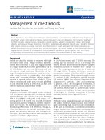
Table 2 American College of Surgeons Advanced Trauma Life Support (ATLS) classification of blood loss based on
initial patient presentation
Class I Class II Class III Class IV
Blood loss* (ml) Up to750 750-1500 1500-2000 >2000
Blood loss (% blood volume) Up to 15% 15%-30% 30%-40% >40%
Pulse rate <100 100-120 120-140 >140
Blood pressure Normal Normal Decreased Decreased
Pulse pressure (mmHg) Normal or increased Decreased Decreased Decreased
Respiratory rate 14-20 20-30 30-40 >35
Urine output (ml/h) >30 20-30 5-15 Negligible
Central nervous system/mental status Slightly anxious Mildly anxious Anxious, confused Confused, lethargic
Fluid replacement Crystalloid Crystalloid Crystalloid and blood Crystalloid and blood
Table reprinted with permission from the American College of Surgeons [37].
*for a 70 kg male.
Rossaint et al. Critical Care 2010, 14:R52
/>Page 4 of 29
also been shown that cerebral tissue lactic acidosis
occurs almost immediately after induction of hypocapnia
in children and adults with TBI and haemorrhagic shock
[44]. In addition, even a modest level of hypocapnia
(<27 mmHg) may result in neuronal depolarisation with
glutamatereleaseandextensionoftheprimaryinjury
via apoptosis [45].
Ventilation with low tidal volume is recommended in
patients with acute lung injury. In patients with normal
lung function, the evidence is scarce, but some obser-
vational studies show that the use of a high tidal
volume is an important risk factor for the development
of lung injury [46,47]. The injurious effect of high tidal
volume may be initiated very early. Randomised studies
demonstrate that short-time ventilation (<five hours)
with high tidal volume (12 ml/kg) without positive
end-expiratory pressure (PEEP) may promote pulmon-
ary inflammation and alveolar coagulation in patients
with normal lung function [48]. Alt hough more studies
are needed, the early use of protective ventilation with
low tidal volume and moderate PE EP is re commended,
particularly in bleeding trauma patients at risk of acute
lung injury.
Immediate intervention
Recommendation 5 We recommend that patients pre-
senting with haemorrhagic shock and an identified
source of bleed ing undergo an immediate bleeding con-
trol procedure unless initial resuscitation measures are
successful (Grade 1B).
Rationale The source of bleeding may be immediately
obvious, and penetrating injuries are more likely to
require surgical bleeding control. In a retrospective
study of 106 abdominal vascular injuries, all 41 patients
arrivi ng in shock follow ing gunshot wounds were candi-
dates for rapid transfer to the operating theatre for sur-
gical bleeding co ntrol [49]. A similar observation in a
study of 271 patients undergoing immediate laparotomy
for gunshot wounds indicates that these wounds
combine d with signs of severe hypovolaemic shock spe-
cifically require early surgical bleeding control. This
observation is also t rue but to a lesser extent for
abdominal stab wounds [50]. Data o n injuries caused by
pene trating metal fragments from explosives or gunshot
wounds in the Vietn am War confirm the need for early
surgical control when patients present in shock [51]. In
blunt trauma, the mechanism of injury can determine to
a certain extent whether the patient in haemorrhagic
shock will be a candidate for surgical bleeding control.
Only a few studies address the relation between the
mechanism of injury and the risk of bleeding, and none
of these publications is a randomised prospective trial of
high evidence [52]. We have found no objective data
describing the relation between the risk of bleeding and
the mechanism of injury of skeletal fractures in general
or of long-bone fractures in particular.
Traffic accidents are the leading cause of pelvic injury.
Motor vehicle crashes cause approximately 60% of pelvic
fractures followed by falls from great heights (23%).
Most of the remainder result from motorbike collisions
and vehicle-pedestrian accidents [53,54]. There is a cor-
relation between ‘unstable’ pelvic fractures and intra-
abdominal injuries [53,55]. An association between
major pelvic fracture s and severe head injuries, conco-
mitant thoracic, abdominal, urological and skeletal inju-
ries is also well described [53]. High-energy injuries
produce greater damage to both the pelvis and organs.
Patients with high- energy injuries requi re more transfu-
sion units, and more than 75% have associated head,
thorax, abdominal or genitourinary injuries [56]. It is
well documented that ‘unstable’ pelvic fractures are
associated with massive haemorrhage [55,57], and hae-
morrhage is the leading cause of death in patients w ith
major pelvic fractures.
Further investigation
Recommendation 6 We recommend that patients pre-
senting with haemorrhagic shock and an unidentified
Table 3 American College of Surgeons Advanced Trauma Life Support (ATLS) responses to initial fluid resuscitation*
Rapid response Transient response Minimal or no response
Vital signs Return to normal Transient improvement, recurrence
of decreased blood pressure and
increased heart rate
Remain abnormal
Estimated blood loss Minimal (10%-20%) Moderate and ongoing (20%-40%) Severe (>40%)
Need for more crystalloid Low High High
Need for blood Low Moderate to high Immediate
Blood preparation Type and crossmatch Type-specific Emergency blood release
Need for operative intervention Possibly Likely Highly likely
Early presence of surgeon Yes Yes Yes
* 2000 ml of isotonic solution in adults; 20 ml/kg bolus of Ringer’s lactate in children.
Table reprinted with permission from the American College of Surgeons [37].
Rossaint et al. Critical Care 2010, 14:R52
/>Page 5 of 29
source of bleeding undergo immediate further investiga -
tion (Grade 1B).
Rationale A patient in haemorrhagic shock with an uni-
dentified source of bleeding should undergo immediate
further assessment of the chest, abdominal cavity and
pelvic ring, which represent the major sources of acute
blood loss in trauma. Aside from a clinical examination,
X-rays of chest and pelvis in conjunction with focused
abdominal sonography for trauma ( FAST) [58] or diag-
nostic peritoneal lavage (DPL) [59] are recommended
diagnostic modalities during the primary survey
[37,60,61]. In selected centres, readily available com-
puted tomography (CT) scanners [62] may replace con-
ventional radiographic imaging techniques during the
primary survey.
Imaging
Recommendation 7 We recommend early imaging
(FAST or CT) for the detection of free fluid in patients
with suspected torso trauma (Grade 1B).
Recommendation 8 Werecommendthatpatientswith
significant free intra-abdominal fluid and haemodynamic
instability undergo urgent intervention (Grade 1A).
Recommendation 9 We recommend further assessment
using CT for haemodynamically stable patients who are
either suspected of having torso bleeding or have a
high-risk mechanism of injury (Grade 1B).
Rationale Blunt abdominal trauma represents a major
diagnostic challenge and an important source of internal
bleeding. FAST has b een established as a rapid and
non-invasive diagnostic approach for the detection of
intra-abdominal free fluid in the emergency room
[63-65]. Large prospective observational studies deter-
mined a high specificity and accuracy but low sensitivity
of initial FAST examination for detecting intra-abdom-
inal injuries in a dults and children [66-72]. Liu and co l-
leagues [73] found a high sensitivity, specificity and
accuracy of initial FAST examination for t he detection
of haemoperitoneum. Although CT scans and DPL were
shown to be more sensitive than sonography for the
detection of haemoperitoneum, these diagnostic modal-
ities are more time-consuming (CT and DPL) and inva-
sive (DPL) [73].
The role of CT scanning of acute trauma patients is
well documented [74-81], and in recent years imaging
for trauma patients has migrated towards multi-slice CT
(MSCT). The integration of modern MSCT scanners in
the emergency room area allows the immediate assess-
ment of trauma victims following admission [76,77].
Using modern MSCT scanners, total whole-body scan-
ning time may be reduced to less than 30 seconds. In a
retrospective study comparing 370 patients in two
groups, Weninger and colleagues [77] showed that faster
diagnosis using MSCT led to shorter emergency room
and operating room time and shorter ICU stays [77].
Huber-Wagner and colleagues [62] also showed the ben-
efit of integration of the whole-body CT into early
trauma care. CT diagnosis significantly increases the
probability of survival in patients with pol ytrauma.
Whole-body CT as a standard diagnostic tool during the
earliest resuscitation phase for polytraumatised patients
provides the added benefit of identifying head and chest
injuries and other bleeding sources in patients with mul-
tiple injuries.
Some authors have shown the benefit of contrast
medium enhanced CT scanning. Anderson and colle a-
gues [82,83] found h igh accuracy in the evaluation of
splenic injuries resulting from trauma after administra-
tion of intravenous contrast material. Delayed phase CT
may be used to d etect active bleeding in solid organs.
Fang and colleagues [84] demonstrated that the pooling
of contrast material within the peritoneal cavity in blunt
liver injuries indicates active and massive bleeding.
Patients with this finding showed rapid deterioration of
haemodynamic status and most of them required emer -
gent surgery. Intraparenchymal pooling of contrast
material with an unruptured liver capsule often indicates
a self-limited haemorrhage, and these patients respond
well to non-operative treatment.
Compared with MSCT, all traditional techniques of
diagnostic and i maging evaluation are associated with
some limitations. The diagnostic accuracy, safety and
effectiveness of immediate MSCT are dependent on
sophisticated pre-hospital treatment by trained and
experienced emergency personnel and short transporta-
tion times [85,86]. If an MSCT is not available in the
emergency room, the realisation of CT scanning implies
transportation of the patient to the CT room, and there-
fore the clinician must evaluate the implications and
potential risks and benefits of the procedure. During
transport, all vital signs should be closely monitored and
resuscitation m easures continued. For those patients in
whom haemodynamic stability is questionable, imaging
techniques such as ultrasound and chest and pelvic
radiography may be useful. Peritoneal lavage is rarely
indicated if ultrasound or CT is available [87]. Transfer
times to and from all forms of diagnostic imaging need
to be considered carefully in any patient who is haemo-
dynamically unstable. In addition to the initial clinical
assessment, near patient testing results, including full
bloo d count, haem atocrit (Hct), blood gases and la ctate,
should be readily available under ideal circumstances.
Hypotensive patients (systolic blood pressure below
90 mmHg) presenting with free intra-abdominal fluid
according to FAST or CT are potential candidates for
earlysurgeryiftheycannotbestabilisedbyinitiated
fluid resuscitation [88-90]. A retrospective study by
Rozycki and colleagues [91] of 1540 patients (1227
blunt, 313 penetrating trauma) assessed with FAST as
Rossaint et al. Critical Care 2010, 14:R52
/>Page 6 of 29
an early diagnostic tool showed that the ultrasound
examination had a sensitivity and specificity close to
100% when the patients were hypotensive.
A number of patients who present with free intra-
abdominal fluid according to FAST can safely undergo
further investigation with MSCT. Under normal circum-
stances, adult patients need to be haemodynami cally
stable when MSCT is performed outside of the emer-
gency room [91]. Haemodynamically stable patients with
a high risk mechanism of i njury, such as high-energy
trauma or even low-energy injuries in the elderly popu-
lation, should be scanned after FAST for additional inju-
ries using MSCT. As CT scanners are integrated in
resuscitation units, whole-body CT diagnosis may
replace FAST as a diagnostic method.
Haematocrit
Recommendation 10 We do not recommend the use of
single Hct measurements as an isolated laboratory mar-
ker for bleeding (Grade 1B).
Rationale Hct assays are part of the basic diagnostic
workupfortraumapatients.Thediagnosticvalueof
the Hct for detecting trauma patients with severe injury
and occult bleeding sources has been a topic of debate
in the past decade [92-94]. A major limit of the diagnos-
tic value of Hct is the confounding influence of resusci-
tative measures on the Hct due to administratio n of
intravenous flui ds and red cell concentrate s [94-97].
A retrospective study of 524 trauma patients determined
a low sensitivity (0.5) of the initial Hct on admission fo r
detecting those patients with traumatic haemorrhage
requiring surgical intervention [94]. Two prospective
observational diagnostic studies de termined the sensitiv-
ity of serial Hct measurements for detecting patients
with severe injury [92,93]. Decreasing serial Hct mea-
surements may reflect continued bleeding, but the
patient with significant bleeding may maintain his or
her serial Hct.
Serum lactate and base deficit
Recommendation 11 We recommend both serum lac-
tate and base deficit me asurements as sensitiv e tests to
estimate and monitor the extent of bleeding and shock
(Grade 1B).
Rationale Serum lactate has been used as a diagnostic
parameter and prognostic marker of haemorrhagic
shock since the 1960s [98]. The amount of lactate pro-
duced by a naerobic glycolysis is an indirect marker of
oxygen debt, tissue hypoperfusion and the severity of
haemorrhagic shock [99-102]. Similarly, base deficit
values derived from arterialbloodgasanalysisprovide
an indirect estimation of global tissue acidosis due to
impaired perfusion [99,101].
Vincent and colleagues [103] showed the value of
serial lactate measurements for predicting survival in a
prospective study in patients with circulat ory shock.
This study showed that changes in lactate concentra-
tions provide an early and objective e valuation of a
patient’s response to therapy and suggested that
repeated lactate determinations represent a reliable
prognostic index for patients with circulatory shock
[103]. Abramson and colleagues [104] performed a pro-
spective observational study in patients with multiple
trauma to evaluate the correlation between lactate clear-
ance and survival. All patie nts in whom lactate levels
returned to the normal range ( ≤2 mmol/l) within
24 hours survived. Survival decreased to 77.8% if nor-
malisation occurred within 48 hours and to 13.6% in
those patients in whom lactate levels we re elevated
above 2 mmol/l for more than 48 hours [104]. These
findings were confirmed in a study by Manikis and c ol-
leagues [105] who showed that the initial lactate levels
were higher in non-survivors after major trauma, and
that the prolonged time for normalisation of lactate
levels of more than 24 hours was associated with the
development of post-traumatic organ failure [105].
Similar to the predictive value of lactate levels, the
initial base deficit has been established as a potent inde-
pendent predictor of mortality in patients with trau-
matic hemorrhagic shock [106]. Davis and c olleagues
[107] stratified the extent of bas e deficit into three cate-
gories, mild (-3 to -5 mEq/l), moderate (-6 to -9 mEq/l)
and severe (<-10 mEq/l), and established a significant
correlation between the admission base deficit and
transfusion requirements within the first 24 hours and
the risk of post-traumatic organ failure or death [107].
The same group of aut hors showed that the base deficit
is a better prognostic marker of death than the pH in
arterial blood gas analyses [108]. Furthermore, the ba se
deficit was shown to represent a highly sensitive marker
for the extent of post-traumatic shock and mortality,
both in adult and paediatric patients [109,110].
In contrast to the data on lactate levels in haemorrha-
gic shock, reliable large-scale pro spec tive studies on the
correlation between base deficit and outcome are still
lacking. Although both the base deficit and serum lac-
tate levels are well correlated with shock and resuscita-
tion, these two parameters do not strictly correlate with
each other in severely injured patients [111]. Therefore,
the i ndependent asse ssment of both para meters is
recommended for the evaluation of shock in trauma
patients [99,101,111,112]. Composite scores that assess
the likelihood of massive transfusion and include base
deficit and other clinical parameters have been devel-
oped but require further validation [112,113]. Callaway
and colleagues [114] performed a seven-year retrospec-
tive analysis of a prospective trauma registry from a
level I trauma centre to determine predictors of mortal-
ity in elderly patients 65 years or older who sustained
blunt trauma and presented with a normal initial
Rossaint et al. Critical Care 2010, 14:R52
/>Page 7 of 29
systolic blood pressure (≥90 mmHg). The odds ratio for
death was increased more than four-fold in those
patients who had either elevated serum lactate levels
above 4 mmol/l or a base deficit below -6 mEq/l,
compared with patients with normal lactate levels
(<2.5 mmol/l) or a base excess (>0 mEq/l). Paladino and
colleagues [115] assessed t he prognostic value of a com-
bination of abnormal vital signs (heart rate >100 beats/
min or a systolic blood pressure <90 mmHg) in con-
junction with serum lactate and base deficit for identify-
ing trauma patients with major injuries, using cut-off
values for lactate at more than 2.2 mmol/l and base def-
icit at less than -2.0 mEq/l, respectively. The authors
found that the addition of the metabolic parameters to
the vital signs increased the sensitivity for identifying
major injury from 40.9% to 76.4%, implying that the
addition of lactate and base deficit to triage vital signs
increases the ability to distinguish major from minor
injury.
Coagulation monitoring
Recommendation 12 We recommend that routine prac-
tice to detect post-traumatic coagulopathy include the
measurement of i nternational normalised ratio (INR),
activated partial thromboplastin time (APTT), fibrinogen
and platelets. INR and APTT alone should not be u sed
to guide haemostatic therapy (Grade 1C). We suggest
that thrombelastometry also be performed to assist in
characterising the coagulopathy and in guiding haemo-
static therapy (Grade 2C).
Rationale Little evidence supports a recomm endation
for the best haemostatic monitoring tool(s). Standard
monitoring comprises INR, APTT, platelets and fibrino-
gen, although there is little direct evidence for the effi-
cacy of these measures. Increasing emphasis focuses on
the importance of fibrinogen and platelet measurements.
It is often assumed that the conventional coagulation
screens (INR and APTT) monitor coagulat ion; however,
these tests monitor only the initiation phase of blood
coa gulation and represent only the first 4% of thrombin
production [116]. It is therefore possible that the con-
ventional coagulation screen appears normal, while the
overall state of blood coagulation i s abnormal. There-
fore, a more complete monitoring of blood coagulation
and fibrinolysis, such as thrombelastometry, may facili-
tate more accurate targeting of therapy. Case series
using thrombelastometry to assess trauma patients have
been published. One stud y applied thrombelastometry
to 23 patients, but without a comparative standard
[117]. Another study found a poor correlation between
thrombelastometry and conventional coagulation para-
meters [10]. Johansson [118] implemented a haemostatic
resuscitation regime (early platelets and fresh frozen
plasma (FFP)) guided using thrombelastometry in
a before-and-after study which showed improved
outcomes. There is insufficientevidenceatpresentto
support the utility of thrombelastometry in the detection
of post-traumatic coagulopathy. More research is
required in this area, and in the meantime physicians
should make their own judgement when developing
local policies.
It is theoretically possible that the pattern of change in
measures of coagulati on such as D-dimers may help to
identify patients with ongoing bleeding. However, there
are no publications relevant to this question, so tradi-
tional methods of detection for ongoing bleeding, such
as serial clinical evaluation of radiology (ultrasound, CT
or angiography) should be used.
III. Rapid control of bleeding
Pelvic ring closure and stabilisation
Recommendation 13 We recommend that patients with
pelvic ring disruption in haemorrhagic shock undergo
immediate pelvic ring closure and stabilisation (Grade
1B).
Packing, embolisation and surgery
Recommendation 14 We recommend that patients with
ongoing haemodynamic instability despite adequate pel-
vic ring stabilisation receive early preperitoneal packing,
angiographic embolisation and/or surgical bleeding con-
trol (Grade 1B).
Rationale The mortality rate of patients with severe
pelvic ring disruptions and haemodynamic instability
remains unacceptably h igh [119-122]. The early detec-
tion of these injuries and initial efforts to reduce disrup-
tion and stabilise the pelvis as well as containing
bleeding is therefore crucial. Markers of pelvic haemor-
rhage include anterior-posterior and vertical shear
deformations, CT ‘blush’ (active arterial extravasation),
bladder compression pressure, pelvic haematoma
volumes of more than 500 ml evident by C T and
ongoing haemodynamic instability despite adequate frac-
ture stabilisation [123-125].
The initial therapy of pelvic fractures includes control
of venous and/or cancellous bone bleeding by pelvic clo-
sure. Some institutions use primarily external fixators to
control haemorrhage from pelvic fractures [124,125] but
pelvic closure may also be achieved using a bed sheet,
pelvic binder or a pelvic C-clamp [126-128]. In addition
to the pelvic closure, fracture stabilisat ion and the tam-
ponade effect of the haematoma, pre, extra or retroperi-
toneal packing will reduce or stop the venous bleeding
[122,129-131]. Preperitoneal packing decreases the need
for pelvic embolisation and may be performed simulta-
neously o r soon after initial pelvic stabilisation
[122,129,131]. The technique can be combined with a
consecutive laparotomy if deemed necessary [122,129 ].
This may decrease the high mortality rate observed in
patients with major p elvic injuries who underwent
Rossaint et al. Critical Care 2010, 14:R52
/>Page 8 of 29
laparotomy as the primary intervention. As a conse-
quence, it was recommended that non-therapeutic lapar-
otomy should be avoided [132].
Angiography and embolisation is currently accepted as
a highly effective means with which to control arterial
bleeding that cannot be controlled by fracture stabilisa-
tion [122-126,131-140]. The presence of sacroiliac joint
disruption, female gen der and du ration of hypotension
can reliably predict patients who would benefit from the
procedure [138]. Controversy exists about the indica-
tions and optimal timing of angiography in haemodyna-
mically unstable patients [131]. Institutional differenc es
in the capacity to perform timely angiography and
embolisation may explain the different treatment algo-
rithms suggested by many authors [119-122,125,129,
131,132,140 ]. Nevertheless, the general consensus is that
a multidisciplinary approach to these severe injuries is
required.
Early bleeding control
Recommendation 15 We recommend that early bleed-
ing control of the abdomen be achieved using packing,
direct surgical bleeding control and the use of local hae-
mostatic procedures. In the exsanguinating patient, aor-
tic cross-clamping may be employed as an adjunct
(Grade 1C).
Rationale Abdominal resuscitative packing is an early
part of the post-traumatic laparotomy to identify major
injuries and sources of haemor rhage [141,142]. If bleed-
ing cannot be controlled using packing and conventional
surgical techniques when the p atient is in extremis or
when proximal vascular control is deemed necessary
before opening the abdomen, aortic cross clamping may
be employed as an adjunct to reduce bleeding and redis-
tribute blood flow to the hea rt and brain [143-145].
When blood l osses are important, when surgical mea-
sures are unsuccessful and/or when the patient is cold,
acidotic and coagulopathic, definitive packi ng may a lso
be the first surgical step within the concept of damage
control [146-155]. Packing aims to compress liver rup-
tures or exert direct pressure on the sources of bleeding
[141,142 ,146-150,152-154]. The definitive packing of the
abdomen may allow further attempts to achieve total
haemostasis through angiography and/or correction of
coagulopathy [155]. The removal of packs should prefer-
ably be performed o nly after 48 hours to lower the risk
of rebleeding [152,153].
Damage control surgery
Recommendation 16 We recommend that damage con-
trol surgery be employed in the severely injured patient
presenting with deep haemorrhagic shock, signs of
ongoing bleeding and coagulopathy. Additional factors
that should trigger a damage control approach are
hypothermia, acidosis, inaccessible major anatomical
injury, a need for time-consuming procedures or conco-
mitant major injury outside the abdomen (Grade 1C).
Rationale The severely injured patient arriving to the
hospital with continuous bleeding or deep haemorrhagic
shock generally has a poor chance of survival unless
early control of bleeding, proper resuscitation and blood
transfusion are achieved. This is particularly true for
patients who present with uncontrolled bleeding due to
multiple penetrating injuries o r patients with multiple
injuries and unstable pelvic fractures with ongoing
bleeding from fracture sites and retroperitoneal vessels.
The common denominator in these patients is the
exhaustion of physiological reserves with resulting pro-
found acidosis, hypothermia and coagulopathy, also
known as t he ‘bloody vicious cycle’. In 1983, Stone and
colleagues described the techniques of abbreviated lapar-
oto my, packing to control haemorrhage and of deferred
definitive surgical repair until coagulation has been
established [156]. Since then, a number of authors have
described the beneficial results of this concept, now
called ‘damage control’ [50,54,121,134,151,156-1 58].
Damage control surgery of the abdomen consists of
three components: the first component is an abbreviated
resuscitative laparotomy for control of bleeding, the res-
titution of blood flow where necessary and the control
of contamination. This should be achieved as rapidly as
possible without spending unnecessary time on trad i-
tional organ repairs t hat can be deferre d to a later
phase. The abdomen is packed and temporary abdom-
inal closure is performed. The second component is
intensive care treatment, focused on core re-warming,
correction of the acid-base imbalance and coagulopathy
as well as optimising the ventilation and the haemody-
namic status. The third component is the definitive sur-
gical repair that is performed only when target
parameters have been achieved [159-162]. Although the
concept of ‘damage control’ intuitively m akes sense, no
RCTs exist to support it. Retrospective studies support
the concept showing reduced morbidity and mortality
rates in selective populations [50,151,157,161].
The same ‘damage control’ principles have been
applied to orthopaedic injuries in severely injured
patients [134,163-166]. Scalea was the first to coin the
term ‘damage control orthopaedics’ [166]. Relevant frac-
tures are primarily stabilised with external fixator s
rather than primary definitive osteosynthesis [134,163].
The less traumatic and shorter duration of the surgical
procedureaimstoreducethesecondarytraumaload.
Definitive osteosy nthesis surgery can be performed after
4 to 14 days when the patient ha s recovered sufficiently.
Retrospective clinical studies and prospective cohort stu-
dies seem to support the concept of damage control
[134,163-165]. The only available randomised study
Rossaint et al. Critical Care 2010, 14:R52
/>Page 9 of 29
shows an advantage for this strategy in ‘borderline’
patients [164].
Local haemostatic measures
Recommendation 17 We recommend the use of topical
haemostatic agents in combination with other surgical
measures or with packing for venous or moderate arterial
bleeding associated with parenchymal injuries (Grade 1B).
Rationale A wide range of local haemostatic agents are
currently available for use as adjuncts to traditional
surgical techniques to obtain haemorrhage control.
These topical agents can be particularly useful when
access to the bleeding area is difficult. Local haemo-
static agents include collagen, gelatin or cellulose-
based products, fibrin and synthetic glues or adhesives
that can be used for both external and internal bleed-
ing while polysaccharide-based and inorganic haemo-
statics are still mainly used and approved for external
bleeding. The use of topical haemostatic agents should
consider several factors such as the type of surgical
procedure, cost, severity of bleeding, coagulation status
and each agent’s specific characteristics. Some of these
agents should be avoided when autotransfusion is
used and several other contraindications need to be
considered [167,168]. The capacity of each agent to
control bleeding was initially studied in animals but
increasing experience from humans is now available
[167-180].
The different types of local haemostatics are briefly
presented according to their basis and haemostatic
capacity:
i) Collagen-based agents trigger platelet aggregation
resulting in clot formation when in contact with a
bleeding surface. They are often combined with a pro-
coagulant substance such as thrombin to enhance the
haemostatic effect. A positive haemostatic effect has
been shown in several human studies [169-172].
ii) Gelatin-based products can be used alone or in
comb ination with a procoagulant substance [167]. Swel-
ling of the gelatin in contact with blood reduces the
blood flow and, in combination with a thrombin-based
component, enhances haemostasis. A similar or superior
haemostatic effect has been observed compared with
collagen-based agents [173-175].
iii) The effect of cellulose-based haemostatic agents on
bleeding has been less well studied and only case reports
that support their use are available.
iv) Fibrin and synthetic glues or adhesives have both
haemostatic and se alant properties and their significant
effect on haemostasis have been shown in several
human RCTs involving vascular, bone, skin and visceral
surgery [176-178].
v) Polysaccharide-based haemostatics can be divided
into two broad categories [167]: N-acetyl-glucosamine-
containing glycosaminoglycans purified fr om microalgae
and diatoms and microporous polysaccharide haemo-
spheres produced from potato starch. The mechani sm of
action is complex and depends on the purity or combina-
tion with other substances such as cellulose or fibrin.
A number of different products are currently available
and have been shown to be efficient for external use.
An observational study showed that haemorrhage control
was achieved using an N-acetylglucosamine-based ban-
dage applied to 10 patients with severe hepatic and
abdominal injuries, acidosis and clinical coagulopathy
[180].
vi) The inorganic haemostatics based on minerals such
as zeolite or smectite have been used and studied mainly
on external bleeding [167,168].
IV. Tissue oxygenation, fluid and hypothermia
Volume replacement
Recommendation 18 We recommend a target systolic
blood pressure of 80 to 100 mmHg until major bleeding
has been stopped in the initial phase following trauma
without brain injury (Grade 1C).
Rationale In order to maintain tissue oxygenation, tra-
ditional treatment of trauma patients uses early and
aggressive fluid administration to restore blood volume.
This approach may, however, increase the hydrostatic
pressure on the wound, cause a dislodgement of blood
clots, a dilution of coagulation factors and undesirable
cooling of the patient. The concept of low-volume fluid
resuscitation, so-called ‘permissive hypotension’,avoids
the adverse effects of early aggressive resuscitati on while
maintaining a level of tissue perfusion that, although
lower than normal, is adequate for short periods [130].
A controlled hypotensive fluid resuscitation should aim
to achieve a mea n arterial pressure of 65 mmHg or
more [181]. Its general eff ectiveness rem ains to be con-
firmed in RCTs; however, studies have demonstrated
increased survival when a low volume fluid resuscitation
concept was used in penetrating trauma [182,183]. In
contrast, no significant difference in survival was found
in patients with blunt trauma [184]. One study con-
cluded that mortality was higher after on-site resuscita-
tion compared with in-hospital resuscitation [185]. It
seems that greater increases in bl ood pressure are toler-
ated without exacerbating haemorrhage when they are
achieved gradually and with a significant delay following
the initial injury [186]. All the same, a recent Cochrane
systematic review concluded that there is no eviden ce
from RCTs for or against early or larger volume intrave-
nous fluids to treat u ncontrolled haemorrhage [187].
However, a recent retrospective analysis demonstrated
that aggressive resuscitation techniques, often initiated
in the prehospital setting, appear to increase the likeli-
hood that patients with severe extremity injuries develop
secondary abdominal compartment syndrome (ACS)
Rossaint et al. Critical Care 2010, 14:R52
/>Page 10 of 29
[188]. In this study, early, large-volume crystalloid
administration was the greatest predictor of secondary
ACS. Moreover, a retrospective a nalysis of the German
Trauma Registry database including 17,200 multiply
injured patients showed that the incidence of coagulopa-
thy increased with increasing volume of intravenous
fluids administered pre-clinically. Coagulopathy was
observed in more than 40% of patients with more than
2000 ml, in more than 50% with more than 3000 ml,
and in more than 70% with more than 4000 ml adminis-
tered [3].
The low-volume approach is contraindicated in TBI
and spinal injuries, because an adequate perfusion pres-
sure is crucial to ensure tissue oxygenation of the
injured central nervous system. In addition, the concept
of permissive hypotension should be carefully consid-
ered in the elderly patient and may be contraindicated if
the patient suffers from chronic arterial hypertension.
A recent analysis from an ongoing multi-centre pro-
spective cohort study suggests that the early use of vaso-
pressors for haemodynamic support after haemorrhagic
shock in comparison to aggressive volume resuscitation
may be deleterious and should be used cautiously [189].
However, this study has several limitations: the study is
a secondary analysis of a prospective cohort study, and
was not designed to answer the specific hypothesis
tested. Thus, it is not possible to separate vasopressor
from the early management of trauma patients. In addi-
tion, although the use of a vasopressor helps to rapidly
restore arterial pressure, it shou ld not be v iewed as a
substitute for fluid resuscitation and the target blood
pressure must be respected.
Fluid therapy
Recommendation 19 We recommend that c rystalloids
be applied initially to treat the bleeding trauma patient
(Grade 1B). We suggest that hypertonic solutions also
be considered during initial treatment (Grade 2B). We
suggest that the addition of colloids be considered
within the prescribed limits for each solution in haemo-
dynamically unstable patients (Grade 2C).
Rationale It is still unclear what type of fluid should be
employed in the initial treatment of the bleeding trauma
patient. Although several meta-analyses have shown an
increased risk of death in patients treated with colloids
compared with patients treated with crystalloids
[190-194] and three of these studies showed that the
effect was particularly significant in a trauma subgroup
[190,193,194], a more recent meta-analysis showed no
difference in mortality between colloids and crystalloids
[195]. If colloids are used, modern hydroxyethyl starch
or gelatin solutions should be used because the risk:ben-
efit ratio of dextran is disadvantageous. Problems in
evaluating and comparing the use o f different resuscita-
tion fluids include the heterogeneity of populations and
therapy strategies, limited quality of analysed studies,
mortality not always being the primary outcome, and
different, often short, observation periods. It is therefore
difficult to reach a definitive conclusion as to the advan-
tage of one type of resuscitation fluid over the other.
The Saline versus Albumin Fluid Evaluation study com-
pared 4% albumin with 0.9% sodium chloride in 6997
ICU patients and showed that albumin administration
was not associated with worse outcomes; however, there
was a trend towards higher mortality in the brain
trauma subgroup that received albumin (P = 0.06) [196].
Promising results have been obtained with hypertonic
solutions. Recentl y, a double-blind, RCT in 209 patients
with blunt traumatic injuries analysed the effect of the
treatment with 250 ml of 7.5% hypertonic saline and 6%
dextran 70 compared with lactated Ringer solution on
organ failure. The intent-to-treat ana lysis demonstrated
no significant difference in organ failure and in acute
respiratory disress syndrome (ARDS)-free survival. How-
ever, there was improved ARDS-free survival in the sub-
set (19% of the population) requiring 10 U or more of
packed red blood cells (RBCs) [197]. One study showed
that the use of hype rtonic saline was associated with
lower intracranial pressure than with normal saline in
brain-injured patients [198] and a meta-analysis compar-
ing hypertonic saline dextran with normal saline for
resuscitation in hypotension from penetrating torso
injuries showed improved survival in the hypertonic sal-
ine dextran group when surgery was required [199].
A clinical trial with brain injury patients found that
hypertonic saline reduced intracranial pressure more
effectively than dextran solution with 20% mannitol
when compared in equimolar dosing [200]. However,
Cooper and colleagues f ound almost no difference in
neurological function six months after TBI in patients
who had received pre-hosp ital hypertonic saline resusci-
tation compared with conventional fluid [201]. In con-
clusion, the evidence suggests that hypertonic saline
solutions are safe, and will improve haemodynamics
during hypovolaemic resuscitation. The evidence for
increased survival with use of hypertonic saline solutions
is inconclusive. It is possible that certain subgroups
might benefit from hypertonic saline solutions, but
further research is required [202].
Normothermia
Recommendation 20 We recommend early application
of measures to reduce heat loss and warm the hypother-
mic patient in order to achieve and mai ntain nor-
mothermia (Grade 1C).
Rationale Hypothermia, defined as a core body tem-
perature below 35°C, is associated with acidosis, hypo-
tension and coagulopathy in severe ly injured patients. In
a retrospective study with 122 patients, hypothermia
was an ominous clinical sign, accompanied by high
Rossaint et al. Critical Care 2010, 14:R52
/>Page 11 of 29
mortality and blood loss [203]. The profound clinical
effects of hypothermia ultimately lead to higher morbid-
ity and mortality, and hypothermic patients require
more blood products [204].
Hypothermia is associated with an increased risk o f
severe bleeding, and hypothermia in trauma patients
represents an independen t risk factor for bleeding and
death [205]. The effects of hypothermia include altered
platelet function, impaired coagulation factor function
(a 1°C drop in temperature is associated with a 10%
drop in function), enzyme inhibition and fibrinolysis
[206,207]. Body temperatures below 34°C compromise
blood co agulation, but this has only been observed
when coagulation tests (prothrombin time (PT) and
APTT) are carried out at the low temperatures seen in
patients with hypothermia, and not when assessed at
37°C as is routine practice for such tests. Steps to pre-
vent hypothermia and the risk o f hypothermia-induced
coagulopathy include removing wet clothing, covering
the patient to avoid additional heat loss, increasing the
ambient temperature, forced air warming, warm fluid
therapy and, in extreme cases, ext raco rpo real re-warm-
ing devices [208,209].
Animal and human studies of controlled hypothermia
in haemorrhage have shown some positive results com-
pared with normothermia [210,211]. Contradictory
results have been observed in meta-analyses that examine
mortality and neurological outcomes associated with
mild hypothermia in patients with TBI, po ssibly due to
the different exclusion and inclusion criteria for the stu-
dies used for the analysis [212-214]. The speed of induc-
tion and duration of hypothermia, which may be very
important factors that influence the benefit associated
with this treatment. It has been shown that five days of
long-term cooling is more efficacious than two days of
short-term cooling when mild hypothermia is used to
control refractory intracranial hypertension in adults
with severe TBI [215]. Obviously, the time span of
hypothermia is crucial, because a recent prospective RCT
in 225 children with severe TBI showed that hypothermic
therapy initiated within 8 hours after injury and contin-
ued for 24 hours did not improve the neurological out-
come and may increase mortality [216]. Furthermore, the
mode of inducing cerebral hypothermia induction may
influence its effectiveness. In a RCT comparing non-inva-
sive selective brain cooling (33 to 35°C) in 66 patients
with severe TBI and mild systemic hypothermia (rectal
temperature 33 to 35°C) and a control group not exposed
to hypothermia, natural rewarming began after three
days. Mean intracranial pressure 24, 48 or 72 hours after
injury was significantly lower in the selective brain cool-
ing group than in the control group [217].
Prolonged hypothermia may be considered in patients
with isolated head trauma after haemorrhage has been
arrested. If mild hypothermia is applied in TBI, cooling
should take place within the first three h ours following
injury, preferably using selective brain cooling by cool-
ing the head and neck, be maintained for a t least
48 hours [218], rewarming should last 24 hours and the
cer ebral perfusion pressure should be maintained above
50 mmHg (systolic blood pressure ≥70 mmHg). Patients
most likely to benefit from hypothermia are those with
a GCS at admission between 4 and 7 [219]. Possible
side effects are hypotension, hypovolaemia, electrolyte
disorders, insulin resistance and reduced insulin secre-
tion and increased risk of infection [220]. Further stu-
dies are warranted to investigate the postulated benefit
of hypothermia in TBI taking these important factors
into account.
V. Management of bleeding and coagulation
Erythrocytes
Recommendation 21 We recommend a target haemo-
globin (Hb) of 7 to 9 g/dl (Grade 1C).
Rationale Erythrocytes contribute to haemostasis by
influencing the biochemical and functional responsive-
ness of activated platelets via the rheological effect on
platelet margination and by supporting thrombin gen-
eration [221]; however, the optimal Hct or Hb concen-
tration required to sustain haemostasis in massively
bleeding patients is unclear. Further investigations into
the role of the Hb concentration on haemostasis in mas-
sively transfused patients are therefore warranted.
The effects of the Hct on blood coagulation have not
been fully elucidated [222]. An acute reduction of the
Hct results in an increase in the bleeding time [223,224]
with restoration upon re-transfusion [223]. This may
relate to the presence of the enzyme elastase on the sur-
face of RBC membranes, which may activate coagulation
factor IX [225,226]. However, a moderate reduction of
theHctdoesnotincreasebloodlossfromastandard
splee n injury [224], and an isolated in vitro reduction of
the Hct did not compromise blood coagulation as
assessed by thrombelastometry [227].
No prospective RCT has compared restrictive and liberal
transfusion regimens in trauma, but 203 trauma patients
from the Tra nsfusion Requirements in Critical Care trial
[228] were re-analysed [229]. A restrictive transfusion regi-
men(Hbtransfusiontrigger<7.0 g/dl) resulted in fewer
transfusions as compared with the liberal transfusion regi-
men (Hb transfusion trigger <10 g/dl) and appeared to be
safe. However, no statistically significant benefit in terms
of multiple organ failure or post-traumatic infections was
observed. It should be emphasised that this study was
neither designed nor powered to answer these q uestions
with precision. In addition, it cannot be ruled out that the
number of RBC units transfused merely reflects the sever-
ity of injury. Nevertheless, RBC transfusions have been
Rossaint et al. Critical Care 2010, 14:R52
/>Page 12 of 29
shown in multiple studies to be associated with increased
mortality [230-234], lung injury [234-236], increased infec-
tion rates [237,238] and rena l failure in trauma victims
[233]. This ill effect may be particularly important with
RBC transfusions stored for more than 14 days [233].
Despite the lack of high-level scientific evidence for a
specific Hb transfusion trigger in patients with TBI,
these patients are currently transfused in many centres
to achieve an Hb of approximately 10 g/dl [239]. This
might be justified by the recent finding that increa sing
the Hb from 8.7 to 10.2 g/dl improved local cerebral
oxygenation in 75% of patients [158]. In another preli-
minary study in patients with TBI, one to two RBC
transf usions at a Hb of approximately 9 g/dl tra nsiently
(three to six hours) increased cerebral oxygenation,
again in approximately 75% of patients [240,241]. A sto-
rage time of more than 19 days precluded this effect
[240]. In another recent study, cerebral tissue oxygena-
tion,onaverage,didnotincre ase due to an increase in
Hb from 8.2 to 10.1 g/dl [242]. Nevertheless, the authors
came to the conclusion based on multivariable statistical
models that the changes in cerebral oxygenation corre-
lated significantly with Hb concentration [242]. This
conc lusion , however, was questioned in the accompany-
ing editorial [243].
In an initial outcome study the lowest Hct was corre-
lated with adverse neuro logical outcome and RBC trans-
fusions were also found to be an independent factor
predicting adverse neurological outcome [244]. Interest-
ingly, the number of d ays with a Hct be low 30% was
found to be correlated with an improved neurological
outcome [244]. In a more recent o utcome study in 1150
patients with TBI, RBC transfusions were found to be
associated with a two-fold increased mortality and a
three-fold increased complication rate [138]. Therefore,
patients with severe TBI should not have an Hb transfu-
sion threshold different than that of other critically ill
patients.
Coagulation support
Recommendation 22 We recommend that monitoring
and measures to support coagulation be initiated as
early as possible (Grade 1C).
Rationale Major trauma results not only in bleeding
from ana tomical sites but also frequently in coagulopa-
thy, which is associated with a several-fold increase in
mortality [3,5,8,9,245]. This early coagulopathy of
trauma is mainly found in patients with hypoperfusion
(base deficit >6 mE/l) [8,245] and is characterised by an
up-regulation of endothelial thrombomodulin, which
forms complexes with thrombin [246].
Early monitoring of coagulation is essential to detect
trauma-induced coagulopathy and to define t he main
causes, includ ing hyperfibrinolysis [10,117]. Early thera-
peutic intervention does improve co agulation tests [247]
and persistent coagulopathy at ICU entry has been
shown to be associated with a increased mortality [248].
Therefore, early aggressive treatment is likely to improve
the outcome of severely injured patients [ 249]. However,
there are also studi es in which no survival benefit could
be shown [247,250].
Calcium
Recommendation 23 We recommend that ionised cal-
cium levels be monitored during massive transfusion
(Grade 1C). We suggest that calcium chloride be admi-
nistered during massive tra nsfusion if ionised calcium
levels are low or electrocardiographic changes suggest
hypocalcaemia (Grade 2C).
Rationale Calcium in the extracellular plasma exists
either in a free ionised state (45%) or bound to proteins
and other molecules in a biologically inactive state
(55%). The normal concentration of the ionised form
ranges from 1.1 to 1.3 mmol/l and is influenced by the
pH. A 0.1 unit increase in pH decreases the ionised cal-
cium con centration by approximately 0.05 mmol/l [181].
The availability of ionised calcium is essential for the
timely formation and stabilisation of fibrin polymerisa-
tion sites, and a decrease in cytosolic calcium concentra-
tion precipitates a decrease in all platelet-related
activities [181]. In addition, contractility of the heart
and systemic vascular resistance are compromised at
low ionised calcium levels. Combining beneficial cardio-
vascular and c oagulation effects, the level for ionised
calcium concentration should therefore be maintained
above 0.9 mmol/l [181].
Early hypocalcaemia following traumatic injury shows
a significant correlat ion with the amount of infused col-
loids, but not with crystalloids, and may be attributable
to colloid-induced haemodilution [251]. Also, hypocal-
caemia develops during massive transfusion as a result
of the citrate employed as an anticoagulant in blood
products. Citrate exerts its anticoagulant activi ty by
binding ionised calcium, and hypocalcaemia is most
common in association with FFP and platelet transfu-
sion because these products contain high citrate concen-
trations. Citrate undergoes rapid he patic metabolism,
and hypocalcaemia is generally transient during standard
transfusion procedures. Citrate metabolism may be dra-
matically impaired by hypoperfusion states, hypothermia
and in patients with hepatic insufficiency [252].
Fresh frozen plasma
Recommendation 24 We recommend early treatment
with thawed FFP in patients with massive bleeding
(Grade 1B). The initial recommended dose is 10 to
15 ml/kg. Further doses will depend on coagulation
monitoring and the amount of other blood products
administered (Grade 1C).
Rationale The clinical eff icacy of FFP is largely unpro-
ven [253]. Never theless, most guidelines recommend the
Rossaint et al. Critical Care 2010, 14:R52
/>Page 13 of 29
use of FFP either in ma ssive bleeding or in significant
bleeding complicated by coagulopathy (PT or APTT
more than 1.5 t imes control) [7,254,255]. Patients trea-
ted with oral anticoagulants (vitamin K antagonists) pre-
sent a particular challenge, and FFP is recommended
[255] only when prothrombin complex concentrate
(PCC) is not available [254]. The most frequently
recommended dose is 10 to 15 ml/kg [254,255], and
further doses may be required [256]. As with all pro-
ducts derived from human blood, the risks associated
with FFP treatment include circulatory overload, ABO
incompatibility, transmission of infectious diseases
(including prion diseases), mild allergic reactions and
transfusion-related acute lung injury [254,257,258]. FFP
and platelet concentrates appear to be the most fre-
quently implicated blood products in transfusion-related
acute lung injury [257-260]. Although the formal link
between the administration of FFP, control of bleeding
and an eventual improvement in the outcome of bleed-
ing patients is lacking, most experts would agree that
FFP treatment is beneficial in patients with massive
bleeding or significant bleeding complicated by
coagulopathy.
There are very few well-designed studies that explore
massive transfusion strategy. The need for massive
transfusion is relatively rare, occurring in less than 2 %
of civilian trauma p atients, but higher (7%) in the mili-
tary setting. Massive transfusion man agement has bee n
based on the concept that coagulopathy associated with
severe trauma was primarily consumptive due to the
dilution of blood clotting factors and the consumption
of haemostasis factors at the site of injury.
FFP was recommended when PT or APTT was 1.5
times normal or after 10 RBC units had been transfused.
Many massive transfusion protocols stipulated one unit
of FFP for every four units of RBCs. In recent years, ret-
rospective data from the US Army combat support hospi-
tals have shown an association between survival and a
higher ratio of transfused FFP and RBC units. These data
show that casualties who received FFP and RBCs at a
ratio of 1:4 or lower, had a three-fold higher mortality
than thos e who received a massive transfusion with a 2:3
ratio. These data have induced many civilian trauma cen-
tres to modify their transfusion approach to incorporate
the early use of thawed FFP in ratios approaching 1:1.
Ten relevant studies addressing FFP:RBC r atio have
been identified, all of which were retrospective studi es,
although some are based on data collected prospectively
for other reasons. None of the studies were clinical
RCTs. The majority of the authors used massive transfu-
sion (10 RBC units within 24 hours) as the entry criter-
ion; however, to limit bias due to FFP unavailability,
one study [261] excluded pa tients who died within the
first 30 minutes. One of the studies [262] took into
consideration only patients alive upon ICU admission,
and a nother defined massive transfusion as 10 units or
more prio r to ICU admission. One report [247] defined
massive transfusion as more than 10 units over 6 hours.
Two of the studies are based on data collected in a
combat setting, while the other eight were performed
based on data collected at civilian trauma centres. The
majority of the studies are single centre; one study is
multi-centre [261] and one is a retrospective analysis of
the German Trauma Registry [3].
Seven studies showed better outcomes using a high
FFP:RBC ratio [3,261-266] and two did not [250,267].
One st udy may be classified as indeterminate because a
high FFP:RBC ratio (average 1:2) was associated with a
better survival than a low ratio (average 1:4), but the
survival curve was U-shaped, with the lowest mortality
at a 1:2 to 1:3 ratio [247]. The two combat studies
showed better outcomes using a high ratio [265,266].
Early empirical infusion of FFP may increase the fre-
quency of delayed traumatic intracerebral haematoma
and the mortality in patients with severe head injury
[268]. Most of the studies calculate FFP:RBC ratio at
24 hours after admission. When Snyder and colleagues
[267] used the FFP:RBC ratio at 24 hours as a fixed
value, patients who received a higher ratio had signifi-
cantly better outcomes, but if the timing of component
product transfusion was taken into account, the differ-
ence was no longer statistically significant.
These combat data are retrospective, refer to young,
previously healthy male patients with penetrating inju-
ries and may be confounded to some extent by treat-
ment biases. Because FFP requires a significant amount
of time before it is thawed and available for transfusion
and many trauma deaths occur soon after hospital
admission, patients who die early may receive RBC units
but die before FFP therapy has begun. These cases may
therefore be included in the low ratio group e ven if a
1:1 strategy was intended. One further ground for criti-
cism of many of these studies is that the number of
RBCsunitstransfusedisanindicatorofseverityof
injury that cannot be complete ly adjusted for by regres-
sion analysis. All of these limitations must be kept in
mind when analysing the available recent literature and
emphasises the need for prospective trials.
Platelets
Recommendation 25 We recommend that platelets be
administered to maintain a platelet count above 50 ×
10
9
/l (Grade 1C). We suggest maintenance of a platelet
count above 100 × 10
9
/l in patients with mult iple
trauma who are severely bleeding or have TBI (Grade
2C). We suggest an initial dose of four to eight platelet
concentrates or one aphaeresis pack (Grade 2C).
Rationale In medical conditions leading to thrombocy-
topaenia, haemorrhage does not often occur until the
Rossaint et al. Critical Care 2010, 14:R52
/>Page 14 of 29
platelet count falls below 5 0 × 10
9
/l, and platelet func-
tion decreases exponentially below this point [269-272].
There is no direct evide nce to support a particular pla-
telet transfusion threshold in the trauma patient. A con-
sensus development conference sponsored by the
National Institutes of Health (NIH; Bethesda, MD, USA)
in 1986 determined that bleeding is unlikely to be
caused by thrombocytopaenia at platelet counts of 50 ×
10
9
/l or greater and agreed that platelet transfusion is
appropriate to prevent or control bleeding associated
with deficiencies in platelet number or function
[273,274]. The NIH consensu s did not consider trauma,
but it seems reasonable to recommend that a platelet
countofatleast50×10
9
/l be maintained following
injury.
An argument can be made for maintaining a higher
level of platelets, perhaps up to 100 × 10
9
/l, following
injury. If a patient has increased fibrin degrad atio n pro-
ducts due to disseminated intravascular coagulation
and/or hyperfibrinolysis, this will interfere with platelet
function and a higher threshold of 75 × 10
9
/l has been
suggested by consensus groups [275,276]. Moreover,
platelet-rich concentrate is an autologo us concentration
of platelets and growth factors (e.g. transforming growth
factor-beta, vascular endothelial growth factor and plate-
let-derived growth factor), and due to the increased con-
centration and release of these factors, platelet-rich
concentrates could potentially enhance bone and soft
tissue healing [277]. Transfusion threshold levels of up
to 100 × 10
9
/l have been suggested for treatment of
severe brain injury and massive haemorrhage, but the
evidence for the higher threshold is weak [275,276]. One
group showed that trauma patients receiving platelets
and RBCs at a ratio of 1:5 or greater had a lower 30-day
mortality when compared with those with who received
less than this rat io (38% vs. 61%, P = 0.001) [264].
Another study of massively transfused trauma patients
has pointed to an early aggressive correction of coagulo-
pathy with platelet transfusion as a possible contributing
factor to good outco me [278]. In this retrospective
cohort study, survivors received one platelet transfusion
for every 7.7 units of blood transfused whereas nonsur-
vivors received only one platelet transfusion for every
11.9 units of blood transfused (P = 0.03).
When platelet transfusion was introduced in the
1950s, no clinical trials were employed to assess t he uti-
lity of platelet therapy compared with placebo, and such
trials today might be considered unethical. The appro-
priate dose of platelets is therefore uncertain. Platelet
concentrate produced from a unit of whole blood con-
tains 7.5 × 10
10
platelets on average and should increase
the platelet count by 5 to 10 × 10
9
/l in a 70 kg recipient.
Aphaeresis platelet concentrates generally contain
approximately 3 to 6 × 10
11
platelets, depending on
local collection practice, and physicians should be cogni-
sant of the doses provided locally. A pool of four to
eight platelet concentrates or a single-donor aphaeresis
unit is usually sufficient toprovidehaemostasisina
thrombocytopaenic, bleeding patient. If r equired, the
dose of platelets (× 10
9
) can be calculated in more detail
from the desired platelet increment, the patient’sblood
volume in litres (estimated by mul tiplying the patient’s
body surface area by 2.5, or 70 ml/kg in an adult), and a
correction factor of 0.67 to allow for pooling of approxi-
mately 33% of transfused platelets in the spleen.
Fibrinogen and cryoprecipitate
Recommendation 26 We recommend treatment with
fibrinogen conc entrate or cryoprecipitate if significant
bleeding is accompanied by thrombelastometric signs of a
functional fibrinogen deficit or a plasma fibrinogen level of
less than 1.5 to 2.0 g/l (Grade 1C). We suggest an initial
fibrinogen concentrate dose of 3 to 4 g or 50 mg/kg of
cryoprecipitate, which is approximately equivalent to 15 to
20 units in a 70 kg adult. Repeat doses may be guided by
thrombelastometric monitoring and laboratory assessment
of fibrinogen levels (Grade 2C).
Rationale The formation of fibrin is a key step in
blood coagulat ion [222,279], and hypofibrinogenemia is
a usual component of complex coagulopathies asso-
ciated with massive bleeding. Coagulopathic civilian
trauma patients had a fibrinogen concentration of
0.9 g/l (interquartile ratio (IQR) 0.5 to 1.5 g/l) in con-
junction with a maximum clot firmness of 6 mm (IQR
0 to 9 mm) using thrombelastometry, whereas only
2.5% of healthy volunteers had a maximum clot firm-
ness of 7 mm or less [10]. In trauma patients, a maxi-
mum clot firmness of 7 mm was associated with a
fibrinogen level of approximately 2 g/l [10]. During
massive blood loss replacement, fibrinogen may be the
first coagulation factor to decrease critically [280].
During postpartum haemorrhage, fibrinogen plasma
concentration is the only coagulation parameter inde-
pendently associated with progress toward severe
bleeding, with a level less than 2 g/l having a positive
predictive value of 100% [281]. Blood loss and blood
transfusion needs were also found to inversely corre-
late with preoperative fibrinogen levels in coronary
artery bypass graft surgery [282].
During serious perioperative bleeding, fibrinogen
treatment (2 g, range 1 to 5 g) was associated with a
reduction in allogeneic blood product transfusion [283].
The fibrinogen concentratio n before treatment was
1.4 g/l (IQR 1.0 to 1.8 g/l) rising to 2. 4 g/l (IQR 2.1 to
2.6 g/l) after fibrinogen substitution [283]. An observa-
tional study suggests that fibrinogen substitution can
improve survival in combat-related trauma [284]. An
RCT in patients undergoing radical cystectomy with
excessive blood loss has shown that postope rative blood
Rossaint et al. Critical Care 2010, 14:R52
/>Page 15 of 29
transfusions could be reduced by the a dministration of
45 mg/kg fibrinogen at a mean pre-treatment fibrinogen
level of 1.7 ± 0.3 g/l rising to 2.4 ± 0.1 g/l following
fibrinogen substitution [285].
Fibrinogen administration using thrombelastometry as
guidance may be preferable to measuring fibrinogen
levels in the laboratory. Some methodological issues in
the various laboratory methods to measure fibrinogen
concentration remain [286,287], and in the presence of
artificial colloids such as hydroxyethyl starch, even the
most frequently recommended method [287], the Clauss
method, significantly overestimates the actual fibrinogen
concentration [288].
It is not known whether the administration of fibrino-
gen via factor concentrate, cryoprecipitate or FFP is
associated with a post-traumatic venous thrombotic risk.
However, fibrinogen levels are expected to rise to a level
of approximately 7 g/l after major surgery and trauma
[289,290] even without intra-operative fibrinogen
administration, and the effect of intra-operative fibrino-
gen administration on postoperative fibrinogen levels is
unknown at the present time. Interestingly, intra-opera-
tive administration of 45 mg/kg fibrinogen concentrate
in patients undergoing cystectomy resulted in higher
early postoperative fibrinogen levels but already at
24 hours post-op eration fibrino gen levels were identical
in patients with and without intra-operative fibrinogen
administration [285]. Similarly, 24 hour fibrinogen levels
were identical in patients who received and those who
did not receive 2 g of fibrinogen prior to coronary artery
bypass graft surgery [291]. This result is in keeping with
the study by Weinkove and Rangarajan, who found no
thrombotic risk in patients treated with fibrinogen con-
centrate due to acquired hypofibrinogenemia (fibrinogen
<1.5 g/l) [292].
Pharmacological agents
An increasingly large body of evidence supports the use
of antifibrinolytic agents for the management of bleed-
ing in elective surgery and cardiac surgery patients. For
the purpose of these guidelines, we have assumed that
these effects are transferable to trauma patients, and our
recommendations are based upon this unproven
assumption. Since the last guidelines were written, apro-
tinin has been associated with patient safety issues, with
an increased rate of renal disease and mortality when
compared with the lysine analogues in a large clinical
trial and is therefore no longer recommended [293-296].
Antifibrinolytic agents
Recommendation 27 Wesuggestthatantifibrinolytic
agents be considered in the bleeding trauma patient
(Grade 2C). We recommend monitoring of fibrinolysis
in all patients and administration of antifibrino lytic
agents in patients with established hyperfibrinolysis
(Grade 1B). Suggested dosages are tranexamic acid 10 to
15 mg/kg followed by an infusion of 1 to 5 mg/kg per
hour or ε-aminocaproic acid 100 to 150 mg/kg followed
by 15 mg/kg/h. Antifibrinolytic therapy should be
guided by thrombelastometric monitoring if possible
and stopped once bleeding has been adequately con-
trolled (Grade 2C).
Rationale Tranexamic acid (trans-4-aminomethylcyclo-
hexane-1-carboxylic acid) is a syntheticlysineanalogue
that is a competitive inhibitor of plasmin and plasmino-
gen. Tranexamic acid is distributed throughout all tis-
sues and the plasma half-life is 120 minutes. There is
large variation in the dose employed. In vitro studies
have suggested that a dose of 10 μg/ml is required to
inhibit fibrinolysis [297]. Studies of plasma levels [298]
confi rmed that the Horrow regimen (10 mg/kg followed
by 1 mg/kg per hour) [299], shown to reduce blood loss
in cardiac surgery, attained these levels. Other studies
have used boluses of up to 5 g per patient with no ill
effect [300].
ε-aminocaproic acid is also a synthetic lysine analogue
that has a potency 10-fold weaker than that of tranexa-
mic acid. It is therefore administered in a loading dose
of 150 mg/kg followed by a continuous infusion of
15 mg/kg/h. The initial elimination half-life is 60 to
75 minutes and it must therefore be administered by
continuous infusion in order to maintain therapeutic
drug levels until the bleeding risk has diminished.
The clear efficacy of antifibrinolytic agents in reducing
bleeding in elective surgery and especially in cardiac sur-
gery has been shown in numerous clini cal trials
[301-305]. The benefits of antifibrinolytics in these
situations where hyperfibrinolysis is not usually seen,
suggests t hat under normal circumstances when a
patient has a bleeding vessel, there is low-grade fibrino-
lytic turnover that exacerbates bleeding. Thus, fibrino ly-
sis is ‘switched off’ and less bleeding results. It may be
possible to extrapolate the benefits of antifibrinolytic
agents to bleeding secondary to trauma, although this
assumption is not backed by any published data that
suggest that the haemostatic response to trauma is simi-
lar to the haemostatic response to elective surgery.
There is insufficient evidence from RCTs of antifibrino-
lytic agents in trauma patients to either support or
refute a clinically important treatment eff ect. The effi-
cacy of tranexamic acid in trauma has been assessed by
the Clinical Randomisation of an Antifibrinolytic in Sig-
nificant Haemorrhage (CRASH) II study, in which
20,000 trauma patients worldwid e were randomly
assigned to 1 g of tranexamic acid for a period of
10 minutes followed by 1 g infused for a period of eight
hours. This results o f this trial are due to be published
in 2010 [306].
The risk of precipitated thrombosis with the use of the
lysine analogues tranexamic acid and ε-aminocaproi c
Rossaint et al. Critical Care 2010, 14:R52
/>Page 16 of 29
acid has been of major theoretical concern; however, the
Cochrane review of antifibrinolytics cites studies that
included more than 8,000 patients receiving lysine ana-
logues and demonstrated no increased risk of either
arterial or venous thrombotic events [307]. The lysine
analogues are re nally excreted and accumulate in indivi-
duals with renal failure, therefore dosage should be
reduced in patients with renal failure. In practice, mild
degrees of renal failure do not seem to affect outcome.
Activated recombinant coagulation factor VII
Recommendation 28 We suggest that the use of recom-
binant activated coagulation factor VII (rFVIIa) be con-
sidered if major bleeding in blunt trauma persists
despite standard at tempts to control bleeding and best-
practice use of blood components (Grade 2C).
Rationale rFVII a is not a first-line treatment for bleed-
ing and will be effective only once sources of major
bleeding have been controlled. Once major bleeding
from damaged vessels has been stopped , rFVIIa may be
helpful to induce coagulation in areas of diffuse small
vessel coagulopathic bleeding. rFVIIa should be consid-
ered only if first-line treatment with a combination of
surg ical approaches, best-practice use of blood products
(RBCs, platelets, FFP and cryoprecipitate/fibrinogen
resulting in Hct above 24%, platelets above 50,000 ×
10
9
/l and fibrinogen above 1.5 to 2.0 g/l), the use o f
antifibrinolytics and correction of severe acidosis, severe
hypothermia and hypocalcaemia fail to control bleeding.
Because rFVIIa acts on the patient’s own coagulation
system, adequate numbers of pla telets and fibrinogen
levels are needed to allow a thrombin burst to be
induced by the pharmacological, supraphysiologic al
doses of rFVIIa through direct binding to activated pla-
telets [308,309]. pH and body temperature should be
restored as near to physiological levels as possible
because even small reductions in pH and temperature
result in slower coagulation enzyme kinetics [206,
207,310]. Moreover, hypocalcaemia is frequent ly present
in severely injured patients [251] and so monitoring of
ionised calcium is necessary and administration of intra-
venous calcium may be required [311].
Despite numerous case studies and series reporting
that treatment with rFVIIa can be beneficial in the treat-
ment of bleeding following trauma, there are few high-
quality studies [312-315]. A multi-centre, randomised,
double-blind, placebo-controlled study examined the
efficacy of rFVIIa in patients with blunt or penetrating
trauma [316] and showed that patients with blunt
trauma who survived for more than 48 hours, assigned
to receive rFVIIa 200 μg/kg, after they had received
eight units of RBCs, and a second and third dose of
100 μg/mg one and three hours later; had a reduction
in RBC transfusion requirements and the need for mas-
sive transfusions (>20 units of RBCs), compared with
placebo. They also had a significantly reduced incidence
of ARDS. In contrast, there were no significant effects
in the penetrating trauma patients in this study,
although trends toward reduced RBC requirements and
fewer massive transfusions were observed.
The required dose(s) of rFVIIa is still under debate.
Whereas the above dosing recommendation is based on
the only published RCT available in trauma patients and
is also recommended by a group of European experts
[317], Israeli guidelines based on findings from a case
series of 36 patients who received rFVIIa on a compas-
sionate-use basis in Israel [313] pro pose an ini tial dose
of 120 μg/kg (between 100 and 140 μg/kg) and (if
required) a second and third dose. Pharmacokinetic
modelling techniques have shown that the dose regimen
forrFVIIatreatmentusedintheabovecitedRCTis
capable of providing adequate plasma levels of drug to
support haemostasis [318].
If rFVIIa is administered, the patien t’snextofkin
should be informed that rFVIIa is being used outside
the currently approved indications (off-label use), espe-
cial ly because the use of rFVIIa may increase the risk of
thromboembolic complications [319]. Recent data from
a meta-analysis performed by the manufacturer on
pooled data from placebo-controlled trials outside cur-
rent approved indications in various clinical settings
included over 2,000 patients and showed a higher risk of
arterial thromboembolic adverse events (5.6% in patients
receiving rFVIIa versus 3.0% in placebo-treated patients)
[320].
Prothrombin complex concentrate
Recommendation 29 We recommend the use of pro-
thrombin complex concentrate for the emergency reversal
of vitamin K-dependent oral anticoagulants (Grade 1B).
Rationale Despite the increasing off-license use of
PCC, there are no studies to support its use other than
in haemophilia [321-323] or for the rapid reversal of
the effect of oral vitamin K antagonsists [324-326].
With an ageing population, more trauma p atients are
likely to be t aking vitamin K antagonists, therefore
every trauma unit should have an established manage-
ment policy for these patients. The comparison
between outcomes other than speed of reversal of
anticoagulation between FFP and PCC has not been
established; several clinical trials are in progress,
although none relates specifically to trauma patients.
Despite some clinical recommendations [327], no clini-
cal studies have been performed t o determine whether
administration of PCC is efficacious a nd safe in mana-
ging bleeding in trauma patients who are not on vita-
min K antagonists, although a swine model suggests
that there may be some advantages [328].
Because the use of PCC carries the theoretical
increased risks of both venous and arterial thrombosis
Rossaint et al. Critical Care 2010, 14:R52
/>Page 17 of 29
during the recovery period [329,330], the use of throm-
boprophylaxis is recommended in patients who have
received PCC. Be cause there are variations in the pro-
ductionofPCC,thedosageshouldbedetermined
acco rding to the instructions of the individual manufac-
turer [331]. Research is urgently required to assess
whether PCC has a place in the management of the
bleeding trauma patient.
Desmopressin
Recommendation 30 We do not suggest that desmo-
pressin be used routinely in th e bleeding trauma patient
(Grade 2C). We suggest that desmopressin be consid-
ered in refractory microvascular bleeding if the patient
has been treat ed with platelet-inhibiting drugs such as
acetylsalicylsalicylic acid (Grade 2C).
Rationale Desmopressin (1-deamino-8-D-arginine)
enhances platelet adherence and platelet aggregate
growth on human artery subendothelium and was ori-
ginally licensed for use in von Willebrand disease [332],
a disease that occurs in roughly 1 in 100 patients and in
whom desmopressin is routinely used. I n 1986 the first
study was published stating that desmopressin reduces
blood loss after cardiac surgery by 30% in comparison
with placebo [333]; however, subsequent studies showed
controversial results. Two recently published meta-ana-
lyses [334,335] were able to demonstrate either a trend
towards a reduced blood loss [334] or a small significant
reduction in blood transfusion requirements (-0.29
(-0.52 to -0.06) units per patient), but neither study
could demonstrate any effect on the course of the dis-
ease or mortality. At the same time, concerns arose with
respect to possible thromboembolic complications of
this procoagulant drug. Whereas Ozal and colleagues
described a 2.4-fold increase in risk for myocardial
infarction with desmopressin [336], the last meta-analy-
sis from 2008 could not identify a significant increase in
myocardial infarction or thrombosis associated with des-
mopressi n. Both meta-analyses stress the need for more
RCTs to allow for clear recommendations. On the other
hand, patients may benefit from desmopressin if they
have been pre-treated with platelet-inhibiting drugs, for
example acetylsalicylsalicylic acid [337].
No studies have investigated the ef fect of desmopres-
sin in the trauma patient, and there is great uncertainty
as to whether the results of studies involving non-
trauma patients can be applied to bleeding following
trauma. Therefore, only a weak recommendation can be
made.
Antithrombin III
Recommendation 31 We do not recommend the use of
antithrombin concentrates in the treatment of the bleed-
ing trauma patient (Grade 1C).
Rationale Antithrombin concentrates are indicated in
inherited and acquired antithrombin deficiency.
Although antithrombin deficien cy does occur in c on-
sumptiv e coagulopathy, this is not an isolated condition;
all coagulation factors and physiological anticoagulants
undergo consumption under these circumstances. The
best replacement therapy is FFP. Clinical studies of
antithrombin concentrate in severe blunt trauma and in
critical care have shown no benefit [338,339].
Discussion
This guideline for the management of the bleeding
trauma patient is based on a critical appraisal of the
published literature, a re-appraisal of the recommenda-
tions we published three years a go and a considerat ion
of current clinica l practice in areas in which RCTs will
never be performed f or practical or ethical reasons. In
the process of generating this updated version of the
guideline, we identified a number of scientific ques-
tions that have emerged or were not addressed pre-
viously and have developed recommendations to cover
these issues. The new and revised recommendations
included here reflect both newly available evidence and
shifts in general clinical practice. As bedside testing,
particularly thrombelastogram-based methodology, and
multi-slice CT have become more established in the
emergency department setting, we felt a need to
update our guideline to discuss the use of these new
technologies. We also include new recommendations
on the use of tourniquets a s an adjunct to halt life-
threatening ope n extremity injuries, ionis ed calcium
monitoring and treatment, and the use of local haemo-
static agents and desmopressin in the bleeding tra uma
patient. Our recommendations have also been updated
to reflect the recent removal of aprotinin as an antifi-
brinolytic agent from the market. The final draft of
this document omitted a draft recommendation on the
use of coagulation factor XIII because, although the
author group feels that this agent may play a role in
the haemostatic management of trauma patients in
future, the present lack of evidence in trauma and
means of monitoring the therapeutic effect of this
compound in many hospitals precludes a specific
recommendation at this time.
Although the level of scientific evidence has improved
in some areas, particulary those that have come under
closer scruitiny in the context of ongoing military con-
flicts, other areas remain devoi d of high-l evel evidence.
Although evidence gathered in a military setting may or
may not be readily transferable to the civilian setting,
recent experience has shown that there is a need for
uniform practices in the management of the traumati-
cally injured patient [340]. This obser vation renders the
need for best-practice guidelines even more acute.
We have excluded animal studies from the evidence
considered here, and maintain our opinion that humans
Rossaint et al. Critical Care 2010, 14:R52
/>Page 18 of 29
Figure 1 F low chart of treatment modalities for the bleeding trauma patient discussed i n this guideline. APTT: activated partial
thromboplastin time; ASS: acetylsalicylsalicylic acid; CT: computed tomography; FAST: focused abdominal sonography for trauma; Hb:
haemoglobin; INR: international normalised ratio.
Rossaint et al. Critical Care 2010, 14:R52
/>Page 19 of 29
are the best subjects in whom to study human post-
traumatic injury [341]. We also continue to concur that
in the absence of evi dence to the contrary, children and
elderly adults, with the exception of those who have
been treated with anticoagulant or antiplatelet agents,
should generally be managed in the same manner as the
normal adult patient. Given theriskofthrombembolic
complications, we suggest that the application of pro-
coagulant measures be ceased once haemostasis has
been achieved.
All of the recommendations presented here were for-
mulated according to a consensus reached by the author
group and the professional societies involved. Figure 1
graphically summarises the recommendations included
in this guideline. We have employed the GRADE
[14-16] hierarchy or evidence to formulate each recom-
mendation because it allows strong recommendations to
be supported by weak clinical evidence in areas in
which the ideal clinical RCTs may never be perf ormed.
To minimise the bias introduced by individual experts,
we employed a nominal group process to develop each
rec ommendation and several Delphi rounds to reach an
agreement on the questions to be considered and to
reach a final consensus on each recommendation. To
ensure that the process included input from all of the
relevant specialties, the group comprised a multidisci-
plinary pan-European group of experts, including the
active involvement of representatives from five of the
most relevant European professional societies.
Conclusions
A multidisciplinary approach to management of the
traumatically injured patient remains the cornerstone of
optimal patient care, and we have made an effort to for-
mulate t his guideline in a manner t hat is widely applic-
able to a variety of settings in clinical practice. As the
volume and level of evidence in this field accumulates,
the current state-of-the-art as reflected in this guideline
will need to evolve accordingly.
Key messages
• This clinical practice guideline provides evidence-
based recommendations developed by a multi disci-
plinary task force with respect to the acute manag e-
ment of the b leeding trauma patient, which when
implemented may improve patient outcomes.
• Coagulation monitoring a nd measures to support
coagulation should be implemented as early as possi-
ble following traumatic injury and used to guide
haemostatic therapy.
• A damage control approach to surgical procedures
should guide patient management, including closure
and stabilisation of pelvic ring disruptions, packing,
embolisation and local haemostatic measures.
• This guideline reviews appropriate physiological
targets and suggested use and dosing of fluids, blood
products and pharmacological agents in the bleeding
trauma patient.
• A multidisciplinary approach to management of
the traumatically injured patient remains the corner-
stone of optimal patient care.
Additional file 1: MeSH terms and limits applied to address
guideline literature queries - 2009. Word file containing MeSH terms
and limits applied to address guideline literature queries.
Abbreviations
ACS: abdominal compartment syndrome; APTT: activated partial
thromboplastin time; ARDS: acute respiratory distress syndrome; ATLS:
Advanced Trauma Life Support; CT: computed tomography; DPL: diagnostic
peritoneal lavage; FAST: focused abdominal sonography for trauma; FFP:
fresh frozen plasma; GCS: Glasgow coma score; GRADE: Grading of
Recommendations Assessment: Development and Evaluation; Hb:
haemoglobin; Hct: haematocrit; INR: international normalised ratio; IQR:
interquartile range; MeSH: medical subject heading; MSCT: multi-slice
computed tomography; NIH: National Institutes of Health; PCC: prothrombin
complex concentrate; PEEP: positive end-expiratory pressure; PT:
prothrombin time; RBC: red blood cell; RCT: randomised controlled trial;
rFVIIa: recombinant activated coagulation factor VII; TBI: traumatic brain
injury.
Acknowledgements
The development of this guideline was initiated and performed by the
authors as members of the Task Force for Advanced Bleeding Care in
Trauma. Members of the task force were compensated for their presence at
face-to-face meetings, but not for the time invested in developing and
reviewing the recommendations or manuscript. Meeting organisation and
medical writing support for literature searches and manuscript preparation
were provided by Physicians World Europe GmbH, Mannheim, Germany.
Costs incurred for travel, hotel accommodation, meeting facilities, honoraria
and preparation of the guideline were supported by unrestricted
educational grants from Novo Nordisk Health Care AG, Zurich, Switzerland.
The sponsor had no authorship or editorial control over the content of the
meetings or any subsequent publication.
Endorsed by the European Society of Anaesthesiology (ESA), the European
Society of Intensive Care Medicine (ESICM), the European Shock Society
(ESS), the European Society of Trauma and Emergency Surgery (ESTES) and
the European Society for Emergency Medicine (EuSEM).
Author details
1
Department of Anaesthesiology, University Hospital Aachen, RWTH Aachen
University, Pauwelsstrasse 30, 52074 Aachen, Germany.
2
Department of
Trauma and Orthopedic Surgery, University of Witten/Herdecke, Hospital
Cologne Merheim, Ostmerheimerstrasse 200, 51109 Cologne, Germany.
3
Faculty of Medicine in Hradec Králové, Department of Anaesthesiology and
Intensive Care Medicine, University Hospital Hradec Králové, 50005 Hradec
Králové, Czech Republic.
4
Accident and Emergency Department, University of
Leicester, Infirmary Square, Leicester LE1 5WW, UK.
5
Department of
Anaesthesia and Intensive Care, University of Paris XI, Faculté de Médecine
Paris-Sud, 63 rue Gabriel Péri, 94276 Le Kremlin-Bicêtre, France.
6
Department
of Emergency and Critical Care Medicine, University Hospital Virgen de las
Nieves, ctra de Jaén s/n, 18013 Granada, Spain.
7
Guy’s & St Thomas’
Foundation Trust, Westminster Bridge Road, London, SE1 7EH, UK.
8
Department of Traumatology, General and Teaching Hospital Celje, 3000
Celje, Slovenia.
9
Shock and Trauma Center, S. Camillo Hospital, I-00152 Rome,
Italy.
10
Institute for Research in Operative Medicine (IFOM),
Ostmerheimerstrasse 200, 51109 Cologne, Germany.
11
Department of
Anaesthesia and Intensive Care, Université Paris Descartes, AP-HP Hopital
Cochin, Paris, France.
12
Department of Surgery and Trauma, Karolinska
University Hospital, 171 76 Solna, Sweden.
13
Ludwig-Boltzmann-Institute for
Rossaint et al. Critical Care 2010, 14:R52
/>Page 20 of 29
Experimental and Clinical Traumatology and Lorenz Boehler Trauma Center,
Donaueschingenstrasse 13, 1200 Vienna, Austria.
14
Department of
Orthopaedic Surgery and Department of Neurosurgery, University of
Colorado Denver School of Medicine, Denver Health Medical Center, 777
Bannock Street, Denver, CO 80204, USA.
15
Department of Intensive Care,
Erasme University Hospital, Université Libre de Bruxelles, Route de Lennik
808, 1070 Brussels, Belgium.
16
Institute of Anesthesiology, University Hospital
Zurich, 8091 Zurich, Switzerland.
Authors’ contributions
All of the authors participated in the formulation of questions to be
addressed in the guideline, screening of abstracts and literature, face-to-face
and remote consensus-finding processes, drafting, review, revision and
approval of the manuscript.
Authors’ information
RR serves as chair of the Advanced Bleeding Care in Trauma (ABC-T)
European medical education initiative. VC is a member of the ABC-T
European medical education initiative faculty. TJC is a member of the ABC-T
European medical education initiative faculty. JD is a member of the ABC-T
European medical education initiative faculty. EF-M is a member of the ABC-
T European medical education initiative faculty. PFS is a member of the
ABC-T European medical education initiative faculty. RK represented the
European Society of Trauma and Emergency Surgery (ESTES) on the ABC-T
Task Force. YO represented the European Society of Intensive Care Medicine
(ESICM) on the ABC-T Task Force. LR represented the European Society for
Emergency Medicine (EuSEM) on the ABC-T Task Force. AS represented the
European Shock Society (ESS) on the ABC-T Task Force. DRS serves as co-
chair of the Advanced Bleeding Care in Trauma (ABC-T) European medical
education initiative and represented the European Society of
Anaesthesiology (ESA) on the ABC-T Task Force.
Competing interests
RR has received honoraria for consulting or lecturing from CSL Behring,
Novo Nordisk, Bayer, Air Liquide and Eli Lilly and has received research grant
funding from AGA-Linde, Air Liquide, Novo Nordisk, Eli Lilly and Glaxo
Wellcome. BB has received honoraria for consulting or lecturing from Novo
Nordisk, CSL Behring and Sangart. VC has received honoraria for consulting
or lecturing from Fresenius (Czech Republic), Schering-Plough (Czech
Republic), B. Braun (Czech Republic) and Novo Nordisk (Czech Republic). TJC
has received research funding from the UK National Institute for Health
Research, BOC Linde and the Mid Anglian GP Accident Service. He is a
trustee of BRAKE (a road safety charity) and the College of Emergency
Medicine. JD has received honoraria for consulting or lecturing from Novo
Nordisk, LFB Biomédicaments and Hutchinson Technology. EF-M has has
received honoraria for consulting or lecturing from Sangart and PULSION
Medical Systems. BJH has received honoraria for consulting or lecturing from
Bayer, Boehringer Ingelheim, Sanofi Aventis and Novo Nordisk. RK has no
competing interests to declare. GN has received honoraria for consulting or
lecturing from Novo Nordisk and Sangart and institutional research grant
funding from Novo Nordisk. EN has received honoraria for consulting or
lecturing from Biotest (Dreieich, Germany), Javelin Pharma (NY, USA), Novo
Nordisk (Denmark), MSD Sharp & Dohme (Haar), Pfizer (Berlin), AstraZeneca
(Wedel), B. Braun (Melsungen) and Bristol Myers Squibb (Munich, Germany)
and has received institutional support from Mundipharma (Limburg), Cook
Ltd. (Bloomington, IN, USA), QRX Pharma (Bedminster, NJ, USA), Ethicon
(Norderstedt), KCI (Amstelveen, NL) and Sanofi (Berlin). YO has received
institutional support from LFB (Laboratoire français du Fractio nnement et
des Biotechnologies), Octapharma and Novo Nordisk. LR been involved in
educational courses on bleeding control supported by Baxter. AS has no
competing interests to declare. PFS has received honoraria for consulting or
lecturing from Synthes, Stryker Spine and Novo Nordisk. JLV has received
honoraria for consulting or lecturing from AstraZeneca, Edwards Lifesciences,
Pfizer, Astellas, Eli Lilly, Ferring, GSK, the Medicines group amd Novo Nordisk
and has received research grant funding from AM Pharma, Artisan, Astellas,
Curacyte, Eli Lilly, Esai and Novo Nordisk. DRS has received honoraria or
travel support for consulting or lecturing from Abbott AG (Baar, Switzerland)
Alliance Pharmaceutical Corp. (San Diego, CA, USA) AstraZeneca AG (Zug,
Switzerland) Bayer (Schweiz) AG (Zürich, Switzerland) B. Braun Melsungen AG
(Melsungen, Germany), Boehringer Ingelheim (Schweiz) GmbH (Basel,
Switzerland), CSL Behring GmbH (Hattersheim am Main, Germany) , Curacyte
AG (Munich, Germany) Fresenius SE (Bad Homburg v.d.H., Germany) ,
Galenica AG ((including Vifor SA, Villars-sur-Glâne) Bern, Switzerland),
GlaxoSmithKline GmbH & Co. KG (Hamburg, Germany), Janssen-Cilag AG
(Baar, Switzerland), Novo Nordisk A/S (Bagsvärd, Denmark), Octapharma AG
(Lachen, Switzerland), Organon AG (Pfäffikon/SZ, Switzerland), Oxygen
Biotherapeutics (Costa Mesa, CA, USA), Pentapharm GmbH (Munich,
Germany), Roche Pharma (Schweiz) AG (Reinach, Switzerland) and Schering-
Plough International, Inc. (Kenilworth, NJ, USA). His academic department
currently receives grant support from the University of Zurich, the Research
Award Center for Zurich Integrative Human Physiology, the Swiss National
Science Foundation, the Swiss Foundation for Anesthesia Research, the
European Society of Anaesthesiology (ESA), the Swiss Society of
Anesthesiology and Reanimation (SGAR), the Gebert Ruef Foundation, the
Swiss Life Foundation, the Olga Mayenfisch Foundation, Abbott AG
Switzerland, B. Braun Switzerland, UBS Switzerland, Stiftung für
Staublungenforschung, Switzerland.
The ABC-T European medical education initiative is managed by Physicians
World Europe GmbH (Mannheim, Germany) and supported by educational
grants from Novo Nordisk.
Received: 18 January 2010 Revised: 23 March 2010
Accepted: 6 April 2010 Published: 6 April 2010
References
1. World Health Organisation: cause-specific mortality and morbidity.
[ />2. Cothren CC, Moore EE, Hedegaard HB, Meng K: Epidemiology of urban
trauma deaths: a comprehensive reassessment 10 years later. World J
Surg 2007, 31:1507-1511.
3. Maegele M, Lefering R, Yucel N, Tjardes T, Rixen D, Paffrath T, Simanski C,
Neugebauer E, Bouillon B: Early coagulopathy in multiple injury: an
analysis from the German Trauma Registry on 8724 patients. Injury 2007,
38:298-304.
4. Brohi K, Singh J, Heron M, Coats T: Acute traumatic coagulopathy.
J Trauma 2003, 54:1127-1130.
5. MacLeod JB, Lynn M, McKenney MG, Cohn SM, Murtha M: Early
coagulopathy predicts mortality in trauma. J Trauma 2003, 55:39-44.
6. Moore EE, Knudson MM, Jurkovich GJ, Fildes JJ, Meredith JW: Emergency
traumatologist or trauma and acute care surgeon: decision time. JAm
Coll Surg 2009, 209:394-395.
7. Spahn DR, Rossaint R: Coagulopathy and blood component transfusion in
trauma. Br J Anaesth 2005, 95:130-139.
8. Hess JR, Brohi K, Dutton RP, Hauser CJ, Holcomb JB, Kluger Y, Mackway-
Jones K, Parr MJ, Rizoli SB, Yukioka T, Hoyt DB, Bouillon B: The
coagulopathy of trauma: a review of mechanisms. J Trauma 2008,
65:748-754.
9. Brohi K, Cohen MJ, Ganter MT, Schultz MJ, Levi M, Mackersie RC, Pittet JF:
Acute coagulopathy of trauma: hypoperfusion induces systemic
anticoagulation and hyperfibrinolysis. J Trauma 2008, 64:1211-1217,
discussion 1217.
10. Rugeri L, Levrat A, David JS, Delecroix E, Floccard B, Gros A, Allaouchiche B,
Negrier C: Diagnosis of early coagulation abnormalities in trauma
patients by rotation thrombelastography. J Thromb Haemost 2007,
5:289-295.
11. Hess JR, Lawson JH: The coagulopathy of trauma versus disseminated
intravascular coagulation. J Trauma 2006, 60:S12-19.
12. Spivey M, Parr MJ: Therapeutic approaches in trauma-induced
coagulopathy. Minerva Anestesiol 2005, 71:281-289.
13. Spahn DR, Cerny V, Coats TJ, Duranteau J, Fernandez-Mondejar E, Gordini G,
Stahel PF, Hunt BJ, Komadina R, Neugebauer E, Ozier Y, Riddez L, Schultz A,
Vincent JL, Rossaint R: Management of bleeding following major trauma:
a European guideline. Crit Care 2007, 11:R17.
14. Guyatt G, Gutterman D, Baumann MH, Addrizzo-Harris D, Hylek EM,
Phillips B, Raskob G, Lewis SZ, Schunemann H: Grading strength of
recommendations and quality of evidence in clinical guidelines: Report
from an American College of Chest Physicians task force. Chest 2006,
129
:174-181.
15. Brozek JL, Akl EA, Alonso-Coello P, Lang D, Jaeschke R, Williams JW,
Phillips B, Lelgemann M, Lethaby A, Bousquet J, Guyatt GH,
Schunemann HJ: Grading quality of evidence and strength of
Rossaint et al. Critical Care 2010, 14:R52
/>Page 21 of 29
recommendations in clinical practice guidelines. Part 1 of 3. An
overview of the GRADE approach and grading quality of evidence about
interventions. Allergy 2009, 64:669-677.
16. Brozek JL, Akl EA, Jaeschke R, Lang DM, Bossuyt P, Glasziou P, Helfand M,
Ueffing E, Alonso-Coello P, Meerpohl J, Phillips B, Horvath AR, Bousquet J,
Guyatt GH, Schunemann HJ: Grading quality of evidence and strength of
recommendations in clinical practice guidelines: Part 2 of 3. The GRADE
approach to grading quality of evidence about diagnostic tests and
strategies. Allergy 2009, 64:1109-1116.
17. [ />18. Advanced Bleeding Care in Trauma Slide Kit. [http://www.
AdvancedBleedingCare.org].
19. Rossaint R, Cerny V, Coats TJ, Duranteau J, Fernández-Mondéjar E,
Gordini G, Stahel PF, Hunt BJ, Neugebauer E, Spahn DR: Key issues in
advanced bleeding care in trauma. Shock 2006, 26:322-331.
20. Brown BB: Delphi process: a methodology used for the elicitation of opinions
of experts Santa Monica: Rand Corp 1968.
21. Hoyt DB, Bulger EM, Knudson MM, Morris J, Ierardi R, Sugerman HJ,
Shackford SR, Landercasper J, Winchell RJ, Jurkovich G, Coffey SC, Chang M,
O’Malley KF, Lowry J, Trevisani GT, Cogbill TH: Death in the operating
room: an analysis of a multi-center experience. J Trauma 1994,
37:426-432.
22. Smith W, Williams A, Agudelo J, Shannon M, Morgan S, Stahel P, Moore E:
Early predictors of mortality in hemodynamically unstable pelvis
fractures. J Orthop Trauma 2007, 21:31-37.
23. Martin M, Oh J, Currier H, Tai N, Beekley A, Eckert M, Holcomb J: An
analysis of in-hospital deaths at a modern combat support hospital.
J Trauma 2009, 66:S51-60, discussion S60-51.
24. Thoburn E, Norris P, Flores R, Goode S, Rodriguez E, Adams V, Campbell S,
Albrink M, Rosemurgy A: System care improves trauma outcome: patient
care errors dominate reduced preventable death rate. J Emerg Med 1993,
11:135-139.
25. Hill DA, West RH, Roncal S: Outcome of patients with haemorrhagic
shock: an indicator of performance in a trauma centre. J R Coll Surg
Edinb 1995, 40:221-224.
26. Jayaraman S, Sethi D: Advanced trauma life support training for hospital
staff. Cochrane Database Syst Rev 2009, CD004173.
27. Lakstein D, Blumenfeld A, Sokolov T, Lin G, Bssorai R, Lynn M, Ben-
Abraham R: Tourniquets for hemorrhage control on the battlefield: a 4-
year accumulated experience. J Trauma 2003, 54:S221-225.
28. Beekley AC, Sebesta JA, Blackbourne LH, Herbert GS, Kauvar DS, Baer DG,
Walters TJ, Mullenix PS, Holcomb JB: Prehospital tourniquet use in
Operation Iraqi Freedom: effect on hemorrhage control and outcomes.
J Trauma 2008, 64:S28-37, discussion S37.
29. Brodie S, Hodgetts TJ, Ollerton J, McLeod J, Lambert P, Mahoney P:
Tourniquet use in combat trauma: UK military experience. J R Army Med
Corps 2007, 153:310-313.
30. Kragh JF Jr, Walters TJ, Baer DG, Fox CJ, Wade CE, Salinas J, Holcomb JB:
Survival with emergency tourniquet use to stop bleeding in major limb
trauma. Ann Surg
2009, 249:1-7.
31. Swan KG Jr, Wright DS, Barbagiovanni SS, Swan BC, Swan KG: Tourniquets
revisited. J Trauma 2009, 66:672-675.
32. Dayan L, Zinmann C, Stahl S, Norman D: Complications associated with
prolonged tourniquet application on the battlefield. Mil Med 2008,
173:63-66.
33. Liu CC, Wang CY, Shih HC, Wen YS, Wu JJ, Huang CI, Hsu HS, Huang MH,
Huang MS: Prognostic factors for mortality following falls from height.
Injury 2009, 40:595-597.
34. Cinelli SM, Brady P, Rennie CP, Tuluca C, Hall TS: Comparative results of
trauma scoring systems in fatal outcomes. Conn Med 2009, 73:261-265.
35. Narci A, Solak O, Turhan-Haktanir N, Aycicek A, Demir Y, Ela Y, Ozkaraca E,
Terzi Y: The prognostic importance of trauma scoring systems in
pediatric patients. Pediatr Surg Int 2009, 25:25-30.
36. Moore L, Lavoie A, Turgeon AF, Abdous B, Le Sage N, Emond M,
Liberman M, Bergeron E: The trauma risk adjustment model: a new
model for evaluating trauma care. Ann Surg 2009, 249:1040-1046.
37. American College of Surgeons Committee on Trauma: Advanced trauma life
support for doctors (ATLS) student course manual Chicago, IL: American
College of Surgeons, 8 2008.
38. Aufderheide TP, Sigurdsson G, Pirrallo RG, Yannopoulos D, McKnite S, von
Briesen C, Sparks CW, Conrad CJ, Provo TA, Lurie KG: Hyperventilation-
induced hypotension during cardiopulmonary resuscitation. Circulation
2004, 109:1960-1965.
39. Davis DP, Hoyt DB, Ochs M, Fortlage D, Holbrook T, Marshall LK, Rosen P:
The effect of paramedic rapid sequence intubation on outcome in
patients with severe traumatic brain injury. J Trauma 2003, 54:444-453.
40. Caulfield EV, Dutton RP, Floccare DJ, Stansbury LG, Scalea TM: Prehospital
hypocapnia and poor outcome after severe traumatic brain injury.
J Trauma 2009, 66:1577-1582, discussion 1583.
41. Davis DP, Idris AH, Sise MJ, Kennedy F, Eastman AB, Velky T, Vilke GM,
Hoyt DB: Early ventilation and outcome in patients with moderate to
severe traumatic brain injury. Crit Care Med 2006, 34:1202-1208.
42. Davis DP: Early ventilation in traumatic brain injury. Resuscitation 2008,
76:333-340.
43. Warner KJ, Cuschieri J, Copass MK, Jurkovich GJ, Bulger EM: Emergency
department ventilation effects outcome in severe traumatic brain injury.
J Trauma 2008, 64:341-347.
44. Manley GT, Hemphill JC, Morabito D, Derugin N, Erickson V, Pitts LH,
Knudson MM: Cerebral oxygenation during hemorrhagic shock: perils of
hyperventilation and the therapeutic potential of hypoventilation.
J Trauma 2000, 48:1025-1032, discussion 1032-1023.
45. Blomgren K, Zhu C, Hallin U, Hagberg H: Mitochondria and ischemic
reperfusion damage in the adult and in the developing brain. Biochem
Biophys Res Commun 2003, 304:551-559.
46. Gajic O, Frutos-Vivar F, Esteban A, Hubmayr RD, Anzueto A: Ventilator
settings as a risk factor for acute respiratory distress syndrome in
mechanically ventilated patients. Intensive Care Med 2005, 31:922-926.
47. Mascia L, Zavala E, Bosma K, Pasero D, Decaroli D, Andrews P, Isnardi D,
Davi A, Arguis MJ, Berardino M, Ducati A: High tidal volume is associated
with the development of acute lung injury after severe brain injury: an
international observational study. Crit Care Med 2007, 35:1815-1820.
48. Wolthuis EK, Choi G, Dessing MC, Bresser P, Lutter R, Dzoljic M, Poll van der
T, Vroom MB, Hollmann M, Schultz MJ: Mechanical ventilation with lower
tidal volumes and positive end-expiratory pressure prevents pulmonary
inflammation in patients without preexisting lung injury. Anesthesiology
2008, 108:46-54.
49. Jackson MR, Olson DW, Beckett WC Jr, Olsen SB, Robertson FM: Abdominal
vascular trauma: a review of 106 injuries. Am Surg 1992, 58:622-626.
50. Johnson JW, Gracias VH, Schwab CW, Reilly PM, Kauder DR, Shapiro MB,
Dabrowski GP, Rotondo MF: Evolution in damage control for
exsanguinating penetrating abdominal injury. J Trauma 2001, 51:261-269,
discussion 269-271.
51. Billy LJ, Amato JJ, Rich NM: Aortic injuries in Vietnam. Surgery 1971,
70:385-391.
52. Dean NR, Ledgard JP, Katsaros J: Massive hemorrhage in facial fracture
patients: definition, incidence, and management. Plast Reconstr Surg 2009,
123:680-690.
53. Frakes MA, Evans T: Major pelvic fractures. Crit Care Nurse 2004, 24:18-30,
quiz 31-12.
54. Grotz MR, Gummerson NW, Gansslen A, Petrowsky H, Keel M, Allami MK,
Tzioupis C, Trentz O, Krettek C, Pape HC, Giannoudis PV: Staged
management and outcome of combined pelvic and liver trauma. An
international experience of the deadly duo. Injury 2006, 37:642-651.
55. Cryer HM, Miller FB, Evers BM, Rouben LR, Seligson DL: Pelvic fracture
classification: correlation with hemorrhage. J Trauma 1988, 28:973-980.
56. Burgess AR, Eastridge BJ, Young JW, Ellison TS, Ellison PS Jr, Poka A,
Bathon GH, Brumback RJ: Pelvic ring disruptions: effective classification
system and treatment protocols. J Trauma 1990, 30:848-856.
57. Eastridge BJ, Starr A, Minei JP, O’Keefe GE, Scalea TM: The importance of
fracture pattern in guiding therapeutic decision-making in patients with
hemorrhagic shock and pelvic ring disruptions. J Trauma 2002,
53:446-450, discussion 450-441.
58. Gillman LM, Ball CG, Panebianco N, Al-Kadi A, Kirkpatrick AW: Clinician
performed resuscitative ultrasonography for the initial evaluation and
resuscitation of trauma. Scand J Trauma Resusc Emerg Med 2009, 17:34.
59. Whitehouse JS, Weigelt JA: Diagnostic peritoneal lavage: a review of
indications, technique, and interpretation. Scand J Trauma Resusc Emerg
Med 2009, 17:13.
60. Stahel PF, Heyde CE, Wyrwich W, Ertel W: [Current concepts of polytrauma
management: from ATLS to “damage control"]. Orthopade 2005,
34:823-836.
Rossaint et al. Critical Care 2010, 14:R52
/>Page 22 of 29
61. Gebhard F, Huber-Lang M: Polytrauma–pathophysiology and
management principles. Langenbecks Arch Surg 2008, 393:825-831.
62. Huber-Wagner S, Lefering R, Qvick LM, Korner M, Kay MV, Pfeifer KJ,
Reiser M, Mutschler W, Kanz KG: Effect of whole-body CT during trauma
resuscitation on survival: a retrospective, multicentre study. Lancet 2009,
373(9673):1455-1461.
63. Rozycki GS, Newman PG: Surgeon-performed ultrasound for the
assessment of abdominal injuries. Adv Surg 1999, 33:243-259.
64. Kretschmer KH, Hauser H: [Radiologic diagnosis of abdominal trauma].
Radiologe 1998, 38:693-701.
65. Brenchley J, Walker A, Sloan JP, Hassan TB, Venables H: Evaluation of
focussed assessment with sonography in trauma (FAST) by UK
emergency physicians. Emerg Med J 2006, 23:446-448.
66. Shackford SR, Rogers FB, Osler TM, Trabulsy ME, Clauss DW, Vane DW:
Focused abdominal sonogram for trauma: the learning curve of
nonradiologist clinicians in detecting hemoperitoneum. J Trauma 1999,
46:553-562, discussion 562-554.
67. Richards JR, Schleper NH, Woo BD, Bohnen PA, McGahan JP: Sonographic
assessment of blunt abdominal trauma: a 4-year prospective study.
J Clin Ultrasound 2002, 30:59-67.
68. Richards JR, Knopf NA, Wang L, McGahan JP: Blunt abdominal trauma in
children: evaluation with emergency US. Radiology 2002, 222:749-754.
69. Rose JS, Levitt MA, Porter J, Hutson A, Greenholtz J, Nobay F, Hilty W: Does
the presence of ultrasound really affect computed tomographic scan
use? A prospective randomized trial of ultrasound in trauma. J Trauma
2001, 51:545-550.
70. Stengel D, Bauwens K, Sehouli J, Porzsolt F, Rademacher G, Mutze S,
Ekkernkamp A: Systematic review and meta-analysis of emergency
ultrasonography for blunt abdominal trauma. Br J Surg 2001, 88:901-912.
71. Stengel D, Bauwens K, Rademacher G, Mutze S, Ekkernkamp A: Association
between compliance with methodological standards of diagnostic
research and reported test accuracy: meta-analysis of focused
assessment of US for trauma. Radiology 2005, 236:102-111.
72. Stengel D, Bauwens K, Porzsolt F, Rademacher G, Mutze S, Ekkernkamp A:
[Emergency ultrasound for blunt abdominal trauma–meta-analysis
update 2003]. Zentralbl Chir 2003, 128:1027-1037.
73. Liu M, Lee CH, P’Eng FK: Prospective comparison of diagnostic peritoneal
lavage, computed tomographic scanning, and ultrasonography for the
diagnosis of blunt abdominal trauma. J Trauma 1993, 35:267-270.
74. Rohrl B, Sadick M, Diehl S, Obertacke U, Duber C:
[Whole-body MSCT of
patients after polytrauma: abdominal injuries]. Rofo 2005, 177:1641-1648.
75. Boehm T, Alkadhi H, Schertler T, Baumert B, Roos J, Marincek B,
Wildermuth S: [Application of multislice spiral CT (MSCT) in multiple
injured patients and its effect on diagnostic and therapeutic algorithms].
Rofo 2004, 176:1734-1742.
76. Becker CD, Poletti PA: The trauma concept: the role of MDCT in the
diagnosis and management of visceral injuries. Eur Radiol 2005, 15(Suppl
4):D105-109.
77. Weninger P, Mauritz W, Fridrich P, Spitaler R, Figl M, Kern B, Hertz H:
Emergency room management of patients with blunt major trauma:
evaluation of the multislice computed tomography protocol exemplified
by an urban trauma center. J Trauma 2007, 62:584-591.
78. Heyer CM, Rduch G, Kagel T, Lemburg SP, Theisinger A, Bauer TT, Muhr G,
Nicolas V: [Prospective randomized trial of a modified standard multislice
CT protocol for the evaluation of multiple trauma patients]. Rofo 2005,
177:242-249.
79. Navarrete-Navarro P, Vazquez G, Bosch JM, Fernandez E, Rivera R, Carazo E:
Computed tomography vs clinical and multidisciplinary procedures for
early evaluation of severe abdomen and chest trauma–a cost analysis
approach. Intensive Care Med 1996, 22:208-212.
80. Atri M, Hanson JM, Grinblat L, Brofman N, Chughtai T, Tomlinson G:
Surgically important bowel and/or mesenteric injury in blunt trauma:
accuracy of multidetector CT for evaluation. Radiology 2008, 249:524-533.
81. Marmery H, Shanmuganathan K: Multidetector-row computed
tomography imaging of splenic trauma. Semin Ultrasound CT MR 2006,
27:404-419.
82. Anderson SW, Soto JA, Lucey BC, Burke PA, Hirsch EF, Rhea JT: Blunt
trauma: feasibility and clinical utility of pelvic CT angiography
performed with 64-detector row CT. Radiology 2008, 246:410-419.
83. Anderson SW, Varghese JC, Lucey BC, Burke PA, Hirsch EF, Soto JA: Blunt
splenic trauma: delayed-phase CT for differentiation of active
hemorrhage from contained vascular injury in patients. Radiology 2007,
243:88-95.
84. Fang JF, Chen RJ, Wong YC, Lin BC, Hsu YB, Kao JL, Chen MF: Classification
and treatment of pooling of contrast material on computed
tomographic scan of blunt hepatic trauma. J Trauma 2000, 49:1083-1088.
85. Linsenmaier U, Krotz M, Hauser H, Rock C, Rieger J, Bohndorf K, Pfeifer KJ,
Reiser M: Whole-body computed tomography in polytrauma: techniques
and management. Eur Radiol 2002, 12:1728-1740.
86. Albrecht T, von Schlippenbach J, Stahel PF, Ertel W, Wolf KJ: [The role of
whole body spiral CT in the primary work-up of polytrauma patients–
comparison with conventional radiography and abdominal sonography].
Rofo 2004, 176:1142-1150.
87. Ollerton JE, Sugrue M, Balogh Z, D’
Amours SK, Giles A, Wyllie P: Prospective
study to evaluate the influence of FAST on trauma patient
management. J Trauma 2006, 60:785-791.
88. Farahmand N, Sirlin CB, Brown MA, Shragg GP, Fortlage D, Hoyt DB,
Casola G: Hypotensive patients with blunt abdominal trauma:
performance of screening US. Radiology 2005, 235:436-443.
89. Kirkpatrick AW, Ball CG, D’Amours SK, Zygun D: Acute resuscitation of the
unstable adult trauma patient: bedside diagnosis and therapy. Can J
Surg 2008, 51:57-69.
90. Wherrett LJ, Boulanger BR, McLellan BA, Brenneman FD, Rizoli SB, Culhane J,
Hamilton P: Hypotension after blunt abdominal trauma: the role of
emergent abdominal sonography in surgical triage. J Trauma 1996,
41:815-820.
91. Rozycki GS, Ballard RB, Feliciano DV, Schmidt JA, Pennington SD: Surgeon-
performed ultrasound for the assessment of truncal injuries: lessons
learned from 1540 patients. Ann Surg 1998, 228:557-567.
92. Paradis NA, Balter S, Davison CM, Simon G, Rose M: Hematocrit as a
predictor of significant injury after penetrating trauma. Am J Emerg Med
1997, 15:224-228.
93. Zehtabchi S, Sinert R, Goldman M, Kapitanyan R, Ballas J: Diagnostic
performance of serial haematocrit measurements in identifying major
injury in adult trauma patients. Injury 2006, 37:46-52.
94. Snyder HS: Significance of the initial spun hematocrit in trauma patients.
Am J Emerg Med 1998, 16:150-153.
95. Greenfield RH, Bessen HA, Henneman PL: Effect of crystalloid infusion on
hematocrit and intravascular volume in healthy, nonbleeding subjects.
Ann Emerg Med 1989, 18:51-55.
96. Kass LE, Tien IY, Ushkow BS, Snyder HS: Prospective crossover study of the
effect of phlebotomy and intravenous crystalloid on hematocrit. Acad
Emerg Med 1997, 4:198-201.
97. Stamler KD: Effect of crystalloid infusion on hematocrit in nonbleeding
patients, with applications to clinical traumatology. Ann Emerg Med 1989,
18:747-749.
98. Broder G, Weil MH: Excess lactate: an index of reversibility of shock in
human patients. Science 1964, 143:1457-1459.
99. Wilson M, Davis DP, Coimbra R: Diagnosis and monitoring of hemorrhagic
shock during the initial resuscitation of multiple trauma patients: a
review. J Emerg Med 2003, 24:413-422.
100. Baron BJ, Scalea TM: Acute blood loss. Emerg Med Clin North Am 1996,
14
:35-55.
101. Porter JM, Ivatury RR: In search of the optimal end points of resuscitation
in trauma patients: a review. J Trauma 1998, 44:908-914.
102. Bilkovski RN, Rivers EP, Horst HM: Targeted resuscitation strategies after
injury. Curr Opin Crit Care 2004, 10:529-538.
103. Vincent JL, Dufaye P, Berre J, Leeman M, Degaute JP, Kahn RJ: Serial lactate
determinations during circulatory shock. Crit Care Med 1983, 11:449-451.
104. Abramson D, Scalea TM, Hitchcock R, Trooskin SZ, Henry SM, Greenspan J:
Lactate clearance and survival following injury. J Trauma 1993,
35:584-588, discussion 588-589.
105. Manikis P, Jankowski S, Zhang H, Kahn RJ, Vincent JL: Correlation of serial
blood lactate levels to organ failure and mortality after trauma. Am J
Emerg Med 1995, 13:619-622.
106. Siegel JH: Immediate versus delayed fluid resuscitation in patients with
trauma. N Engl J Med 1995, 332:681, author reply 682-683.
107. Davis JW, Parks SN, Kaups KL, Gladen HE, O’Donnell-Nicol S: Admission
base deficit predicts transfusion requirements and risk of complications.
J Trauma 1996, 41:769-774.
108. Davis JW, Kaups KL, Parks SN: Base deficit is superior to pH in evaluating
clearance of acidosis after traumatic shock. J Trauma 1998, 44:114-118.
Rossaint et al. Critical Care 2010, 14:R52
/>Page 23 of 29
109. Davis JW, Kaups KL: Base deficit in the elderly: a marker of severe injury
and death. J Trauma 1998, 45:873-877.
110. Randolph LC, Takacs M, Davis KA: Resuscitation in the pediatric trauma
population: admission base deficit remains an important prognostic
indicator. J Trauma 2002, 53:838-842.
111. Mikulaschek A, Henry SM, Donovan R, Scalea TM: Serum lactate is not
predicted by anion gap or base excess after trauma resuscitation.
J Trauma 1996, 40:218-222, discussion 222-214.
112. Yucel N, Lefering R, Maegele M, Vorweg M, Tjardes T, Ruchholtz S,
Neugebauer EA, Wappler F, Bouillon B, Rixen D: Trauma Associated Severe
Hemorrhage (TASH)-Score: probability of mass transfusion as surrogate
for life threatening hemorrhage after multiple trauma. J Trauma 2006,
60:1228-1236, discussion 1236-1227.
113. Ruchholtz S, Pehle B, Lewan U, Lefering R, Muller N, Oberbeck R,
Waydhas C: The emergency room transfusion score (ETS): prediction of
blood transfusion requirement in initial resuscitation after severe
trauma. Transfus Med 2006, 16:49-56.
114. Callaway DW, Shapiro NI, Donnino MW, Baker C, Rosen CL: Serum lactate
and base deficit as predictors of mortality in normotensive elderly blunt
trauma patients. J Trauma 2009, 66:1040-1044.
115. Paladino L, Sinert R, Wallace D, Anderson T, Yadav K, Zehtabchi S: The
utility of base deficit and arterial lactate in differentiating major from
minor injury in trauma patients with normal vital signs. Resuscitation
2008, 77:363-368.
116. Mann KG, Butenas S, Brummel K: The dynamics of thrombin formation.
Arterioscler Thromb Vasc Biol 2003, 23:17-25.
117. Levrat A, Gros A, Rugeri L, Inaba K, Floccard B, Negrier C, David JS:
Evaluation of rotation thrombelastography for the diagnosis of
hyperfibrinolysis in trauma patients. Br J Anaesth 2008, 100:792-797.
118. Johansson PI, Stensballe J: Effect of haemostatic control resuscitation on
mortality in massively bleeding patients: a before and after study. Vox
Sang 2009, 96:111-118.
119. Flint L, Babikian G, Anders M, Rodriguez J, Steinberg S: Definitive control of
mortality from severe pelvic fracture. Ann Surg 1990, 211:703-706,
discussion 706-707.
120. Agnew SG: Hemodynamically unstable pelvic fractures. Orthop Clin North
Am 1994, 25:715-721.
121. Ertel W, Keel M, Eid K, Platz A, Trentz O: Control of severe hemorrhage
using C-clamp and pelvic packing in multiply injured patients with
pelvic ring disruption. J Orthop Trauma 2001, 15:468-474.
122. Cothren CC, Moore EE: Emergency department thoracotomy for the
critically injured patient: Objectives, indications, and outcomes. World J
Emerg Surg 2006, 1:4.
123. Miller PR, Moore PS, Mansell E, Meredith JW, Chang MC: External fixation
or arteriogram in bleeding pelvic fracture: initial therapy guided by
markers of arterial hemorrhage. J Trauma 2003, 54:437-443.
124. Hagiwara A, Minakawa K, Fukushima H, Murata A, Masuda H, Shimazaki S:
Predictors of death in patients with life-threatening pelvic hemorrhage
after successful transcatheter arterial embolization. J Trauma 2003,
55:696-703.
125. Heetveld MJ, Harris I, Schlaphoff G, Sugrue M: Guidelines for the
management of haemodynamically unstable pelvic fracture patients.
ANZ J Surg 2004, 74:520-529.
126. Tiemann AH, Schmidt C, Gonschorek O, Josten C: [Use of the “c-clamp” in
the emergency treatment of unstable pelvic fractures]. Zentralbl Chir
2004, 129:245-251.
127. Witschger P, Heini P, Ganz R: [Pelvic clamps for controlling shock in
posterior pelvic ring injuries. Application, biomechanical aspects and
initial clinical results]. Orthopade 1992, 21:393-399.
128. Bottlang M, Simpson T, Sigg J, Krieg JC, Madey SM, Long WB: Noninvasive
reduction of open-book pelvic fractures by circumferential compression.
J Orthop Trauma 2002, 16:367-373.
129. Totterman A, Madsen JE, Skaga NO, Roise O: Extraperitoneal pelvic
packing: a salvage procedure to control massive traumatic pelvic
hemorrhage. J Trauma 2007, 62:843-852.
130. Smith WR, Moore EE, Osborn P, Agudelo JF, Morgan SJ, Parekh AA,
Cothren C: Retroperitoneal packing as a resuscitation technique for
hemodynamically unstable patients with pelvic fractures: report of two
representative cases and a description of technique. J Trauma 2005,
59:1510-1514.
131. Osborn PM, Smith WR, Moore EE, Cothren CC, Morgan SJ, Williams AE,
Stahel PF: Direct retroperitoneal pelvic packing versus pelvic
angiography: A comparison of two management protocols for
haemodynamically unstable pelvic fractures. Injury 2009, 40:54-60.
132. Verbeek D, Sugrue M, Balogh Z, Cass D, Civil I, Harris I, Kossmann T,
Leibman S, Malka V, Pohl A, Rao S, Richardson M, Schuetz M, Ursic C,
Wills V: Acute management of hemodynamically unstable pelvic trauma
patients: time for a change? Multicenter review of recent practice. World
JSurg2008, 32:1874-1882.
133. Hoffer EK, Borsa JJ, Bloch RD, Fontaine AB: Endovascular techniques in the
damage control setting. Radiographics 1999, 19:1340-1348.
134. Giannoudis PV, Pape HC: Damage control orthopaedics in unstable pelvic
ring injuries. Injury 2004, 35:671-677.
135. Agolini SF, Shah K, Jaffe J, Newcomb J, Rhodes M, Reed JF: Arterial
embolization is a rapid and effective technique for controlling pelvic
fracture hemorrhage. J Trauma 1997, 43:395-399.
136. Hak DJ: The role of pelvic angiography in evaluation and management
of pelvic trauma.
Orthop Clin North Am 2004, 35:439-443, v.
137. Velmahos GC, Toutouzas KG, Vassiliu P, Sarkisyan G, Chan LS, Hanks SH,
Berne TV, Demetriades D: A prospective study on the safety and efficacy
of angiographic embolization for pelvic and visceral injuries. J Trauma
2002, 53:303-308, discussion 308.
138. Salim A, Hadjizacharia P, DuBose J, Brown C, Inaba K, Chan L, Margulies DR:
Role of anemia in traumatic brain injury. J Am Coll Surg 2008,
207:398-406.
139. Holting T, Buhr HJ, Richter GM, Roeren T, Friedl W, Herfarth C: Diagnosis
and treatment of retroperitoneal hematoma in multiple trauma patients.
Arch Orthop Trauma Surg 1992, 111:323-326.
140. Geeraerts T, Chhor V, Cheisson G, Martin L, Bessoud B, Ozanne A,
Duranteau J: Clinical review: initial management of blunt pelvic trauma
patients with haemodynamic instability. Crit Care 2007, 11:204.
141. Hirschberg A, Mattox KL: The crush laparotomy. Top Knife Shropshire: tfm
Publishing LtdAllen MK 2006.
142. The trauma laparotomy. Manual of Definitive Surgical Trauma London:
Hodder ArnoldBoffard K 2007.
143. Ledgerwood AM, Kazmers M, Lucas CE: The role of thoracic aortic
occlusion for massive hemoperitoneum. J Trauma 1976, 16:610-615.
144. Millikan JS, Moore EE: Outcome of resuscitative thoracotomy and
descending aortic occlusion performed in the operating room. J Trauma
1984, 24:387-392.
145. Hunt PA, Greaves I, Owens WA: Emergency thoracotomy in thoracic
trauma-a review. Injury 2006, 37:1-19.
146. Sharp KW, Locicero RJ: Abdominal packing for surgically uncontrollable
hemorrhage. Ann Surg 1992, 215:467-474, discussion 474-465.
147. Feliciano DV, Mattox KL, Burch JM, Bitondo CG, Jordan GL Jr: Packing for
control of hepatic hemorrhage. J Trauma 1986, 26:738-743.
148. Carmona RH, Peck DZ, Lim RC Jr: The role of packing and planned
reoperation in severe hepatic trauma. J Trauma 1984, 24:779-784.
149. Cue JI, Cryer HG, Miller FB, Richardson JD, Polk HC Jr: Packing and planned
reexploration for hepatic and retroperitoneal hemorrhage: critical
refinements of a useful technique. J Trauma 1990, 30:1007-1011,
discussion 1011-1003.
150. Adam DJ, Fitridge RA, Raptis S: Intra-abdominal packing for
uncontrollable haemorrhage during ruptured abdominal aortic
aneurysm repair. Eur J Vasc Endovasc Surg 2005, 30:516-519.
151. Rotondo MF, Schwab CW, McGonigal MD, Phillips GR, Fruchterman TM,
Kauder DR, Latenser BA, Angood PA: ’Damage control’: an approach for
improved survival in exsanguinating penetrating abdominal injury.
J Trauma 1993, 35:375-382, discussion 382-373.
152. Nicol AJ, Hommes M, Primrose R, Navsaria PH, Krige JE: Packing for control
of hemorrhage in major liver trauma. World J Surg 2007, 31:569-574.
153. Stylianos S: Abdominal packing for severe hemorrhage. J Pediatr Surg
1998, 33:339-342.
154. Aydin U, Yazici P, Zeytunlu M, Coker A: Is it more dangerous to perform
inadequate packing? World J Emerg Surg 2008, 3:1.
155. MacKenzie S, Kortbeek JB, Mulloy R, Hameed SM: Recent experiences with
a multidisciplinary approach to complex hepatic trauma. Injury 2004,
35:869-877.
156. Stone HH, Strom PR, Mullins RJ: Management of the major coagulopathy
with onset during laparotomy. Ann Surg 1983, 197:532-535.
Rossaint et al. Critical Care 2010, 14:R52
/>Page 24 of 29
157. Carrillo EH, Spain DA, Wilson MA, Miller FB, Richardson JD: Alternatives in
the management of penetrating injuries to the iliac vessels. J Trauma
1998, 44:1024-1029, discussion 1029-1030.
158. Smith MJ, Stiefel MF, Magge S, Frangos S, Bloom S, Gracias V, Le Roux PD:
Packed red blood cell transfusion increases local cerebral oxygenation.
Crit Care Med 2005, 33:1104-1108.
159. Shapiro MB, Jenkins DH, Schwab CW, Rotondo MF: Damage control:
collective review. J Trauma 2000, 49:969-978.
160. Moore EE: Thomas G. Orr Memorial Lecture. Staged laparotomy for the
hypothermia, acidosis, and coagulopathy syndrome. Am J Surg 1996,
172:405-410.
161. Rotondo MF, Zonies DH: The damage control sequence and underlying
logic. Surg Clin North Am 1997, 77:761-777.
162. Braslow B: Damage control in abdominal trauma. Contemp Surgery 2006,
62:65-74.
163. Hildebrand F, Giannoudis P, Kretteck C, Pape HC: Damage control:
extremities. Injury 2004, 35:678-689.
164. Pape HC, Rixen D, Morley J, Husebye EE, Mueller M, Dumont C, Gruner A,
Oestern HJ, Bayeff-Filoff M, Garving C, Pardini D, van Griensven M, Krettek C,
Giannoudis P: Impact of the method of initial stabilization for femoral
shaft fractures in patients with multiple injuries at risk for complications
(borderline patients). Ann Surg 2007, 246:491-499, discussion 499-501.
165. Rixen D, Grass G, Sauerland S, Lefering R, Raum MR, Yucel N, Bouillon B,
Neugebauer EA: Evaluation of criteria for temporary external fixation in
risk-adapted damage control orthopedic surgery of femur shaft fractures
in multiple trauma patients: “evidence-based medicine” versus “reality”
in the trauma registry of the German Trauma Society. J Trauma 2005,
59:1375-1394, discussion 1394-1375.
166. Scalea TM, Boswell SA, Scott JD, Mitchell KA, Kramer ME, Pollak AN: External
fixation as a bridge to intramedullary nailing for patients with multiple
injuries and with femur fractures: damage control orthopedics. J Trauma
2000, 48:613-621, discussion 621-613.
167. Seyednejad H, Imani M, Jamieson T, Seifalian AM: Topical haemostatic
agents. Br J Surg 2008, 95:1197-1225.
168. Recinos G, Inaba K, Dubose J, Demetriades D, Rhee P: Local and systemic
hemostatics in trauma: a review. Ulus Travma Acil Cerrahi Derg 2008,
14:175-181.
169. A novel collagen-based composite offers effective hemostasis for
multiple surgical indications: Results of a randomized controlled trial.
Surgery 2001, 129:445-450.
170. Smith KJ, Skelton HG, Barrett TL, Welch M, Beard J:
Histologic and
immunohistochemical features in biopsy sites in which bovine collagen
matrix was used for hemostasis. J Am Acad Dermatol 1996, 34:434-438.
171. Chapman WC, Clavien PA, Fung J, Khanna A, Bonham A: Effective control
of hepatic bleeding with a novel collagen-based composite combined
with autologous plasma: results of a randomized controlled trial. Arch
Surg 2000, 135:1200-1204, discussion 1205.
172. Sherman R, Chapman WC, Hannon G, Block JE: Control of bone bleeding
at the sternum and iliac crest donor sites using a collagen-based
composite combined with autologous plasma: results of a randomized
controlled trial. Orthopedics 2001, 24:137-141.
173. Oz MC, Cosgrove DM, Badduke BR, Hill JD, Flannery MR, Palumbo R,
Topic N: Controlled clinical trial of a novel hemostatic agent in cardiac
surgery. The Fusion Matrix Study Group. Ann Thorac Surg 2000,
69:1376-1382.
174. Weaver FA, Hood DB, Zatina M, Messina L, Badduke B: Gelatin-thrombin-
based hemostatic sealant for intraoperative bleeding in vascular surgery.
Ann Vasc Surg 2002, 16:286-293.
175. Pursifull NF, Morris MS, Harris RA, Morey AF: Damage control management
of experimental grade 5 renal injuries: further evaluation of FloSeal
gelatin matrix. J Trauma 2006, 60:346-350.
176. Schenk WG, Burks SG, Gagne PJ, Kagan SA, Lawson JH, Spotnitz WD: Fibrin
sealant improves hemostasis in peripheral vascular surgery: a
randomized prospective trial. Ann Surg 2003, 237:871-876, discussion 876.
177. Molloy DO, Archbold HA, Ogonda L, McConway J, Wilson RK, Beverland DE:
Comparison of topical fibrin spray and tranexamic acid on blood loss
after total knee replacement: a prospective, randomised controlled trial.
J Bone Joint Surg Br 2007, 89:306-309.
178. Drake DB, Wong LG: Hemostatic effect of Vivostat patient-derived fibrin
sealant on split-thickness skin graft donor sites. Ann Plast Surg 2003,
50:367-372.
179. Schwartz M, Madariaga J, Hirose R, Shaver TR, Sher L, Chari R, Colonna JO,
Heaton N, Mirza D, Adams R, Rees M, Lloyd D: Comparison of a new fibrin
sealant with standard topical hemostatic agents. Arch Surg 2004,
139:1148-1154.
180. King DR, Cohn SM, Proctor KG: Modified rapid deployment hemostat
bandage terminates bleeding in coagulopathic patients with severe
visceral injuries. J Trauma 2004, 57:756-759.
181. Lier H, Krep H, Schroeder S, Stuber F: Preconditions of hemostasis in
trauma: a review. The influence of acidosis, hypocalcemia, anemia, and
hypothermia on functional hemostasis in trauma. J Trauma 2008,
65:951-960.
182. Dutton RP, Mackenzie CF, Scalea TM: Hypotensive resuscitation during
active hemorrhage: impact on in-hospital mortality. J Trauma 2002,
52:1141-1146.
183. Bickell WH, Wall MJ Jr, Pepe PE, Martin RR, Ginger VF, Allen MK, Mattox KL:
Immediate versus delayed fluid resuscitation for hypotensive patients
with penetrating torso injuries. N Engl J Med 1994, 331:1105-1109.
184. Turner J, Nicholl J, Webber L, Cox H, Dixon S, Yates D:
A randomised
controlled trial of prehospital intravenous fluid replacement therapy in
serious trauma. Health Technol Assess 2000, 4:1-57.
185. Sampalis JS, Tamim H, Denis R, Boukas S, Ruest SA, Nikolis A, Lavoie A,
Fleiszer D, Brown R, Mulder D, Williams JI: Ineffectiveness of on-site
intravenous lines: is prehospital time the culprit? J Trauma 1997,
43:608-615, discussion 615-607.
186. Burris D, Rhee P, Kaufmann C, Pikoulis E, Austin B, Eror A, DeBraux S,
Guzzi L, Leppaniemi A: Controlled resuscitation for uncontrolled
hemorrhagic shock. J Trauma 1999, 46:216-223.
187. Kwan I, Bunn F, Roberts I: Timing and volume of fluid administration for
patients with bleeding. Cochrane Database Syst Rev 2003, CD002245.
188. Madigan MC, Kemp CD, Johnson JC, Cotton BA: Secondary abdominal
compartment syndrome after severe extremity injury: are early,
aggressive fluid resuscitation strategies to blame? J Trauma 2008,
64:280-285.
189. Sperry JL, Minei JP, Frankel HL, West MA, Harbrecht BG, Moore EE, Maier RV,
Nirula R: Early use of vasopressors after injury: caution before
constriction. J Trauma 2008, 64:9-14.
190. Velanovich V: Crystalloid versus colloid fluid resuscitation: a meta-analysis
of mortality. Surgery 1989, 105:65-71.
191. Bisonni RS, Holtgrave DR, Lawler F, Marley DS: Colloids versus crystalloids
in fluid resuscitation: an analysis of randomized controlled trials. JFam
Pract 1991, 32:387-390.
192. Schierhout G, Roberts I: Fluid resuscitation with colloid or crystalloid
solutions in critically ill patients: a systematic review of randomised
trials. BMJ 1998, 316:961-964.
193. Human albumin administration in critically ill patients: systematic review
of randomised controlled trials. Cochrane Injuries Group Albumin
Reviewers. BMJ 1998, 317:235-240.
194. Choi PT, Yip G, Quinonez LG, Cook DJ: Crystalloids vs. colloids in fluid
resuscitation: a systematic review. Crit Care Med 1999, 27:200-210.
195. Roberts I, Alderson P, Bunn F, Chinnock P, Ker K, Schierhout G: Colloids
versus crystalloids for fluid resuscitation in critically ill patients. Cochrane
Database Syst Rev 2004, CD000567.
196. Finfer S, Bellomo R, Boyce N, French J, Myburgh J, Norton R: A comparison
of albumin and saline for fluid resuscitation in the intensive care unit. N
Engl J Med 2004, 350:2247-2256.
197. Bulger EM, Jurkovich GJ, Nathens AB, Copass MK, Hanson S, Cooper C,
Liu PY, Neff M, Awan AB, Warner K, Maier RV: Hypertonic resuscitation of
hypovolemic shock after blunt trauma: a randomized controlled trial.
Arch Surg 2008, 143:139-148, discussion 149.
198. Simma B, Burger R, Falk M, Sacher P, Fanconi S: A prospective,
randomized, and controlled study of fluid management in children with
severe head injury: lactated Ringer’s solution versus hypertonic saline.
Crit Care Med 1998, 26:1265-1270.
199. Wade CE, Grady JJ, Kramer GC: Efficacy of hypertonic saline dextran fluid
resuscitation for patients with hypotension from penetrating trauma.
J Trauma 2003, 54:S144-148.
200. Battison C, Andrews PJ, Graham C, Petty T: Randomized, controlled trial
on the effect of a 20% mannitol solution and a 7.5% saline/6% dextran
solution on increased intracranial pressure after brain injury. Crit Care
Med 2005, 33:196-202, discussion 257-258.
Rossaint et al. Critical Care 2010, 14:R52
/>Page 25 of 29