Báo cáo y học: " Low hemoglobin is associated with poor functional outcome after non-traumatic, supratentorial intracerebral hemorrhag" docx
Bạn đang xem bản rút gọn của tài liệu. Xem và tải ngay bản đầy đủ của tài liệu tại đây (611.74 KB, 8 trang )
Diedler et al. Critical Care 2010, 14:R63
/>Open Access
RESEARCH
BioMed Central
© 2010 Diedler et al.; licensee BioMed Central Ltd. This is an open access article distributed under the terms of the Creative Commons
Attribution License ( which permits unrestricted use, distribution, and reproduction in
any medium, provided the original work is properly cited.
Research
Low hemoglobin is associated with poor functional
outcome after non-traumatic, supratentorial
intracerebral hemorrhage
Jennifer Diedler*
†1
, Marek Sykora
†1,2
, Philipp Hahn
1
, Kristin Heerlein
1
, Marion N Schölzke
1
, Lars Kellert
1
, Julian Bösel
1
,
Sven Poli
1
and Thorsten Steiner
1
Abstract
Introduction: The impact of anemia on functional outcome and mortality in patients suffering from non-traumatic
intracerebral hemorrhage (ICH) has not been investigated. Here, we assessed the relationship between hemoglobin
(HB) levels and clinical outcome after ICH.
Methods: One hundred and ninety six patients suffering from supratentorial, non-traumatic ICH were extracted from
our local stroke database (June 2004 to June 2006). Clinical and radiologic computed tomography data, HB levels on
admission, mean HB values and nadir during hospital stay were recorded. Outcome was assessed at discharge and 3
months using the modified Rankin score (mRS).
Results: Forty six (23.5%) patients achieved a favorable functional outcome (mRS ≤ 3) and 150 (76.5%) had poor
outcome (mRS 4 - 6) at discharge. Patients with poor functional outcome had a lower mean HB (12.3 versus 13.7 g/dl, P
< 0.001) and nadir HB (11.5 versus 13.0 g/dl, P < 0.001). Ten patients (5.1%) received red blood cell (RBC) transfusions. In
a multivariate logistic regression model, the mean HB was an independent predictor for poor functional outcome at
three months (odds ratio (OR) 0.73, 95% confidence interval (CI) 0.58-0.92, P = 0.007), along with National Institute of
Health Stroke Scale (NIHSS) at admission (OR 1.17, 95% CI 1.11 - 1.24, P < 0.001), and age (OR 1.08, 95% CI 1.04 - 1.12, P <
0.001).
Conclusions: We report an association between low HB and poor outcome in patients with non-traumatic,
supratentorial ICH. While a causal relationship could not be proven, previous experimental studies and studies in brain
injured patients provide evidence for detrimental effects of anemia on brain metabolism. However, the potential risk of
anemia must be balanced against the risk of harm from red blood cell infusion.
Introduction
Intracerebral hemorrhage (ICH) accounts for approxi-
mately 10 to 15% of acute strokes and is still associated
with a mortality up to 30 to 50% [1]. ICH volume, neuro-
logical status on admission, age above 80 years and the
presence of intraventricular blood were found to be
strong predictors of 30-day mortality [2]. Around 50% of
the patients require mechanical ventilation [3] and most
are admitted to an ICU [4]. A study including medical
and surgical ICU patients found a high incidence of ane-
mia in critically ill patients and the nadir hemoglobin
(HB) level of less than 9 g/dl as a predictor of increased
mortality and length of hospital stay [5]. At the same
time, the number of red blood cell (RBC) transfusions a
patient received was independently associated with
increased mortality. The current literature supports the
idea that many critically ill patients tolerate HB levels as
low as 7 g/dl and that a liberal transfusion strategy may in
fact lead to worse clinical outcome [5,6]. However, it
remains unclear whether a restrictive transfusion thresh-
old is also suited for neurocritical care patients. Studies
including patients with subarachnoid hemorrhage (SAH)
[7-9] or traumatic brain injury (TBI) [10-12] provide evi-
dence that low HB is associated with poor functional out-
* Correspondence:
1
Department of Neurology, University of Heidelberg, Im Neuenheimer Feld
400, 69120 Heidelberg, Germany
†
Contributed equally
Full list of author information is available at the end of the article
Diedler et al. Critical Care 2010, 14:R63
/>Page 2 of 8
come. A recent study in SAH patients reports that higher
HB levels (11.7 ± 1.5 g/dl vs. 10.9 ± 1.2 g/dl) were related
with better outcome at discharge and at three months [7].
The effects of anemia in patients suffering from supraten-
torial non-traumatic ICH have not yet been investigated.
In the current study, we assessed the impact of anemia on
functional outcome and mortality after ICH.
Materials and methods
Patients
We retrieved all patients suffering from supratentorial
ICH that were admitted to our stroke unit or neurological
ICU between June 2004 and June 2006 from our local
stroke database (n = 247). Complete datasets including
computed tomography (CT) data, baseline National Insti-
tutes of Health Stroke Scale (NIHSS), modified Rankin
Scale (mRS) at discharge and laboratory tests were avail-
able for 196 patients. ICH was diagnosed by CT. Hema-
toma volume was calculated from the first CT scan using
the a × b × c × 0.5 method [13]. Stroke severity on admis-
sion was assessed using the NIHSS. Functional outcome
at discharge was assessed by the attending physician
using the mRS. Functional outcome at three months was
assessed by a standardized telephone interview using the
mRS or by assessing the final reports after end of rehabil-
itation. Outcome scores were dichotomized into favor-
able (mRS ≤ 3) and poor functional outcome (mRS 4-6).
The study was approved by the local ethics committee (S-
406/2009). The data was collected in an anonymized
database, therefore the need for informed consent was
waived.
HB levels and transfusions
All HB measurements during hospital stay were extracted
from the laboratory database. Routinely, ICU patients
have daily laboratory controls, stroke unit patients every
other day. In the mean, 7.5 HB values were available for
each patient (SD ± 8.0). For every patient, HB on admis-
sion and the nadir (lowest value during hospital stay) and
mean HB levels (calculated from all available values) were
recorded for further analysis. The records of all patients
with a nadir of 10 g/dl or less were screened for RBC
transfusions and the reason for transfusion. At the
moment there is no predefined institutional standard for
RBC transfusion in ICH patients. Anemia was defined as
a HB level of less than 12.1 g/l for women and less than
13.1 g/l for men [14].
Statistical analysis
Distribution of the data was tested using the one-sample
Kolmogorov-Smirnov test. Comparisons between groups
of patients were made using the chi-squared, Student's t
test or the Mann-Whitney U test where appropriate.
Mean HB values in the different outcome categories were
compared by using a one-way analysis of variance
(ANOVA), post-hoc analyses were performed applying
Bonferroni adjustment. Homogeneity of variance was
tested using the Levene's homogeneity-of-variance test
when applying the Student's t test and ANOVA. Logistic
regression was used to create a model to predict poor
outcome (mRS 4 to 6) at discharge and at three months
and in-hospital mortality. The variables age, hemorrhage
volume, NIHSS at admission, the presence of intraven-
tricular blood, ICU stay, the need for mechanical ventila-
tion, RBC transfusion, the mean HB level during hospital
stay and admission HB were entered into the multivariate
model. Because of co-linearity of the included variables, a
forward stepwise regression analysis was employed to
select which of the variables should be included in a
regression model to predict the dependent variable. The
procedure applied the variables one by one using a crite-
rion such that the F-statistic for the variable to be added
must exceed the level of 0.05. After a variable has been
added to the model, the procedure analyzes all included
variables and deletes any variable that fails to produce an
F-value less than 0.1. After necessary deletions are
accomplished, further variable can be added to the
model. The procedure terminates when no variable out-
side the model exceeds the necessary threshold and every
variable included is significant. A Cox proportional
model was used in order to analyse the risk to develop
anemia during hospital stay for both outcome groups.
Values of P < 0.05 were considered statistically significant
in all tests. Statistical analyses were performed using the
SPSS software package (SPSS 16.0, SPSS Inc., Chicago,
Illinois, USA).
Results
The mean age was 67.1 years (range 29 to 96, standard
deviation (SD) 13.5). The median hemorrhage volume
was 26.9 ml (range 0.4 to 383, interquartile range (IQR)
60.8) and 78 patients (39%) had intraventricular hemor-
rhage extension. The most frequent etiologies were
hypertension (43%) and ICH due to oral anticoagulants
(20%); other etiologies included amyloid angiopathy
(16%) and arteriovenous malformations or cavernous
angioma (6%). For 7.1% of patients the etiology remained
undetermined at the time of discharge.
The mean admission HB was 13.7 g/dl (range 7.4 to
18.4, SD 1.9), the mean HB during hospital stay was 12.6
g/dl (range 6.3 to 18.4, SD 2.1) and the mean nadir HB
was 11.9 g/dl (range 6.3 to 18.4, SD 2.3). Only 10 patients
(5%) received RBC transfusions during hospital stay.
Mean admission HB of transfused patients was 12.4 g/dl
(7.6 to 16.3, SD 2.7) and mean nadir HB triggering trans-
fusion was 7.9 g/dl (6.3 to 10, SD 1.2). Reasons for trans-
fusion were surgery (n = 5), erosive gastritis (n = 1),
anemia due to chronic nephropathy (n = 1), coronary
Diedler et al. Critical Care 2010, 14:R63
/>Page 3 of 8
artery disease (n = 1) and undetermined (n = 2). None of
the included patients encountered major bleedings with
the need for mass transfusion.
Table 1 shows the univariate analysis of outcome at dis-
charge. Fourty-six (24%) patients achieved a favorable
functional outcome (mRS ≤ 3) while 150 (77%) had a poor
functional outcome (mRS 4 to 6). Patients with poor
functional outcome were significantly older (62.5 vs. 68.6
years, P = 0.007) had larger hemorrhages (8.6 vs. 30 ml, P
< 0.001), and a lower mean HB (13.7 vs. 12.3 g/dl, P <
0.001) and nadir HB (13.0 vs. 11.5 P < 0.001) during hos-
pital stay. In addition, significantly more patients with
poor outcome had intraventricular hemorrhage exten-
sion (11% vs. 48%, P < 0.001), developed anemia during
hospital stay (22% vs. 56%, P < 0.001), needed ICU treat-
ment (15 vs. 51%, P < 0.001), and mechanical ventilation
(4% vs. 37%, P < 0.001). There was no significant differ-
ence in the median duration of hospital stay (seven vs. six
days, P = 0.32) or the number of transfused patients
between both groups (2% vs.6%, P = 0.302).
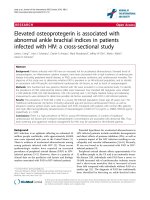
Figure 1 shows the risk to develop anemia during hospi-
tal stay for patients with favorable (mRS 0 to 3) versus
poor (mRS 4 to 6) functional outcomes. Poor functional
outcome at discharge was associated with a higher risk of
developing anemia throughout hospital stay (P = 0.029).
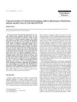
Figure 2 shows patients stratified by mRS category (1 to
6), excluding the patients that did not survive the first day
in hospital (n = 17). Mean HB levels were significantly
different between groups (14.0, 14.0, 13.4, 13.0, 11.9, and
11.2 mg/dl for mRS 1 to 6 respectively, F (5,172) = 7.71, P
< 0.001). Post-hoc analysis to compare the different out-
come groups separately was performed using Bonferroni
adjustment for multiple comparisons; P values are indi-
cated in Figure 2.
We performed a stepwise logistic regression model to
predict unfavorable outcome (mRS 4 to 6) at discharge,
including age, hemorrhage volume, baseline NIHSS, the
Table 1: Univariate analysis of functional outcome at discharge
Variable mRS 0 to 3
(n = 46)
mRS 4 to 6
(n = 150)
P
Age (years)
(mean, SD)
62.5 (13.4) 68.6 (13.1) 0.007
a
Sex (male, n, %) 29 (63.0) 101 (67.3) 0.136
c
Hemorrhage volume (ml)
(median, IQR)
8.6 (24.0) 30 (66.4) <0.001
b
Intraventricular hemorrhage extension (n, %) 5 (10.9) 72 (48.0) <0.001
c
NIHSS on admission
(median, IQR)
4 (3) 16 (23) <0.001
b
Admission HB (g/dl)
(mean, SD)
14.2 (1.6) 13.6 (2.0) 0.052
a
Mean HB (g/dl)
(mean, SD)
13.7 (1.8) 12.3 (2.0) <0.001
a
Nadir HB (g/dl)
(mean, SD)
13.0 (1.9) 11.5 (2.3) <0.001
a
Anemia during hospital stay
(n, %)
10 (21.7) 84 (56.0) <0.001
c
HB measurements (n)
(median, IQR)
4 (4.3) 5 (8.5) 0.22
b
Hospital stay (days)
(median, IQR)
7 (8) 6 (8) 0.32
b
ICU stay (n, %) 7 (15.2) 76 (50.7) <0.001
c
Mechanical ventilation (n, %) 2 (4.3) 55 (36.9) <0.001
c
RBC transfusion (n, %) 1 (2.2) 9 (6.0) 0.302
c
a
Student t-test
b
Mann-Whitney-U test
c
Chi squared test
HB, hemoglobin; IQR, interquartile range; mRS, modified Rankin Scale; NIHSS, National Institutes of Health Stroke Scale; RBC, red blood cell;
SD, standard deviation.
Diedler et al. Critical Care 2010, 14:R63
/>Page 4 of 8
presence of intraventricular hemorrhage extension, ICU
stay, the need for mechanical ventilation, RBC transfu-
sion and the mean HB level during hospital stay. In the
final model, only NIHSS on admission (odds ratio (OR)
1.30, 95% confidence interval (CI) 1.17 to 1.45, P < 0.001),
presence of intraventricular hemorrhage extension (OR
4.50, 95% CI 1.23 to 15.83, P = 0.019), age (per year, OR
1.04, 95% CI 1.01 to 1.08, P = 0.021) and the mean HB
(per mg/dl, OR 0.76, 95% CI 0.59 to 0.97, P = 0.027)
remained independent predictors for poor functional
outcome at discharge (Table 2).
In-hospital mortality was 22% (43 patients). In univari-
ate analysis, hemorrhage volume, intraventricular hemor-
rhage extension, admission status, duration of hospital
stay, the need for mechanical ventilation and ICU stay,
and admission HB levels were associated with in-hospital
mortality (Table 3). In the multivariable logistic regres-
sion model, NIHSS on admission (OR 1.17, 95% CI 1.10
to 1.24, P < 0.001), hemorrhage volume (per ml, OR 1.02,
95% CI 1.01 to 1.03, P = 0.003) and age (per year, OR 1.05,
95%CI 1.02 to 1.09, P = 0.006) remained independent pre-
dictors for in-hospital mortality (Table 4).
Outcome at three months was available for 176 (90%)
patients, 20 patients were lost to follow up. Stepwise
logistic regression revealed NIHSS on admission (OR
1.17, 95% CI 1.11 to 1.24, P < 0.001), age (per year, OR
1.08, 95% CI 1.04 to 1.12, P < 0.001) and the mean HB
(per mg/dl, OR 0.73, 95% CI 0.58 to 0.92, P = 0.007) as
independent predictors of outcome at three months
(Table 5).
Discussion
In the current study we found lower HB concentrations to
be an independent predictor of poor functional outcome
in patients suffering from supratentorial, non-traumatic
ICH. Anemia during hospital stay was more likely to
occur in patients with poor rather than with favorable
functional outcomes.
The design of the current study does not allow for the
determination of whether the relation between low HB
levels and functional outcome is causative. Based on the
current data, it cannot be excluded that anemia simply
represents a marker for severity of illness, rather than
causing additional brain injury. Critically ill patients usu-
ally receive more intravenous fluids, have more blood
drawn for laboratory tests, and more frequently undergo
invasive procedures, all factors leading to lower HB con-
centrations. However, we found lower HB concentrations
to be associated with poor outcome, independent of
stroke severity, which is the most powerful predictor of
outcome of acute ICH.
The absolute difference of mean HB levels between
both outcome groups was 1.4 mg/dl, corresponding to a
reduction of blood oxygen content of around 10% ((1.39 ×
HB concentration - O
2
Sat/100) + (0.003 × PaO
2
)). A
reduction of mean HB levels from 14 to 11.9 mg/dl as
found for patients with mRS 1 versus 6 respectively
Figure 1 Cumulative hazard to develop anaemia during hospital
stay for both outcome groups separately. During their hospital stay,
patients with poor functional outcome (modified Rankin score (mRS) 4
to 6) had a higher risk to develop anemia compared with those with fa-
vorable outcome at discharge.
Duration of hospital stay
6050403020100
Cum Hazard
5
4
3
2
1
0
mRS at discharge 4-6
mRS at discharge 0-3
Figure 2 Mean hemoglobin values and outcome at discharge.
Mean hemoglobin (HB) levels were significantly different between all
groups (F (5,172) = 7.71, P < 0.001). Significant differences between
group means are indicated by the bars (post-hoc analysis using Bonfer-
roni adjustment for multiple comparisons). mRS, modified Rankin
score.
mRS at discharge
654321
Mean HB (g/dl)
17,5
15,0
12,5
10,0
7,5
<0.01 <0.01
<0.01 <0.01
<0.05 <0.05
<0.05
Diedler et al. Critical Care 2010, 14:R63
/>Page 5 of 8
makes a difference in blood oxygen content of 20%,
roughly assuming similar partial pressure of arterial oxy-
gen (PaO
2
) levels. Previous studies in SAH patients report
a comparable magnitude of absolute differences in HB
levels between outcome groups [7,8]. Moreover, there is a
large body of literature including patients with TBI [10-
12], SAH [7-9], or ischemic stroke [15-17] suggesting that
anemia or even relative anemia may not be tolerated in
the setting of acute brain injury. In patients with acute
brain injury, physiological compensatory mechanisms
such as an increase in cerebral blood flow [18,19] may
fail, rendering them more vulnerable to fluctuations in
blood oxygen content. Possible mechanisms include
impairment of cerebrovascular autoregulation and meta-
Table 2: Final stepwise logistic regression model to predict poor outcome (mRS 4 to 6) at discharge. Included variables:
age, hemorrhage volume, NIHSS at admission, the presence of intraventricular blood, ICU stay, the need for mechanical
ventilation, RBC transfusion and the mean HB level during hospital stay
Variable Coefficient P OR (95% CI)
NIHSS on admission (per point) 0.261 <0.001 1.30 (1.17-1.45)
Presence of intraventricular
hemorrhage extension
1.503 0.019 4.50 (1.23-15.83)
Age (per year) 0.043 0.021 1.04 (1.01-1.08)
Mean HB (per mg/dl) -0.281 0.027 0.76 (0.59-0.97)
CI, confidence interval; HB, hemoglobin; mRS, modified Rankin Scale; NIHSS, National Institutes of Health Stroke Scale; OR, odds ratio; RBC,
red blood cell.
Table 3: Univariate analysis of factors associated with in-hospital mortality
Variable mRS 0 to 5
(n = 153)
mRS 6
(n = 43)
P
Age (years)
(mean, SD)
66.1 (13.8) 71.1 (11.1) 0.028
a
Sex (male, n, %) 100 (65.4) 30 (69.8) 0.589
c
Hemorrhage volume (ml)
(median, IQR)
17.1 (36.5) 88 (108.9) <0.001
b
Intraventricular hemorrhage extension (n, %) 49 (32.9) 28 (65.1) <0.001
c
NIHSS on admission
(median, IQR)
10 (12) 34 (10) <0.001
b
Admission HB (g/dl)
(mean, SD)
13.9 (1.8) 13.2 (2.3) 0.040
a
Mean HB (g/dl)
(mean, SD)
12.8 (2.0) 12.2 (2.4) 0.150
a
Nadir HB (g/dl)
(mean, SD)
11.9 (2.2) 11.7 (2.7) 0.516
a
Anemia during hospital stay
(n, %)
71 (46.4) 23 (53.5) 0.411
c
Hospital stay(days)
(median, IQR)
7 (9) 2 (43) <0.001
b
ICU stay (n, %) 53 (34.6) 30 (69.8) <0.001
c
Mechanical ventilation (n, %) 33 (21.6) 24 (57.1) <0.001
c
RBC transfusion (n, %) 7 (4.6) 3 (7) 0.527
c
a
Student t-test
b
Mann-Whitney-U test
c
Chi squared test
HB, hemoglobin; IQR, interquartile range; mRS, modified Rankin Scale; NIHSS, National Institutes of Health Stroke Scale; RBC, red blood cell;
SD, standard deviation.
Diedler et al. Critical Care 2010, 14:R63
/>Page 6 of 8
bolic disturbances of the injured brain [20]. Although
some studies have reported intact autoregulation in the
perihematomal region in the acute and subacute phase of
ICH [21,22], it has recently been demonstrated that
global cerebral autoregulation can be impaired in ICH
patients. Additionally, loss of cerebrovascular pressure
reactivity was associated with poor outcome [23]. Failure
of autoregulation may impede a compensatory increment
in cerebral blood flow as response to anemia and thus
render ICH patients more vulnerable to a decrease in
blood oxygen content.
In addition, animal studies provide evidence that ane-
mic hypoxia may exacerbate primary neurological injury
[24,25]. Although the concept of an ischemic penumbra
in hemorrhagic stroke has increasingly been challenged
[26,27], new hypotheses claim the presence of a meta-
bolic penumbra [28,29]. Recent studies do not point
towards a general lack of oxygen in the perihematomal
region, but rather to a changed metabolism [29] with low
rates of oxygen use [26,30]. However, the exact timing of
metabolic and inflammatory processes in the perihema-
tomal zone remains to be elucidated. Moreover, it is con-
ceivable that the metabolic and oxygen demand may
change during the course of the disease. So far, there are
no studies in humans suffering from acute ICH investi-
gating the effect of low HB levels on the perihematomal
zone. Only animal studies in dogs that were exposed to
chronic anemia before the induction of experimental ICH
provide evidence for an altered brain metabolism in ane-
mic animals [31,32]. Although chronic anemic hypoxia
promoted a decrease in metabolic demands and an
increase in cerebral blood flow, these adaptive responses
deteriorated at induction of ICH [31]. In the acute phase
of ICH, anemia was associated with an increased critical
threshold of brain oxygenation and progressive deteriora-
tion of cerebral hemodynamics.
Although we found an independent association
between low HB and worse functional outcomes, HB lev-
els during hospital stay were not predictive of in-hospital
mortality. Only hemorrhage volume, age and admission
status remained independent predictors for in-hospital
mortality in the multivariate model. This may be
explained by the overwhelming influence of hemorrhage
volume on mortality and the fact that 40% of patients
who died, died on the first day of hospital stay. Of inter-
est, admission HB levels differed significantly between
both groups in an univariate analysis. This finding is in
line with a recent study including almost 700 patients
with non-traumatic ICH investigating the role of anemia
on admission (day 1) on the clinical course of acute ICH
[33]. Although patients with anemia on admission (25.8%
of patients) were at higher risk of death at 30 days in uni-
variate analysis, this effect did not persist in a multivari-
ate model including hemorrhage volume. Interestingly,
the authors report that the presence of anemia on admis-
sion was associated with larger ICH volume and thereby
hypothesize that the presence of anemia may contribute
to hemorrhage growth. Another explanation may be that
admission HB levels rather are a marker for poor physio-
logical status on admission. Unfortunately, scores for
Table 4: Final stepwise logistic regression model to predict in-hospital mortality. Included variables: age, hemorrhage
volume, NIHSS at admission, the presence of intraventricular blood, ICU stay, the need for mechanical ventilation, RBC
transfusion and the admission HB
Variable Coefficient P OR (95% CI)
NIHSS on admission (per
point)
0.109 <0.001 1.17 (1.11-1.24)
Hemorrhage volume (per ml) 0.015 0.003 1.02 (1.01-1.03)
Age (per year) 0.052 0.006 1.05 (1.02-1.09)
CI, confidence interval; HB, hemoglobin; NIHSS, National Institutes of Health Stroke Scale; OR, odds ratio; RBC, red blood cell.
Table 5: Final stepwise logistic regression model to predict poor outcome (mRS 4 to 6) at three months. Included variables:
age, hemorrhage volume, NIHSS at admission, the presence of intraventricular blood, ICU stay, the need for mechanical
ventilation, RBC transfusion and the mean HB level during hospital stay)
Variable Coefficient P OR (95% CI)
NIHSS on admission (per point) 0.157 <0.001 1.17 (1.11-1.24)
Age (per year) 0.076 <0.001 1.08 (1.04-1.12)
Mean HB (per mg/dl) -0.319 0.007 0.73 (0.58-0.92)
CI, confidence interval; HB, hemoglobin; NIHSS, National Institutes of Health Stroke Scale; OR, odds ratio; RBC, red blood cell.
Diedler et al. Critical Care 2010, 14:R63
/>Page 7 of 8
physiologic illness such as the acute physiology and
chronic health evaluation (APACHE) II score were not
available due to the retrospective design of our study.
The main limitations of the current study include the
small number of transfused patients and the retrospec-
tive, observational design that does not shed light on the
underlying metabolic processes. However, while the latter
was beyond the scope of the current study, further studies
invasively assessing the parenchymal metabolic effects of
anemia and RBC transfusion in ICH patients seem justi-
fied. In the current study, only 10 (5.1%) patients received
RBC transfusions during their hospital stay. RBC transfu-
sion was included as a variable in multivariable models;
however, the number of transfused patients was too low
to provide solid data on the effect of RBC transfusion on
outcome parameters. In order to exclude the possibility
that poor outcomes were primarily related to transfusion,
rather than anemia, we repeated our analysis after
excluding the 10 transfused patients from the multivari-
ate models. In this repeat analysis, mean HB did not
remain an independent predictor for poor outcome at
discharge but stayed an independent predictor in the
model for outcome at three months [see Additional file
1]. However, evidence exists from previous studies
including patients with TBI [10,12] or SAH [34,35] that
despite some putative beneficial physiological effects,
RBC transfusion was associated with additional risks and
poorer outcome. In addition, Zygun and colleagues
recently have assessed the effect of RBC transfusion on
cerebral oxygenation and metabolism in TBI patients
[36]. They report that transfusion of RBC resulted in
improved brain tissue oxygenation, but without notice-
able effect on cerebral metabolism as measured by lac-
tate-pyuvate ratio. There is currently no data available for
ICH patients.
Conclusions
In summary, in the current study we found an association
between low HB and poor functional outcome in patients
with non-traumatic ICH, as was previously reported for
patients with SAH, TBI and ischemic stroke. Although
none of the studies in brain-injured patients has so far
proven a causative relation between anemia and poor
outcome, physiological and observational studies provide
evidence for possible detrimental effects of anemia on
brain metabolism. However, the potential risk of anemia
must be balanced against the potential risk of harm from
allogenic RBC infusion. Further trials are needed to
investigate the local metabolic effects of anemia and RBC
transfusion in ICH patients.
Key messages
• Poor functional outcome at discharge and at 90 days
was associated with lower mean HB levels during
hospital stay in patients with non-traumatic,
supratentorial ICH.
• Based on the currently available data it could not be
elucidated if the presence of anemia promotes further
brain injury or if it represents a marker of severe ill-
ness.
• Further trials are needed to investigate if RBC trans-
fusion in acute ICH may lead to improved outcome.
Additional material
Abbreviations
ANOVA: analysis of variance; APACHE: acute physiology and chronic health
evaluation; CI: confidence interval; CT: computed tomography; HB: hemoglo-
bin; ICH: intracerebral hemorrhage; mRS: modified Rankin score; NIHSS:
National Institute of Health Stroke Scale; OR: odds ratio; PaO
2
: partial pressure
of arterial oxygen; RBC: red blood cell; SAH: subarachnoid hemorrhage; SD:
standard deviation; TBI: traumatic brain injury.
Competing interests
The authors declare that they have no competing interests.
Authors' contributions
JD and MS contributed equally and planned and designed the study and per-
formed the statistical analysis. JD wrote the first draft of the manuscript. PH
performed data acquisition. MS, KH, MS, LK, JB and SP critically revised the
manuscript. TS contributed to conception of the study and critically revised
the manuscript.
Author Details
1
Department of Neurology, University of Heidelberg, Im Neuenheimer Feld
400, 69120 Heidelberg, Germany and
2
Department of Neurology, Comenius
University, Mickiewiczova 13, 813 69 Bratislava, Slovakia
References
1. Steiner T, Diringer MN, Schneider D, Mayer SA, Begtrup K, Broderick J,
Skolnick BE, Davis SM: Dynamics of intraventricular hemorrhage in
patients with spontaneous intracerebral hemorrhage: risk factors,
clinical impact, and effect of hemostatic therapy with recombinant
activated factor VII. Neurosurgery 2006, 59:767-773. discussion 773-764
2. Hemphill JC, Bonovich DC, Besmertis L, Manley GT, Johnston SC: The ICH
score: a simple, reliable grading scale for intracerebral hemorrhage.
Stroke 2001, 32:891-897.
3. Gujjar AR, Deibert E, Manno EM, Duff S, Diringer MN: Mechanical
ventilation for ischemic stroke and intracerebral hemorrhage:
indications, timing, and outcome. Neurology 1998, 51:447-451.
4. Diringer MN, Edwards DF: Admission to a neurologic/neurosurgical
intensive care unit is associated with reduced mortality rate after
intracerebral hemorrhage. Crit Care Med 2001, 29:635-640.
5. Corwin HL, Gettinger A, Pearl RG, Fink MP, Levy MM, Abraham E,
MacIntyre NR, Shabot MM, Duh MS, Shapiro MJ: The CRIT Study: Anemia
and blood transfusion in the critically ill current clinical practice in the
United States. Crit Care Med 2004, 32:39-52.
6. Vincent JL, Baron JF, Reinhart K, Gattinoni L, Thijs L, Webb A, Meier-
Hellmann A, Nollet G, Peres-Bota D: Anemia and blood transfusion in
critically ill patients. Jama 2002, 288:1499-1507.
7. Naidech AM, Jovanovic B, Wartenberg KE, Parra A, Ostapkovich N,
Connolly ES, Mayer SA, Commichau C: Higher hemoglobin is associated
with improved outcome after subarachnoid hemorrhage. Crit Care Med
2007, 35:2383-2389.
Additional file 1 Additional logistic regression models. Logistic regres-
sion models after exclusion of 10 patients who had received red blood cell
transfusions.
Received: 22 October 2009 Revised: 18 February 2010
Accepted: 14 April 2010 Published: 14 April 2010
This article is available from: 2010 Diedler et al.; licensee BioMed Central Ltd. This is an open access article distributed under the terms of the Creative Commons A ttribution License ( which permits unrestricted use, distribution, and reproduction in any medium, provided the original work is properly cited.Critical Care 2010, 14:R63
Diedler et al. Critical Care 2010, 14:R63
/>Page 8 of 8
8. Kramer AH, Zygun DA, Bleck TP, Dumont AS, Kassell NF, Nathan B:
Relationship between hemoglobin concentrations and outcomes
across subgroups of patients with aneurysmal subarachnoid
hemorrhage. Neurocrit Care 2009, 10:157-165.
9. Wartenberg KE, Schmidt JM, Claassen J, Temes RE, Frontera JA,
Ostapkovich N, Parra A, Connolly ES, Mayer SA: Impact of medical
complications on outcome after subarachnoid hemorrhage. Crit Care
Med 2006, 34:617-623. quiz 624
10. Carlson AP, Schermer CR, Lu SW: Retrospective evaluation of anemia
and transfusion in traumatic brain injury. J Trauma 2006, 61:567-571.
11. Van Beek JG, Mushkudiani NA, Steyerberg EW, Butcher I, McHugh GS, Lu J,
Marmarou A, Murray GD, Maas AI: Prognostic value of admission
laboratory parameters in traumatic brain injury: results from the
IMPACT study. J Neurotrauma 2007, 24:315-328.
12. Salim A, Hadjizacharia P, DuBose J, Brown C, Inaba K, Chan L, Margulies DR:
Role of anemia in traumatic brain injury. J Am Coll Surg 2008,
207:398-406.
13. Broderick JP, Brott T, Duldner J, Tomsick T, Huster G: Volume of
intracerebral hemorrhage: a powerful and easy-to-use predictor of 30-
day mortality. Stroke 1993, 24:987-993.
14. Beutler E, Waalen J: The definition of anemia: what is the lower limit of
normal of the blood hemoglobin concentration? Blood 2006,
107:1747-1750.
15. Wade JP, Taylor DW, Barnett HJ, Hachinski VC: Hemoglobin
concentration and prognosis in symptomatic obstructive
cerebrovascular disease. Stroke 1987, 18:68-71.
16. Huang WY, Chen IC, Meng L, Weng WC, Peng TI: The influence of anemia
on clinical presentation and outcome of patients with first-ever
atherosclerosis-related ischemic stroke. J Clin Neurosci 2009,
16:645-649.
17. Nybo M, Kristensen SR, Mickley H, Jensen JK: The influence of anaemia
on stroke prognosis and its relation to N-terminal pro-brain natriuretic
peptide. Eur J Neurol 2007, 14:477-482.
18. van Bommel J, Trouwborst A, Schwarte L, Siegemund M, Ince C, Henny Ch
P: Intestinal and cerebral oxygenation during severe isovolemic
hemodilution and subsequent hyperoxic ventilation in a pig model.
Anesthesiology 2002, 97:660-670.
19. Tomiyama Y, Jansen K, Brian JE Jr, Todd MM: Hemodilution, cerebral O2
delivery, and cerebral blood flow: a study using hyperbaric
oxygenation. Am J Physiol 1999, 276:H1190-1196.
20. Vespa PM: The implications of cerebral ischemia and metabolic
dysfunction for treatment strategies in neurointensive care. Curr Opin
Crit Care 2006, 12:119-123.
21. Powers WJ, Zazulia AR, Videen TO, Adams RE, Yundt KD, Aiyagari V, Grubb
RL Jr, Diringer MN: Autoregulation of cerebral blood flow surrounding
acute (6 to 22 hours) intracerebral hemorrhage. Neurology 2001,
57:18-24.
22. Kuwata N, Kuroda K, Funayama M, Sato N, Kubo N, Ogawa A:
Dysautoregulation in patients with hypertensive intracerebral
hemorrhage. A SPECT study. Neurosurg Rev 1995, 18:237-245.
23. Diedler J, Sykora M, Rupp A, Poli S, Karpel-Massler G, Sakowitz O, Steiner T:
Impaired cerebral vasomotor activity in spontaneous intracerebral
hemorrhage. Stroke 2009, 40:815-819.
24. Todd MM, Wu B, Warner DS: The hemispheric cerebrovascular response
to hemodilution is attenuated by a focal cryogenic brain injury. J
Neurotrauma 1994, 11:149-160.
25. Reasoner DK, Ryu KH, Hindman BJ, Cutkomp J, Smith T: Marked
hemodilution increases neurologic injury after focal cerebral ischemia
in rabbits. Anesth Analg 1996, 82:61-67.
26. Zazulia AR, Diringer MN, Videen TO, Adams RE, Yundt K, Aiyagari V, Grubb
RL Jr, Powers WJ: Hypoperfusion without ischemia surrounding acute
intracerebral hemorrhage. J Cereb Blood Flow Metab 2001, 21:804-810.
27. Miller CM, Vespa PM, McArthur DL, Hirt D, Etchepare M: Frameless
stereotactic aspiration and thrombolysis of deep intracerebral
hemorrhage is associated with reduced levels of extracellular cerebral
glutamate and unchanged lactate pyruvate ratios. Neurocrit Care 2007,
6:22-29.
28. Vespa PM: Metabolic penumbra in intracerebral hemorrhage. Stroke
2009, 40:1547-1548.
29. Zazulia AR, Videen TO, Powers WJ: Transient focal increase in
perihematomal glucose metabolism after acute human intracerebral
hemorrhage. Stroke 2009, 40:1638-1643.
30. Kim-Han JS, Kopp SJ, Dugan LL, Diringer MN: Perihematomal
mitochondrial dysfunction after intracerebral hemorrhage. Stroke
2006, 37:2457-2462.
31. Lee EJ, Hung YC: Marked anemic hypoxia deteriorates cerebral
hemodynamics and brain metabolism during massive intracerebral
hemorrhage. J Neurol Sci 2001, 190:3-10.
32. Lee EJ, Hung YC, Lee MY: Anemic hypoxia in moderate intracerebral
hemorrhage: the alterations of cerebral hemodynamics and brain
metabolism. J Neurol Sci 1999, 164:117-123.
33. Kumar MA, Rost NS, Snider RW, Chanderraj R, Greenberg SM, Smith EE,
Rosand J: Anemia and hematoma volume in acute intracerebral
hemorrhage. Crit Care Med 2009, 37:1442-1447.
34. Tseng MY, Hutchinson PJ, Kirkpatrick PJ: Effects of fluid therapy following
aneurysmal subarachnoid haemorrhage: a prospective clinical study.
Br J Neurosurg 2008, 22:257-268.
35. Kramer AH, Gurka MJ, Nathan B, Dumont AS, Kassell NF, Bleck TP:
Complications associated with anemia and blood transfusion in
patients with aneurysmal subarachnoid hemorrhage. Crit Care Med
2008, 36:2070-2075.
36. Zygun DA, Nortje J, Hutchinson PJ, Timofeev I, Menon DK, Gupta AK: The
effect of red blood cell transfusion on cerebral oxygenation and
metabolism after severe traumatic brain injury. Crit Care Med 2009,
37:1074-1078.
doi: 10.1186/cc8961
Cite this article as: Diedler et al., Low hemoglobin is associated with poor
functional outcome after non-traumatic, supratentorial intracerebral hemor-
rhage Critical Care 2010, 14:R63