Báo cáo y học: "Preoperative diastolic function predicts the onset of left ventricular dysfunction following aortic valve replacement in high-risk patients with aortic stenosis" docx
Bạn đang xem bản rút gọn của tài liệu. Xem và tải ngay bản đầy đủ của tài liệu tại đây (1.21 MB, 11 trang )
Licker et al. Critical Care 2010, 14:R101
/>Open Access
RESEARCH
© 2010 Licker et al.; licensee BioMed Central Ltd. This is an open access article distributed under the terms of the Creative Commons
Attribution License ( which permits unrestricted use, distribution, and reproduction in
any medium, provided the original work is properly cited.
Research
Preoperative diastolic function predicts the onset
of left ventricular dysfunction following aortic
valve replacement in high-risk patients with aortic
stenosis
Marc Licker*
1
, Mustafa Cikirikcioglu
2
, Cidgem Inan
1
, Vanessa Cartier
1
, Afksendyios Kalangos
2
, Thomas Theologou
2
,
Tiziano Cassina
3
and John Diaper
1
Abstract
Introduction: Left ventricular (LV) dysfunction frequently occurs after cardiac surgery, requiring inotropic treatment
and/or mechanical circulatory support. In this study, we aimed to identify clinical, surgical and echocardiographic
factors that are associated with LV dysfunction during weaning from cardiopulmonary bypass (CPB) in high-risk
patients undergoing valve replacement for aortic stenosis.
Methods: Perioperative data were prospectively collected in 108 surgical candidates with an expected operative
mortality ≥9%. All anesthetic and surgical techniques were standardized. Reduced LV systolic function was defined by
an ejection fraction <40%. Diastolic function of the LV was assessed using standard Doppler-derived parameters, tissue
Doppler Imaging (TDI) and transmitral flow propagation velocity (Vp).
Results: Doppler-derived pulmonary flow indices and TDI could not be obtained in 14 patients. In the remaining 94
patients, poor systolic LV was documented in 14% (n = 12) and diastolic dysfunction in 84% of patients (n = 89), all of
whom had Vp <50 cm/s. During weaning from CPB, 38 patients (40%) required inotropic and/or mechanical circulatory
support. By multivariate regression analysis, we identified three independent predictors of LV systolic dysfunction: age
(Odds ratio [OR] = 1.11; 95% confidence interval (CI), 1.01 to 1.22), aortic clamping time (OR = 1.04; 95% CI, 1.00 to 1.08)
and Vp (OR = 0.65; 95% CI, 0.52 to 0.81). Among echocardiographic measurements, Vp was found to be superior in
terms of prognostic value and reliability. The best cut-off value for Vp to predict LV dysfunction was 40 cm/s (sensitivity
of 72% and specificity 94%). Patients who experienced LV dysfunction presented higher in-hospital mortality (18.4% vs.
3.6% in patients without LV dysfunction, P = 0.044) and an increased incidence of serious cardiac events (81.6 vs. 28.6%,
P < 0.001).
Conclusions: This study provides the first evidence that, besides advanced age and prolonged myocardial ischemic
time, LV diastolic dysfunction characterized by Vp ≤ 40 cm/sec identifies patients who will require cardiovascular
support following valve replacement for aortic stenosis.
Introduction
More than 200,000 aortic valve replacements are per-
formed annually worldwide and this number will con-
tinue to increase with the aging population. Over the last
two decades, the operative mortality rate has steadily
declined from 10% to 4% along with improvements in
surgical and anesthetic techniques [1-3]. However, left
ventricular (LV) dysfunction requiring the administration
of inotropic drugs often occurs after separation from car-
diopulmonary bypass (CPB) and has been associated with
prolonged ICU and hospital stay [3,4]. Although this
myocardial stunning usually resolves within 48 hours, it
may lead to low cardiac output syndrome that has
become the leading cause of postoperative death [5,6].
* Correspondence:
1
Faculty of Medicine (University of Geneva) and Department of
Anaesthesiology, Pharmacology and Intensive Care, University Hospital, rue
Gabrielle-Perret-Gentil, CH-1211 Geneva 14, Switzerland
Full list of author information is available at the end of the article
Licker et al. Critical Care 2010, 14:R101
/>Page 2 of 11
In large cohorts of patients undergoing cardiac surgery,
post-CPB LV dysfunction has been linked to age, female
gender, history of heart failure, recent myocardial infarct,
low LV ejection fraction, prolonged aortic cross-clamping
and complexity of surgery [7-11]. More recently, echocar-
diographic markers of preoperative LV diastolic dysfunc-
tion have been associated with difficulties in weaning
patients from CPB [12,13].
Although clinical signs (for example, pulmonary con-
gestion, New York Heart Association [NYHA] classes)
and markers of systolic LV function (for example, LV
ejection fraction) have been studied extensively and
incorporated in scoring algorithms for predicting periop-
erative risk, the prognostic value of diastolic dysfunction
assessed by transoesophageal echocardiography (TEE)
has not been examined in patients undergoing aortic
valve replacement [1,2,5,14,15]. Besides pulsed-wave
Doppler measurements of mitral inflow and pulmonary
venous flow, evaluation of diastolic function has recently
been improved with color M-mode transmitral flow
propagation velocity (Vp) and mitral valve annular veloci-
ties recorded by tissue Doppler imaging (TDI) [16,17].
The main purpose of this study was to identify predic-
tors of LV dysfunction in high-risk patients with aortic
stenosis undergoing valvular replacement. Secondarily,
we analyzed different Doppler parameters of diastolic
function regarding their ability to predict post-CPB LV
dysfunction.
Materials and methods
Study design and settings
This prospective cohort study was conducted in a tertiary
reference center, from January 2006 to December 2008.
The study was approved by the Institutional Research
Board of the University Hospital of Geneva and informed
consent was obtained from each patient with severe aor-
tic valvular stenosis who met the eligibility criteria. The
Bernstein-Parsonnet algorithm was used to assess the
operative risk of mortality [18]. During the study period,
108 patients were selected among a cohort of 145 surgical
candidates undergoing elective aortic valve replacement,
either isolated or combined with coronary artery bypass
grafting or aortic root replacement. A predicted risk of
mortality exceeding 9% was considered as an entry crite-
ria. Exclusion criteria consisted of atrial fibrillation or
flutter, implanted pacemaker, severe mitral stenosis or
regurgitation, severe pulmonary hypertension (mean pul-
monary artery pressure ≥45 mmHg), moderate-to-severe
valvular aortic insufficiency and preoperative inotropic
or ventilatory support. Patients were secondarily
excluded if poor image quality precluded echocardio-
graphic measurements. All patients were operated on by
one of three board certified cardiac surgeons and were
managed by the same team of cardiothoracic anesthesiol-
ogists.
Perioperative patient management
The usual medications were continued on the morning of
the procedure, except diuretics and angiotensin-convert-
ing enzyme inhibitors or angiotensin II antagonists that
were interrupted one day before. In the operating theatre,
all patients were equipped with a noninvasive oscillomet-
ric monitor (brachial artery pressure), a radial arterial
catheter, a central venous line and a bispectral monitor of
the electroencephalogram (BIS Aspect Medical Systems
A-2000 XP, Newton, Maryland, USA). Anesthesia man-
agement consisted of intra-thecal morphine, low doses of
intravenous sufentanyl and an infusion of propofol to tar-
get BIS values between 40 and 60. Cardiac precondition-
ing was also provided with inhaled isoflurane (1% to
1.5%) before CPB.
A TEE probe (T6210 Omniplane II Philips Medical Sys-
tem, Andover, MA, USA) was introduced after anesthesia
induction and images were digitally acquired before CPB
and stored on a Philips Sonos 5500 Ultrasound Imaging
system (Philips Medical Systems).
After full heparinization, normothermic CPB was insti-
tuted with a nonpulsatile flow (2.2 to 2.5 L/minute/m
2
)
and alpha-stat control for acid-base management. The
circuit and the membrane oxygenator were primed with 2
L of normal saline solution and mean arterial pressure
(MAP) was maintained between 50 and 70 mmHg with
vasoactive medications as necessary. During aortic cross-
clamping, myocardial protection was achieved by inter-
mittent antegrade infusion of cold blood. The aortic valve
prosthesis (Carpentier-Edwards Perimount; Jt. Jude Med-
ical Inc. Minneapolis, Minnesota, USA) was implanted in
the supra-annular position with interrupted mattress
sutures. All patients received tranexamic acid (20 mg/kg)
and the transfusion threshold was a hematocrit less than
18 to 20% during CPB and less than 25% before/after
CPB.
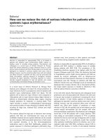
At the end of the procedure, weaning from CPB was
guided by TEE assessment and hemodynamic measure-
ments. After de-airing the cardiac cavities and resump-
tion of mechanical ventilation, the pump flow was
gradually reduced allowing filling of the cardiac cham-
bers. In addition to fluid loading, electrical atrio-ventric-
ular pacing, vasopressors and inotropes drugs as well as
intra-aortic balloon pump (IABP) were eventually intro-
duced to target the specific hemodynamic endoints: LV
end-diastolic diameter (up to preoperative values or 2.2
and 2.8 cm/m
2
), MAP between 65 and 100 mmHg and
heart rate between 70 and 100 beats/minute (see Figure
1).
The investigators performing the TEE were not
involved in any therapeutic decision during the weaning
Licker et al. Critical Care 2010, 14:R101
/>Page 3 of 11
process and the attending anesthesiologist in charge of
the patient was blinded to the diastolic measurements.
Pulmonary artery catheters were inserted in patients
receiving inotropic support at the admission on the
Intensive Care Unit (ICU).
Study endpoints
The diagnostic criteria for post-CPB LV dysfunction was
based on the need of inotropic support for at least two
hours (dobutamine ≥5 mcg/kg/min, epinephrine >0.05
mcg/kg/min, milrinone >0.25 mcg/kg/min, norepineph-
rine >0.02 mcg/kg/min) in the presence of low MAP (<60
mmHg ascertained by both invasive and noninvasive
pressure monitors) and with persistent, new or worsen-
ing LV functional impairment (for example, FAC (frac-
tional area change) <40%). Secondary outcome variables
were any postoperative cardiac adverse event occurring
in the ICU such as myocardial infarct (troponin-I ≥1.5
ng/ml associated with new Q waves or ST segment
abnormalities on the ECG, or with coronary artery inter-
vention), supra-ventricular or ventricular arrhythmias
(requiring anti-arrhythmic drugs or electrical cardiover-
sion) and low cardiac output syndrome (cardiac index
<2.2 L/min/m
2
, need for inotropic and/or IABP support
to maintain MAP >65 mmHg).
Measurements
During primary hospitalization, data related to patient
demographic information, comorbidities, current medi-
cations, intraoperative TEE examination, indexed effec-
tive orifice area [19], anesthetic and surgical management
as well as postoperative cardiac outcome were prospec-
tively collected on a case report form and entered in a
dedicated database.
A comprehensive TEE examination was performed
before CPB using two-dimensional, M-mode, pulsed
Doppler and TDI to assess systolic and diastolic LV func-
tion. In the transgastric short axis view, posterior wall
thickness (PWT), LV end-diastolic and end-systolic areas
(EDA and ESA, respectively) were measured. FAC of the
LV was computed as (LVEDA -LVESA)/LVEDA. From a
mid-esophageal four-chamber view, peak early (E) and
late (A) mitral inflow velocities, deceleration time (DT)
and isovolumic relaxation time (IVRT) were derived from
recordings obtained with the pulsed Doppler sample vol-
ume positioned at the tip of the mitral leaflets. Peak sys-
Figure 1 Weaning protocol from Cardio-Pulmonary-Bypass.
Licker et al. Critical Care 2010, 14:R101
/>Page 4 of 11
tolic (S), diastolic (D) and atrial reversal velocities (Ar)
were measured with the pulsed Doppler sample volume
positioned within 1 to 2 cm of the left upper pulmonary
vein. Thereafter, the TDI function was activated for
recording early and late diastolic velocities of the mitral
annulus (E' and A', respectively) by positioning the 5-mm
sample volume within the septal and lateral insertion
sites of the mitral leaflets to cover the longitudinal excur-
sion of the mitral annulus. Finally, a color M-mode map
was displayed from a mid-oesophageal four-chamber
view, to obtain the longest column of flow from the mitral
annulus to the apex. The M-mode cursor was aligned
through the center, parallel to the transmitral inflow and
a clear propagation wave front was obtained by adjusting
the Nyquist limit and baseline shift. Vp was defined as the
slope of the first aliasing velocity during early filling, mea-
sured from the mitral valve opening to 4 cm into the LV
cavity.
Cardiac stroke volume was calculated as the flow sur-
face area multiplied by the velocity time integral through
the LV outflow tract obtained by pulsed wave Doppler.
Cardiac index (CI) was calculated as the product of SV
and HR divided by body surface area. All recorded values
were averaged from three consecutive beats.
Poor systolic LV function was considered if the LV ejec-
tion was <40% on the preoperative transthoracic echocar-
diographic examination. According to the working group
of the European Association of Echocardiography and the
American Society of Echocardiography, LV diastolic
function was graded into four classes: normal (E/A > 0.8,
DT < 200 ms, and E'/A' > 1 or S/D 1 to 1.5), impaired
relaxation (E/A < 0.8, DT > 200 ms, IVRT ≥ 100 ms and
E'/A' <1 or S/D >1.5), pseudo-normalization (E/A = 1 to 2,
DT = 150 to 200 ms, and E'/A' <1 or S/D <1.2), and
restrictive pattern (E/A >2, DT <150 ms and E'/A' <1 or S/
D <0.8) [19].
To test the intra- and interobserver variabilities, E and
A, E' and A' as well as Vp were measured twice by two
independent operators, in 10 randomly selected cases.
Statistical analysis
Perioperative clinical, surgical and echocardiographic
characteristics of patients with and without post-CPB LV
dysfunction were compared with the χ
2
test for categori-
cal variables (expressed in percentage) and the Student t
test (normal distribution) or Wilcoxon rank test (non-
Gaussian distribution) for continuous variables (all
expressed as mean ± SD).
Variables that had a univariate probability value <0.20
or those judged to be clinically important were selected
for inclusion in a logistic regression model by stepwise
selection. To avoid multi-colinearity, only one variable
was retained in a set of variables with a correlation coeffi-
cient greater than 0.5. Independent predictors of LV dys-
function and factor-adjusted odds ratios (ORs) with 95%
confidence interval (CI) were calculated. Model discrimi-
nation was evaluated by the area under the receiver-oper-
ator-characteristic (ROC) curve, and calibration was
assessed with the Hosmer-Lemeshow goodness-of-fit sta-
tistic. Receiver operating characteristics (ROC) curves
were constructed to determine the best cut-off of
echocardiographic parameters (with P < 0.2) to predict
post-CPB LV dysfunction. All analyses were performed
using SPSS software (version 14.0 for Microsoft Win-
dows; SPSS, Chicago, IL, USA) and statistical significance
was specified as a two-tailed type I error (P value) set
below the 0.05 level.
Results
Over a three-year period, 108 high-risk patients under-
went valve replacement for severe aortic stenosis and 14
were excluded since Doppler-derived pulmonary flow
indices and TDI could not be obtained (9 and 11 patients,
respectively). In the remaining 94 patients, all presented
LV hypertrophy (PWT >11 mm), poor systolic LV func-
tion was found in 14% of patients (n = 12) whereas dia-
stolic dysfunction was diagnosed in 84% of patients (n =
89), all of whom had Vp < 50 cm/s. Regarding echocar-
diographic measurements, intra-and interobserver vari-
abilities were lowest for Vp and highest for E' and A'
measurements (Table 1).
During weaning from CPB, LV dysfunction occurred in
38 patients (40.4%). Inotropic support consisted in the
administration of dobutamine (5.6 ± 2.7 mcg/kg/h, over
10 ± 5 hours), epinephrine (0.52 ± 0.41 mcg/kg/h over 6 ±
3 hours), norepinephrine (0.08 ± 0.04 mcg/kg/h over 12 ±
7 hours) and/or milrinone (0.27 ± 0.14 mcg/kg/h over 6 ±
2 hours). Five patients were also treated with an IABP in
combination with inotropes. As shown in Table 2, MAP
and CI were significantly lower in patients with post-CPB
LV dysfunction. Of the 31 preoperative and intraopera-
tive variables subjected to univariate analysis, eight dem-
onstrated a significant association with the occurrence of
post-CPB LV dysfunction (Table 3). Patients with post-
CPB LV dysfunction were significantly older, they had
lower LV ejection fraction, more severe grades of LV dia-
stolic dysfunction, lower Vp as well as prolonged duration
of CPB and aortic clamping.
Stepwise logistic regression analyses identified three
independent predictors of LV dysfunction: age (OR =
1.11; 95% CI, 1.01 to 1.22), aortic clamping time (OR =
1.04; 95% CI, 1.00 to 1.08) and Vp (OR = 0.65; 95% CI,
0.52 to 0.81). This multivariate model for predicting LV
dysfunction was robust, with an area under the ROC
curve of 0.96 (95% CI, 0.89 to 0.99) and a Hosmer-Leme-
show goodness-of-fit probability value of 0.49 indicating
good model calibration and discrimination. Substitution
of aortic clamping time for CPB time and diastolic classes
Licker et al. Critical Care 2010, 14:R101
/>Page 5 of 11
(1 to 4) for Vp, did not improve the area under the ROC
curve. There was no evidence that additional covariates
would improve the model (P = 0.21 by the Wald link spec-
ification test).
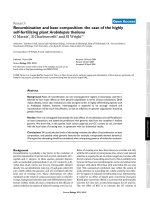
As shown in Figure 2, the best cut-off value for Vp to
predict LV dysfunction was 40 cm/s as it maximized both
sensitivity (73%; 95% CI, 55% to 87%) and specificity
(96%; 95% CI, 87% to 99%).
The expected mortality of the whole cohort was 22%
whereas the observed mortality was only 10.6%. As
shown in Table 4, compared with patients without post-
CPB LV dysfunction, those experiencing LV dysfunction
presented higher in-hospital mortality (18.4% vs. 3.6%, P
= 0.044) and an increased incidence of serious cardiac
events (81.6 vs. 28.6%, P < 0.001). These patients also
required prolonged mechanical ventilation and longer
stay in the ICU and in the hospital.
The incidence of LV dysfunction and cardiac complica-
tions increased significantly with the severity of diastolic
dysfunction, particularly in patients with a restrictive fill-
ing pattern and those with Vp less than 40 cm/s (Figure
3). Noteworthy, LV dysfunction was observed in 28 out of
30 patients (90%) with low Vp (≤40 cm/sec) as opposed to
7 out of 64 patients with normal-to-high Vp.
Discussion
In this prospective study, 40% of high-risk patients under-
going aortic valve replacement required inotropic sup-
Table 1: Intra- and interobserver characteristics
Characteristics Intra-observer variability Inter-observer variability
E wave 7.8 8.7
A wave 8.1 9.4
Deceleration time 8.9 9.6
PV S 7.1 8.2
PV D 8.3 9.1
E' wave lateral 7.9 8.8
A' wave lateral 8.1 9.2
E' wave septal 9.4 11.6
A' wave septal 9.7 10.9
Vp 3.2 4.8
PV S, peak systolic velocity of pulmonary venous flow; PV D, peak diastolic velocity of pulmonary venous flow; E, peak early mitral inflow; A,
late mitral inflow; DT, deceleration time; E' and A', early and late diastolic annular velocities, Vp, transmitral flow velocity
Table 2: Hemodynamic in patients with and without post-CPB left ventricular dysfunction
No LV dysfunction (n = 56) With LV dysfunction (n = 38) P value
Mean Arterial Pressure, mmHg
Before CPB (10 minutes) 93 (12) 90 (15) 0.847
After CPB (10 minutes) 82 (14) 68 (16)* 0.012
Heart Rate, b/min
Before CPB (10 minutes) 70 (8) 73 (9) 0.912
After CPB (10 minutes) 78 (13) 82(14) 0.634
Central Venous Pressure, cm H
2
O
Before CPB (10 minutes) 6 (3) 7 (4) 0.879
After CPB (10 minutes) 8 (4) 9 (5) 0.953
Cardiac Index, L/min/m
2
Before CPB (10 minutes) 2.4 (1.0) 2.2 (1.3) 0.597
After CPB (10 minutes) 3.6 (1.3) 2.1 (0.9)* < 0.001
LV, left ventricular; CPB, cardiopulmonary bypass.
*P < 0.05, between the two groups; χ2 test with Yates correction or unpaired Student t test.
Licker et al. Critical Care 2010, 14:R101
/>Page 6 of 11
Table 3: Distribution of perioperative variables according to the presence of post-CPB left ventricular dysfunction
No LV dysfunction (n = 56) With LV dysfunction (n = 38) P value
Preoperative clinical and biological variables
Age (y) 66 (9) 74 (10) 0.004
Male Gender (%) 61.8 66.7 0.288
Body Mass Index (kg/m2)
28 (5) 27 (6) 0.281
Hypertension (%) 85.1 82.3 0.654
Diabetes Mellitus (%) 30.9 27.5 0.745
Coronary Artery Disease (%) 29.1 38.5 0.412
Peripheral Vascular Disease (%) 12.1 10.9 0.511
Dyslipemia (%) 71.8 54.5 0.098
Renal Insufficiency1 (%)
3.6 5.2 0.532
Anemia2 (%)
7.1 10.1 0.789
Beta-blockers (%) 49.1 61.5 0.295
ACE Inhibitors or AII antagonists (%) 20.1 35.9 0.352
Calcium channel antagonists (%) 23.6 33.3 0.101
Diuretics (%) 21.9 17.9 0.796
Nitrate (%) 12.7 17.9 0.562
LV Ejection Fraction (%) 56 (8) 49 (11) 0.001
Parsonnet score (u) 15 (9) 21 (9) 0.001
Intraoperative echocardiographic data
LV Mass Index (g/m2)
135 (34) 140 (29) 0.188
LV Fractional Area Changes (%) 54 (14) 46 (14) 0.015
LV Septal Thickness (mm) 16 (3) 17 (4) 0.899
Transmitral E/A ratio 1.2 (0.6) 1.3 (0.7) 0.415
Deceleration Time (ms) 181 (81) 158 (65) 0.173
Isovolemic Relaxation Time (ms) 108 (30) 106 (32) 0.897
PV S/D ratio 1.2 (0.4) 1.1 (0.5) 0.067
PV Ar (ms) 14 (9) 16 (7) 0.198
E'/A' lateral 1.0 (0.5) 0.9 (0.5) 0.736
E'/A' septal 1.0 (0.4) 1.0 (0.6) 0.716
Vp (cm/s) 53 (11) 37 (7) <0.001
Diastolic Functional Class <0.001
Normal (%) 26.8 0
Impaired Relaxation (%) 50.0 34.2
Pseudo-normalization (%) 23.2 42.1
Restrictive pattern (%) 0.0 23.7
Surgical variables 11 16
Type of Procedure (%) 0.280
Isolated Valve Replacement 65.9 51.1
Associated Coronary Artery Bypass 21.6 34.1
Licker et al. Critical Care 2010, 14:R101
/>Page 7 of 11
port and/or an intraortic balloon pump for weaning from
CPB. Advanced age, preoperative LV diastolic dysfunc-
tion and prolonged aortic clamping time were identified
as independent risk factors of post-CPB LV dysfunction.
Among the echocardiographic markers of LV diastolic
dysfunction, the transmitral flow propagation wave (Vp)
was found superior in terms of prognostic value and reli-
ability. Below a cut-off value of 40 cm/s, 90% of patients
required inotropic support after weaning from CPB as
opposed to only 11% among those with preoperative Vp
>40 cm/s.
The anesthetic and surgical techniques were all stan-
dardized and protocol-driven hemodynamic treatments
were based on information gathered from pressure moni-
tors and TEE examination. In contrast to previous large
cohort studies, we focused on aortic valvular patients
with an expected operative mortality ≥9% based on the
Bernstein-Parsonnet algorithm [20]. The higher opera-
tive risk profile was mainly related to hypertension (84%
of patients), advanced age (62% ≥70 years) hyperlipidemia
(62%) and diabetus mellitus (28%), all factors known to
participate in the development of LV hypertrophy and
diastolic LV dysfunction [21].
Predictors of LV dysfunction after aortic valvular
replacement have been investigated in four other studies
which largely differ in their case-mix, hemodynamic
treatments and criteria to define the main study endpoint
[6-8,22]. In these cohort studies, inotropic therapy varied
from 4% to 52% and was mainly related to advanced age,
congestive heart failure, low LV ejection fraction, ele-
vated LV end-diastolic pressure and prolonged aortic
cross-clamping time. Interestingly, we found that patients
with post-CPB LV dysfunction experienced higher
plasma levels of troponin and a two-to-three fold
increase in postoperative cardiac complications. Consis-
tent with these data, Müller et al. reported a higher 30-
day mortality rate among patients receiving inotropic
drugs following cardiac surgery [22].
Our study is the first investigation assessing the prog-
nostic implication of echocardiographic markers in addi-
tion to clinical and surgical variables in patients
undergoing aortic valve replacement. Based on standard
Doppler-derived measurements, more than 80% of
patients presented LV diastolic dysfunction and, all of
them had Vp <50 cm/s. This was consistent with previous
reports identifying abnormal LV relaxation and filling
patterns in more than 50% of elderly, in patients with aor-
tic stenosis and those undergoing coronary artery bypass
surgery [23,24]. As reported in longitudinal population-
based studies, LV diastolic dysfunction often precedes the
development of LV systolic impairment, conveying a poor
prognosis, particularly after myocardial infarct, in con-
gestive heart failure and in cardiac amyloidosis [25-27].
Preoperative LV diastolic dysfunction associated with
myocardial hypertrophic and fibrotic changes could pre-
dispose patients to LV dysfunction during weaning from
CPB for several reasons. First, patients with enlarged car-
Associated Aortic Root Replacement 12.5 14.8
Aortic Clamping Time (minutes) 77 (28) 98 (40) 0.010
Cardiopulmonary Bypass Time (minutes) 101 (33) 135 (57) 0.004
Indexed Effective Orifice Area 1.05 (0.11) 0.98 (0.13) 0.634
LV, left ventricular; CPB, cardiopulmonary bypass.
*P < 0.05, between the two groups; χ
2
test with Yates correction or unpaired Student t test.
LV, left ventricule; ACE, angiotensin converting enzyme; AII, angiotensin II; PV Ar, pulmonary vein atrial reversal velocity; PV S/D, ratio of peak
systolic to diastolic pulmonary vein flow velocities; E'/A', ratio of early to late diastolic velocities of the mitral annulus determined by tissue
Doppler imaging.
1
Renal insufficency defined as creatinine clearance <10 ml/minute.
2
Anemia defined as Hemoglobin <110 g/L in female and <120 g/L in male.
Table 3: Distribution of perioperative variables according to the presence of post-CPB left ventricular dysfunction
Figure 2 Receiver operating characteristic (ROC) curves assessing
the association of transmitral propagation velocities (Vp) with
post-cardiopulmonary bypass left ventricular dysfunction (mean
and 95% coinfidence limits).
Licker et al. Critical Care 2010, 14:R101
/>Page 8 of 11
diac muscular mass and reduced capillary density are
prone to develop ischemic lesions due to suboptimal
delivery of the cardioplegic solution particularly after
prolonged aortic cross-clamping time [28,29]. Second,
accelerated apoptosis of hypertrophied cardiomyocytes
may further decrease mechanical cardiac efficiency and
has been shown to correlate with increased release of tro-
ponin following aortic valve surgery [30,31]. Third, LV
diastolic dysfunction often coexists with latent or patent
alterations in systolic LV function that corresponds to the
clinical syndrome of congestive heart failure and the func-
tional states of elevated LV end-diastolic pressure or low
LV ejection fraction which are all considered strong pre-
dictors of LV dysfunction, cardiac complications and
mortality after cardiac surgery [2,3,5-8,32].
Although Doppler-derived mitral inflow and pulmo-
nary venous flow measurements as well as TDI currently
provide the cornerstones of the assessment of LV dia-
stolic function, their practical application in the operating
room may be hampered by difficulties in recording and
measuring each of these parameters within a short time
in anesthetized cardiac patients. In addition, most of
these echo-Doppler parameters are highly influenced by
age, heart rate and loading conditions [33]. Therefore,
dynamic tests such as the Valsalva manoeuvre are neces-
sary to unmask impaired LV relaxation and to distinguish
pseudo-normalization patterns. In our experience, these
measurements were less reproducible (intra- and interob-
server variabilities ranging from 7% to 10% and 8% to
12%, respectively) and could not be obtained in 13% of
patients. In addition, extensive calcifications of the aortic
valve likely restrain the downward excursion of the mitral
annulus resulting in low peak annular velocities (E')
which underestimates LV longitudinal relaxation. Like-
wise, the success rate of Doppler-derived pulmonary flow
measurements has been reported within a wide range
(37% to 99%) and with considerable inter-reader variabil-
ity (3% to 21%) [34,35].
In agreement with other studies, we could easily deter-
mine Vp in all patients without post-acquisition manipu-
lation and with minimal inter-and intraoperator
variability (<5%) [34,36]. Basically, Vp reflects the spatio-
temporal distribution of early diastolic blood flow gener-
ated by atrio-ventricular pressure gradients and vorticity
resulting from shear between inflowing and stationary
blood in the LV. A significant negative correlation has
been demonstrated between Vp and the gold standard
parameter of LV diastolic function, the time constant of
relaxation (τ) [35]. Besides simplicity and reproductibil-
ity, Vp is less dependent on loading conditions and heart
rate changes. Consistent with previous studies [24,37,38],
below a threshold value of 50 cm/sec, Vp reliably detected
all grades of diastolic dysfunction. In addition, analysis of
ROC curves indicated that a cut-off value of 40 cm/sec
was helpful to discriminate patients experiencing LV dys-
function after weaning from CPB that was also paralleled
by an increased incidence of adverse cardiac events in the
ICU. Likewise, Matyal et al. [39] confirmed the impor-
tance of LV diastolic dysfunction for risk stratification in
vascular surgery. Below a Vp threshold of 45 cm/sec,
Table 4: Postoperative clinical outcome
No post-CPB LV dysfunction
(n = 56)
With post-CPB LV
dysfunction (n = 38)
P value
In-Hospital Mortality (%) 3.6 18.4* 0.044
Adverse Cardiac events (%)
Myocardial Infarct (%) 0 13.2* 0.022
Arrhythmia's (%) 19.6 50* 0.029
Low Cardiac Output (%) 1.8 60.5* <0.001
Wound infection (%) 3.6 5.3 0.665
Pneumonia (%) 3.6 7.9 0.368
Re-operation (%) 8.9 5.3 0.773
Duration of Mechanical Ventilation (h) 9 (7) 38 (28)* 0.032
Peak Serum Troponin (ng/L) 1.7 (1.2) 12.3 (9.2)* 0.026
Peak Serum Creatinin (mg/L) 88 (24) 102 (46) 0.092
Duration of stay in ICU (d) 3.1 (1.5) 6.9 (5.5)* <0.001
Duration of stay in Hospital (d) 10 (3) 14 (7)* 0.022
LV, left ventricular; CPB, cardiopulmonary bypass.
*P < 0.05, between the two groups; χ
2
test with Yates correction or unpaired Student t test.
Licker et al. Critical Care 2010, 14:R101
/>Page 9 of 11
patients were twice as likely to experience at least one
postoperative adverse events than patients with Vp >45
cm. Taken together, these data suggest that, in the periop-
erative settings where hemodynamic conditions are
changing often rapidly, Vp is better suited to evaluate LV
diastolic function than the traditional echo-Doppler
parameters.
We are mindful of several limitations. First, being con-
ducted in a single centre with a relatively small popula-
tion sample focusing mainly on LV function, this
observational study requires further validation in a larger
group of patients with a combined assessment of left and
right ventricular function. Patients with arrhythmias and
pulmonary hypertension were excluded from the study
Figure 3 Incidence of post-cardiopulmonary bypass left ventricular dysfunction (black square) and postoperative cardiac complications
(open square. Myocardial infarct, arrhythmias and/or low cardiac output syndrome) in relation with the severity of left ventricular diastolic dysfunc-
tion are expressed by standard classification (a) and by transmitral flow propagation velocity (Vp) (b).
Licker et al. Critical Care 2010, 14:R101
/>Page 10 of 11
and the low prevalence of systolic LV failure, anemia and
renal failure precluded any conclusion regarding these
potential risk factors (type II error, false negative results).
Second, Vp might underestimate the severity of diastolic
dysfunction in cases presenting LV chamber dilation due
to swirlings of the inflow along the LV wall [19,34]. Since
less than 15% of our patients presented low LV ejection,
we presume that low Vp values correctly reflect impair-
ments in LV relaxation and filling. Third, although
increased LV wall thickness was documented in all
patients, we did not examine the influence of LV geome-
try (for example, excentric or concentric hypertrophy,
remodelling) and plasma biomarkers of cardiac disten-
sion (for example, brain natriuretic peptides (BNP) on LV
diastolic function. Interestingly, several reports have
stressed the negative impact of concentric LV geometries
(with or without enlarged cardiac mass) and of elevated
BNP levels on in-hospital mortality and early cardiac
complications [40-42].
Conclusions
This study provides the first evidence that diastolic dys-
function as defined by Vp <40 cm/s, in addition to
advanced age and prolonged ischemic time, identifies
patients at risk of LV dysfunction after valvular aortic sur-
gery. Clinicians should anticipate a greater impact of
perioperative TEE to identify high-risk cardiac patients
while improving fluid and inotropic/lusitropic drug treat-
ments. The association of preoperative diastolic dysfunc-
tion with adverse cardiac outcome begs the question as to
whether trials of specific perioperative strategies to
improve LV relaxation and filling patterns should be con-
sidered in patients undergoing aortic valve surgery.
Key messages
• Advanced age, preoperative LV diastolic dysfunc-
tion and prolonged aortic clamping time are signifi-
cant predictors of LV dysfunction following CPB
requiring inotropic support in patients undergoing
valve replacement for aortic stenosis.
• Among several echocardiographic parameters,
transmitral flow propagation velocity (Vp) less than
40 cm/sec best identified patients at higher risk of LV
dysfunction after CPB and was associated with more
frequent cardiac complications in the ICU.
Abbreviations
BNP: brain natriuretic peptides; CI: confidence interval; CPB: cardiopulmonary
bypass; DT: deceleration time; E' and A': early and late diastolic velocities of the
mitral annulus; EDA: end-diastolic area; ESA: end-systolic area; FAC: fractional
area change; IABP: intra-aortic balloon pump; IVRT: isovolumic relaxation time;
LV: left ventricular; MAP: mean arterial pressure; ORs: odds ratios; PWT: posterior
wall thickness; ROC: receiver-operator-characteristic; S: D and Ar: peak systolic,
diastolic and atrial reversal velocities of pulmonary venous flow; TDI: tissue
Doppler imaging; TEE: transoesophageal echocardiography; Vp: transmitral
flow propagation velocity.
Competing interests
The authors declare that they have no competing interests.
Authors' contributions
ML and JD participated in the study design, data analysis, interpretation of the
data as well as the writing of the manuscript. JD, VC and CI participated in data
collection, literature search and data interpretation. AK, TC, TT and MC partici-
pated in revising the bibliography, and correcting and editing the manuscript.
All the authors have read and approved the final manuscript.
Acknowledgements
The Lancardis Foundation in Sion (Switzerland) granted partial support for this
study. No other sources have influenced the study design, data analysis or
decision to submit the manuscript for publication.
Author Details
1
Faculty of Medicine (University of Geneva) and Department of
Anaesthesiology, Pharmacology and Intensive Care, University Hospital, rue
Gabrielle-Perret-Gentil, CH-1211 Geneva 14, Switzerland,
2
Department of
Cardiovascular Surgery, University Hospital, rue Gabrielle-Perret-Gentil, CH-
1211 Geneva 14, Switzerland and
3
Departement of Anesthesia and Critical
Care, Cardiocentro Ticino, via Tesserete 48, CH- 6900 Lugano, Switzerland
References
1. Wendt D, Osswald BR, Kayser K, Thielmann M, Tossios P, Massoudy P,
Kamler M, Jakob H: Society of Thoracic Surgeons score is superior to the
EuroSCORE determining mortality in high risk patients undergoing
isolated aortic valve replacement. Ann Thorac Surg 2009, 88:468-474.
2. Hannan EL, Samadashvili Z, Lahey SJ, Smith CR, Culliford AT, Higgins RS,
Gold JP, Jones RH: Aortic valve replacement for patients with severe
aortic stenosis: risk factors and their impact on 30-month mortality.
Ann Thorac Surg 2009, 87:1741-1749.
3. Brown JM, O'Brien SM, Wu C, Sikora JA, Griffith BP, Gammie JS: Isolated
aortic valve replacement in North America comprising 108,687
patients in 10 years: changes in risks, valve types, and outcomes in the
Society of Thoracic Surgeons National Database. J Thorac Cardiovasc
Surg 2009, 137:82-90.
4. Kastrup M, Markewitz A, Spies C, Carl M, Erb J, Grosse J, Schirmer U:
Current practice of hemodynamic monitoring and vasopressor and
inotropic therapy in post-operative cardiac surgery patients in
Germany: results from a postal survey. Acta Anaesthesiol Scand 2007,
51:347-358.
5. Vanky FB, Hakanson E, Tamas E, Svedjeholm R: Risk factors for
postoperative heart failure in patients operated on for aortic stenosis.
Ann Thorac Surg 2006, 81:1297-1304.
6. Maganti MD, Rao V, Borger MA, Ivanov J, David TE: Predictors of low
cardiac output syndrome after isolated aortic valve surgery. Circulation
2005, 112:I448-I452.
7. Butterworth JF, Legault C, Royster RL, Hammon JW Jr: Factors that predict
the use of positive inotropic drug support after cardiac valve surgery.
Anesth Analg 1998, 86:461-467.
8. Ahmed I, House CM, Nelson WB: Predictors of inotrope use in patients
undergoing concomitant coronary artery bypass graft (CABG) and
aortic valve replacement (AVR) surgeries at separation from
cardiopulmonary bypass (CPB). J Cardiothorac Surg 2009, 4:24.
9. Rao V, Ivanov J, Weisel RD, Ikonomidis JS, Christakis GT, David TE:
Predictors of low cardiac output syndrome after coronary artery
bypass. J Thorac Cardiovasc Surg 1996, 112:38-51.
10. Royster RL, Butterworth JF, Prough DS, Johnston WE, Thomas JL, Hogan
PE, Case LD, Gravlee GP: Preoperative and intraoperative predictors of
inotropic support and long-term outcome in patients having coronary
artery bypass grafting.
Anesth Analg 1991, 72:729-736.
11. McKinlay KH, Schinderle DB, Swaminathan M, Podgoreanu MV, Milano CA,
Messier RH, El-Moalem H, Newman MF, Clements FM, Mathew JP:
Predictors of inotrope use during separation from cardiopulmonary
bypass. J Cardiothorac Vasc Anesth 2004, 18:404-408.
12. Denault AY, Couture P, Buithieu J, Haddad F, Carrier M, Babin D, Levesque
S, Tardif JC: Left and right ventricular diastolic dysfunction as predictors
Received: 1 September 2009 Revised: 20 November 2009
Accepted: 3 June 2010 Published: 3 June 2010
This article is available from: 2010 Licker et al.; licensee BioMed Central Ltd. This is an open access article distributed under the terms of the Creative Commons A ttribution License ( which permits unrestricted use, distribution, and reproduction in any medium, provided the original work is properly cited.Critical Care 2010, 14:R101
Licker et al. Critical Care 2010, 14:R101
/>Page 11 of 11
of difficult separation from cardiopulmonary bypass. Can J Anaesth
2006, 53:1020-1029.
13. Bernard F, Denault A, Babin D, Goyer C, Couture P, Couturier A, Buithieu J:
Diastolic dysfunction is predictive of difficult weaning from
cardiopulmonary bypass. Anesth Analg 2001, 92:291-298.
14. Nakagawa D, Suwa M, Ito T, Kono T, Kitaura Y: Postoperative outcome in
aortic stenosis with diastolic heart failure compared to one with
depressed systolic function. Int Heart J 2007, 48:79-86.
15. Dewey TM, Brown D, Ryan WH, Herbert MA, Prince SL, Mack MJ: Reliability
of risk algorithms in predicting early and late operative outcomes in
high-risk patients undergoing aortic valve replacement. J Thorac
Cardiovasc Surg 2008, 135:180-187.
16. Bruch C, Stypmann J, Grude M, Gradaus R, Breithardt G, Wichter T: Tissue
Doppler imaging in patients with moderate to severe aortic valve
stenosis: clinical usefulness and diagnostic accuracy. Am Heart J 2004,
148:696-702.
17. Oh JK, Hatle L, Tajik AJ, Little WC: Diastolic heart failure can be diagnosed
by comprehensive two-dimensional and Doppler echocardiography. J
Am Coll Cardiol 2006, 47:500-506.
18. Gabrielle F, Roques F, Michel P, Bernard A, de Vicentis C, Roques X, Brenot
R, Baudet E, David M: Is the Parsonnet's score a good predictive score of
mortality in adult cardiac surgery: assessment by a French multicentre
study. Eur J Cardiothorac Surg 1997, 11:406-414.
19. Nagueh SF, Appleton CP, Gillebert TC, Marino PN, Oh JK, Smiseth OA,
Waggoner AD, Flachskampf FA, Pellikka PA, Evangelisa A:
Recommendations for the evaluation of left ventricular diastolic
function by echocardiography. Eur J Echocardiogr 2009, 10:165-193.
20. Berman M, Stamler A, Sahar G, Georghiou GP, Sharoni E, Brauner R,
Medalion B, Vidne BA, Kogan A: Validation of the 2000 Bernstein-
Parsonnet score versus the EuroSCORE as a prognostic tool in cardiac
surgery. Ann Thorac Surg 2006, 81:537-540.
21. Ruilope LM, Schmieder RE: Left ventricular hypertrophy and clinical
outcomes in hypertensive patients. Am J Hypertens 2008, 21:500-508.
22. Muller M, Junger A, Brau M, Kwapisz MM, Schindler E, Akinturk H, Benson
M, Hempelmann G: Incidence and risk calculation of inotropic support
in patients undergoing cardiac surgery with cardiopulmonary bypass
using an automated anaesthesia record-keeping system. Br J Anaesth
2002, 89:398-404.
23. Phillip B, Pastor D, Bellows W, Leung JM: The prevalence of preoperative
diastolic filling abnormalities in geriatric surgical patients. Anesth
Analg 2003, 97:1214-1221.
24. Djaiani GN, McCreath BJ, Ti LK, Mackensen BG, Podgoreanu M, Phillips-
Bute B, Mathew JP: Mitral flow propagation velocity identifies patients
with abnormal diastolic function during coronary artery bypass graft
surgery. Anesth Analg 2002, 95:524-530. table of contents
25. Whalley GA, Gamble GD, Doughty RN: Restrictive diastolic filling
predicts death after acute myocardial infarction: systematic review and
meta-analysis of prospective studies. Heart 2006, 92:1588-1594.
26. Persson H, Lonn E, Edner M, Baruch L, Lang CC, Morton JJ, Ostergren J,
McKelvie RS: Diastolic dysfunction in heart failure with preserved
systolic function: need for objective evidence:results from the CHARM
Echocardiographic Substudy-CHARMES. J Am Coll Cardiol 2007,
49:687-694.
27. Badano LP, Albanese MC, De Biaggio P, Rozbowsky P, Miani D, Fresco C,
Fioretti PM: Prevalence, clinical characteristics, quality of life, and
prognosis of patients with congestive heart failure and isolated left
ventricular diastolic dysfunction. J Am Soc Echocardiogr 2004,
17:253-261.
28. Natsuaki M, Itoh T, Okazaki Y, Rikitake K, Ohtubo S, Furukawa K: Risk
factors associated with perioperative myocardial damage in patients
with severe aortic stenosis. J Cardiovasc Surg (Torino) 2004, 45:271-277.
29. Ascione R, Caputo M, Gomes WJ, Lotto AA, Bryan AJ, Angelini GD,
Suleiman MS: Myocardial injury in hypertrophic hearts of patients
undergoing aortic valve surgery using cold or warm blood
cardioplegia. Eur J Cardiothorac Surg 2002, 21:440-446.
30. Laine H, Katoh C, Luotolahti M, Yki-Jarvinen H, Kantola I, Jula A, Takala TO,
Ruotsalainen U, Iida H, Haaparanta M, Nuutila P, Knuuti J: Myocardial
oxygen consumption is unchanged but efficiency is reduced in
patients with essential hypertension and left ventricular hypertrophy.
Circulation 1999, 100:2425-2430.
31. Gaudino M, Anselmi A, Abbate A, Galiuto L, Luciani N, Glieca F, Possati G:
Myocardial apoptosis predicts postoperative course after aortic valve
replacement in patients with severe left ventricular hypertrophy. J
Heart Valve Dis 2007, 16:344-348.
32. Salem R, Denault AY, Couture P, Belisle S, Fortier A, Guertin MC, Carrier M,
Martineau R: Left ventricular end-diastolic pressure is a predictor of
mortality in cardiac surgery independently of left ventricular ejection
fraction. Br J Anaesth 2006, 97:292-297.
33. Khouri SJ, Maly GT, Suh DD, Walsh TE: A practical approach to the
echocardiographic evaluation of diastolic function. J Am Soc
Echocardiogr 2004, 17:290-297.
34. Bess RL, Khan S, Rosman HS, Cohen GI, Allebban Z, Gardin JM: Technical
aspects of diastology: why mitral inflow and tissue Doppler imaging
are the preferred parameters? Echocardiography 2006, 23:332-339.
35. Masuyama T, Nagano R, Nariyama K, Lee JM, Yamamoto K, Naito J, Mano T,
Kondo H, Hori M, Kamada T: Transthoracic Doppler echocardiographic
measurements of pulmonary venous flow velocity patterns:
comparison with transesophageal measurements. J Am Soc
Echocardiogr 1995, 8:61-69.
36. Palmieri V, Arezzi E, Sabatella M, Celentano A: Interstudy reproducibility
of parameters of left ventricular diastolic function: a Doppler
echocardiography study. J Am Soc Echocardiogr 2003, 16:1128-1135.
37. Mahmood F, Matyal R, Subramaniam B, Mitchell J, Pomposelli F, Lerner AB,
Maslow A, Hess PM: Transmitral flow propagation velocity and
assessment of diastolic function during abdominal aortic aneurysm
repair. J Cardiothorac Vasc Anesth 2007, 21:486-491.
38. Hettwer S, Panzner-Grote B, Witthaut R, Werdan K: Isolated diastolic
dysfunction diagnostic value of tissue Doppler imaging, colour M-
mode and N-terminal pro B-type natriuretic peptide. Clin Res Cardiol
2007, 96:874-882.
39. Matyal R, Hess PE, Subramaniam B, Mitchell J, Panzica PJ, Pomposelli F,
Mahmood F: Perioperative diastolic dysfunction during vascular
surgery and its association with postoperative outcome. J Vasc Surg
2009, 50:70-76.
40. Duncan AI, Lowe BS, Garcia MJ, Xu M, Gillinov AM, Mihaljevic T, Koch CG:
Influence of concentric left ventricular remodeling on early mortality
after aortic valve replacement. Ann Thorac Surg 2008, 85:2030-2039.
41. Orsinelli DA, Aurigemma GP, Battista S, Krendel S, Gaasch WH: Left
ventricular hypertrophy and mortality after aortic valve replacement
for aortic stenosis. A high risk subgroup identified by preoperative
relative wall thickness. J Am Coll Cardiol 1993, 22:1679-1683.
42. Pedrazzini GB, Masson S, Latini R, Klersy C, Rossi MG, Pasotti E, Faletra FF,
Siclari F, Minervini F, Moccetti T, Auricchio A: Comparison of brain
natriuretic peptide plasma levels versus logistic EuroSCORE in
predicting in-hospital and late postoperative mortality in patients
undergoing aortic valve replacement for symptomatic aortic stenosis.
Am J Cardiol 2008, 102:749-754.
doi: 10.1186/cc9040
Cite this article as: Licker et al., Preoperative diastolic function predicts the
onset of left ventricular dysfunction following aortic valve replacement in
high-risk patients with aortic stenosis Critical Care 2010, 14:R101