Báo cáo y học: "Prognostic value of continuous EEG monitoring during therapeutic hypothermia after cardiac arrest" docx
Bạn đang xem bản rút gọn của tài liệu. Xem và tải ngay bản đầy đủ của tài liệu tại đây (770.03 KB, 8 trang )
RESEARC H Open Access
Prognostic value of continuous EEG monitoring
during therapeutic hypothermia after cardiac
arrest
Andrea O Rossetti
1†
, Luis A Urbano
2†
, Frederik Delodder
2
, Peter W Kaplan
3
, Mauro Oddo
2*
Abstract
Introduction: Continuous EEG (cEEG) is increasingly used to monitor brain function in neuro-ICU patients.
However, its value in patients with coma after cardiac arrest (CA), particularly in the setting of therapeutic
hypothermia (TH), is only beginning to be elucidated. The aim of this study was to examine whether cEEG
performed during TH may predict outcome.
Methods: From April 2009 to April 2010, we prospectively studied 34 consecutive comatose patients treated with
TH after CA who were monitored with cEEG, initiated during hypothermia and maintained after rewarming. EEG
background reactivity to painful stimulation was tested. We analyzed the associatio n between cEEG findings and
neurologic outcome, assessed at 2 months with the Glasgow-Pittsburgh Cerebral Performance Categories (CPC).
Results: Continuous EEG recording was started 12 ± 6 hours after CA and lasted 30 ± 11 hours. Nonreactive cEEG
background (12 of 15 (75%) among nonsurvivors versus none of 19 (0) survivors; P < 0.001) and prolonged
discontinuous “burst-suppression” activity (11 of 15 (73%) versus none of 19; P < 0.001) were significantly
associated with mortality. EEG seizures with absent background reacti vity also differed significantly (seven of 15
(47%) versus none of 12 (0); P = 0.001). In patients with nonreactive background or seizures/epileptiform discharges
on cEEG, no improvement was seen after TH. Nonreactive cEEG background during TH had a positive predictive
value of 100% (95% confidence interval (CI), 74 to 100%) and a false-positive rate of 0 (95% CI, 0 to 18%) for
mortality. All survivors had cEEG background reactivity, and the majority of them (14 (74%) of 19) had a favorable
outcome (CPC 1 or 2).
Conclusions: Continuous EEG monitoring showing a nonreactive or discontinuous background during TH is
strongly associated with unfavorable outcome in patients with coma after CA. These data warrant larger studies to
confirm the value of continuous EEG monitoring in predicting prognosis after CA and TH.
Introduction
Therapeutic hypothermia (TH) improves outcome in
comatose survivors of cardiac arrest (CA) [1-3]. TH also
alters the predictive value of neurologic progno stication
in patients with postanoxic coma [4]. We and o thers
recently demonstrated that, compared with previous stu-
dies performed before the introduction of TH [5], neu-
rologic examination per formed at 72 hours may be
unreliable to predict outcome after CA, and that stan-
dard EEG may significantly improve prognostication at
this time [6,7].
Continuous EEG monitoring (cEEG) provides impor-
tant information regarding brain function, particularly in
comatose patients [8,9], and is increasingly used to
monitor early on-line changes of cerebral electrophysiol-
ogy at the bedside in critically ill patients. Only a few
studies have evaluated the role of cEEG performed dur-
ing TH in the early phase of postresuscitation care.
These studies, however, either included pediatric popu-
lations only [10] or were focused primarily on the preva-
lence of postanoxic seizures [11]. However, the exact
prognostic value of cEEG findings during TH in patients
* Correspondence:
† Contributed equally
2
Department of Intensive Care Medicine, Lausanne Universi ty Hospital and
Faculty of Biology and Medicine, BH-08, Rue du Bugnon 46, CHUV, 1011
Lausanne, Switzerland
Full list of author information is available at the end of the article
Rossetti et al. Critical Care 2010, 14:R173
/>© 2010 Oddo et al.; licensee BioMed Central Ltd. This is an open access article distributed under the terms of the Creative Commons
Attribution License ( which permits unrestricted use, distribution, and reproduction in
any medium, provided the original work is properly cited.
with postanoxic coma has not been investigated . In this
prospective study, we sou ght to examine the relation
between cEEG findings during TH and outcome in
comatose survivors of CA. We primarily tested the
hypothesis that the type and reactivity of cEEG back-
ground during TH may reliably predict patient
prognosis.
Materials and methods
Patients
We prospectively studied consecutive coma tose adult
patients (older than 16 year s) admitted from April 2009
to April 2010 to the medicosurgical intensive care unit
(ICU) of the University Hospital of Lausanne, w ho were
treated with TH after successful resuscitation from CA
and were monitored with cEEG, initiated during
hypothermia. Approval for the study was ob tained by the
local Institutional Review Board with waiver of informed
consent, because cEEG was part of standard pat ient care.
All patients were resuscitated according to current
recommendations [2] and treated with mild TH to 33°C
for 24 hours. Therapeutic hypothermia was started
immediately after admission to the emergency depart-
ment an d was applied by using a cooling technique com-
bining the administration of intravenous ice-cold fluids
and the application of a surface cooling device (Arctic
Sun System; Medivance, Louisville, CO, USA), according
to the pr otocol in use in our institution [6,12]. Midazo-
lam (0.1 mg/kg/h) and fentanyl (1.5 μg/kg/h) were given
for sedation-analgesia, and vecuronium (0.1 mg/kg
boluses) was administered to control shivering.
Continuous EEG data
Video-cEEG (Viasys Neurocare, Madison, WI, USA ) was
started as soon as possible after ICU admission and dur-
ing TH, by using nine to 21 electrodes arranged accord-
ing to the international 10-20 system, and was
maintained up to at least 6 hours after rewarming. Back-
ground reactivity on cEEG was tested with repetitive
auditory, visual, and nociceptive stimulations performed
by an experienced neurologist during and after TH, as
described in our previous study [6]. Within 4 hours
after the end of cEEG, all recordings were interpreted
by two EEG-certified neurologists; cEEG backg round
reactivity was considered present if cerebral electrical
activity of at least 10 μV (regardless of frequency range)
was observed, and EEG background showed any clear
and r eproducible change in amplitude or frequency on
simulation, excluding “stimulus-induced rhythmic, peri-
odic, or irritative discharges” (SIRPIDS) or induction of
muscle artifact alone. Stimulation and EEG background
activity were assessed in all patients after at least
12 hours after the start of TH (that is, during the m ain-
tenance phase of TH) and within 24 hours from CA:
thus, EEG background reactivity was tested before the
72-hour d elay recommended by the American Academy
of Neurology [5]. EEG background interrupted by f lat
periods was labeled as “discontinuous” (in this setting,
also known as “burst-suppression”)ifthispatternwas
found over the whole recor ding. Repetitive or rhythmic,
focal or generalized spikes, sharp waves, spike and
waves, or rhythmic waves evolving in amplitude, f re-
quency, or field were categorized as “epileptiform,” as
detailed in our previous studies [6,13,14].
Additional standard assessments and treatment
The following investigations were performed shortly after
rewarming, at l east 36 hours after CA, at a patient core
temperature >35°C and off sedation, as previously
reported [6]: repeated neurologic examination, a standard
(30 minute) EEG with the previously mentioned stimula-
tions, and cortical somatosensory evoked potentials
(SSEPs). Patients with EE G evidence of status epilepticus
were treated with intravenous antiepileptic drugs (includ-
ing levetirac etam, midazola m, va lproate, o r pro pofol for
at least 24 hours), as reported in our previous study [14].
Treatment was discontinued if no clinical improvement
was noted after at least 72 hours, together with incom-
plete recovery of all brainstem reflexes (pupillary, oculo-
cephalic, corneal), and/or bilaterally absent cortical
response of SSEPs, in accordance with current recom-
mendations [5]. Physicians were not blinded to the cEEG
results; however, cEEG findings were not used to guide
therapy or to decide withdrawal of care.
Data collection
Baseline demographics, including type of CA (ventricu-
lar fibrillation (VF) versus non-VF, including asystole
and pulseless electrical activity), time from CA to return
of spontaneous circulation (ROSC), etiology of CA (car-
diac versus noncardiac), and t ime from CA to tempera-
ture target of 33°C were prospectively collected. The
following cEEG data were recorded during TH and
included in the analysis: presence/absence of back-
ground reactivity, presence/absence of discontinuous
EEG background, and presence/absence of epileptiform
abnormalities.
Outcome assessment
In-hospital mortality was used as primary outcome. Neu-
rologic outcome was assessed at 2 months by review of the
computerized dat abase of our hospital or a phone inter-
view, and categorized according to the Glasgow-Pittsburgh
Cerebral Performance Categories (CPC), in which 1 =
good recovery, 2 = moderate disability, 3 = severe disability
with dependency for daily-life activity, 4 = vegetative state,
and 5 = death [15], and outcome was dichotomized as
good (CPC 1 and 2) versus poor (CPC 3 to 5).
Rossetti et al. Critical Care 2010, 14:R173
/>Page 2 of 8
Statistical analysis
Quantitative parameters are reported as median and
range, and dichotomous variables, as number and per-
centage. Two-sided t tests, Fisher Exact, and Mann-
Whitney U tests were used as needed. Significance was
assumed at a level of P < 0.01, applying conservative
analysis for multiple comparisons between variables
(Bonferroni co rrections, with five tests). Positive (PPV)
and negative (NPV) predictive values for mortality and
false-positive rates (FPR; 1-specificity) were calculated
by using a binomial 95% CI. Area under the receiver
operating characteristic (ROC) curve was used to assess
the predictive values for mortality, and comparisons
were analyzed by using nonparametric tests. Calcula-
tions were performed with Stata software, version 9
(College Station, TX, USA).
Results
Patients
We studied 34 comatose CA survivors treated with TH
for 24 hours and monitored with cEEG during TH.
Mean patient age was 61 ± 13 years; median time from
CA to ROSC was 20 (interquartile range, 10 to 30) min-
utes; mean time from CA to cEEG recording was 12 ± 6
hours; cEEG lasted a mean of 30 ± 11 hours. No com-
plication related to the cEEG was observed; shivering,
muscle, or electrode artifacts were tr ansient and did not
interfere with interpretation.
Relation between baseline clinical variables and outcome
At 2 months, 15 pat ients died, and 19 patients survived.
Themajorityofsurvivors(14(74%)of19patients)had
a good outcome (n = 8 with CPC 1; n = 6 with CPC 2),
whereas the remaining five patients had CPC 3. No
patient remained in a vegetative state. Baseline demo-
graphic variables, including gender, initial arrest rhythm,
CA etiology, and time from CA to ROSC were compar-
able between survivors and nonsurvivors (Table 1).
Early continuous EEG findings and outcome
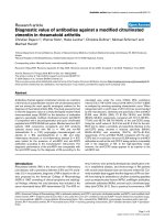
Representative examples of EEG recordings during TH
are given in Figure 1, showing one patient with a reac-
tive cEEG background who eventually had a good recov-
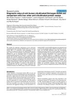
ery (Figure 1) and another patient with a persistent
dis continuous EEG backgroun d activity alternating with
generalized, electrical seizures ("seizure- suppression pat-
tern”), who eventually died (Figure 2).
The associati on between outcome and cEEG findings
during TH is shown in Table 2. After adjusting for mul-
tiple compa risons, nonreactive EEG background, persis-
tent discontinuous EEG pattern, and presence of
seizures/epileptiform discharges were strongly associated
with mortality. Importantly, all patients with epilepti-
form abnormalities or persistent discontinuous EEG
background or both also showed absent EEG reactivity.
Predictive values for mortality for these three cEEG fea-
tures, as well as SSEPs, are shown in Table 3. Despite
relatively wide confidence intervals due to the small
sample size, the positive predictive value (PPV) was
100%, and the false-posit ive rate (FPR) was 0, thus indi-
cating excellent prognostic value for early cEEG fea-
tures. Of note, compared with patients w ith a reactive
cEEG background, those with nonreactive cEEG back-
grounds received similar weight-adjusted doses of mida-
zolam (P = 0.49; t test) and fentanyl (P = 0.33; t test).
Association between outcome and neurologic and
electrophysiological examinations at 72 hours
Neurologic examination and SSEPs were performed at
72 hours in normothermic conditions, as per protocol at
our institution and according to actual recommenda-
tions [5]. All nonsurvivors with absent cEEG reactive
background during TH also had absent SSEPs at 72
hours. Although the PPV for mortality of absent cEEG-
reactive background and bilaterally absent SSEPs was
1.00, the NPV of cEEG was higher than that of SSEP
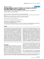
(0.83 versus 0.70; Table 3). In addition, when using the
area under the ROC c urve (Figure 3), cEEG reactivity
yielded better prediction than did SSEP, with a statisti-
cally significant difference in the predictive ability in
favor of EEG background reactivity over SSEPs (0.88
versus 0.69; P = 0.006).
Incomplete recovery of brainstem reflexes (pupillary,
oculocephalic, corneal) and absent or extension motor
reaction to pain also differed among survivors and non-
survivors(threeof19versus11of15,andthreeof19
versus 15 of 15, respectively); however, the false-positive
rate was greater than zero for both, confirming that
Table 1 Patient baseline characteristics in survivors versus nonsurvivors
Survivors (n = 19) Nonsurvivors (n = 15)
Female gender, number (%) 6 (32%) 3 (20%)
Median age, years (range) 62 (35-84) 64 (32-73)
Initial CA rhythm ventricular fibrillation, number (%) 14 (73%) 10 (67%)
CA of cardiac etiology, number (%) 16 (84%) 11 (73%)
Median time from CA to ROSC, minutes (range) 20 (5-40) 22 (8-180)
CA, cardiac arrest; ROSC, return of spontaneous circulation.
Rossetti et al. Critical Care 2010, 14:R173
/>Page 3 of 8
neurologic examination alone may not be reliable in
predicting the outcome after CA and TH.
Postanoxic seizures and epileptiform discharges
The total number of patients with epileptiform EEG fea-
tures during the entire study period was eight (26%) of
34. Five had generalized electrographic seizures alternat-
ing w it h diffuse sup press ion ("seizure-suppr essi on” pat-
tern), and two had generalized, sustained periodic
epilep tiform discharges (G-PEDs), again alternating with
generalized background suppressions. One patient had
delayed seizures that became apparent only after TH
and rewarming. None of the seven patients with early
(that is, during TH) epileptiform abnormalities showed a
significant improvement on the standard EEG per-
formed after TH in normothermic conditions. Further-
more, all had a nonreactive EEG background and died.
In contrast, in the single patient with delayed (that is,
after TH, at normothermia) postanoxic seizures, cEEG
became diffusely epileptiform with multifocal myoclonia
only after weaning of sedation: of note, cEEG back-
ground remained reactive despite epileptiform activity,
and the patient regained consciousness and survived.
Discussion
The main results of this single-center prospect ive study
can be summarized as follows: (1) absent EEG back-
ground reactivity observed during the maintenance
phase of TH a ppeared to be st rongly associated with
poor outcome in patients with coma after CA; (2) all
patients in whom cEEG showed background reactivity
to painful stimuli survived, and the large majority (74%)
awoke and had a favorable outcome; (3) persistent dis-
continuous background and the presence of seizures or
epileptiform discharges on cEEG were also strong risk
factors for poor outcome; (4) nonreactive cEEG back-
ground yielded a significantly better prognostic value
than SSEPs, mostly becaus e of a higher negative predic-
tive value; (5) EEG reactivity to painful stimulation did
not seem to be affected by TH, because all patients with
Figure 1 EEG recording performed during therapeutic hypothermia from one representative patient who had a good outcome
(Cerebral Performance Category 1 at 2 months). EEG shows a reactive EEG background activity to sound ("claps”); recording, 30 mm/sec, 10
μV/mm.
Rossetti et al. Critical Care 2010, 14:R173
/>Page 4 of 8
absent background reactivity during TH had similar
findings on the EEG performed in normothermic condi-
tions, and it was not influenced by sedation-analgesia.
To our knowledge, this is the first clinical study show-
ing that nonreactive EEG background activity during
TH is an early predictor of poor outcome in patients
with postanoxic coma. Before TH beca me a widely used
treatment of hypoxic/ischemic encephalopathy, diffuse
EEG background suppression below 20 μV, burst-sup-
pression with generalized epileptifor m activity, or
generalized periodic complexes on a flat background
have been associated with poor outcome [16,17]. This
was recently confirmed by our group in patients treated
with TH, in whom standard EEG was performed a t the
end of treatment in normothermic conditions [6]. More-
over, prolonged epileptiform EEG features are indepen-
dently correlated with mortality after postanoxic coma
[13], in patients assessed both after [6] and during
[10,13] TH. However, none of these studies formally
addressed the predictive value of any of the EEG
Figure 2 EEG recording performed during therapeutic hypothermia from one representati ve patient who died.EEGshows
discontinuous EEG background activity, alternating with generalized, electrical seizures ("seizure-suppression pattern”). EEG was nonreactive to
painful stimuli; recording, 20 mm/sec, 10 μV/mm.
Table 2 Continuous EEG characteristics in survivors versus nonsurvivors
Survivors (n = 19) Nonsurvivors (n = 15) P value (test)
Time from CA to initiation of cEEG, hours (range) 16 (3-23) 10 (1-21) 0.11 (U)
Median cEEG duration, hours (range) 26 (19-48) 26 (22-66) 0.17 (U)
Nonreactive cEEG background, number (%) 0 (0) 12 (75%) <0.001 (Fisher)
Prolonged discontinuous activity ("burst-suppression”), number (%) 0 (0) 11 (73%) <0.001 (Fisher)
EEG seizures or epileptiform discharges, number (%) 0 (0) 7 (47%) 0.001 (Fisher)
CA, cardiac arrest.
Rossetti et al. Critical Care 2010, 14:R173
/>Page 5 of 8
findings during TH or compared the value of EEG with
that of neurologic examination or SSEPs, the latter
being regarded as reliable predictors of p oor prognosis
[5]. We have recently shown that background reactivity
performed after TH in no rmothermic conditions is a
strong outcome predictor of postano xic coma [6], and
thus undertook this study to examine the prognostic
value o f EEG background performed during TH in the
early phase after CA. Our present findings c onfirm our
previous study and indeed seem to suggest that reactive
bac kgrou nd on cEEG has a strong prognosti c predictive
value, even when monitoring is performed during TH.
They also suggest that background reactivity is not sig-
nificantly influenced by core temperature or by sedation.
After e arlier reports on favora ble outcome for patients
showing continuous amplitude-integrated EEG after TH
[18], a recent study on 30 patients showed that quantita-
tive EEG features during TH (burst-suppression ratio,
response entropy, state entropy) were signi ficantly asso-
ciated with long-term functional outcome [19].
Although our results are in line with these findings, we
add important concomitant clinical information and
describe a much easier appr oach for EEG interpretation,
without the need for more-complicated and not easily
available software analysis.
Although our study was not primarily focused on the
epidemiology of postanoxic seizures, this issue deserves
further discussion. Previous studies reported a variable
prevalence of post anoxic seizures from 10% [11] to 47 %
[10]. We observed a 21% prevalence (seven of 34
patients) of epileptiform abnormalities during TH, of
whom five patients (15% of the entire cohort) had sus-
tained EEG seizures. Because mild hypothermia and
sedation (midazolam in our study) have antiepileptic
action, the occurrence of electrical seizures during TH
may reflect more-severe and diffuse brain injury. This
might explain why none of the seven patients with sei-
zures during TH survived, in line with previous observa-
tions [11]. In contrast, it appears that seizures occurring
only at the end of TH, after rewarming and off sedation,
Table 3 Prognostic predictive value of continuous EEG (30-day mortality)
PPV NPV FPR
Nonreactive background 1.00 (0.74-1.00) 0.83 (0.65-0.97) 0 (0-0.18)
Prolonged discontinuous activity ("burst-suppression”) 1.00 (0.71-1.00) 0.86 (0.61-0.95) 0 (0-0.18)
Seizures/epileptiform discharges 1.00 (0.59-1.00) 0.70 (0.50-0.86) 0 (0-0.18)
Bilaterally absent SSEPs 1.00 (0.48-1.00) 0.70 (0.50-0.86) 0 (0-0.18)
FPR, false-positive rate; NPV, negative predictive value; PPV, positive predictive value; SSEPs, somatosensory evoked potentials.
Figure 3 Area under the receiver operating characte ristic (ROC ) curve for mortality prediction of cEEG reactivity (performed during
therapeutic hypothermia, blue line) and of somatosensory evoked potentials (SSEPs, performed in normothermic conditions, red line).
Continuous EEG yielded better prediction than SSEPs (ROC area, 0.88 versus 0.69; P = 0.006).
Rossetti et al. Critical Care 2010, 14:R173
/>Page 6 of 8
carry a better prognosis, possibly because brain injury is
less severe (thus they are effectively treated with induced
hypothermia and sedatives). Indeed, one patient in our
cohort, treated for status epilepticus that developed after
TH, survived. Altogether, these data underline the value
of early cEEG fo r the treatment of comatose CA
patients treated with TH.
Study limitations
This study has several limitations. First, the sample size
is limited; thus our results are to be considered preli-
minary and will need further confirmation by other
groups and larger studies. However, for this reason, we
applied conservative statistical corrections for multiple
comparisons (Bonferroni). Second, it was a single-center
study, thus data cannot be generalized. Some subjectivity
may also be related to the scoring of EEG reactivity;
however, we used the same method described in our
recent report, which included more than 100 patients.
Time from CA to initiation of cEEG did not differ sig-
nificantly between survivors and nonsurvivors (Table 2);
thus it is unlikely that timing of cEEG affected the pre-
dictive value of the test. Finally, because the cEEG was
interpreted before knowing final patient prognosis, it is
unlikely that it influenced outcome. Furthermore,
although clinicians were aware of cEEG results, EEG
findings (both during TH and at normothermia) were
not used to guide therap y or decisions for withdrawal of
care; thus we believe that this contributed to minimize
the so-called “self-fulfilling prophecy” phenomenon [6].
Conclusions
Continuous EEG background abnormalities during TH
seem to be strongly associated with outcome after CA
and appear to yield excellent point estimates for positive
predictive values and false-positive rates for mortality.
Our data suggest that continuous EEG may be of value
in predicting outcome after CA and TH. Add itional lar-
ger prospective studies are needed to confirm our find-
ings a nd to verify further whether continuous EEG ca n
be helpful for the prognostic assessment of postanoxic
coma.
Key messages
• The results of this single-center study show that
the presence of back ground reactivity on continuous
EEG monitoring (cEEG) performed during therapeu-
tic hypothermia is associated with 30-day survival
and favorable neurologic outcome after cardiac
arrest.
• Our preliminary data suggest that nonreactive EEG
background carries a dismal outcome and is 100%
predictive of mortality in comatose cardiac-arrest
patients.
• Early cEEG findings appear to have a significantly
better predictive value than somatosensory evoked
potentials performed after TH.
• Additional larger p rospective studies are nee ded to
confirm whether continuous EEG may be a helpful
tool for the prognostic assessment of postanoxic
coma.
Abbreviations
CA: cardiac arrest; cEEG: continuous electroencephalography; CPC: Glasgow-
Pittsburgh Cerebral Performance Categories; EEG: electroencephalography;
FPR: false-positive rate; G-PEDS: generalized, sustained periodic epileptiform
discharges; ICU: intensive care unit; NPV: negative predictive value; PPV:
positive predictive value; ROC: receiver operating characteristic; ROSC: return
of spontaneous circulation; SIRPIDS: stimulus induced rhythmic, periodic, or
irritative discharges; SSEPs: somatosensory evoked potentials; TH: therapeutic
hypothermia; VF: ventricular fibrillation.
Acknowledgements
This study was supported by departmental funding from the Service de
Médecine Intensive Adulte and the Département des Neurosciences
Cliniques, Centre Hospitalier Universitaire Vaudois (CHUV), University Hospital,
Lausanne, Switzerland.
The authors thank Malin Maeder-Ingvar, MD, for her help in the data
collection and express their gratitude to all ICU fellows, residents, and
nurses, as well as to all EEG technicians for their valuable help.
Author details
1
Department of Clinical Neurosciences, Lausanne University Hospital and
Faculty of Biology and Medicine, BH-07, Rue du Bugnon 46, CHUV, 1011
Lausanne, Switzerland.
2
Department of Intensive Care Medicine, Lausanne
University Hospital and Faculty of Biology and Medicine, BH-08, Rue du
Bugnon 46, CHUV, 1011 Lausanne, Switzerland.
3
Department of Neurology,
Johns Hopkins Bayview Medical Center, 4940 Eastern Avenue, Baltimore,
Maryland 21224, USA.
Authors’ contributions
AOR conceived the study, collected the data, carried out part of the data
analysis, and drafted the manuscript. LAU carried out part of the data
analysis and drafted the manuscript. FD helped with data collection and
study coordination and revised the manuscript. PWK revised the manuscript
and gave important intellectual contributions. MO conceived the study, was
responsible for study coordination, and revised and helped to draft the
manuscript.
Competing interests
The authors declare that they have no competing interests.
Received: 27 March 2010 Revised: 24 June 2010
Accepted: 29 September 2010 Published: 29 September 2010
References
1. Hypothermia after Cardiac Arrest Study Group: Mild therapeutic
hypothermia to improve the neurologic outcome after cardiac arrest. N
Engl J Med 2002, 346:549-556.
2. ECC Committee, Subcommittees and Task Forces of the American Heart
Association: American Heart Association Guidelines for Cardiopulmonary
Resuscitation and Emergency Cardiovascular Care. Circulation 2005, 112:
IV1-IV203.
3. Bernard SA, Gray TW, Buist MD, Jones BM, Silvester W, Gutteridge G,
Smith K: Treatment of comatose survivors of out-of-hospital cardiac
arrest with induced hypothermia. N Engl J Med 2002, 346:557-563.
4. Young GB: Clinical practice: neurologic prognosis after cardiac arrest. N
Engl J Med 2009, 361:605-611.
5. Wijdicks EF, Hijdra A, Young GB, Bassetti CL, Wiebe S: Practice parameter:
prediction of outcome in comatose survivors after cardiopulmonary
resuscitation (an evidence-based review): report of the Quality
Rossetti et al. Critical Care 2010, 14:R173
/>Page 7 of 8
Standards Subcommittee of the American Academy of Neurology.
Neurology 2006, 67:203-210.
6. Rossetti AO, Oddo M, Logroscino G, Kaplan PW: Prognostication after
cardiac arrest and hypothermia: a prospective study. Ann Neurol 2010,
67:301-307.
7. Al Thenayan E, Savard M, Sharpe M, Norton L, Young B: Predictors of poor
neurologic outcome after induced mild hypothermia following cardiac
arrest. Neurology 2008, 71:1535-1537.
8. Friedman D, Claassen J, Hirsch LJ: Continuous electroencephalogram
monitoring in the intensive care unit. Anesth Analg 2009, 109:506-523.
9. Rossetti AO, Oddo M: The neuro-ICU patient and electroencephalography
paroxysms: if and when to treat. Curr Opin Crit Care 2010, 16:105-109.
10. Abend NS, Topjian A, Ichord R, Herman ST, Helfaer M, Donnelly M,
Nadkarni V, Dlugos DJ, Clancy RR: Electroencephalographic monitoring
during hypothermia after pediatric cardiac arrest. Neurology 2009,
72:1931-1940.
11. Legriel S, Bruneel F, Sediri H, Hilly J, Abbosh N, Lagarrigue MH, Troche G,
Guezennec P, Pico F, Bedos JP: Early EEG monitoring for detecting
postanoxic status epilepticus during therapeutic hypothermia: a pilot
study. Neurocrit Care 2009, 11:338-344.
12. Oddo M, Ribordy V, Feihl F, Rossetti AO, Schaller MD, Chiolero R, Liaudet L:
Early predictors of outcome in comatose survivors of ventricular
fibrillation and non-ventricular fibrillation cardiac arrest treated with
hypothermia: a prospective study. Crit Care Med 2008, 36:2296-2301.
13. Rossetti AO, Logroscino G, Liaudet L, Ruffieux C, Ribordy V, Schaller MD,
Despland PA, Oddo M: Status epilepticus: an independent outcome
predictor after cerebral anoxia. Neurology 2007, 69:255-260.
14. Rossetti AO, Oddo M, Liaudet L, Kaplan PW: Predictors of awakening from
postanoxic status epilepticus after therapeutic hypothermia. Neurology
2009, 72:744-749.
15. Booth CM, Boone RH, Tomlinson G, Detsky AS: Is this patient dead,
vegetative, or severely neurologically impaired? Assessing outcome for
comatose survivors of cardiac arrest. JAMA 2004, 291:870-879.
16. Bassetti C, Bomio F, Mathis J, Hess CW: Early prognosis in coma after
cardiac arrest: a prospective clinical, electrophysiological, and
biochemical study of 60 patients. J Neurol Neurosurg Psychiatry 1996,
61:610-615.
17. Zandbergen EG, Hijdra A, Koelman JH, Hart AA, Vos PE, Verbeek MM, de
Haan RJ: Prediction of poor outcome within the first 3 days of
postanoxic coma. Neurology 2006, 66:62-68.
18. Rundgren M, Rosen I, Friberg H: Amplitude-integrated EEG (aEEG) predicts
outcome after cardiac arrest and induced hypothermia. Intensive Care
Med 2006, 32:836-842.
19. Wennervirta JE, Ermes MJ, Tiainen SM, Salmi TK, Hynninen MS, Sarkela MO,
Hynynen MJ, Stenman UH, Viertio-Oja HE, Saastamoinen KP, Pettilä VY,
Vakkuri AP: Hypothermia-treated cardiac arrest patients with good
neurological outcome differ early in quantitative variables of EEG
suppression and epileptiform activity. Crit Care Med 2009, 37:2427-2435.
doi:10.1186/cc9276
Cite this article as: Rossetti et al.: Prognostic value of continuous EEG
monitoring during therapeutic hypothermia after cardiac arrest. Critical
Care 2010 14:R173.
Submit your next manuscript to BioMed Central
and take full advantage of:
• Convenient online submission
• Thorough peer review
• No space constraints or color figure charges
• Immediate publication on acceptance
• Inclusion in PubMed, CAS, Scopus and Google Scholar
• Research which is freely available for redistribution
Submit your manuscript at
www.biomedcentral.com/submit
Rossetti et al. Critical Care 2010, 14:R173
/>Page 8 of 8