Báo cáo y học: " A stochastic model of oncogene expression and the relevance of this model to cancer therapy" ppt
Bạn đang xem bản rút gọn của tài liệu. Xem và tải ngay bản đầy đủ của tài liệu tại đây (275.09 KB, 7 trang )
BioMed Central
Page 1 of 7
(page number not for citation purposes)
Theoretical Biology and Medical
Modelling
Open Access
Research
A stochastic model of oncogene expression and the relevance of this
model to cancer therapy
Francis D Alfano*
Address: The Harold Leever Cancer Center, 1075 Chase Parkway, Waterbury, Connecticut, 06708, USA
Email: Francis D Alfano* -
* Corresponding author
Abstract
Background: Ablation of an oncogene or of the activity of the protein it encodes can result in
apoptosis and/or inhibit tumor cell proliferation. Therefore, if the oncogene or set of oncogenes
contributing maximally to a tumor cell's survival can be identified, such oncogene(s) are the most
appropriate target(s) for maximizing tumor cell kill.
Methods and results: A mathematical model is presented that describes cellular phenotypic
entropy as a function of cellular proliferation and/or survival, and states of transformation and
differentiation. Oncogenes become part of the cellular machinery, block apoptosis and
differentiation or promote proliferation and give rise to new states of cellular transformation. Our
model gives a quantitative assessment of the amount of cellular death or growth inhibition that
result from the ablation of an oncogene's protein product. We review data from studies of chronic
myelogenous leukemia and K562 cells to illustrate these principles.
Conclusion: The model discussed in this paper has implications for oncogene-directed therapies
and their use in combination with other therapeutic modalities.
Background
For the past thirty years, cancer research has elucidated a
family of genes that are integrally involved in the cancer
process. These genes comprise two subsets. One subset,
termed oncogenes, gives rise to proteins that modulate
such processes as cell cycle progression, signaling, cellular
growth and apoptosis [1-3]. The other consists of genes
that can suppress tumor activity and their absence can
lead to the initiation and/or progression of cancer [4].
There has been a body of work discussing the view that
cancer arises from genetic instability. Evidence for genetic
instability that has been cited includes the occurrence of
chromosomal abnormalities, microsatellite DNAs and
aberrant gene expression through hypermethylation of
DNA [5-7]. Moreover, recent work using RNA microarray
analysis has shown that there are key genes that are over-
expressed as a result of malignant transformation, and
others that are under expressed, compared to RNA tran-
scripts in nonmalignant counterparts [8]. Many of these
gene expression changes illustrated by gene microarray
analysis may be secondary or even far distal to the primary
changes determined by the actual oncogene or suppressor
gene. In order to define the complex behavior of a tumor
cell population associated with these complex gene
expressions, we have chosen to define entropies of apop-
tosis, cellular differentiation and survival or growth inhi-
bition. We hypothesize that a cell's phenotypic entropy is
determined as a function of the survival fraction or prolif-
eration rate of a tumor ;and also, the number of trans-
formed and differentiated states that arise within a
Published: 31 January 2006
Theoretical Biology and Medical Modelling 2006, 3:5 doi:10.1186/1742-4682-3-5
Received: 02 December 2005
Accepted: 31 January 2006
This article is available from: />© 2006 Alfano; licensee BioMed Central Ltd.
This is an Open Access article distributed under the terms of the Creative Commons Attribution License ( />),
which permits unrestricted use, distribution, and reproduction in any medium, provided the original work is properly cited.
Theoretical Biology and Medical Modelling 2006, 3:5 />Page 2 of 7
(page number not for citation purposes)
particular cell population. The mathematical relations
that we have formulated can quantitatively determine
how ablation of an oncogene's protein activity can result
in apoptosis and/or a decrease in proliferation within a
population of tumor cells. The goal is then to determine
which oncogene or set of oncogenes contributes maxi-
mally to a tumor cell's survival; and thereby, to predict
which oncogene(s) are the most appropriate target(s) for
maximizing tumor cell kill.
The model
The cellular phenotypic entropy is determined by first
defining all allowable phenotypes. These phenotypes
include the set of transformed states associated with aber-
rant gene expression, the set of differentiated states that
have been defined as a result of stochastic gene expression
and the distribution of cells between living and dead or
growth inhibited. Therefore
f
s
Ent(phenotype) = Ent(transform/differentiation) f
s
+
Ent(cell survival) (1)
where Ent(transform/differentiation) is the entropy of
transformation and differentiation per observed popula-
tion and Ent(cell survival) is the entropy of cell death or
growth inhibition per total population; f
s
is the ratio of
observed cells to predicted cells.
The transformed states are the phenotypes that a cell can
access which provide a hyperproliferative advantage over
the cell's normal counterpart. This includes phenotypes
that have a better growth advantage as well as phenotypes
that have antiapoptotic behaviors in environments that
would normally lead to cellular apoptosis. The total
number of states that a cell can access is determined by the
number of end-differentiated states and by the number of
new "environments" that a transformed cell can inhabit
over and above its normal counterpart. Let Ω represent the
number of transformed states that a cell can access as a
transformed cell and let ω represent the number of states
that the cell's normal counterpart can access via differen-
tiation. We presume that ω is equal to the number of end-
differentiated states in an undifferentiated cell or equal to
1 in an end-differentiated cell. We will define the entropy
of the combined states of transformation and differentia-
tion as
f
s
Ent(transform/differentiation) = c f
s
ln(ω Ω + ω). (2)
Equation (2) is motivated by the classic definition of
entropy as applied to biological systems [9]. If a system
can exist in N equivalent configurations, then the entropy
of that system is given by
Entropy = c ln N
where "c" is a constant of proportionality.
In equation (2), we presume that each purely transformed
state will ultimately interact with each differentiated state
giving rise to a new and unique transformed state. For
example, an undifferentiated cell containing a trans-
formed phenotype and differentiating into two differenti-
ated cells will occupy one of two possible transformed
phenotypes.
Each transformed state confers a survival advantage to the
transformed cell as compared with the cell's normal coun-
terpart. More practically, transformed cells are also
defined by the set of oncogene/suppressor genes that are
active within the cell and the number of different cellular
mechanisms that this set of genes acts upon to change the
cell's behavior with respect to growth and apoptosis. To
simplify further analysis, let us consider the action of
oncogenes only and disregard the action of suppressor
genes. Also, we will assume that there is a direct correla-
tion between the set of 'environments' that a transformed
cell can inhabit and the unique cellular mechanism that
an oncogene affects. Therefore, let us correlate each trans-
formed state Ω with the number of oncogenes multiplied
by the unique cellular mechanisms that each oncogene
affects (see Fig. 1).
The term Ent(cell survival) in equation (1) can be calcu-
lated by defining cell death (or decreased proliferation)
and cell viability (or enhanced viability) as two states that
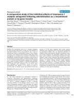
Oncogenes can affect multiple cellular pathways, which result in modulating proliferation and apoptosisFigure 1
Oncogenes can affect multiple cellular pathways, which result
in modulating proliferation and apoptosis. Depicted here are
three oncogenes. Oncogene 2's activity overlaps with the
activities of the other two. Therefore, ablation of oncogene
2's activity would not result in any measurable change in pro-
liferation or apoptosis.
Theoretical Biology and Medical Modelling 2006, 3:5 />Page 3 of 7
(page number not for citation purposes)
are independent of the number of transformed states but
still nonetheless contribute to the overall phenotypic
entropy. Define the total number of cells, which is deter-
mined by a suitable control, as N
c
and the measured
number of observed cells as n
s
. The difference, N
c
-n
s
, in
some cases would represent apoptosis or cell death, and in
other cases would represent decreased proliferation meas-
ured against a suitable standard. If Ent(cell survival) is
defined in a canonicalthermodynamic formalism [9],
then
N
c
Ent(cell survival) = c ln(N
c
!/n
s
!(N
c
-n
s
)!). (3)
Utilizing Stirling's approximation [10] and defining f
s
=
n
s
/N
c
, Equation (3) becomes
Ent(cell survival) - c [(1-f
s
) ln(1-f
s
) +f
s
ln f
s
] for 0<f
s
<1
(4)
and
Ent(cell survival) = 0 for f
s
= 0 or 1 by continuity.
Combining equations (1), (2) and (4) gives us
f
s
[Ent(phenotype)] = - c [(1-f
s
) ln(1-f
s
) +f
s
ln f
s
] + c f
s
ln(ω
Ω + ω) or
Ent(phenotype) = - c [((1-f
s
)/f
s
)ln(1-f
s
)+ ln f
s
] + c ln(Ω +
1)+ c ln(ω). (5)
Equation (5) is the general statement of the model, which
relates the phenotypic entropy to the processes of differ-
entiation, transformation and cellular growth and/or
apoptosis.
Since the entropy in equation (5) is defined along the clas-
sic definition of entropy, we can apply the second law of
thermodynamics to our analysis. Consider a modulator of
differentiation and/or oncogene activity that either
reduces or completely eliminates the action of the onco-
gene or changes the number of available differentiated
phenotypes. We know that Ent(phenotype) should
increase or remain equal with time, and therefore this
entropy, after administration of the oncogene modulator,
should be greater than or equal to the entropy prior to its
administration. However, if the inhibitor is removed,
then the entropy after removal should return to that of the
premodulator's environment, i.e.
Ent(phenotype)
premodulator
≤ Ent(phenotype)
postmodulator
≥
Ent(phenotype)
premodulator
. (6)
The result is self-consistent only if equation (6) is consid-
ered with the equal signs. Therefore, if we take times pre
and post modulator administration that most closely
approximate the equality of entropies pre and post the
administration of the modulator, then
{- [((1-f
s
)/f
s
) ln(1-f
s
)+ ln f
s
] + ln(Ω + 1)+ ln(ω)}
premodulator
= {- [((1-f
s
)/f
s
) ln(1-f
s
) + ln f
s
] + ln(Ω + 1)+
ln(ω)}
post_modulator
. (7)
In Table 1, we consider multiple examples where changes
of Ω and ω occur as a result of the modulator; f
s
(pre mod-
ulator) is for most of the examples taken as 1 but we con-
sider examples of modulator given in the setting of
cytotoxic drugs, which can reduce f
s
(premodulator) to
less than 1. The intent of these substitutions is to calculate
f
s
(post modulator) to determine the effect of the modula-
tor in different settings. We can solve for f
s
(post modula-
tor) in equation (7) by using the root function of
MATHCAD version 11 [25].
Chronic mylogenous leukemia and K562
Our intent is to study oncogenic behavior in realistic
models to determine whether the principles outlined
above can predict outcomes of therapy. As an example, let
us consider the Bcr-Abl oncogenic protein, which is the
transforming agent for chronic myelogenous leukemia
(CML) [11,12]. The Bcr-Abl protein is the result of the
fusion of sequences from the Abl proto-oncogene on
chromosome 9 with the sequences from the proto-onco-
gene, Bcr, on chromosome 22. The two major forms of
Bcr-Abl, p210 and p190, can each cause chronic myeloge-
nous leukemia (CML) in humans. The Abl component of
this protein encodes a nonreceptor tyrosine kinase that is
constitutively active and activates a number of signal
transduction pathways involved with cell proliferation
and apoptosis. Bcr-Abl can inhibit apoptosis and decrease
cell proliferation by its kinase action in experimental sys-
tems and myeloid cells. These mechanisms have been
shown to be mediated for the most part through (1) acti-
vation of phosphatidylinositol 3-kinase (PI-3K) and (2)
Jak-Stat kinases. In addition, Bcr-Abl can affect p53 and
MYC in a RAS-dependent manner [12-14] and can acti-
vate Jun N-terminal kinase (JNK).
In CML, Marley et al. [15,16] found that the antiprolifera-
tive effect of the Bcr-Abl inhibitor Imatinib correlated
most closely with the inhibition of PI-3K within chronic
myeloid leukemia progenitor cells, and also found that
AG490, a Jak2 kinase inhibitor and FTI II, a farnesyltrans-
ferase inhibitor and an inhibitor of RAS activation, could
also reduce the proliferation of clonogenic CML cells. This
suggests that Bcr-Abl can influence at least three separate
proliferation or antiapoptotic mechanisms within CML
cells and effect transformation by activating three separate
cellular mechanisms (see Fig. 2). There is evidence that
CML is a disease of stem cells that can undergo self-
Theoretical Biology and Medical Modelling 2006, 3:5 />Page 4 of 7
(page number not for citation purposes)
renewal as well as differentiate into committed progenitor
cells capable of proliferating. Laboratory evidence has
shown that drugs such as interferon or Imatinib, the
inhibitor of the Bcr-Abl kinase, have different antiprolifer-
ative effects on CML stem cells and committed progenitor
cells [17-19]. Therefore, any analysis of CML proliferation
and apoptosis needs to take into account these two dis-
tinct cellular types.
Imatinib is a tyrosine kinase inhibitor that specifically
binds the ATP pocket of Bcr-Abl tyrosine kinase, inhibit-
ing the activity of the kinase. Moreover, it is known to
induce apoptosis in Bcr-Abl positive cells [15,16]. In CML
cells, the predominant effect of Imatinib is not to induce
apoptosis but to decrease proliferation of the committed
progenitor cells and to a lesser extent the CML stem cells
[17,19]. We can use equation (7) and Table 1 to calculate
the effect of Imatinib on the stem cell and committed pro-
genitor populations. The committed progenitor popula-
tion is a differentiated system, and therefore
ω(preImatinib) = ω(postImatinib). The term [(1-f
s
)/f
s
ln(1-f
s
) +ln f
s
]
preImatinib
is taken to be zero since f
s
(preImat-
inib) is taken to be nearly equal to one. Since the Bcr-Abl
kinase predominantly affects three enzyme mechanisms,
the Ω(preImatinib) is equal to three. We will assume the
maximum effect of Imatinib and so take Ω(postImatinib)
to be zero. Therefore, symbolically, we have {Ω(Pre)-
>Ω(Post): 3->0; ω(Pre)->ω(Post): 1->1; f
s
(Pre) = 1}. By
referring to Table 1, we find f
s
(Post) to be 0.5. In CML
stem cells, Imatinib has been shown to have much less of
an impact on cellular proliferation. One postulated mech-
anism for this is the presence of an enhanced multidrug
resistance protein (MDR) which extrudes the drug from
the interior of the cell [20]. Therefore, Imatinib may not
maximally inhibit the cellular mechanisms outlined
above. We can reasonably postulate that Ω(Pre)-
>Ω(Post): 3->1. Furthermore, the effect of Imatinib on dif-
ferentiation of the CML stem cell is not clear. Schuster et
al. [21] were able to show that the block of differentiation
on a murine hematopoietic progenitor line by Bcr-Abl
kinase was reversed by Imatinib but Angstreich et al. [22]
were unable to show any effect on the differentiation of
CML progenitor stem cells by Imatinib. For our analysis of
CML stem cells, we will consider a mixing of two states;
i.e. {Ω(Pre)->Ω(Post): 3->1; ω(Pre)->ω(Post): 2->2;
f
s
(Pre) = 1} and {Ω(Pre)->Ω(Post): 3->1; ω(Pre)-
>ω(Post): 2->1; f
s
(Pre) = 1} to reflect this duplicity of dif-
ferentiation data. Table 1 shows that f
s
= 0.77 and 0.5 for
these two situations, respectively. This establishes the
range of f
s
to be between 0.5 and 0.77 for CML stem cells.
Holtz et al. [19] studied the separate effects of Imatinib on
CML stem cells and committed progenitor cells and
derived an index of inhibition that is appropriate for our
analysis. They found a progenitor frequency that was
decreased by 52 ± 5 % for committed progenitors and 43
± 12% for primitive progenitors (stem cells). If we associ-
ate one minus the percent decrease in progenitor fre-
quency with f
s
, then f
s
is equal to 0.48 ± 0.05 and 0.57 ±
0.12 by their data, in agreement with our theoretical pre-
dictions.
Imatinib and the chemotherapy drug cytosine-arabino-
side have been shown to induce apoptosis and erythroid
differentiation in the Bcr-Abl positive cell line K562 [23].
Fang et al. [23] also demonstrated a strong influence by
Imatinib on the Akt kinase system of K562 cells. Imatinib
induced erythroid differentiation in 37.5 % of K562 cells.
Arnaud et al. [24] also observed erythroid differentiation
in response to Imatinib and furthermore observed meg-
akaryocytic differentiation with respect to phorbol esters.
Therefore, for the K562 system, one can consider a mix of
the states {Ω(Pre)->Ω(Post): 3->2; ω(Pre)->ω(Post): 3->2;
f
s
(Pre) = 1} and {Ω(Pre)->Ω(Post): 3->2; ω(Pre)-
>ω(Post): 3->3; f
s
(Pre) = 1} to represent this system. We
can compute values of f
s
(Post) as 0.77 and 0.92, respec-
Table 1: Tumor cell survival fraction as a function of changes in the states of transformation, differentiation, and the initial tumor
survival fraction.
f
s
(Pre) Ω(Pre)-> Ω(Post) ω(Pre)->ω(Post) f
s
(Post)
1.0 3->01->1.50
1.0 3->12->2.77
1.0 3->12->1.50
1.0 3->23->2.77
1.0 3->23->3.92
.85 3->23->3.74
.85 3->23->2.57
The term f
s
(Pre) is the survival fraction of a tumor cell's population prior to the application of a targeted therapy; Ω(Pre)-> Ω(Post) represents the
change in transformed states resulting from oncogene inhibition ; ω(Pre)->ω(Post) represents the change in differentiation states resulting from
oncogene inhibition. The term f
s
(Post) is the survival fraction or reduced proliferation of a tumor cell population that results from oncogene
inhibition and is calculated from equation (7).
Theoretical Biology and Medical Modelling 2006, 3:5 />Page 5 of 7
(page number not for citation purposes)
tively. Fang et al. found that the percentage of nonapop-
totic cells measured by an Annexin V assay was 81.7 ±
2.4% and by a morphology assay was 84.9 ± 1.6%.
When cytosine-arabinoside was added to the system, dif-
ferentiation remained about the same at 38.8% but the
percent of nonapoptotic cells decreased to 71.2 ± 1.8%
and 65 ± 0.3% by the Annexin and morphology assays,
respectively. Since cytosine-arabinoside alone induced an
apoptosis of 15%, we have that f
s
(PreImatinib but in the
presence of cytosine-arabiniside) was0.85. Substituting
this into the above states, we have {Ω(Pre)->Ω(Post): 3-
>2; ω(Pre)->ω(Post):3->3; f
s
(Pre) = .85} and {Ω(Pre)-
>Ω(Post): 3->2; ω(Pre)->ω(Post): 3->2; f
s
(Pre) = .85}. By
referring to Table 1, we find that f
s
(Post) is equal to 0.74
and 0.57, respectively. The theoretical values are within
the range of the experimental data.
Discussion
We have developed a model of cellular behavior that
interprets cellular transformation, apoptosis/proliferation
and differentiation as stochastic processes. The model
defines the necessity of considering all these mechanisms
of cellular behavior together because there is interdepend-
ence amongst them. For example, differentiation may
lead to the generation of apoptosis or decreased cellular
proliferation; and transformation can result in enhanced
proliferation when compared to the transformed cell's
normal counterpart. Others have interpreted cellular
transformation as a stochastic process [9,26] and several
lines of evidence have been developed to explain the
underlying cause of the stochastic behavior of cancer.
Genetic instability as a cause for cancer has been a recur-
ring theme since the classic paper of Boveri [27] and is
defined by most authors as the generation of altered cellu-
lar behavior because of an altered protein network sec-
ondary to the introduction of a new oncogene protein or
the removal of a tumor suppressor protein. In either case,
definite outcomes are thought to be predicted by either
event. Furthermore, over a long enough period of time,
cellular behavior can evolve within a transformed popula-
tion of cells, leading to a heterogeneous set of cellular
behaviors.
We have used entropy as a measure of change for transfor-
mation; not only because entropy is a linear function and
often different items of interest can simply be added
together, but also because this approach is supported by
past analyses that have used entropy to model cancer
behavior in the context of chemical carcinogenesis [9].
Furthermore, recent work on the differentiation of mye-
loid colony-forming cells has shown that experimental
data best fit a stochastic model [28]. Because of the sim-
plicity of the entropy function, we can collect components
of cellular behavior that best fit our knowledge of the cel-
lular phenotype; i.e. the cell's growth capacity and sur-
vival, the cell's differentiation status and the cell's
transformation status. Each of these quantities can be
defined within the context of an entropy function and
combined to serve as an index of cellular phenotypic
entropy.
We consider the cellular phenotypic entropy to remain
constant during therapies that are observed to be reversi-
ble. As an example for study, we chose chronic myeloge-
nous leukemia because this model is well defined in terms
of the oncogenes involved. Imatinib induces a high rate of
remissions when given in a clinical context, but when
Imatinib is discontinued the disease returns to a clinical
state identical to that observed before the inhibitor. This
observation also has been made in vitro [29]. Such would
not be the case with most chemotherapies since they are
often mutagenic and exert their effect by modifying cellu-
lar DNA permanently [30].
In our analysis of Bcr-Abl kinase action in CML, we sur-
mised that the protein is acting predominantly over three
kinase systems to enhance cellular proliferation. These
three systems involve pathways that have already been
elucidated such as (1) the Akt kinase system, (2) the RAS
dependent p53 system, and (3) the Jak-Stat kinase system
[12,13]. However, these systems are not totally independ-
ent and their interdependence may serve to reduce the
number of transforming states that we have designated
within our mathematical computations. As such, the Jun
pathway was also recognized to be affected by the BCR-
Bcr-Abl modulates up to four cellular pathways, but because of the interdependence of these pathways, we conclude that 3 pathways best represent the number of transformed states incurred by the oncogene's behaviorFigure 2
Bcr-Abl modulates up to four cellular pathways, but because
of the interdependence of these pathways, we conclude that
3 pathways best represent the number of transformed states
incurred by the oncogene's behavior.
Theoretical Biology and Medical Modelling 2006, 3:5 />Page 6 of 7
(page number not for citation purposes)
ABL kinase, but it is not clear that this is strongly impli-
cated in Bcr-Abl kinase action within the context of CML.
Even if it were appropriate to consider the Jun pathway,
the interdependence of these pathways may still justify
equating Ω(Pre), the number of transformed states deter-
mined by the Bcr-Abl kinase, to three (see Fig. 2).
By our model, each enzyme system is correlated with a
transformed state, and the more each system is affected by
the inhibitor, the greater the effect the inhibitor has on
reducing cellular growth and/or inducing apoptosis. Fur-
thermore, if the inhibitor contributes to the differentia-
tion of the transformed cell, then that also will contribute
to a greater reduction of cellular proliferation and a possi-
ble increase in apoptosis. In all the instances we cited, the
data supports our theoretical model.
Our model does not address the issue of how a normal
cell with low phenotypic entropy becomes transformed to
a cell with higher entropy. Even if Imatinib fully ablates
the activity of the Bcr-Abl kinase, the cell remains trans-
formed and the phenotypic entropy does not change.
Therefore, transformation by our model is considered
independent of oncogene expression (see Figure 3). How
can this be? The answer lies in the fact that the signature
of transformation is not in the expression of the oncogene
protein but in the alteration in the DNA by the oncogene.
In the case of Bcr-Abl, it is the observed 9–22 chromo-
some translocation that affects the behavior of other nor-
mal cellular proteins [31]. Such a conclusion is supported
by the observations of Keating et al., who observed varia-
ble expression of Bcr-Abl transcripts in early CML progen-
itor cells that exhibited the chromosome translocation
[32].
More studies will be needed to determine whether this is
a specific feature of Bcr-Abl positive disease or a manifes-
tation of a more general principle of cellular transforma-
tion and cancer. Namely, does an oncogene act in a
similar fashion within a set of oncogenes as it does when
it acts alone? If so, then it would be important to know
how to measure an oncogene's action so that one could
target the specific oncogene with the greatest impact on a
tumor cell's survival. Within the context of our model, we
can provide a recipe for calculating the extent to which
inhibiting an oncogene's action can reduce tumor cell sur-
vival by answering the following questions:
(1) How many proliferation/apoptosis mechanisms are
active in the tumor cell's normal counterpart? (In CML,
we argued for three mechanisms.)
(2) What oncogenes inhibit which mechanisms? The
answer would most likely be specific to the oncogenes
that are active within the tumor cell.
(3) How many end-differentiated states apply to the
tumor's specific environment, and does the oncogene
inhibitor change the number of differentiated states
expressed after oncogene ablation, and does the inhibitor
completely ablate the oncogene's action with respect to all
proliferation/apoptosis mechanisms? And finally,
(4) What is the initial survival fraction of the tumor cell's
population prior to oncogene inhibition? The initial sur-
vival may vary depending upon other therapies applied
such as radiation and/or chemotherapy. Table (1) yields
theoretical calculations of survival fractions as a function
of changes of oncogene expression, differentiation and
the initial survival fraction before a targeted therapy is
applied, demonstrating the synergy between oncogene
specific therapies and other modalities such as chemo-
therapy.
Competing interests
The author(s) declare that they have no competing inter-
ests.
Acknowledgements
The author thanks two anonymous reviewers for constructive comments;
and thanks Dr. Paul Agutter for his help. We also thank Dr Greg Angstreich
for discussions concerning CML growth and differentiation.
References
1. Wynford-Thomas D: Oncogenes and anti-oncogenes; the
molecular basis of tumour behavior. J Pathol 1991, 165:187-201.
A normal cell, N, is transformed thereby increasing its phe-notypic entropyFigure 3
A normal cell, N, is transformed thereby increasing its phe-
notypic entropy. When the oncogene inhibitor is applied to
the transformed cell, T, the cell maintains constant pheno-
typic entropy and therefore does not return to its normal
state. As a result of the loss of the oncogene's protective
actions, the transformed cell, Ti, is less adapted to its envi-
ronment and undergoes either growth inhibition or apopto-
sis.
Publish with BioMed Central and every
scientist can read your work free of charge
"BioMed Central will be the most significant development for
disseminating the results of biomedical research in our lifetime."
Sir Paul Nurse, Cancer Research UK
Your research papers will be:
available free of charge to the entire biomedical community
peer reviewed and published immediately upon acceptance
cited in PubMed and archived on PubMed Central
yours — you keep the copyright
Submit your manuscript here:
/>BioMedcentral
Theoretical Biology and Medical Modelling 2006, 3:5 />Page 7 of 7
(page number not for citation purposes)
2. Evan GI, Wyllie AH, Gilbert CS, Littlewood TD, Land H, Brooks M,
Waters CM, Penn LZ, Hancock DC: Induction of apoptosis in
fibroblasts by c-myc protein. Cell 1992, 69:119-128.
3. Marin MC, Hsu B, Stephens LC, Brisbay S, McDonnell TJ: The func-
tional basis of c-myc and bcl-2 complementation during mul-
tistep lymphomagenesis in vivo. Exp Cell Res 1995, 217:240-247.
4. Stanbridge EJ: Functional evidence for human tumor suppres-
sor genes: Chromosome and molecular genetic studies. Can-
cer Surv 1992, 12:5-24.
5. Modrich P, Lahue R: Mismatch repair in replication fidelity,
genetic recombination and cancer biology. Annu Rev Biochem
1996, 65:101-133.
6. Lengauer C, Kinzler KW, Vogelstein B: DNA methylation and
genetic instability in colorectal cancer cells. Proc Natl Acad Sci
USA 1997, 94:2545-2550.
7. Duesberg PH: Cancer genes: rare recombinants instead of
activated oncogenes. Proc Natl Acad Sci USA 1987, 84:4.
8. Ross DT, Scherf U, Eisen MB, Perou CM, Rees C, Spellman P, Iyer V,
Jeffrey SS, Van deRijn M, Waltham M, Pergamschikov A, Lee JC,
Lashkari D, Shalon D, Myers TG, Weinstein JN, Botstein D, Brown
PO: Systemic variation in gene expression patterns in human
cancer cell lines. Nat Genet 2000, 403:503-509.
9. Alfano FD: A stochastic model of cellular transformation and
its relevance to chemical carcinogenesis. Math Biosci 1998,
149:95-106.
10. Whitaker ET, Watson GN: A Course of Modern Analysis Cambridge:
Cambridge University Press; 1963. Section 12.33
11. Groffen J, Stephenson JR, Heisterkamp N, Bartram C, deKlein A, Gro-
eveld G: Philadelphia chromosomal breakpoints are clustered
within a limited region, Bcr, on chromosome 22. Cell 1984,
36:93-99.
12. Gordon MY, Goldman JM: Cellular and molecular mechanisms
in chronicmyeloid leukemia: biology and treatment. Br J Hae-
matol 1996, 95:10-20.
13. Barnes DJ, Melo JV: Cytogenetic and molecular genetic aspects
of chronic myeloid leukemia. Acta Haematol 2002, 108:180-202.
14. Deininger MWN, Goldman JM, Melo JV: The molecular biology of
chronic myeloid leukemia. Blood 2000, 96:3343-3356.
15. Marley SB, Lewis JL, Schneider H, Rudd CE, Gordon MY: Phosphati-
dylinositol-3 kinase inhibitors reproduce the selective anti-
proliferative effects of imatinib on chronic myeloid leukemia
progenitor cells. Br J Haematol 2004, 125:500-511.
16. Marley SB, Davidson JR, Goldman JM, MY Gordon MY: Effects of
combinations of therapeutic agents on the proliferation of
progenitor cells in chronic myeloid leukemia. Br J Haematol
2002, 116:162-165.
17. Michor F, Hughes TP, Iwasa Y, Branford S, Shah NP, Sawyers CL,
Nowak MA: Dynamics of chronic myeloid leukemia. Nature
2005, 435:1267-1270.
18. Huntly BJP, Gilliland DG: Summing up cancer stem cells. Nature
2005, 435:1169-1170.
19. Holtz MS, Slovak ML, Zhang F, Sawyers CL, Forman SJ, Bhatia R:
Imatinib mesylate(STI571) inhibits growth of primitive
malignant progenitors in chronic myelogenous leukemia
through reversal of abnormally increased proliferation. Blood
2002, 99:3792-3800.
20. Mahon FX, Belloc F, Lagarde V, Chollet C, Moreau-Gaudry F, Reiffers
J, Goldman JM, Melo JV: MDR1 gene overexpression confers
resistance to imatinib mesylate in leukemia cell line models.
Blood 2003, 102:2368-2373.
21. Schuster C, Forster K, Dierks H, Elsasser A, Behre G, Simon N, Dan-
hauser-Reidi , Hallek M, Warmuth M: The effects of Bcr-Abl on C/
EPB transcription-factor regulation and neutrophilic differ-
entiation are reversed by the Abl kinase inhibitor imatinib
mesylate. Blood 2003, 101:655-663.
22. Angstreich GR, Matsui W, Huff CA, Vala MS, Barber J, Hawkins AL,
Griffin CA, Smith BD, Jones RJ: Effects of imatinib and interferon
on primitive chronic myeloid leukemia progenitors. Br J Hae-
matol 2005, 130:373-381.
23. Fang G, Kim CN, Perkins CL, Ramadevi N, Winton E, Wittman S,
Bhalla KN: CGP57148B(STI-571) induces differentiation and
apoptosis and sensitizes Bcr-Abl-positive human leukemia
cells to apoptosis due to antileukemic drugs. Blood 2000,
96:2246-2253.
24. Arnaud J, Herrant M, Legros L, Belhacene N, Luciano F, Pages G, Hof-
man P, Auberger P: Imatinib induces mitochondria-dependent
apoptosis of the Bcr-Abl positive K562 cell line and its differ-
entiation toward the erythroid lineage. FASEB 2003,
17:2160-2162.
25. Mathcad version 11, Mathsoft Engineering & Education, Inc 101 Main St,
Cambridge, MA 02142 .
26. Lengaur C, Kinzler KW, Vogelstein B: Genetic instabilities in
human cancers. Nature 1998, 396:643-9.
27. Boveri T: Zur Frage der Entstehung maligner tummoren.
Gustav; 1914.
28. Marley SB, Lewis JL, Gordon MY: Progenitor cells divide sym-
metrically to generate new colony-forming cells and clonal
heterogeneity. Br J of Haematol 2003, 121:643-648.
29. Deininger MW, Goldman JM, Lydon N, Meio JV: The tyrosine
kinase inhibitor CGP57148B selectively inhibits the growth
of Bcr-Abl positive cells. Blood 1997, 90:3691-3698.
30. Pletsa V, Valavanis C, van Delft JHM, Steenwinkel MST, Kyrtopoulos
SA: DNA damage and mutagenesis induced by procarbazine
in _lacZ transgenic mice: Evidence that bone marrow muta-
tions do not arise primarily through miscoding by O
6
-meth-
ylguanine. Carcinogen 1997, 18:2191-2196.
31. Salesse S, Verfaillie CM: Bcr/Abl-mediated increased expression
of multiple known novel gene that may contribute to the
pathogenesis of chronic myelogenous leukemia. Mol Canc Ther
2003, 2:173-182.
32. Keating A, Wang X, Laraya P: Variable transcription of Bcr-Abl
by Ph
+
cells arising from hematopoetic progenitors in
chronic myeloid leukemia. Blood 1994, 83:1744-1749.