Báo cáo y học: " Indications that "codon boundaries" are physico-chemically defined and that protein-folding information is contained in the redundant exon bases" pot
Bạn đang xem bản rút gọn của tài liệu. Xem và tải ngay bản đầy đủ của tài liệu tại đây (991.44 KB, 11 trang )
BioMed Central
Page 1 of 11
(page number not for citation purposes)
Theoretical Biology and Medical
Modelling
Open Access
Research
Indications that "codon boundaries" are physico-chemically defined
and that protein-folding information is contained in the redundant
exon bases
Jan Charles Biro*
Address: Homulus Foundation, San Francisco, CA 94 105, USA
Email: Jan Charles Biro* -
* Corresponding author
Abstract
Background: All the information necessary for protein folding is supposed to be present in the
amino acid sequence. It is still not possible to provide specific ab initio structure predictions by
bioinformatical methods. It is suspected that additional folding information is present in protein
coding nucleic acid sequences, but this is not represented by the known genetic code.
Results: Nucleic acid subsequences comprising the 1st and/or 3rd codon residues in mRNAs
express significantly higher free folding energy (FFE) than the subsequence containing only the 2nd
residues (p < 0.0001, n = 81). This periodic FFE difference is not present in introns. It is therefore
a specific physico-chemical characteristic of coding sequences and might contribute to
unambiguous definition of codon boundaries during translation. The FFEs of the 1st and 3rd
residues are additive, which suggests that these residues contain a significant number of
complementary bases and that may contribute to selection for local RNA secondary structures in
coding regions. This periodic, codon-related structure-formation of mRNAs indicates a connection
between the structures of exons and the corresponding (translated) proteins. The folding energy
dot plots of RNAs and the residue contact maps of the coded proteins are indeed similar. Residue
contact statistics using 81 different protein structures confirmed that amino acids that are coded
by partially reverse and complementary codons (Watson-Crick (WC) base pairs at the 1st and 3rd
codon positions and translated in reverse orientation) are preferentially co-located in protein
structures.
Conclusion: Exons are distinguished from introns, and codon boundaries are physico-chemically
defined, by periodically distributed FFE differences between codon positions. There is a selection
for local RNA secondary structures in coding regions and this nucleic acid structure resembles the
folding profiles of the coded proteins. The preferentially (specifically) interacting amino acids are
coded by partially complementary codons, which strongly supports the connection between
mRNA and the corresponding protein structures and indicates that there is protein folding
information in nucleic acids that is not present in the genetic code. This might suggest an additional
explanation of codon redundancy.
Published: 07 August 2006
Theoretical Biology and Medical Modelling 2006, 3:28 doi:10.1186/1742-4682-3-28
Received: 16 December 2005
Accepted: 07 August 2006
This article is available from: />© 2006 Biro; licensee BioMed Central Ltd.
This is an Open Access article distributed under the terms of the Creative Commons Attribution License ( />),
which permits unrestricted use, distribution, and reproduction in any medium, provided the original work is properly cited.
Theoretical Biology and Medical Modelling 2006, 3:28 />Page 2 of 11
(page number not for citation purposes)
Background
The protein folding problem has been one of the grand
challenges in computational molecular biology. The
problem is to predict the native three-dimensional struc-
ture of a protein from its amino acid sequence. It is widely
believed that the amino acid sequence contains all the
information necessary to make up the correct three-
dimensional structure, since protein folding is apparently
thermodynamically determined; i.e., given a proper envi-
ronment, a protein will fold up spontaneously. This is
called Anfinsen's thermodynamic principle [1].
The thermodynamic principle has been confirmed many
times on many different kinds of proteins in vitro. Critics
says that the in vivo chemical conditions are different
from those in vitro, the correct folding is determined by
interactions with other molecules (chaperons, hormones,
substrate, etc.) and protein folding is much more complex
than re-naturation of denatured poly-amino acids. The
fact that many naturally-occurring proteins fold reliably
and quickly to their native state, despite the astronomical
number of possible configurations, has come to be known
as Levinthal's Paradox [2].
Anfinsen's principle was formulated in the 1960s using
purely chemical experiments and a lot of intuition. Today,
many sequences and structures are available to establish a
logical and understandable link between sequence, struc-
ture and function. But it is still not possible to predict the
structure (or a range of possible structures) correctly from
the sequence alone, ab initio and in silico [3].
There are two potential, external sources of additional and
specific protein folding information: (a) the chaperons
(other proteins that assist in the folding of proteins and
nucleic acids [4]; and (b) the protein-coding nucleic acid
sequences themselves (which are templates for protein
synthesis, but are not defined as chaperons).
The idea that the nucleotide sequence itself could modu-
late translation and hence affect co-translational folding
and assembly of proteins has been investigated in a
number of studies [5-7]. Studies on the relationships
between synonymous codon usage and protein secondary
structural units are especially popular [8-10]. The genetic
code is redundant (61 codons code 20 amino acids) and
as many as 6 synonymous codons can code the same
amino acid (Arg, Leu, Ser). The "wobble" base has no
effect on the meaning of most codons, but nevertheless
codon usage (wobble usage) is not randomly defined
[11,12] and there are well-known, stable species-specific
differences in codon usage. It seems logical to search for
some meaning (biological purpose) for the wobble bases
and try to associate them with protein folding.
Another observation concerning the code redundancy
dilemma is that there is a widespread selection (prefer-
ence) for local RNA secondary structure in protein coding
regions [13]. A given protein can be encoded by a large
number of distinct mRNA species, potentially allowing
mRNAs to optimize desirable RNA structural features
simultaneously, in addition to their protein coding func-
tion. The immediate question is whether there is some
logical connection between the possible optimal RNA
structures and the possible optimal biologically active
protein structures.
Methods
Single-stranded RNA molecules can form local secondary
structures through the interactions of complementary seg-
ments. Watson-Crick (WC) base pair formation lowers the
average free energy, dG, of the RNA and the magnitude of
change is proportional to the number of base pair forma-
tions. Therefore the free folding energy (FFE) is used to
characterize the local complementarity of nucleic acids
[13]. The free folding energy is defined as FFE = (dG
shuffled
- dG
native
)/L × 100, where L is the length of the nucleic
acid, i.e., free energy difference between native and shuf-
fled (randomized) nucleic acids per 100 nucleotides.
Higher positive values indicate stronger bias toward sec-
ondary structure in the native mRNA, and negative values
indicate bias against secondary structure in the native
mRNA.
We used a nucleic acid secondary structure predicting
tool, the mfold [14] to obtain dG values and the lowest dG
was used to calculate the FFE. The mfold also provided the
folding energy dot plots, which are very useful for visual-
izing the energetically most favored structures in a 2D
matrix.
A series of JAVA tools were used: SeqX to visualize the pro-
tein structures in 2D as amino acid residue contact maps
[15]; SeqForm to select sequence residues in predefined
phases (every third in our case) [16]. Structural data were
downloaded from PDB [17], NDB [18], and the Inte-
grated Sequence-Structure Database (ISSD) [19].
Structures were generally randomly selected regarding
species and biological function (a few exceptions are men-
tioned in the Results). Care was taken to avoid very simi-
lar structures in the selections. A propensity for alpha
helices was monitored during selection and structures
with very high and very low alpha helix contents were also
selected to ensure a wide range of structural representa-
tions.
Linear regression analyses and Student's t-tests were used
for statistical analysis of the results.
Theoretical Biology and Medical Modelling 2006, 3:28 />Page 3 of 11
(page number not for citation purposes)
Results
A selection of 81 different protein structures together with
the corresponding protein and coding sequences was used
for this study. These 81 proteins represented different
(randomly selected) species and different (also randomly
selected) protein functions and therefore the results might
be regarded as more generally valid. The propensity for
different secondary structure elements was recorded (as
annotated in different databases). The proportion of
alpha helices ranged from 0 to 90% in the 81 proteins and
showed a significant negative correlation to the propor-
tion of beta sheets (not shown). The coding sequences
were phase separated by SeqForm into three subse-
quences, each containing only the 1
st
, 2
nd
and 3
rd
letters of
the codons. Similar phase separation was made for
intronic sequences immediately before and after the exon.
There are, of course, no known codons in the intronic
sequences, therefore we continued the same phase that we
applied to the exon, assuming that this kind of selection is
correct, and maintained the denotation of the phase even
for non-coding regions. Subsequences corresponding to
the 1
st
and 3
rd
codon letters in the coding regions had sig-
nificantly higher FFEs than subsequences corresponding
to the 2
nd
codon letters. No such difference was seen in
non-coding regions (Figure 1A–C).
Higher FFEs in subsequences of 1
st
and 3
rd
codon residues
than in the 2
nd
indicate the presence of a larger number of
complementary bases at the right positions of these sub-
sequences. However, this might be the case only because
the first and last codon residues form simpler subse-
quences and contain longer repeats of the same nucle-
otide than the 2
nd
residues. This would not be surprising
for the 3
rd
(wobble) base but would not be expected for
the 1
st
residue, even though it is known that the central
codon letters are the most important for distinguishing
among amino acids (as shown in the in the Common Peri-
odic Table of Codons and Amino Acids [20]). It is more sig-
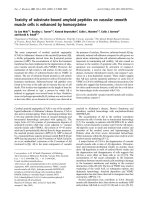
Free folding energies in different codon residuesFigure 1
Free folding energies in different codon residues. Free folding energies (FFE) were determined in phase-selected subse-
quences of 81 different genes. The original nucleic acids contained the intact three-letter codons (1
st
+2
nd
+3
rd
). Subsequences
were constructed by periodic removal of one letter from the codon and maintaining the other two (1
st
+2
nd
, 1
st
+3
rd
, 2
nd
+3
rd
)
or removing two letters and maintaining only one (1
st
, 2
nd
, 3
rd
). Distinction was made between exons (B and D) and the pre-
ceding (-1, A) and following (+1, C) sequences (introns). The dG values were determined by mfold and the FFE was calculated.
Each bar represents the mean ± SEM, n = 81.
Theoretical Biology and Medical Modelling 2006, 3:28 />Page 4 of 11
(page number not for citation purposes)
nificant that the FFEs for the 1
st
and 3
rd
residues are
additive and together they represent the entire FFE of the
intact mRNA (Figure 1D).
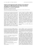
There is a correlation between the protein structure and
the FFEs associated with codon residues. This correlation
is especially prominent when the FFE ratios are compared
to the helix/sheet ratios (Figure 2).
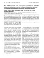
The unique, codon-related FFE pattern and its correlation
to alpha helix content suggested some similarity between
protein structures and the possible structures of the cod-
ing sequences. This possibility was examined by visual
comparison of 16 randomly selected protein residue con-
tact maps and the energy dot plots of the corresponding
RNAs. We could see similarities between the two different
kinds of maps (Figure 3). However, this type of compari-
son is not quantitative and direct statistical evaluation is
not possible.
Another similar, but still not quantitative, comparison of
protein and coding structures was performed on four pro-
teins that are known to have very similar 3D structures
although their primary structures (sequences), and their
mRNA sequences, are less than 30% similar. These four
proteins exemplify the fact that the tertiary structures of
proteins are much more conserved than the amino acid
sequences. We asked whether this is also true for the RNA
structures and sequences. We found that there are signs of
conservation even in the RNA secondary structure (as
indicated by the energy dot plots) and there are similari-
ties between the protein and nucleic acid structures (Fig-
ure 4). Comparisons of the protein residue contact map
with the nucleic acid folding maps suggest similarities
between the 3D structures of these different kinds of mol-
ecules. However, this is a semi-quantitative method.
More direct statistical support might be obtained by ana-
lyzing and comparing residue co-locations in these struc-
tures. Assume that the structural unit of mRNA is a tri-
nucleotide (codon) and the structural unit of the protein
is the amino acid. The codon may form a secondary struc-
ture by interacting with other codons according to the WC
base complementary rules, and contribute to the forma-
tion of a local double helix. The 5'-A1U2G3-3' sequence
(Met, M codon) forms a perfect double string with the 3'-
U3A2C1-5' sequence (His, H codon, reverse and comple-
mentary reading). Suboptimal complexes are 5'-A1X2G3-
3' partially complemented by 3'-U3X2C1-5' (AAG, Lys;
AUG, Met; AGG, Arg; ACG, Thr; and CAU, His; CUU, Leu;
CGU, Arg; CCU, Pro, respectively).
I searched for some pattern in the codons of co-locating
amino acids and analyzed the frequencies of the 8 possi-
ble patterns in the 64 nucleic acid triplets (Figure 5). The
codons were either complementary to each other in all
three (-123-) or at least 2 (-12X-, 1X3-, -X23-) codon posi-
tions. In these latter cases the codon complementarity was
partial, because complementarity was not required for
one codon position (X). The complementary codons were
translated in the same (5'>3' & 3'>5', only complemen-
tary, C) or reversed and complementary (5'>3' & 5'>3',
RC) directions.
These perfectly or partially complementary codon pat-
terns, read in direct or opposite directions, defined 8 dif-
ferent ways in which amino acids can be paired on the
basis of their codon complementarities: the 8 possible
amino acid – amino acid (or protein-protein) interaction
codes.
FFE associated with codon positions vs. protein structureFigure 2
FFE associated with codon positions vs. protein
structure. Free Folding Energies associated with 1
st
, 2
nd
and
3
rd
codon residues in 78 different mRNA sequences were
calculated and compared to the helix/sheet ratios of the cor-
responding protein structures. Linear regression analyses,
where pink symbols represent the linear regression line.
Theoretical Biology and Medical Modelling 2006, 3:28 />Page 5 of 11
(page number not for citation purposes)
Our experiments with FFE indicate that local nucleic acid
structures are formed under this suboptimal condition,
i.e., when the 1
st
and 3
rd
codon residues are complemen-
tary but the 2
nd
is not. If this is the case, and there is a con-
nection between nucleic acid and protein 3D structures,
one might expect that the 4 amino acids coded by 5'-
A1X2G3-3' codons will preferentially co-locate with 4
other amino acids coded by 3'-U3X2C1-5' codons. We
have constructed 8 different complementary codon com-
binations and found that the codons of co-locating amino
acids are often complementary at the 1
st
and 3
rd
positions
and follow the D-1X3/RC-3X1 formula but not the 7 other
formulae (Figure 6A–B). This means that amino acids that
are coded by partially reverse and complementary codons
(WC base pairs at the 1
st
and 3
rd
codon positions and
translated in reverse orientation) are preferentially co-
located in protein structures.
Discussion
It is well known that coding and non-coding DNA
sequences (exons/introns) are different and this differ-
ence is somehow related to the asymmetry of the codons,
Comparison of protein and corresponding mRNA structuresFigure 3
Comparison of protein and corresponding mRNA structures. Residue contact maps (RCM) were obtained from the
PBD files of protein structures using the SeqX tool (left triangles). Energy dot plots (EDP) for the coding sequences were
obtained using the mfold tool (right triangles). The two kinds of maps were aligned along a common left diagonal axis to make
possible an easy visual comparison of the different kind of representations. The black dots in the RCMs indicate amino acids
that are within 6Å of each other in the protein structure. The colored (grass-like) areas in the EDPs indicate the energetically
mostly likely RNA interactions (color code in increasing order: yellow, green, red, black).
Theoretical Biology and Medical Modelling 2006, 3:28 />Page 6 of 11
(page number not for citation purposes)
Comparison of protein and mRNA secondary structuresFigure 4
Comparison of protein and mRNA secondary structures. Residue contact maps (RCM) were obtained from the PBD
files of 4 protein structures (1CBI, 1EIO, 1IFC, 1OPA) using the SeqX tool (left column). Energy dot plots (EDP) for the coding
sequences were obtained using the mfold tool (right column). The left diagonal portions of these two kinds of maps are com-
pared in the central part of the figure. Blue horizontal lines in the background correspond to the main amino acid co-location
sites in the RCM. Intact RNA (123) as well as subsequences containing only the 1st and 3rd codon letters (13) are compared.
The black dots in the RCMs indicate amino acids that are within 6Å of each other in the protein structure. The colored (grass-
like) areas in the EDPs indicate the energetically most likely RNA interactions (color code in increasing order: yellow, green,
red, black).
Theoretical Biology and Medical Modelling 2006, 3:28 />Page 7 of 11
(page number not for citation purposes)
Amino acid pairs coded by complementary codonsFigure 5
Amino acid pairs coded by complementary codons. Two optimal (perfect) and six suboptimal (partial) codon comple-
mentarity situations (codon codes) are listed. In the perfect complementarity situation a codon (AUG), which is transcribed
from the sense (pos) DNA strand in the 5'>3' direction, is complemented with the UAC codon that is transcribed from the
antisense (neg) DNA strand in complementary (C-123) or reverse-complementary (RC-321) orientations. In the suboptimal
codon codes, one codon residue is undefined (X) and may or may not complemented in the corresponding codon on the neg-
ative strand in that residue position. Translation of the codon and its complementary pair will result in different amino acid
pairs, depending on the codon pattern. This is illustrated in examples where the undefined X residues uniformly replaced by A
in the positive and U in the negative strands. For example, the meaning of 5'-AAG-3'/3'-UUC-5' (from the D_1X3/RC_3X1
codon code pattern) is that this codon pair will be translated into the amino acids Lys (K) and Leu (L) and will result in K><L
residue pairs. Letter -P at the end of a codon pattern indicates the presence (-P), in contrast to the non-presence (-N), of that
particular codon code to determine a specific amino acid co-location in a concrete protein structure (this is used in Figure 6).
Theoretical Biology and Medical Modelling 2006, 3:28 />Page 8 of 11
(page number not for citation purposes)
i.e. that the third codon letter (wobble) is less important
in defining the meaning of the codon than the first and
second letters. Many Markov models have been formu-
lated to find this asymmetry and predict coding sequences
(genes) de novo. These in silico methods work rather well
but not perfectly and some scientists remain unconvinced
that codon asymmetry explains the exon-intron differ-
ences satisfactorily.
Another codon-related problem is that the well-known,
non-overlapping, triplet codon translation process is
extremely phase-dependent and there is theoretically no
tolerance for any phase shift. There are famous examples
of single nucleotide deletions that destroy the meaningful
translation of a sequence and are incompatible with life.
However, considering the magnitude and complexity of
the eukaryotic proteome, the precision of translation is
astonishingly good. Such physical precision is not possi-
ble without a massive and consistent physico-chemical
underpinning. Therefore, discovery of the existence of sec-
ondary structure bias (folding energy differences) in cod-
ing regions of many organisms [13] was very welcome
because it clearly defined codon boundaries on a physico-
chemical basis.
Our experiments with free folding energy (FFE) confirmed
that this bias exists. In addition, there is a very consistent
and very significant pattern of FFE distribution along the
Complementary codes vs. amino acid co-locationsFigure 6
Complementary codes vs. amino acid co-locations. A: The propensities for the 400 possible amino acid pairs were
monitored in 81 different protein structures with the SeqX tool. The tool detected co-locations when two amino acids were
closer than 6Å to each other (neighbors on the same strand were excluded). The total number of co-locations was 34,630.
Eight different complementary codes were constructed for the codons (two optimal and six suboptimal). In the two optimal
codes all three codon residues (123) were complementary (C) or reverse-complementary (RC) to each other. In the subopti-
mal codes only two of three codon residues were C or RC to each other (12, 13, 23), while the third was not necessarily com-
plementary (X). (For example, complementary code RC_3X1 means that the first and third codon letters are always
complementary (to D_1X3), but not the second, and the possible codons are read in reverse orientation). The 400 co-loca-
tions were divided into 20 subgroups corresponding to 20 amino acids (one of the co-locating pairs), each group containing 20
amino acids (corresponding to the other amino acids in each co-locating pair). If the codons of the amino acid pairs followed
the predefined complementary code, the co-location was regarded as positive (P); if not, the co-location was regarded as neg-
ative (N). Each symbol represents the mean frequency of P or N co-locations corresponding to the indicated amino acid.
Paired Student's t-test, n = 20. B: The ratio of positive (P) and negative (N) co-locations was calculated on data from (A). Each
bar represents the mean ± SEM, n = 20.
Theoretical Biology and Medical Modelling 2006, 3:28 />Page 9 of 11
(page number not for citation purposes)
nucleotide sequence. Comparing the FFEs of phase-
selected subsequences, those subsequences comprising
only the 1
st
or only the 3
rd
codon letters showed signifi-
cantly higher FFE than those consisting only of the 2
nd
let-
ters. This FFE difference was not present in the intronic
sequences preceding and following the exons, but it was
present in exons from different species. This is an interest-
ing observation because these phenomena might not only
distinguish between exons and introns on a physico-
chemical basis, but might also clearly define the tri-nucle-
otide codons and thus the phase of the translation. This
codon-related phase-specific variation in FFE may explain
why mRNAs have greater negative free folding energies
than shuffled or codon choice randomized sequences
[21].
Free folding energy in nucleic acids is always associated
with WC base pair formation. A higher FFE indicates more
WC pairs (presence of complementarity) and a lower FFE
indicates fewer WC pairs (less complementarity). The
FFEs in the 1
st
and 3
rd
codon positions were additive,
while the 2
nd
letter did not contribute to the total FFE; the
total FFE of the entire (intact) nucleic acid was the same as
that of subsequences containing only the 1
st
and 3
rd
codon
letters (2
nd
deleted). This indicates that local RNA second-
ary structure bias is caused by complementarity of the 1
st
and 3
rd
codon residues in local sequences. This partial,
local complementarity is more optimal in reverse orienta-
tion of the local sequences, as expected with loop forma-
tions.
FFEs are obtained by considering free folding energies of
substrings that do not represent the real molecule: forcing
nucleotides to be consecutive is an extreme methodologi-
cal approach to measuring the structure features of coding
sequences. However, this bioinformatical method has
been successfully used by others [13,21]. In addition, the
behaviors of the 1
st
, 2
nd
and 3
rd
codon bases separately are
useful for showing that the 2
nd
codon position does not
have the same significance in the codons as the other two
positions [20]. Intronic sequences do not contain codons
and consequently thy show no position-related periodic-
FFE variation.
It is known that single-stranded RNA molecules can form
local secondary structures through the interactions of
complementary segments. The novel observation here is
that these interactions preferentially involve the 1
st
and 3
rd
codon residues. This connection between the RNA sec-
ondary structure and codons immediately directs atten-
tion toward the question of protein folding and its long-
suspected connection to RNA folding [22,23].
Only about one-third (20/64) of the genetic code is used
for protein coding, i.e., there is a great excess of informa-
tion in the mRNA. At the same time, the information car-
ried by amino acids seems to be insufficient (as stated by
some scientists) to complete unambiguous protein fold-
ing. Therefore, it is believed that the third codon residue
(wobble base) contains information additional to that
already present in the genetic code. A specialized data-
base, the ISSD [19], was established in an effort to connect
different features of protein structure to wobble bases [24]
with more or less success.
We found a significant correlation between FFE ratios and
the helix/sheet contents of protein structures. It was possi-
ble to make direct visual comparison of mRNA structures
(as statistically predicted by mfold energy dot-plots) and
protein structures (as 2D residue contact maps). This
method suggests similarity between nucleic acid and pro-
tein structures.
It is known that some complex protein structures are very
similar even if there is less than 30% sequence similarity.
It was interesting to see whether the same principle might
apply to nucleic acids, and structural similarity might exist
even when the sequence similarity is low. Furthermore,
significant similarity between nucleic acid and protein
structures might exist even without translational connec-
tion. Structure seems to be more preserved, even in
nucleic acids, than sequence. However, although the
matrix comparisons are suggestive, they remain semi-
quantitative. Better support was necessary.
A working hypotheses grew out of these observations,
namely that (a) partial, local reverse-complementarity
exists in nucleic acids that form the nucleic acid structure;
(b) there is some degree of similarity between the folding
of nucleic acids and proteins; (c) protein structure deter-
mines the amino acid co-locations; (4) in consequence,
amino acids coded by interacting (partially reverse com-
plementary) codons might show preferential co-locations
in the protein structures. And it seems to be the case:
codons that contain complementary bases at the 1
st
and
3
rd
positions and are translated in reverse orientation
result in preferentially co-located (interacting) amino
acids in the 3D protein structure. Other complementary
residue combinations or translation in the same (not
reverse) direction (as many as seven combinations in
total) did not result in any preferentially co-locating sub-
set of amino acid pairs.
Construction of residue contact maps for protein struc-
tures and statistical evaluation of residue co-locations is a
frequently used method for visualization and analysis of
spatial connections among amino acids [25-27]. The
amino acid co-locations in real protein structures are
clearly not random [28,29] and therefore residue co-loca-
tion matrices are often used to assist in the prediction of
Theoretical Biology and Medical Modelling 2006, 3:28 />Page 10 of 11
(page number not for citation purposes)
novel protein structures [30,31]. We have carefully exam-
ined the physico-chemical properties of specifically inter-
acting amino acids in and between protein structures, and
concluded that these interactions follow the well-known
physico-chemical rules of size, charge and hydrophobic
compatibility (unpublished data), well in line with Anfin-
sen's prediction. The recent study supports the conclusion
that there is a previously unknown connection between
the codons of specifically interacting amino acids; those
codons are complementary at the 1
st
and 3
rd
(but not the
2
nd
) codon positions.
The idea that sequence complementarity might explain
the nature of specific protein-protein interactions is not
new and was suggested as long ago as 1981 [32,35,36]. I
was never able to confirm my own original theory experi-
mentally, the suggestion of perfect complementarity
between codons of interacting amino acids [32,33],
though others were more successful [34]. The explanation
is that codon complementarity is suboptimal and does
not involve the 2
nd
codon residue. Experimental in vitro
confirmation is required to validate this recent theoretical
and in silico prediction.
Availability: />: SeqX,
SeqForm.
Acknowledgements
The author of this article (J.C.B.) believes that he was the first scientist sug-
gesting the existence of a "proteomic code". The original idea was published
in 1981 in the Medical Hypotheses [32,35,36] as well as some aspects of the
recent concept of a "protein-protein interaction code" [37] that was fur-
ther developed in this article.
References
1. Anfinsen CB, Redfield RR, Choate WI, Page J, Carroll WR: Studies
on the gross structure, cross-linkages, and terminal
sequences in ribonuclease. J Biol Chem 1954, 207:201-210.
2. Levinthal C: How to fold graciously in Mossbauer spectroscopy
in biological systems. In Proceedings of a Meeting held at Allerton
House, Monticello, IL Edited by: Debrunner P, Tsibris JCM, Munck E.
Urbana, IL: University of Illinois Press; 1969:22-24.
3. Klepeis JL, Floudas AC: ASTRA-FOLD: a combinatorial and glo-
bal optimization framework for ab initio prediction of three-
dimensional structures of proteins from the amino acid
sequence. Biochem J 2003, 85:2119-2146.
4. Walter S, Buchner J: Molecular chaperones – cellular machines
for protein folding. Angew Chem Int Ed Engl 2002, 41:1098-1113.
5. Komar AA, Kommer A, Krasheninnikov IA, Spirin AS: Cotransla-
tional folding of globin. J Biol Chem 1997, 272:10646-10651.
6. Thanaraj TA, Argos P: Protein secondary structural types are
differentially coded on messenger RNA. Protein Sci 1996,
5:1973-1983.
7. Brunak S, Engelbrecht J: Protein structure and the sequential
structure of mRNA: alpha-helix and beta-sheet signals at the
nucleotide level. Proteins 1996, 25:237-252.
8. Gupta SK, Majumdar S, Bhattacharya TK, Ghosh TC: Studies on the
relationships between the synonymous codon usage and pro-
tein secondary structural units. Biochem Biophys Res Commun
2000, 269:692-696.
9. Chiusano ML, Alvarez-Valin F, Di Giulio M, D'Onofrio G, Ammirato
G, Colonna G, Bernardi G: Second codon positions of genes and
the secondary structures of proteins. Relationships and
implications for the origin of the genetic code. Gene 2000,
261:63-69.
10. Gu W, Zhou T, Ma J, Sun X, Lu Z: The relationship between syn-
onymous codon usage and protein structure in Escherichia
coli and Homo sapiens. Biosystems 2004, 73:89-97.
11. Ermolaeva O: Synonymous codon usage in bacteria. Curr Issues
Mol Biol 2001, 3:91-97.
12. Biro JC, Biro JM, Biro AM: Hidden massages in hidden sub-
sequences: a study on collagens. 30th FEBS Congress – 9th IUBMB
Conference, Budapest, Hungary, 2–7 July 2005 2005. abstract.
13. Katz L, Burge CB: Widespread selection for local RNA second-
ary structure in coding regions of bacterial genes. Genome Res
2003, 13:2042-2051.
14. Zuker M: Mfold web server for nucleic acid folding and hybrid-
ization prediction. Nucleic Acids Res 2003, 31:3406-3415.
15. Biro JC, Fordos G: SeqX: a tool to detect, analyze and visualize
residue co-locations in protein and nucleic acid structures.
BMC Bioinformatics 2005, 6:170 [ />].
16. Biro JC: SeqForm. 2005 [ />].
17. Berman HM, Westbrook J, Feng Z, Gilliland G, Bhat TN, Weissig H,
Shindyalov IN, Bourne PE: The Protein Data Bank. Nucleic Acids
Res 2000, 28:235-242 [ />].
18. Berman HM, Olson WK, Beveridge DL, Westbrook J, Gelbin A,
Demeny T, Hsieh SH, Srinivasan AR, Schneider B: The Nucleic Acid
Database: a comprehensive relational database of three-
dimensional structures of nucleic acids. Biophys J 1992,
63:751-759 [ />].
19. Adzhubei IA, Adzhubei AA: ISSD Version 2.0: taxonomic range
extended. Nucleic Acids Res 1999, 27:268-271 [
tein.bio.msu.su/issd/].
20. Biro JC, Benyo B, Sansom C, Szlavecz A, Fordos G, Micsik T, Benyo
Z: A common periodic table of codons and amino acids. Bio-
chem Biophys Res Commun 2003, 306:408-415.
21. Seffens W, Digby D: mRNA has greater negative folding free
energies than shuffled or codon choice randomized
sequences. Nucleic Acids Res 1999, 27:1578-1584.
22. Oresic M, Dehn M, Korenblum D, Shalloway D: Tracing specific
synonymous codon-secondary structure correlations
through evolution. J Mol Evol 2003, 56:473-4840.
23. D'Onofrio G, Ghosh TC, Bernardi G: The base composition of
the genes is correlated with the secondary structures of the
encoded proteins. Gene 2002, 300:179-187.
24. Xie T, Ding D: The relationship between synonymous codon
usage and protein structure. FEBS Lett 1998, 434:93-96.
25. Kumarevel TS, Gromiha MM, Ponnuswamy MN: Distribution of
amino acid residues and residue-residue contacts in molecu-
lar chaperons. Prep Biochem Biotechnol 2001, 31:163-183.
26. Eilers M, Patel AB, Liu W, Smith SO: Comparison of helix inter-
actions in membrane and soluble alpha-bundle proteins. Bio-
chem J 2002, 82:2720-2736.
27. Glaser F, Steinberg DM, Vakser IA, Ben-Tal N: Residue frequencies
at protein-protein interfaces. Proteins Struct Funct Genet 2001,
43:89-102.
28. Naor D, Fisher D, Jernigan RL, Wolfson H, Nussinov R: Amino acid
pair interchanges at spatially conserved locations. J Mol Biol
1996, 256:924-938.
29. Azarya-Sprinzak E, Naor D, Wolfson HJ, Nussinov R: Interchanges
of spatially neighboring residues in structurally conserved
environment. Protein Eng 1997, 10:1109-1122.
30. Singer MS, Vriend G, Bywater RP: Prediction of protein residue
contacts with a PDB-derived likelihood matrix. Protein Eng
2002, 15:721-725.
31. Shao Y, Bystroff C: Predicting inter-residue contacts using
templates and pathways. Proteins Struct Funct Genet 2003,
53:497-502.
32. Biro J: Comparative analysis of specificity in protein-protein
interactions. Part II: The complementary coding of some
proteins as the possible source of specificity in protein-pro-
tein interactions. Med Hypotheses 1981, 7:981-993.
33. Segersteen U, Nordgren H, Biro JC: Frequent occurrence of
short complementary sequences in nucleic acids. Biochem Bio-
phys Res Commun 1986, 139:94-101.
34. Hela JR, Roberts GW, Raynes JG, Bhakoo A, Miller AD: Specific
interactions between sense and complementary peptides:
the basics for the proteomic code. Chembiochem 2002,
3:136-151.
Publish with BioMed Central and every
scientist can read your work free of charge
"BioMed Central will be the most significant development for
disseminating the results of biomedical research in our lifetime."
Sir Paul Nurse, Cancer Research UK
Your research papers will be:
available free of charge to the entire biomedical community
peer reviewed and published immediately upon acceptance
cited in PubMed and archived on PubMed Central
yours — you keep the copyright
Submit your manuscript here:
/>BioMedcentral
Theoretical Biology and Medical Modelling 2006, 3:28 />Page 11 of 11
(page number not for citation purposes)
35. Biro J: Comparative analysis of specificity in protein-protein
interactions. Part I: A theoretical and mathematical
approach to specificity in protein-protein interactions. Med
Hypotheses 1981, 7:969-79.
36. Biro J: Comparative analysis of specificity in protein-protein
interactions. Part III: Models of the gene expression based on
the sequential complementary coding of some pituitary pro-
teins. Med Hypotheses 1981, 7:995-1007.
37. Biro JC: A novel intra-molecular protein-protein interaction
code based on partial complementary coding of co-locating
amino acids. Med Hypotheses 2006, 66:137-42.