Báo cáo y học: "The contemporary role of blood products and components used in trauma resuscitation" pdf
Bạn đang xem bản rút gọn của tài liệu. Xem và tải ngay bản đầy đủ của tài liệu tại đây (1.1 MB, 17 trang )
REVIEW Open Access
The contemporary role of blood products and
components used in trauma resuscitation
David J Dries
Abstract
Introduction: There is renewed interest in blood product use for resuscitation stimulated by recent military
experience and growing recognition of the limitations of large-volume crystalloid resusci tation.
Methods: An editorial review of recent reports published by investigators from the United States and Europe is
presented. There is little prospective data in this area.
Results: Despite increasing sophistication of trauma care systems, hemorrhage remains the major cause of early
death after injury. In patients receiving massive transfusion, defined as 10 or more units of packed red blood cells
in the first 24 hours after injury, administration of plasma and platelets in a ratio equivalent to packed red blood
cells is becoming more common. There is a clear possibility of time dependent enrollment bias. The early use of
multiple types of blood products is stimulated by the recognition of coagulopathy after reinjury which may occur
as many as 25% of patients. These patients typically have large-volume tissue injury and are acidotic. Despite early
enthusiasm, the value of administration of recombinant factor VIIa is now in question. Another dilemma is
monitoring of appropriate component administration to control coagulopathy.
Conclusion: In patients requiring large volumes of blood products or displaying coagulopathy after injury, it
appears that early and aggressive administration of blood component therapy may actually reduce the aggregate
amount of blood required. If recombinant factor VIIa is given, it shoul d be utilized in the fully resuscitated patient.
Thrombelastograp hy is seeing increased application for real-time assessment of coagulation changes after injury
and directed replacement of components of the clotting mecha nism.
Pathogenesis of Acute Coagulopathy After
Trauma
Historical Perspective
Hemorrhagic shock accounts for a significant number of
deaths in patients arriving at hospital with acute injury
[1,2]. Patients with uncontrolled hemorrhage continue
to succumb despite adoption of damage control techni-
ques and improved transpo rt and emergency care. Coa-
gulopathy, occurring even before resuscitation,
contributes significantly to the morbidity associated with
bleeding [3,4]. Recognition of the morbidity associated
with bleeding and coagulation abnormality goes back t o
the work of Simmons and coworkers during the Viet-
nam conflict [5]. Even at that time, standard tests
including prothro mbin time (PT) and partial thrombo-
plastin time (PTT) correlated poorly with acute
resuscitation efforts. Similar work in the late 1970s was
performed in civilian patients receiving massive transfu-
sion. Again, PT, PTT and bleeding time were only help-
ful if markedly prolonged [6].
Lucas and Ledgerwood performed a variety of studies
in large animals and patients to determine changes in
the coagulation profile with hemorrhagic shock [7]. In
patient studies, platelet count fell until 48 hours after
injury and increased dramatically during convalescence.
Bleeding times and platelet aggregation studies mirrored
platelet levels. Re ductions in fibrinogen, Factor V and
Factor VIII were noted with hemorrhagic shock which
normalized by day one after bleeding. By day four after
bleeding, fibrinogen increased to supranormal levels.
Clotting times mirrored fibrinogen, Factor V and Factor
VIII levels. These investigators then studied the role of
Fresh Frozen Plasma (FFP) supplementation in hemor-
rhagic shock with two studies. In animal studies, sub-
jects received shed blood and crystalloid with some
Correspondence:
Regions Hospital, 640 Jackson Street, St. Paul, MN 55101 University of
Minnesota, 420 Delaware Street SE, Minneapolis, MN 55455, USA
Dries Scandinavian Journal of Trauma, Resuscitation and Emergency Medicine 2010, 18:63
/>© 2010 Dries; lice nsee BioMed Central Ltd. This is an O pen Access article distributed under the terms of the Creative Commons
Attribution License (http://creativecommons.o rg/licenses/by/2.0), whi ch pe rmits unrestricted use, distribution, and reproduction in
any medium, provided the original work is properly cited.
animals receiving Fresh Frozen Plasma. In this animal
work, Fresh Frozen Plasma did no t improve coagulation
factors, fibrinogen and Factors II, V, VII and VIII. In a
second controlled study, fresh frozen plasma was given
not only during blood volume restoration but also for
an additional hour during ongoing controlled hemor-
rhage without shock. Fresh Frozen Plasma prevented
reduction in coagulat ion factors compared to animals
not receiving fresh frozen plasma. Clotting times paral-
leled coagulation factor levels. From this work, Lucas
and Ledgerwood ultimately concluded that hemorrhagic
shock resuscitation requires restoration of blood loss
with packed cells and crystall oid while FFP is approp ri-
ately added due to losses of coagulation proteins [7].
Studies in the 1970s and 1980s provided additional
detail regarding the limitation of simple laboratory para-
meters and factor levels in evaluation of patient
response to massive transfusion [6,8]. In a study of 27
patients requiring massive transfusion, platelet counts
fell in proportion to the size of transfusion while Factors
V and VIII correlated poorly with the volume of blood
transfused. Where coagulopathy appeared, the majority
of patients responded to platelet administration. In this
early work, the most useful laboratory test for predicting
abnormal bleeding was the platelet count. A falling fibri-
nogen level was felt to b e indicative of DIC. The bleed-
ing time, prothrombin time and partial thromboplastin
time were not helpful in assessing the cause of bleeding
unless they were greater than 1.5 times the control
value [6]. In a subsequent series of studies from the
same investigative group, 36 massively transfused
patients were followed for microvascular bleeding. Mod-
erate deficiencies in the clotting factors evaluated were
comm on but they were not as sociated with m icrovascu-
lar bleeding. Microvascular bleeding was associated with
severe coagulation abnormalities such as clotting factor
levels less than 20% of control. In statistical analysis,
clotting factor activities less than 20% of control were
reliably reflected by significant prolongation of PT and
PTT. These investigators also suggested that e mpiric
blood replacement formulas available at the time were
not likely to prevent microvascular bleeding because
consumption of platelets or clotting factors did not con-
sistently appear and simple dilution frequently did not
correspond to microvascular bleeding [8].
The attention of the American trauma community was
drawn to coagulopathy after trauma with description of
the “blo ody vicious cycle” by the Denver Health team
over 20 years ago [3]. These investigators noted the con-
tribution of hypothermia, acidosis and hemo dilution
associated with inadequate resuscitation and excessive
use of crystalloid. Subsequent work extended these
observations describing early coagulopathy which could
be independent of clotting factor deficiency (consistent
with scattered earlier observations) [9]. Moore and
others, in a recent multicenter trial of hemoglobin oxy-
gen carriers, observed ea rly coagulopathy in the setting
of severe injury, which was present in the field, prior to
Emergen cy Department arrival and initiation of resusci-
tation. Coagulopathic patients were at increased risk f or
organ failure and mortality. One concern in the presen-
tation of these patients was inconsistency in available
laboratory data which identified patients at risk [10].
Dating to development of Advanced Trauma Life Sup-
port, trauma teams have used fixed guidelines for
plasma and platelet replacement during massive transfu-
sion to prevent and correct dilutional coagulopathy.
Empiric plasma and platelet replacement was based on
washout physiology, a mathematical model of exchange
transfusion. The model assumes stab le blood volume
and calc ulates exponential decay of each blood compo-
nent with bleeding. In severe injury, however, these
assumptions may not apply: blood volume fluctuates
widely and bleeding rates vary with blood pressure and
replacement frequently lags behind blood loss. Replace-
ment guidelines based on simple washout physiology
may be inadequate [11-14].
In one of the first papers to question historical trans-
fusion practice in the setting of massive trauma, Hirsh-
berg, Mattox and coworkers, utilizing clinical data,
developed a computer model designed to capture inter-
actions between bleeding, hemodynamics, hemodilution
and blood component replacement during severe
hemorrhage. Replacement options were offered in the
model and their effectiveness evaluated [11].
In the computer model, an intravascular compartment
was created accepting crystalloid in fusion and calculat-
ing the exchange of free water between intravascular
and interstitial spaces. The basic compartment model
was a “leaky bucket” where inflow is determined by a
clinical scenario and outflow (bleeding rate) is propor-
tional to systolic blood pressure. The effectiveness of
crystalloid resuscitation decreases during massive
hemorrhage in proportio n to the volume of blood lost.
In this computer simulation, an exponential model of
effectiveness for crystalloid resuscitation is employed.
Hemostasis was modeled by a relationship sensitive to
blood pressure with 90 mmHg associated with ongoing
bleeding and 50 mmHg associated with minimal blood
loss. The impact of dilution on prothrombin time, fibri-
nogen and plate lets were based on data obtained from
dilution curves in the hospital coagulation laboratory
from patients with significant hemorrhage. Standard
product replacement quantities were assumed [11,15,16].
After setting thresholds for acceptable loss of clotting
factors, platelets and fibrinogen, the authors modeled
behavior of coagulation during rapid exsanguination
without clotting factor or platelet replacement. The
Dries Scandinavian Journal of Trauma, Resuscitation and Emergency Medicine 2010, 18:63
/>Page 2 of 17
prothrombin time reached a critical level first followed by
fibrinogen and platelets. If patients were resuscitated with
smaller amounts of crystalloid, leaving overall blood
volume reduced, the effective life of components of the
coagulation cascade was increased. More aggressive Fresh
Frozen Plasma (FFP) replacement was indicated by this
model. The optimal ratio for administration of FFP to
packed red blood cells (PRBCs) in this analysis was 2:3.
Delayed administration of FFP led to critical clotting factor
deficiency regardless of subsequent administration of FFP.
Fibrinogen depletion was easier to correct. Even after
administration of 5 units of PRBCs, the hemostatic thresh-
old for fibrinogen was not exceeded if a FFP to PRBC ratio
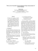
of 4:5 was employed. Analysis of platelet dilution show
that even if platelet replacement was delayed until 10 units
of PRBCs were infused, critical platelet dilution was pre-
vented with a subsequent platelet to PRBC ratio of 8:10
[11] (Figure 1).
The essential message of this work is that massive
transfusion protocols in existence when this study was
performed provide inadequate clotting factor replace-
ment during exsanguinating hemorrhage and neither
prevent or correct dilutional coagulopathy.
Acute Coagulopathy of Trauma
Brohi and coworkers from the United Kingdom helped
to reinvigorate discussion of scattered seminal
observations regarding coagulopathy after injury by
adding new coagulation laboratory techniques to earlier
clinical observations [17]. R eviewing over 1,000 c ases,
patients with acute coagulopathy had higher mortality
throughout the spectrum of Injury Severity Scores (ISS).
Contrary to historic teaching that coagulopathy was a
function of hemodilution with massive crystalloid
resuscitation, these authors noted that the incidence of
coagulopathy increased with severity of injury but not
necessarily in relationship to the volume of intravenous
fluid administered to patients. Brohi and others helped
to reemphasize the obse rvation that acute coagulopathy
could occur before significant fluid administration which
was a ttributable to the injury itself and proportional to
the volume of injured tissue. Development of coagulopa-
thy was an independent predictor of poor outcome.
Mediators associated with tissue trauma includi ng
humoral and cellular immune system activation with
coagulation, fibrinolysis, complement and kallikrein cas-
cades have since been associated with changes in hemo-
static mechanisms in the body similar to those identified
in the setting of sepsis [17-19,1].
MacLeod, in a recent commentary, discussed factors
contributing to coagulo pathy in the setting of t rauma
[20]. That hypothermia relates to development of coagu-
lopathy has been demonstrated in vitro and in clinical
studies. Temperature reduction impairs platelet aggre ga-
tion and decreases function of coagulation factors in
non-diluted blood. Patients with temperature reduction
below 34°C had elevated prothrombin and partial
thromboplastin times. Coagulation, like most biological
enzyme systems, works best at normal temperature.
Similarly, acidosis occurring in the setting of trauma as
a result of bleeding and hypotension also contributes to
clotting failure. Animal work shows that a pH <7.20 is
associated with hemostatic impairment. Platelet dysfunc-
tion and coagulation enzyme system changes are noted
when blood from healthy volunteers is subjected to an
acidic environment [21,22].
We are now noting that with or without hypothermia
and a cidosis post-traumatic coagulopathy may develop
in a significant number of patients. Possible explanations
for this phenomenon include factor dilution, clotting
system depletion and disseminated intravascular coagu-
lation. Interplay of these and other factors in the face of
ongoing blood loss is still not understood. Crystalloids
and colloids can dilute available clotting factors. Increas-
ing microvascular tissue injury may deplete the coagula-
tion system due to demands of hemorrhage control at
multiple sites. Third, and most interesting, loss of c lot-
ting factors associated with exaggerated inflammation is
now being reported in association with injury. The pre-
sence of predictors of coa gulopathy has been suggested
by historical data from the United States and the
Figure 1 Behavior of the computer model for massive bleeding
without replacement of clotting factors or platelets. Bleeding
fraction is the volume of blood lost divided by the estimated blood
volume (4,900 mL). Early loss of clotting factors is seen. (Dotted line
is threshold for critical component deficit.)
Dries Scandinavian Journal of Trauma, Resuscitation and Emergency Medicine 2010, 18:63
/>Page 3 of 17
European Union. While flaws exist in this p reliminary
epidemiologic data, it is now clear that coagulation
changes after injury reflect more than the amount of
crystalloid given [21-24].
Hess and coworkers as part of an international medi-
cal collaboration (The Educational Initiative on Critical
Bleeding in Trauma) developed a literature review to
increase awareness of coagulopathy independent of crys-
talloid administration following trauma [19]. The key
initiating factor is tissue injury. This is borne out by ori-
ginal work demonstrating the c lose association between
tissue injury and the degree of coagulopathy. Patients
with severe tissue injury but no physiologic derange-
ment, however, rarely present with coagulopathy and
have a lower mortality rate [25,26]. Tissue damage initi-
ates coagulation as endothelial injury at the site of
trauma leads to exposure of subendothelial collagen and
Tissue Factor which bind von Willebrand factor, plate-
lets and activated Factor VII (FVII). Tissue Factor or
FVII activate plasma coagulation and thrombin and
fibrin are formed. A subsequent amplification process
mediated by factor IX may take place on the surfac e of
activated platelets [27].
Hyperfibrinolysis is seen as a direct consequence of
the combination of tissue injury and shock. Endothelial
injury accelerates fibrinolysis because of direct release of
Tissue Plasminogen Activator [19,28]. Tissue Plasmino-
gen Activator expression by endothelium is increased in
the presence of thrombin. Fibrinolysis is accelerated
because of the combined affects of endothelial Tissue
Plasminogen Activator release due to ischemia and inhi-
bition of Plasminogen Activator Inhibitor i n shock.
While hyperfibrinolysis may focus c lot propagation on
the sites of actual vascular injury, with widespread
insults, this localization may be lost. Specific organ inju-
ries have been associated with coagulopathy. Traumatic
brain injury has been noted with increased bleeding
thought due to release of brain-specific thromboplastins
with subsequent inappropriate clotting factor cons ump-
tion. Hyperfibrinolysis has also been seen in more recent
studies of head-injured patients. Long bone fractures
along with brain and massive soft tissue injury also may
prime the patient f or coagulopathy [29,30]. These con-
tributing factors, however, are inadequate to lead to cat-
astrophic coagulopathy if present in isolation.
A n umber of important cofactors must be present to
stimulate coagulopathy in t he setting of trauma [19].
Shock is a dose -dependent cause of tissue hypoperfu-
sion. Elevated base deficit has been associated with
coagulopathy in as many as 25% of patient s in one large
study. Progression of shock appears to result in hyper fi-
brinolysis. The exact processes involved are unclear.
One mediator implicated in coagulopathy after
injury is Activated Protein C. Immediate post-injury
coagulopathy is likely a combination of effects caused by
large volume tissue trauma and hypoperfusion.
Several other historic factors are acknowledged for
their contribution to coagulopathy after trauma. Hess
and others continue to acknowledge the impact of dilu-
tion of coagulation factors with crystalloid resuscitation
aft er injury [19]. While ackno wledging inadequat e clini-
cal data at present, equivalent ratios of FFP, PRBCs and
platelets must be considered for management of coagu-
lopathy after injury. Hypothermia and acidemia are con-
trolled to reduce their impact on enzyme systems [ 31].
Inflammation is receiving greater attention as a conse-
quence of severe injury. Recent data suggests earlier
activation of the immune system after injury than pre-
viously proposed. Similar to sepsis, cross-talk has been
noted between coagulation and inflammation systems.
Activation of coagulation proteases may induce inap-
propriate inflammatory response t hrough cell surface
receptors and activation of cascades such as Comple-
ment and platelet degranulation [32-34]. Trauma
patients are initially coagulopathic with increased bleed-
ing but may prog ress to a hypercoagulable state putting
them at increased risk for thrombotic events. This late
thrombotic state bears similarities with coagulopathy of
severe sepsis and depletion of Protein C. Injured and
septic patients share a propensity toward multiple organ
failure and prothrombotic states. A diagram displaying
the interrelated mechanisms contributing to coagulopa-
thy after trauma is presented (Figure 2).
Blood Component Therapy and the “Ra tio”
Despite work from multiple groups suggesting that sim-
ple replacem ent of packed red blood cells was not a suf-
ficient answer to the most severely injured patient,
particularly in the setting of coagulopathy, the concept
of combination blood component replacement remained
outside the mainstream of trauma care for ove r 20 years
[7,8,3]. In part, this may reflect the difficulty in cha rac-
terizing coagulopathy after injury due to limitations of
Figure 2 Diag ram showing some of mechanisms leading to
coagulopathy in the injured. ACoTS = Acute Coagulopathy of
Trauma-Shock.
Dries Scandinavian Journal of Trauma, Resuscitation and Emergency Medicine 2010, 18:63
/>Page 4 of 17
static testing as described above. It took additional con-
flicts in the Middle East and experience in a multina-
tional group of trauma centers to bring awareness of the
need for multiple blo od component therapy in massive
bleeding to the level of general trauma practice.
The 1970s and 1980s saw several groups propose
resuscitation of significant hemorrhage with combina-
tions of blood components. Kas huk and Moore pro-
posed multicomponent blood therapy in patients with
significant vascular injury [3]. In a study of patients with
major abdominal vascular injury, Kashuk and coworkers
noted frequent deviation from a standard ratio of 4:1 or
5:1 for units of packed red blood cells to units of Fresh
Frozen Plasma. The ratio was 8:1 in nonsurvivors and
9:1 where overt coagulopathy was noted. Fifty-one per-
cent of patients in this series were coagulopathic after
vascular control was obtained. Using multivariat e analy-
sis, Ciavarella and coworke rs from the Puget Sound
Blood Center and Harborview Medical Center proposed
aggressive supplementation of platelets in the setting o f
massive transfusio n. These investigators no ted that pla-
telet counts below 50 × 10
9
per liter correlated highly
with mi crovascular ble eding in t rauma and sur gery
patients. Fibrinogen repletion w as also empha sized.
Other guides to resuscitation included fibrinogen level,
prothrombin time and partial thromboplastin time. Sup-
plemental Fresh Frozen Plasma or cryoprecipitate was
recommended for low fibrinogen levels [8]. Lucas and
Ledgerwood, summarizing extensive preclinical and clin-
ical studies, suggested administration of Fresh Frozen
Plasma after 6 units of packed red blood cells had been
infused. Additional Fresh Frozen Plasma was recom-
mended for every five additional packed red blood cell
transfusions. Monitoring included platelet count, PT
and PTT after each 5 units of packed red blood cells are
administered. Platelet transfusion is generally unneces-
sary unless the platelet count falls below 50,000 [7].
Despite this early work, blood loss continues to be the
major cause of early death after injury accounting for
50% of deaths occurring during the initial 48 hours after
hospitalization. Bleeding remains a common cause of
preventable deaths after injury [35-37]. Many centers
are beginning to establish protocols for massive transfu-
sion practice but criteria and co mpliance continues to
vary. Trauma centers are examining approaches to com-
prehensive hemostatic resuscitation as a replacement
strategy for earlier approaches based on rapid, early
infusion of crystalloids and PRBCs alone [17-20].
Rhee and coworkers, using the massive database of the
Los Angeles County Level I Trauma Center, examined
transfusion practices in 25,000 patients [38]. Approxi-
mately 16% of these patients received a blood tr ansfu-
sion. Massive transfusion (≥10 units of PRBCs per day)
occurred in 11.4% of transfused patients. After excluding
head-injured patients, these authors studied approxi-
mately 400 individuals. A trend toward increasing FFP
use was noted during the six years of data which was
reviewed (January 2000 to December 2005). Logistic
regression identified the ratio of FFP to PRBC use as an
independent predictor of survival. With a higher the
ratio of FFP:PRBC, a greater probability of survival was
noted. The optimal ratio in this analysis was an FFP:
PRBC ratio of 1:3 or less. R hee and coworkers provide a
large retrospective dataset demonstrating that earlier
more aggressive plasma replacement can be associated
with improved outcomes after bleeding requiring mas-
sive transfusion. Ratios derived in this massive retro-
spective data review support the observations of
Hirshberg, Mattox and coworkers [11]. Like the data
presented by Kashuk and coworkers in another widely
cited report, this retrospective dataset suggests improved
clinical outcome with increased administration of FFP
[39] (Figure 3).
Another view o f damage control hematology comes
from Vanderbilt University Medical Center in Nashville,
Tennessee. This group implemented a Trauma Exsan-
guination Protocol involving acute administration of 10
units PRBC with 4 units FFP and 2 units platelets. In an
18 month period, 90 patients received this resuscitation
and were compared to a historic set of controls. The
group of patients receiving the Trauma Exsanguination
Protocol as des cribed by these investigators had lower
mortality, much hi gher blood product use in ini tial
operative procedures and higher use of products in the
initial 24 hours though overall blood product consump-
tion during hospitalization was decreased [40].
The strongest multicenter civilian data examining the
impact of plasma and platelet admini strat ion along with
red blood cells on outcome in massive transfusion
comes from Holcomb and coworkers [41]. These inves-
tigators report over 450 patients obtained from 16 adult
Figure 3 Mortality Decrease with Higher FFP:PRBC Ratios.
Dries Scandinavian Journal of Trauma, Resuscitation and Emergency Medicine 2010, 18:63
/>Page 5 of 17
and pediatric cente rs. Overall survival in this group is
59%. Patients were gravely ill as reflected by an admis-
sion base deficit of -11.7, pH 7.2, Glasgow Coma Score
of 9 and a mean Injury Severity Score of 32. Examina-
tion of multicenter data reflects an improvement in out-
come as the ratio of Fresh Frozen Plasma to packed red
blood cells administered approaches 1. Fresh Frozen
Plasma, however, is not the sole solution to improved
coagulation response in acute injury. These workers also
examined the relationship of aggr essive plasma and pla-
telet administrat ion in these patients. Opt imal outcome
in this massive transfusion group was obtained with
aggressive platelet as well as plasma administration.
Worst outcomes were seen when aggr essive administra-
tion of plasma and platelets did not take place. Where
either F FP or platelets were given in higher proportion
in relationship to packed red cells intermediate results
were obtained. Not surprisingly, t he cause of death
which was favorably affected was trunc al hemorrhage.
Examination of the Kaplan-Meier curves provided by
these workers demonstrates that the impact of early
blood product administration on mortality is seen in
improved outcomes immediately after injury (Figure 4).
A summary statement comes from Holcomb and a
combination of military and civilian investigators
[18,19]. These w orkers i dentify a patient group at high
risk for coagulopathy and resuscitation failure due to
hypothermia, acidosis, hypoperfusion, inflammation and
volume of tissue injury. In the paradigm proposed by
these writers, resuscitation begins with prehospital lim-
itation of blood pressure at approximately 90 mmHg
preventing renewed bleeding from recently clotted ves-
sels. Intravascular volume resuscitation is accomplished
using thawed plasma in a 1:1 or 1:2 ratio with PRBCs.
Acidosis is managed by use of THAM and vol ume load-
ing with blood components as hemostasis is obtained.
These workers utilize rFVIIa “ occasionally” along with
early units of red cells. A massive transfusion protocol
for these investigators included delivery of packs of 6
units of plasma, 6 units of PRBC, 6 units of platelets
and 10 units of cryoprecipitate in stored individual cool-
ers. These coolers are continued until notification
comes from the trauma team. Even in causalit ies requir-
ing resuscitation with 10-40 units of blood products,
Holco mb and coworkers found that as little as 5-8 liters
of crystalloid are utilized during the first 24 hours repre-
senti ng a decrease of at least 50% compared to standard
practice. The lack of intraoperative coagulopathic bl eed-
ing allows surgeons to focus on surgical hemorrhage.
ThegoalisarrivalofthepatientinICUinawarm,
euvolemic and nonacidotic state. INR approaches nor-
mal and edema is minimized. Subjectively, pa tients trea-
ted in this way are more easily ventilated and easier to
extubate than patients with a similar blood loss treated
with standard crystalloid resuscitation and smaller
amounts of blood products. Clearly, these clinical obser-
vations warrant development of hypothesis-driven
research. Holcomb and others suggest that massive
transfusion will be required in 6 -7% of military practice
and 1-2% of civilian trauma patients.
An intriguing evaluation of the relationship of blood
product administration to mortality comes from the
Alabama School of Medicine in Birmingham [42].
Again, patients requiring massive transfusion defined as
>10 units PRBCs within 24 hours were studied. One
hundred thirty-four individuals met this definition
between 2005 and 2007. This study, however, defined
FFP:PRBC ratios in two ways; first, as a fixed value at 24
hours and then as a time varying covariate. High ratio
was defined as >1:2 with low ratio as <1:2 units of FFP:
PRBCs. Using 2 4 hour mortalit y comparison, patients
with a high ratio of FFP:PRBCs administered had a sig-
nificant improvement in outcome. As is the case in
other studies of massive transfusion, mortal ity occurred
early in hospital course.
In a telling second analysis, the Alabama investigators
examined temporal mortality among low and high ratio
patient groups [42]. During early time intervals, most
deaths occurred in the group receiving a low ratio for
that interval while during the later time intervals more
Figure 4 30-day survival using Kaplan-Meier curves comparing
patients receiving high ratios of fresh frozen plasma (FFP) and
platelets to PRBCs versus patients receiving low ratios of either
FFP or platelets. Patients with best outcomes had high ratios of
both FFP and platelets to PRBCs while worst outcomes came with
low ratios of both FFP and platelets to PRBCs. Where one
component, either FFP or platelets was low, intermediate outcomes
were obtained.
Dries Scandinavian Journal of Trauma, Resuscitation and Emergency Medicine 2010, 18:63
/>Page 6 of 17
deaths occurred in the group receiving a high FFP:PRBC
ratio. The pattern of mortality in this data includes the
potential for survival bias as the majority of deaths
occurred when most patients resided in the low ratio
group, before the accumulation of patients in the high
ratio group. These investigators t hen performed Cox
regression modeling with FFP:PRBC ratio as a time
dependent coordinate. In this assessment, the survival
advantage associated with the high ratio group a s
demonstrated previously disappeared. Adjustment for
platelet, cryoprecipitate and rFVIIa administration did
not change this result. Because many deaths, those asso-
ciated with hemorrhage, occurred early in the hospital
course, many patients in these time intervals were in the
low ratio group (low FFP use) rather than the high ra tio
group. Survival bias w as introduced as patie nts in the
low ratio group died early which fixed them at a low
FFP:PRBC ratio and prevented them from transitioning
to the high ratio group. These observations are also
reflected in a p aper from the Stanford group by Riskin
and coworkers. Riskin and others identified improved
outcomes with rapid administration of blood products
to appropriate patients even if equivalent amounts of
FFP and PRBCs were employed [43]. This important
analysis of retrospective data reinforces the need for
carefully orchestrated prospective studies.
Complications of Massive Transfusion
There are many clinical issues beyond compon ent
“ratios” for the injured patient.
TRALI
While summary data suggests that increased use of
plasma and platelets may improve outcome in the set-
ting of massive transfusion, use of these addition al com-
ponents should be done thoughtfully [44-47]. A growing
body of work describing Transfusion-Related Acute
Lung Injury (TRALI) identifies early and late respiratory
failure secondary to this problem as the major complica-
tion of transfusion. The likelihood of TRALI increases
with plasma-based products;thus,FreshFrozenPlasma
and platelets may place patients at increased risk. At
present, we can only provide supportive care for the
patient with TRALI, though use of fresh products may
reduce the risk of late TRALI which appears to be a sto-
rage lesion. We must also be aware that giving packed
red cells, platelets and plasma in a 1:1:1 ratio does not
replace fresh whole blood which may be the optimal
blood product for resu scitation. In a recent review, Sih-
ler and Napolitano point out that administration of
stored components in a 1:1:1 ratio provides reduced
amounts of red cells, clotting factors and platelets rela-
tive to fresh whole blood. FFP, however, may provide
secondary benefit as a fibrinogen source [45,47,48].
Transfusion Risks May Be Increased With “Old” Blood
Modern blo od banking is based on component therapy.
Blood components undergo changes during storage
which may affect the recipient including release of
bioactive agents with immune consequences. Generation
of inflammatory mediators is related to durat ion of unit
storage. Small datasets note an increased risk of multiple
organ failure where the age of units of transfused blood
is increased. Thus, fresh blood may be the most appro-
priate initial resuscit ation product for trauma patients
requiring transfusion [49-52].
Other age-related changes o f stored blood have been
identified. For example, red cell deformability is reduced
not only after injury but in stored blood as the duration
of storage increases. Supernatants from stored red blood
cells have been documented to prime inflammatory cells
in vitro and induce expression of adhe sion molecu les in
neutrophils and proinflammatory cytokines. Among
proinflammatory cytokines identified are IL-6, IL-8 and
TNF-a. Finally, with increased length of red b lood cell
storage, free hemog lobin concentrations in red cell pro-
ducts are increased. Free hemoglobin in units of stored
red blood cells can bind nitric oxide and cause vasocon-
striction. Local vascular effects related to the vasocon-
strictive properties of stored red blood cells may limit
off-loading of oxygen to tissues, the principle rationale
for transfusion [49,50].
What is the Effect of Giving Uncross-matched Blood?
Many centers initiate blood product resuscitation with
uncross-matched blood. Lynn and coworkers have
examined their clinical experience with administration
of uncross-matched type-O red blood cells [53]. This
product is given at the discretion of attending physicians
to patients with active hemorrhagic shock and need for
immediate transfusion before the availability of cross-
matched blood. Frequently, the decision for giving
uncross-matched type-O PRBCs is a subjective assess-
ment based on vital signs, physical examination and
experience. In a review of over 800 patients from a five
year period, approximately 3,000 units of uncross-
matched type-O blood were given. The mean Injury
Severity Score in the patients receiving this blood was
32. The univariate analysis based on amount of uncross-
matched type-O blood demonstrated a linear correlation
between the number of units given and the pr obability
of death. Obviously, quantity of uncross-matched type-
O blood given is a lso a surrogate for dept h of shock,
rate of hemorrhage and is a marker for mortality due to
injury. These observations were confirmed by Inaba and
coworkers who examined use of over 5,000 uncross-
matched units over six years. Administration of uncross-
matched blood was indicative of the need for massive
transfusion and higher mortality [54].
Dries Scandinavian Journal of Trauma, Resuscitation and Emergency Medicine 2010, 18:63
/>Page 7 of 17
When Should We Employ a Massive Transfusion Protocol?
Little is written about the criteria for activation of a
massive t ransfusion protocol. In our trauma center, we
use the classification of shock, secondary to hemorrhage,
promoted by the American College of Surgeons and the
Advanced Trauma Life Support (ATLS) progra m [55].
Patients presenting with persistent hypotension in con-
junction with other signs of Class III shock are candi-
dates for administration of o ur massive transfusion
protocol. Repeated determination of vital signs and the
appropriate clinical setting is necessary to trigger the
massive transfusion protocol. Despite using this time-
honored set of criteria, many patients who do not
require massive transfusion may be started on this pro-
tocol. We clearly need better criteria to determine initia-
tion of a massive transfusion protocol. As noted above,
historical data and rece nt reports from the military, sug-
gest that in the military setting, 6-7%% of patients will
require massive transfusion, and in the civilian setting,
only 1-2% of patients will require massive transfusion
[18].
A recent analysis from the German Trauma Registry
examined parameters available within the first 10 min-
utes after hospital admission as predictors of the need
for massive transfusion [56]. Massive transfusion was
defined in this analysis as administration of at least 10
units o f PRBCs during the initia l phase of therapy. The
result was a simple scoring system called TASH
(Trauma-Associated Severe Hemorrhage) using hemo-
globin (2-8 points), base e xcess (1-4 points), systolic
blood pressure (1-4 points), heart rate (2 points), free
fluid on abdominal ultrasound (3 points), open and/or
dislocated fractures of extremities (3 points), pelvic frac-
ture with blood loss (6 points) and male gender (1
point). A score of 15 points in t he TASH Scale predicts
a50%riskofmassivetransfusion.Lynnsuggeststhat
similar indicators emerged in a review of the Miami
Trauma Registry [53].
Cotton and the group at V anderbilt in the United
States propose a similar predictive score reflecting the
need for massive transfusion in trauma [57]. These
authors identify four dichotomous components available
at the bedside of injured patients early in evaluation.
The presence of any one component contributes one
point to the total score f or a possible range of scores
from 0 t o 4. Para meters include penetrating mechanism
(0 = no, 1 = yes); Emergency Departm ent systoli c blood
pressure of 90 mmHg or less (0 = no, 1 = yes); Emer-
gency Department heart rate of 120 beats/min or greater
(0 = no, 1 = yes); and positive abdominal sonogram (0 =
no, 1 = yes). When all of these factors are present, the
Nashville group suggests that the likelihood of massive
transfusion is very high (Figure 5). Examination of con-
tribution from individual components to the ABC
(Assessment of Blood Consumption) Score of these
investigators reveals that each contributes in r oughly
equal proportion (Figure 6). In a second multicenter
study, Cotton and coworkers validated the ABC Score
with data obtained from Parkland Hospital in Dallas, the
Johns Hopkins Institutions in Baltimore and a dataset
for Vanderbilt University. The predictive value of the
ABC Score was consistent across the three trauma cen-
ters examined. In fact, the negative predictive value was
97% across this trial. From this data, the authors argue
that less than 5% of patients who will require massive
transfusion will be missed using the ABC Score [58].
In another recent study, Cotton and coworkers evalu-
ated the ability of uncross-matched blood transfusion in
the Emergency Depart ment to pred ict early (<6 hours)
massive transfusion of red blood cells and blood compo-
nents. Massive transfusion was defined as the need for
10 units or more of packed red blood cells in the first
AB
C
Sco
r
e
Figure 5 Rate of Massive Transfusion by ABC Score.
AB
C
Sco
r
e
Figure 6 Individual contribution of each component of ABC
Score to the likelihood of massive transfusion.
Dries Scandinavian Journal of Trauma, Resuscitation and Emergency Medicine 2010, 18:63
/>Page 8 of 17
six hours. Early massive transfusion of plasma was
defined as six units or more of plasma in the first six
hours. Early massive transfusion of platelets was defined
as two or more a pheresis platelet transfusions in the
first six hours. These authors studied 485 patients who
received Emergency Department transfusions and 956
patients who did not receive Emergency Department
transfusions after trauma. Patients receiving uncross-
matched red blood cells in the Emergency Department
were more than three times more likely to receive early
massive transfusion of red blood cells. These authors
recommend considering Emergency Department trans-
fusion of uncross-matched red blood cells as a trigger
for activation of an institution’s massive transfusion pro-
tocol [59].
What is a Massive Transfusion Protocol?
Massive transfusion is most commonly defined as
administration of ten units of packed red blood cells in
the first 24 hours after admission to hospital. Generally,
this does not include emergency department uncross-
matched products. Cotton, Holcomb and c oworkers
define massive transfusion of p lasma as the administra-
tion of six units or more in the first 24 hours after
admission. Massive transfusion of pl atelets is defined as
the transfusion of two or more apheresis units in the
first 24 hours after admission. These workers distinguish
between “ early” massive transfusion and massive trans-
fusion in recent writings. Early massive transfusion of
redbloodcellsisdefinedastransfusion of ten units or
more of packed red blood cells in the first six hours
after admission. Early massive transfusion of plasma is
defined as administration of six units of plasma or more
in the first six hours after admission. Early massive
transfusion of platelets is defined as transfusion of two
or more apheresis units in the first six hours after
admission. In defining massive transfusion and early
massive transfusion in this way, the authors address the
time bias which may be associated with the pattern of
blood product administration and attempt to distinguish
between the patient requiring therapy for early emergent
bleeding as opposed as to the patien t requiring ongoing
stabilization with blood product administration [59].
Role of Recombinant Factor VIIa
Recombinant FVIIa (rFVIIa) was introduced in the
1980s as a hemostatic agent [60]. Recombinant FVIIa is
thought to act locally at the site of tissue injury and vas-
cular wall disruption by injury with presentation of Tis-
sue Factor and production of Thrombin sufficient to
activat e platelets. The activ ated platelet surface can then
form a template on which rFVIIa can directly or indir-
ect ly mediate further coagulation resulting in additional
thrombin generation and ultimately fibrinogen conver-
sion to fibrin. Clot formation is stabilized by inhibition
of fibrinolysis due to rFVIIa-mediated activation of
Thrombin Activatable Fibrinolysis Inhibitor. Initially,
rFV IIa was used in patients with congen ital or acqui red
hemophilia and inhibiting antibodies toward factor VIII
or IX and it has been licensed in t he United States and
other parts of the world for this purpose. There is sig-
nificant off-label use of rFVIIa in surgical applications
including uncontrolled bleeding in the operating room
or following injury.
Other recent investigations suggest that rFVIIa act s by
binding activated platelets and activating Factor Xa on
platelet surface independent of its usual co-factor, Tis-
sue Factor. The activation of Factor X (FX) on the plate-
let surface would n ormally be via the FIXa-FVIIIa
complex which is deficient in hemophilia. Factor Xa
produces a “burst” of thrombin generation required for
effective clot formation. At high doses, rFVIIa can par-
tially restore platelet surface FX activation and thrombin
generation [61,62].
Until recently, much of the literature associated with
rFVIIa comes from case reports or uncontrolled series.
In fact, a literature review published in 2005 by Levi and
coworkers identified publications with rFVIIa noted
until July, 2004. The majority of publications were case
reports or case series. Twenty-eight clinical trials repre-
sented 6% of publications. Eleven of the clinical trials
addressed the needs of hemophiliacs, three t rials
reflected patients with other coagulation defects while
seven trials were devoted to patients with liver disease.
Only one study at the time of this review was conducted
in surgical patients. Thus, much of the work of the
trauma community with rFVIIa is recent and the num-
ber of studies is small [63,64].
Physiologic limits for the use of rF VIIa in the setting
of injury are being identified [65]. Meng and coworkers
examined the effectiveness of high dose rFVIIa in
hypothermic and acidotic patients. This group studied
blood collected from h ealthy, consenting adult volun-
teers. For temperature studies, blood reactions with
rFVIIa were kept at 24°C, 33°C and 37°C. For pH stu-
dies, the pH of the reaction was adjusted by solutions of
saline buffered to obtain the desired pH. In tempera-
tures studies, rFVIIa activity on phospholipids and plate-
lets was not reduced significantly at the 33°C compared
to37°C.Inall,theactivityofrFVIIaandTissueFactor
was reduced by approximately 20% at 33°C in compari-
son to 37°C. However, a physiologic pH decrease from
7.4 to 7.0 reduced the activity of rFVIIa with Tissue Fac-
tor by over 60%. These observations are consistent with
clinical data, reviewed below, suggesting reduced efficacy
of rFVIIa in the setting of acidosis.
The largest clinical data set with regard to manage-
ment of trauma comes from Boffard and the NovoSeven
Trauma Study Group [66,67]. These investigators, in a
Dries Scandinavian Journal of Trauma, Resuscitation and Emergency Medicine 2010, 18:63
/>Page 9 of 17
prospective, randomized trial, enrolled 301 patients of
whom 143 patients with blunt trauma and 134 patients
with penetrating trauma were eligible for analysis.
Examination of the primary endpoint, red blood cell
transfusion requirements during the initial 48 hour
observation period after the initial dose of study drug,
reveals that administration of rFVIIa in the setting of
blunt trauma significantly reduced 48 hour red blood
cell requirements by approximately 2.6 units. The need
for massive transfusion was reduced from 20 o f 61
patients in the placebo group to 8 of 56 patients in the
group receiving rFVIIa. In patients with penetrating
trauma, no significant effect of rFVIIa was observed
with respect to 48 hour red blood cell transfusion
requirements with an aggregate red blood cell reduction
of approximately one unit over the study course. The
need for massive transfusion in penetrating trauma was
reduced from 10 of 54 patients in the placebo group to
4 of 58 patients with r FVIIa. No difference between
treatment groups was observed in either blunt or pene-
trating trauma patient populations with respect to
administration of FFP, platelets or cryoprecipitate.
Despite the reduced need for massive transfusion, there
was no difference in mortality in either the blunt or
penetrating trauma groups.
There are three additional multicenter trials reporting
use of rFVIIa in injured patients [68-70]. Raobaikady and
others examined blood product use in 48 patients treated
for pelvic fractures. The rFVIIa dose employed was 90
μg/kg and the primary outcome examined was periopera-
tive blood loss during reconstruction. No difference was
noted in comparison to patients receiving placebo. In the
recently reported CONTROL Trial, Hauser and cowor-
kers, in a randomized prospective format, studied 573
patients [69]. The majority of t hese individuals sustained
blunt trauma. Protocol administr ation for factor VII and
initial trauma care were carefully employed. In patients
with both penetrating and blunt trauma, rFVIIa reduced
blood product use but did not affect mortality compared
with placebo. Thrombotic events were similar across
study groups. This trial was stopped early because of lack
of efficacy for rFVIIa demonstrated on interim statistical
analysis. The largest clinical experience with rFVIIa
comes from the Unit ed States military [70] . Wade and
others recently reviewed experience with over 2,000 sol-
diers. A subset of this group, 271 patients, w as matched
by epidemiologic criteria to injured soldiers who did not
receive rFVIIa. Fifty-one percent of patients in each
group rece ived massive transfusion. There was no differ-
ence in complications or mortality with administration of
rFVIIa (Table 1).
The largest reported single center N orth American
experience with rFVIIa comes from the Shock Trauma
Institute at the University of Maryland [71]. In this
retrospective study, experience with 81 coagu lopathic
trauma patients treated with rFVIIa during the y ears
2001 to 2003 is compared with controls matched from
the Trauma Registry during a comparable period. A
number of causes for coagulopathy were noted. The lar-
gest group of patients (46 patients), suffered acute trau-
matic hemorrhage. Traumatic brain injury (20 patients),
warfarin use (9 patients) and 6 patients with various
hematologic defects including 2 individuals with FVII
deficiency were included in this review. Coagulopathy
was reversed, based on clinical response in 61 of 81
cases. Significant reduction in prothrombin time was
seen in patients receiving rFVIIa. Overall mortality in
the patients receiving rFVIIa was 42% versus 43% in a
group of patients identified as coagulopathic with com-
parable injuries and lactate levels identified from the
Trauma Registry. In comparing patients who appeared
to be responders to non-responders to rFVIIa, the
Maryland group noted poorer outcomes in acidotic
patients consistent with previous preclinical work. These
authors did note a small number of severely acidotic
patients who did survive with administration of rFVIIa.
Thus, simple acidosis may warrant reconsideration if
use of rF VIIa is otherwise appropriate. The only throm-
botic complications observed in this series, segmental
bowel necrosis in 3 patients with mesenteric injury after
rFVIIa therapy, was also seen in 2 individuals who did
not receive rFVIIa.
One additional recent trial in hemorrhagic stroke is
worthy of comment. Eight hundred and forty-one
patients with intracerebral hemorrhage were randomized
to placebo, low dose or high dose rFVIIa within 4 hours
of onset of stroke. Endpoints studied were impor tant;
disability and death. Low dose rFVIIa was 20 μg/kg
body weight and high dose rFVIIa was 80 μg/kg body
weight. While scheduled follow-up CT scans demon-
strated reduced volume of hemorrhage in patients
receiving rFVIIa, no difference in functional outcome or
mortality was identified. Serious thromboembolic events
were similar in all three groups. Arterial adverse events
were more frequent in the high dose rFVII a gro up than
in placebo (9% versus 4%, p = 0.04). Adverse events
were closely followed. The frequency of elevated tropo-
nin I values was 1 5%, 13% and 22% a nd the frequency
of ST elevation myocardial infarction was 1.5%, 0.4%
and 2.0% in the placebo group and the groups receiving
20 μg and 80 μg of rFVIIa per kilogram respectively. CT
evidence of acute cerebral in farction was identified in
2.2%, 3.3% and 4.7% of patients in the placebo group
and the groups receiving 20 μgand80μgofrFVIIaper
kilogram respectively. Age was identified as a risk factor
for thromboembolic events in a post hoc analysis. rFVIIa
is cost effective but has not changed outcomes in trau-
matic brain injury in a more recent trial [72].
Dries Scandinavian Journal of Trauma, Resuscitation and Emergency Medicine 2010, 18:63
/>Page 10 of 17
Most concerning in recent discussion regarding the
useofrFVIIainthesettingofinjuryisapotentialrole
for this material in magnifying early traumatic coagulo-
pathy. Administration of rFVIIa in supraphysiologic
doses may increase combined activity of Thrombin and
Thrombomodulin. Within the coagulation cascade,
Thrombomodulin from endothelium complexes with
Thrombin in association with activation of Protein C
and its cofactor Protein S. Through consumption of
Plasminogen Activator Inhibitor I, fibrinolysis is
increased and Tissue Plasminogen Activator is also
released by endothelium in shock states contributing to
fibrinolysis (discussed above). In addition to effects just
listed, increased binding of Thrombin to Thrombomo-
dulin reduces conversion of Fibrinogen to Fibrin and
platelet activation. If, therefore, in the setting of hypo-
perfusion, administration of rFVIIa increases Thrombin
production, addi tional activation of Protein C (with coa-
gulopathy) may occur rather than generation of Fibrin.
Administration of rFVIIa in the setting of hypoperfusion
may contribute to rather than control coagulopathy
[1,60].
Two recent metaanalyses also suggest a cautious
approach [73,74]. Hsia and others conclude that the use
of rFVIIa may reduce t he need for blood tra nsfusion
and possibly reduce mortality [73]. The dose of rFVIIa
should be limited to 90 μ g/k g and an increased risk of
arterial thrombosis may exist. A more pessimistic view
comes from Hardy and two coauthors in a recent review
from the Annals of Thoracic Surgery [74]. These workers
conclude that generalized use of rFVIIa to pr event or
control b leeding i n nonhemophiliac patients cannot be
recommended [66,74].
Newer Products
Prothrombin Complex Concentrate
Currently, Fresh Frozen Plasma (FFP) is the standard
choice to correct coagulopathy after major injury. Draw-
backs associated with FFP such as the need for thawing
and the requirement for ABO compatibility may be lim-
ited by holding thawed plasma or administering Type
AB or Type A plasma in emergencies. These resources
mayonlybeavailableinmajortraumacenters.Amore
readily available and concentrated coagulation factor
replacement such as Prothrombi n Complex Concentrate
(PCC) could provide advantages in emergent situations
[75-77]. In addition to factor VII, PCC contains coagula-
tion factors II, IX and X and the anticoagulation pro-
teins C and S. PCC in combination with fibrinogen has
been shown to enhance coagulation and final clot
strength in a porcine model of dilutional coagulopathy
[78]. More recent work using controlled splenic injury
and hemodilution demonstrates more rapid hemostasis
and augmented thrombin generation in comparison to
rFVIIa. Notably, time to splenic hemostasis was not sig-
nificantly reduced by rFVIIa in comparison to placebo
[79].
Tranexamic Acid
Part of the respo nse to surgery and trauma is clot
breakdown (fibrinolysis), which may become pathologi-
cal in the setting of injury. Antifibrinolytic agents reduce
blood loss in patients with both normal and exaggerated
fibrinolytic response to surgery and do so without
apparent increase in postoperative complications
[80-82].
Tranexamic acid is a synthetic derivative of the ami-
noacid lysine which inhibits fibrinolysis by blocking the
lysine binding sites on plasminogen. Fifty-three studies
including 3,836 participants have involved tranexamic
acid in patients undergoing elective surgery. Tranexamic
acid reduced the need for blood transfusion by a third
in these patients with no significant reduction in mortal-
ity. Tranexamic acid was recently investigated as a
means to reduce blood product utilization and mor tality
in trauma patients [83,29,36,84].
In a massive r andomized, control trial spanning 40
countries, over 20,000 adult trauma patients with or risk
of significant bleeding were r andomly assigned w ithin
eight hours of injury to either tranexamic acid (loading
dose 1 gram over 10 minutes and then infusion of 1
gram over 8 hours) or matc hing placebo [84]. R andomi-
zation was balan ced by center and pa rticipants and
Table 1 Summary of Important Trials Published*
Author and Year Patient Group rFVIIa Dosing Primary Endpoint Outcomes
Boffard; [67] J Trauma 2005; 59:8-18 Penetrating and blunt
trauma (301)
200+100+100
μg/kg
RBC units first 24 hours Reduction in RBCs (blunt)
Raobaikady; [68] Br J Anaesth 2005;
94:586-591
Pelvic fractures (48) 90 μg/kg Perioperative blood
loss
No difference
Hauser; [69] J Trauma 2010; 69:489-
500
Blunt and penetrating
trauma (573)
200+100+100
μg/kg
Mortality, blood
product use
No mortality difference, Less
product use
Wade; [70] J Trauma 2010; 69:353-
359**
Military trauma (2,050) Varied Complications,
mortality
No difference
*Modified from Ann Emerg Med 2009; 54:737-744.
**Large retrospective case control analysis.
Dries Scandinavian Journal of Trauma, Resuscitation and Emergency Medicine 2010, 18:63
/>Page 11 of 17
study staff were blinded to treatment allocation. The
primary outcome was death in hospital within four
weeks of injury described with complications including
bleeding, vascular occlusion, multiorgan failure, trau-
matic brain injury and others. All cause mortality was
significantly reduced with tranexamic acid (14.5%) in
comparison to placebo (16.0%). The risk of death speci-
fic to bleeding was also significantly reduced (4.9% with
tranexamic acid vs 5.7% with placebo). This is by far the
largest outcome study related to bleeding in the setting
of injury. The use of tranexamic acid is supporte d by
this large dataset. Remarkably, there were no adverse
events regarded as serious, unexpected, or suspected to
be related to the study treatment. Even more important,
the results of this trial were not dependent on the
results of laboratory tests. Study admission was based
on clinical criteria. One can speculate that administra-
tion of this material guided by appropriate laboratory
testing might lead to even stronger support for its use.
The authors freely admit that this trial provides lim-
ited insight into the mechanism by which tranexamic
acid reduces the risk of death in bleeding patients after
injury. Previous workers have demonstrated, however,
that hyperfibrinolysis is a frequent feature of coagulopa-
thy after injury and raise the possibility that antifibrino-
lytic agents such as tranexamic acid might operate via
this mechanism. Unfortunately, this trial did not mea-
sure fibrinolytic activity. Finally, the authors note that
additional work is required to determine if tranexamic
acid is beneficial in the setting of traumatic brain injury.
Monitoring of Coagulopathy
Up to 25% of multiple trauma patients suffer from coa-
gulopathy. Coagulopathy may be associated with hemo-
dilution, transfusion of blood products, hypothermia,
acidosis and shock. As Fresh Frozen Plasma, coagulation
factors and other pharmacologic therapies are adminis-
tered, it is of great value to monitor the effects of these
interventions on coagulation. The current standard of
care for coagulation assessment is a series of tests
including prothrombin time expressed as international
normalized ratio (INR), activated partial thromboplastin
time (APTT), thrombin time (TT) and platelet counts.
This monitoring is often flawed because of differences
between laboratory conditions in the clinical environ-
ment together with significant intervals between drawing
of blood and obtaining results which may render these
tests useless [85,86].
One approach to this problem would be to improve
point of care monitoring of coagulation using the tech-
nique of thrombelastography (TEG). TEG offers the
advantage of provi ding a real-time graphic representa-
tion of clot formation and whole blood. Unlike standard
laboratory t ests, T EG off ersanalysisofthewhole
coagulation cascade permitting identification of depleted
components and directed therapy to correct coagulopa-
thy. The procedure involves placing a small volume of
blood in an oscillating cup at 37°C or at patient tem-
perature. As the blood in the cup clots, the motion of
the cup as rotated is transmitted to a pin dipped in the
blood. TEG has been used in preliminary st udies to
evaluate changes in coagulation in injured patients
[85-88].
Carroll and coworkers evaluated a TEG system and
platelet mapping, which can also be performed using a
TEG technology, and correlated these values with tr ans-
fusion and fatality in a series of trauma patients. Initial
blood samples in this study were obta ined at ac cident
scenes and in the Emergency Department. Overall, little
difference was seen in TEG parameters between the
accident scene and Emergency Department. Standard
TEG parameters and the platelet mapping assay
employed did not correlate with the need for transfusion
except in patients where poor platelet function was
identified. However, abnormality in TEG parameters
and platelet mapping studies were strongly c orrelated
with mortality. In this respect, TEG and platelet map-
ping parameters were more sensitive than standard clot-
ting tests such as PT, aPTT and platelet count [89].
Thrombelastography (TEG) may also facilitate detec-
tion of hypercoagulable states. In an ICU study of
burned and traumatized patients, Park and coworkers
found a significant number of non-bleeding injured
patients developed a hypercoagulable state within the
initial days after injury. In comparison of TEG to PT
and aPTT, TEG demonstrated increased coagulation
while PT and aPTT did not. Despite aggressive throm-
boprophylaxis i n patients followed during this study, 3
of 58 patients suffered pulmonary emboli [90].
As discussed in an excellent review by Kashuk, Moore
and others, TEG was first described in 1948 [1,91]. It
assesses clot strength from the time of initial fibrin for-
mation to clot retraction ending in fibrinolysis. TEG is
theonlysingletestprovidinginformationonthebal-
ance between the opposing components of coagulation,
thrombosis and lysis while the battery of traditional coa-
gulation tests, which include bleeding time, PT, aPTT,
thrombin time, fibrinogen levels, factor assays, platelet
counts and functional assays are based on isolated, static
data points [92,93]. TEG examines interaction of the
entire clotting cascade and platelet function in whole
blood. PT measures only the extrinsic clotting system
while aPTT exam ines an enzymatic reaction in the
intrinsic clotting cascade. Hypothermia, a common com-
plication of injury also affects the coagulation process
and leads to functional abnormalities. Platelet dysfunc-
tion is influenced by thrombin and fibrinogen concen-
trations and can be affected by hypothermia, acidosis
Dries Scandinavian Journal of Trauma, Resuscitation and Emergency Medicine 2010, 18:63
/>Page 12 of 17
and hypocalcemia [1]. Much of the recent experience
with TEG comes from Europe. Some European centers
use the ROTEM device which differs f rom classic TEG
in that t he blood specimen is stationary while the pin is
rotated instead of the cup. Like TEG, ROTEM has been
useful in providing global evaluation of the coagulation
process including fibrinolysis [94].
A variant of TEG reported by the Denver group is
rapid Thrombelastography (rTEG). rTEG differs from
conventional TEG in that T issue Factor is added to the
whole blood specimen allowing a more rapid coagula-
tion reaction and subsequent eva luation. A rece nt Eur-
opean report also suggests that rTEG is useful in
evaluation of patients after injury [1,88,23].
The most sophisticated North American program of
blood component resuscitation guided by rTEG has
been developed by investigators in Denver [1]. The Den-
ver group uses component infusion therapy based on
rTEG findings. They a nticipate use of FFP to provide a
final ratio of 1:2 to 1:3 units of FFP to Packed Red
Blood Cells and propose that goal-directed therapy
using rTEG facilitates stepwise correction of coagulation
abnormalities by comparative asses sment of serial rTEG
tracings (Figure 7). A particular benefit of this approach
is identification of fibrinolysis which may be treated
with epsilonaminocaproic acid. Hyperfibrinolysis may
also be identified with ROTEM technol ogy. The Denver
protocol is depicted based on a series of rTEG measures
[94,88,23].
Two recent European consensus statements reflect on
the dilemma of monitoring blood component therapy in
the setting of resuscitation. Gaarder and coworkers in
the Scandinavian Guidelines - “The Massively Bleed-
ing Patient” suggest a relationship between administra-
tion of FFP and red cell products given the dose
adjustment by laboratory measuremen t of fibrinogen,
coagulation parameters and by thrombelasto graphy [95].
In the setting of uncontrolled bleeding, recommended
administration of plasma is in a 1:1 ratio with red cell
products with guida nce by the paramet ers described
above. These authors f urther acknowledge limitation of
conventional coagulation assays to describe t he dynamic
bleeding condition of injured patients [96,97]. TEG is,
therefore, recommended by the group as a whole blood
analysis providing quantitative information regarding
hemostasis and changes occurring in coagulation
response during product infusion. These writers hold
TEG superior with regarding to identification of clini-
cally relevant coagulopathy and as a predictor of the
need for product administration in trauma patients [87].
A more conservative stance is found in the recent
European Guideline (Management of Bleeding Follow-
ing Major Trauma: An Updated European Guide-
line). Rossaint a nd the authors of this guideline
recommend routine measure of INR, aPTT, fibrinogen
levels and platelet counts. They also suggest that TEG
be performed to ass ist in characterizing coagulopathy
and in guiding hemostatic therapy [98].
The updated European guideline notes little evidence
supporting optimal hemostatic monitoring tools in the
setting of bleeding with trauma [98]. INR and a PTT
monitor only the initiation of blood coagulation and
represent a small fraction of thrombin production.
Thus, conventional coagulation scre ens may be normal
while overall blood coagulation is abnormal. Authors of
the European consensus statement acknowledge TEG as
a means to provide more complete monitoring of blood
coagulation and fibrinolysis. Case series usin g TEG as
reviewed by these authors have mixed results. Some
authors utilize TEG to guide resuscitation with early
platelet and Fresh Frozen Plasma administration and
suggestimprovedoutcomes.Otherworkdemonstrates
poo r correlation between TEG and conventional coagu-
lation parameters (however, this may be appropriate).
Another possible approach is more frequent
Figure 7 Denver rTEG Protocol - G is a computer-generated
value reflecting the complete strength of the clot from initial
fibrin burst through fibrinolysis and is calculated from
amplitude which begins at the bifurcation of the tracing. This is
based on a curvilinear relationship: G = (5,000 × amplitude)/(100
minus amplitude). Conceptually, G is the best measure of clot
strength as it reflects the contributions of the enzymatic and
platelet components of hemostasis. Normal coagulation is defined
as G between 5.3 and 12.4 dynes/cm
2
.
Dries Scandinavian Journal of Trauma, Resuscitation and Emergency Medicine 2010, 18:63
/>Page 13 of 17
measurement of coagul atio n parameters with identifica-
tion of trends which may predict coagulation outcomes
after injury [99,85,100].
Conclusion
Our understanding of the coagulopathy of trauma has
changed significantly in recent years. In the setting of
under perfusion and significant volume of tissue injury,
coagulation abnormality may occur before fluid adminis-
tration contrary to historical teaching which emphasizes
hemodilution in the setting of massive crystalloid resusci-
tation. Development of early coagulopathy after trauma is
an independent predictor of poor outcome. Growing
recognition o f early coagulopathy after injury has led to
renewed emphasis on early blood product administration
in the injured patient with bleeding [101,102].
While much important work has been done, we have
more questions than answers in this area [103]. A number
of simple observations can be made. Hemorrhage is still a
common factor in the majority of patients sustaining early
mortality after trauma [35]. Early use of blood products
decreases the use of blood [47]. Criteria to identify patients
appropriate for blood product administration are being
developed [56,57]. The most promising of these criteria
are the TASH Score from German investigators and the
ABC Score from Cotton and coworkers. We continue to
invest igate the optimal combination of blood component
therapy. In civilian practice, however, a ratio of packed red
cells, Fresh Frozen Plasma and platelets of 1:1:1 is not
equivalent to fresh whole blood, a clinical gold standard
[44,47] (Table 2). Most investigators now agree that ratios
of red blood cell units to plasma units should be no more
than 2:1 to 3:1. Platelets must also be given but the dose
varies with collection technique. An apheresis unit from
one blood bank may be equivalent to several platelet
“packs” from another source. Finally, rapid use of massive
transfusion in appropriate patients is important.
The limitations of static clotting parameters and factor
levels to characterize bleeding are now better recog-
nized. TEG, ROTEM and rTEG offer real-time multifac-
torial evaluation of the clotting response to injury.
Whether these new techniques also improve our ability
to provide hemostatic resuscitation is unclear [102].
Acknowledgements
The author acknowledges the technical assistance of Ms. Sherry Willett in
preparation of this manuscript.
Author information
David J. Dries, MSE, MD, FACS, FCCM, FCCP is the Assistant Medical Director
of Surgical Care for HealthPartners Medical Group and Division Head for
Surgery at Regions Hospital, the Level I Trauma and Burn Center, in St. Paul,
Minnesota, USA. He is also Professor of Surgery, Professor of Anesthesiology
and Clinical Adjunct Professor of Emergency Medicine at the University of
Minnesota. Dr. Dries also holds the John F. Perry, Jr. Chair of Trauma Surgery
at the University of Minnesota.
Competing interests
The author declares that they have no competing interests.
Received: 26 April 2010 Accepted: 24 November 2010
Published: 24 November 2010
References
1. Kashuk JL, Moore EE, Sawyer M, Le T, Johnson J, Biffl WL, Cothren CC,
Barnett C, Stahel P, Sillman CC, Sauaia A, Banerjee A: Postinjury
coagulopathy management: Goal directed resuscitation via POC
thrombelastography. Ann Surg 2010, 251:604-614.
2. Sauaia A, Moore FA, Moore EE, Moser KS, Brennan R, Read RA, Pons PT:
Epidemiology of trauma deaths: A reassessment. J Trauma 1995,
38:185-193.
3. Kashuk JL, Moore EE, Millikan JS, Moore JB: Major abdominal vascular
trauma–a unified approach. J Trauma 1982, 22:672-679.
4. Cosgriff N, Moore EE, Sauaia A, Kenny-Moynihan M, Burch JM, Galloway B:
Predicting life-threatening coagulopathy in the massively transfused
trauma patient: Hypothermia and acidoses revisited. J Trauma 1997,
42:857-862.
5. Simmons RL, Collins JA, Heisterkamp CA, Mills DE, Andren R, Phillips LL:
Coagulation disorders in combat casualties. I. Acute changes after
wounding. II. Effects of massive transfusion. III Post-resuscitative
changes. Ann Surg 1969, 169:455-482.
6. Counts RB, Haisch C, Simon T L, Maxwell NG, Heimbach DM, Carrico CJ:
Hemostasis in massively transfused trauma patients. Ann Surg 1979,
190:91-99.
7. Ledgerwood AM, Lucas CE: A review of studies on the effects of
hemorrhagic shock and resuscitation on the coagulation profile.
J Trauma 2003, 54(suppl):S68-S74.
8. Ciavarella D, Reed RL, Counts RB, Baron L, Pavlin E, Heimbach DM,
Carrico CJ: Clotting factor levels and the risk of diffuse microvascular
bleeding in the massively transfused patient. Br J Haematol 1987,
67:365-368.
9. MacLeod JB, Lynn M, McKenney MG, Cohn SM, Murtha M: Early
coagulopathy predicts mortality in trauma. J Trauma 2003, 55:39-44.
10. Moore EE, Moore FA, Fabian TC, Bernard AC, Fulda GJ, Hoyt DB, Duane TM,
Weireter LJ Jr, Gomez GA, Cipolle MD, Rodman GH Jr, Malangoni MA,
Hides GA, Omert LA, Gould SA, PolyHeme Study Group: Human
polymerized hemoglobin for the treatment of hemorrhagic shock when
blood is unavailable: The USA multicenter trial. J Am Coll Surg 2009,
208:1-13.
Table 2 Whole Blood Composition Compared with Component Therapy
Whole Blood (500 mL) Component Therapy (660 mL)
Hematocrit 38-50% 1 unit PRBC = 335 mL with hematocrit 55%
Platelets 150-400 K/μL 1 unit platelets = 50 mL with 5.5 × 10
10
platelets
Plasma Coagulation Factors = 100% 1 unit plasma = 275 mL with 80% of the coagulation activity compared with whole blood
1 unit PRBCs + 1 unit platelets + 1 unit FFP = 660 mL with hematocrit 29%, platelets 88 K/μL and coagulation activity 65% compared with whole blood.
(Component volumes reported are based on data provided from a North American reference.)
Reproduced with permission from Chest 2010; 137:209-220.
Dries Scandinavian Journal of Trauma, Resuscitation and Emergency Medicine 2010, 18:63
/>Page 14 of 17
11. Hirshberg A, Dugas M, Banez EI, Scott BG, Wall MJ Jr, Mattox KL:
Minimizing dilutional coagulopathy in exsanguinating hemorrhage: A
computer simulation. J Trauma 2003, 54:454-463.
12. Fresh-Frozen Plasma, Cryoprecipitate, and Platelets Administration Practice
Guidelines Development Task Force of the College of American
Pathologists: Practice parameters for the use of fresh-frozen plasma,
cryoprecipitate and platelets. JAMA 1994, 271:777-781.
13. Lundsgaard-Hansen P: Treatment of acute blood loss. Vox Sang 1992,
63:241-246.
14. Smetannikov Y, Hopkins D: Intraoperative bleeding: A mathematical
model for minimizing hemoglobin loss. Transfusion 1996, 36:832-835.
15. Milles G, Koucky CJ, Zacheis HG: Experimental uncontrolled arterial
hemorrhage. Surgery 1966, 60:434-442.
16. Bickell WH, Bruttig SP, Wade CE: Hemodynamic response to abdominal
aortotomy in the anesthetized swine. Circ Shock 1989, 28:321-332.
17. Brohi K, Singh J, Heron M, Coats T: Acute traumatic coagulopathy.
J Trauma 2003, 54:1127-1130.
18. Holcomb JB, Jenkins D, Rhee P, Johannigman J, Mahoney P, Mehta S,
Cox ED, Gehrke MJ, Beilman GJ, Schreiber M, Flaherty SF, Grathwohl KW,
Spinella PC, Perkins JG, Beekley AC, McMullin NR, Park MS, Gonzalez EA,
Wade CE, Dubick MA, Schwab CW, Moore FA, Champion HR, Hoyt DB,
Hess JR: Damage control resuscitation: Directly addressing the early
coagulopathy of trauma. J Trauma 2007, 62:307-310.
19. Hess JR, Brohi K, Dutton RP, Hauser CJ, Holcomb JB, Kluger Y, Mackway-
Jones K, Parr MJ, Rizoli SB, Yukioka T, Hoyt DB, Bouillon B: The
coagulopathy of trauma: A review of mechanisms. J Trauma 2008,
65:748-754.
20. MacLeod JB: Trauma and coagulopathy. A new paradigm to consider.
Arch Surg 2008, 143:797-801.
21. Martini WZ: Coagulopathy by hypothermia and acidosis: Mechanisms of
thrombin generation and fibrinogen availability. J Trauma 2009,
67:202-209.
22. Duchesne JC, Islam TM, Stuke L, Timmer JR, Barbeau JM, Marr AB, Hunt JP,
Dellavolpe JD, Wahl G, Greiffenstein P, Steeb GE, McGinness C, Baker CC,
McSwain NE Jr: Hemostatic resuscitation during surgery improves
survival in patients with traumatic-induced coagulopathy. J Trauma 2009,
67:33-39.
23. Schöchl H, Frietsch T, Pavelka M, Jámbor C: Hyperfibrinolysis after major
trauma: Differential diagnosis of lysis patterns and prognostic value of
thrombelastometry. J Trauma 2009, 67:125-131.
24. Chesebro BB, Rahn P, Carles M, Esmon CT, Xu J, Brohi K, Frith D, Pittet JF,
Cohen MJ: Increase in activated protein C mediates acute traumatic
coagulopathy in mice. Shock 2009, 32:659-665.
25. Brohi K, Cohen MJ, Ganter MT, Matthay MA, Mackersie RC, Pittet JF:
Acute
traumatic coagulopathy: Initiated by hypoperfusion: Modulated through
the protein C pathway? Ann Surg 2007, 245:812-818.
26. Niles SE, McLaughlin DF, Perkins JG, Wade CE, Li Y, Spinella PC, Holcomb JB:
Increased mortality associated with the early coagulopathy of trauma in
combat casualties. J Trauma 2008, 64:1459-1465.
27. Lau HK: The interaction between platelets and factor VII/VIIa. Transfus
Apher Sci 2003, 28:279-283.
28. Brohi K, Cohen MJ, Ganter MT, Schultz MJ, Levi M, Mackersie RC, Pittet JF:
Acute coagulopathy of trauma: Hypoperfusion induces systemic
anticoagulation and hyperfibrinolysis. J Trauma 2008, 64:1211-1217.
29. Hulka F, Mullins RJ, Frank EH: Blunt brain injury activates the coagulation
process. Arch Surg 1996, 131:923-928.
30. Cohen MJ, Brohi K, Ganter MT, Manley GT, Mackersie RC, Pittet JF: Early
coagulopathy after traumatic brain injury: The role of hypoperfusion and
the protein C pathway. J Trauma 2007, 63:1254-1262.
31. Reed RL, Bracey AW Jr, Hudson JD, Miller TA, Fischer RP: Hypothermia and
blood coagulation: Dissociation between enzyme activity and clotting
factor levels. Circ Shock 1990, 32:141-152.
32. Ganter MT, Cohen MJ, Brohi K, Chesebro BB, Staudenmayer KL, Rahn P,
Christiaans SC, Bir ND, Pittet JF: Angiopoietin-2, marker and mediator of
endothelial activation with prognostic significance early after trauma?
Ann Surg 2008, 247:320-326.
33. Landis RC: Protease activated receptors: Clinical relevance to hemostasis
and inflammation. Hematol Oncol Clin North Am 2007, 21:103-113.
34. Ganter MT, Brohi K, Cohen MJ, Shaffer LA, Walsh MC, Stahl GL, Pittet JF:
Role of the alternative pathway in the early complement activation
following major trauma. Shock 2007, 28:29-34.
35. Kauvar DS, Lefering R, Wade CE: Impact of hemorrhage on trauma
outcome: An overview of epidemiology, clinical presentations, and
therapeutic considerations. J Trauma 2006, 60:S3-S11.
36. Acosta JA, Yang JC, Winchell RJ, Simons RK, Fortlage DA, Hollingsworth-
Fridlund P, Hoyt DB: Lethal injuries and time to death in a level I trauma
center. J Am Coll Surg 1998, 186:528-533.
37. Baker CC, Oppenheimer L, Stephens B, Lewis FR, Trunkey DD:
Epidemiology of trauma deaths. Am J Surg 1980, 140:144-150.
38. Teixeira PG, Inaba K, Shulman I, Salim A, Demetriades D, Brown C,
Browder T, Green D, Rhee P: Impact of plasma transfusion in massively
transfused trauma patients. J Trauma 2009, 66:693-697.
39. Kashuk JL, Moore EE, Johnson JL, Haenel J, Wilson M, Moore JB,
Cothren CC, Biffl WL, Banerjee A, Sauaia A: Postinjury life threatening
coagulopathy: Is 1:1 fresh frozen plasma:packed red blood cells the
answer? J Trauma
2008, 65:261-271.
40. Cotton BA, Gunter OL, Isbell J, Au BK, Robertson AM, Morris JA Jr, St
Jacques P, Young PP: Damage control hematology: The impact of a
trauma exsanguination protocol on survival and blood product
utilization. J Trauma 2008, 64:1177-1183.
41. Holcomb JB, Wade CE, Michalek JE, Chisholm GB, Zarzabal LA,
Schreiber MA, Gonzalez EA, Pomper GJ, Perkins JG, Spinella PC, Williams KL,
Park MS: Increased plasma and platelet to red blood cell ratios improves
outcome in 466 massively transfused civilian trauma patients. Ann Surg
2008, 248:447-458.
42. Snyder CW, Weinberg JA, McGwin G Jr, Melton SM, George RL, Reiff DA,
Cross JM, Hubbard-Brown J, Rue LW III, Kerby JD: The relationship of blood
product ratio to mortality: Survival benefit or survival bias? J Trauma
2009, 66:358-364.
43. Riskin DJ, Tsai TC, Riskin L, Hernandez-Boussard T, Purtill M, Maggio PM,
Spain DA, Brundage SI: Massive transfusion protocols: The role of
aggressive resuscitation versus product ratio in mortality reduction. JAm
Coll Surg 2009, 209:198-205.
44. Sihler KC, Napolitano LM: Complications of massive transfusion. Chest
2010, 137:209-220.
45. Marik PE, Corwin HL: Acute lung injury following blood transfusion:
Expanding the definition. Crit Care Med 2008, 36:3080-3084.
46. Gajic O, Rana R, Winters JL, Yilmaz M, Mendez JL, Rickman OB, O’Byrne MM,
Evenson LK, Malinchoc M, DeGoey SR, Afessa B, Hubmayr RD, Moore SB:
Transfusion-related acute lung injury in the critically ill: Prospective
nested case-control study. Am R Respir Crit Care Med 2007, 176:886-891.
47. Sihler KC, Napolitano LM: Massive transfusion: New insights. Chest 2009,
136:1654-1667.
48. Stinger HK, Spinella PC, Perkins JG, Grathwohl KW, Salinas J, Martini WZ,
Hess JR, Dubick MA, Simon CD, Beekley AC, Wolf SE, Wade CE, Holcomb JB:
The ratio of fibrinogen to red cells transfused affects survival in
casualties receiving massive transfusions at an army combat support
hospital. J Trauma 2008, 64:S79-S85.
49. Napolitano L: Cumulative risks of early red blood cell transfusion. J
Trauma 2006, 60:S26-S34.
50. Schulman CI, Nathe K, Brown M, Cohn SM: Impact of age of transfused
blood in the trauma patient. J Trauma 2002, 52:1224-1225.
51. Makley AT, Goodman MD, Friend LA, Deters JS, Johannigman JA,
Dorlac WC, Lentsch AB, Pritts TA: Resuscitation with fresh whole blood
ameliorates the inflammatory response after hemorrhagic shock. J
Trauma 2010, 68:305-311.
52. Spinella PC, Perkins JG, Grathwohl KW, Beekley AC, Holcomb JB: Warm
fresh whole blood is independently associated with improved survival
for patients with combat-related traumatic injuries. J Trauma 2009,
66:
S69-S76.
53. Dutton RP, Lefering R, Lynn M: Database predictors of transfusion and
mortality. J Trauma 2006, 60:S70-S77.
54. Inaba K, Teixeira PG, Shulman I, Nelson J, Lee J, Salim A, Brown C,
Demetriades D, Rhee P: The impact of uncross-matched blood
transfusion on the need for massive transfusion and mortality: Analysis
of 5,166 uncross-matched units. J Trauma 2008, 65:1222-1226.
55. Committee on Trauma, American College of Surgeons: Advanced Trauma
Life Support (ATLS). Chicago;, 8 2008, 55-84.
56. Yücel N, Lefering R, Maegele M, Vorweg M, Tjardes T, Ruchholtz S,
Neugebauer EA, Wappler F, Bouillon B, Rixen D, Polytrauma Study Group of
the German Trauma Society: Trauma Associated Severe Hemorrhage
(TASH) Score: Probability of mass transfusion as surrogate for life
Dries Scandinavian Journal of Trauma, Resuscitation and Emergency Medicine 2010, 18:63
/>Page 15 of 17
threatening hemorrhage after multiple trauma. J Trauma 2006,
60:1228-1237.
57. Nunez TC, Voskresensky IV, Dossett LA, Shinall R, Dutton WD, Cotton BA:
Early prediction of massive transfusion in trauma: Simple as ABC
(Assessment of Blood Consumption)? J Trauma 2009, 66:346-352.
58. Cotton BA, Dossett LA, Haut ER, Shafi S, Nunez TC, Au BK, Zaydfudim V,
Johnston M, Arbogast P, Young PP: Multicetner validation of a simplified
score to predict massive transfusion in trauma. J Trauma 2010, 69(suppl):
S33-S39.
59. Nunez TC, Dutton WD, May AK, Holcomb JB, Yound PP, Cotton BA:
Emergency department blood transfusion predicts early massive
transfusion and early blood component requirement. Transfusion 2010,
50:1914-1920.
60. Al-Ruzzeh S, Navia JL: The “off-label” role of recombinant factor VIIa in
surgery: Is the problem deficient evidence or defective concept? JAm
Coll Surg 2009, 209:659-667.
61. Veldman A, Hoffman M, Ehrenforth S: New insights into the coagulation
system and implications for new therapeutic options with recombinant
factor VIIa. Curr Med Chem 2003, 10:797-811.
62. Viles-Gonzalez JF, Anand SX, Zafar MU, Fuster V, Badimon JJ: Tissue factor
coagulation pathway: A new therapeutic target in atherothrombosis. J
Cardiovasc Pharmacol 2004, 43:669-676.
63. Levi M, Peters M, Büller HR: Efficacy and safety of recombinant factor VIIa
for treatment of severe bleeding: A systematic review. Crit Care Med
2005, 33:883-890.
64. O’Connell KA, Wood JJ, Wise RP, Lozier JN, Braun MM: Thromboembolic
adverse events after use of recombinant human coagulation factor VIIa.
JAMA 2006, 295:293-298.
65. Meng ZH, Wolberg AS, Monroe DM, Hoffman M: The effect of
temperature and pH on the activity of factor VIIa: Implications for the
efficacy of high-dose factor VIIa in hypothermic and acidotic patients. J
Trauma 2003, 55:886-891.
66. Nishijima DK, Zehtabchi S: Evidence-based Emergency Medicine/Critically
Appraised Topic. The efficacy of recombinant activated factor VII in
severe trauma. Ann Emerg Med 2009, 54:737-744.
67. Boffard KD, Riou B, Warren B, Choong PI, Rizoli S, Rossaint R, Axelsen M,
Kluger Y, for the NovoSeven Trauma Study Group: Recombinant factor VIIa
as adjunctive therapy for bleeding control in severely injured trauma
patients: Two parallel randomized, placebo-controlled, double-blind
clinical trials. J Trauma 2005, 59:8-18.
68. Raobaikady R, Redman J, Ball JA, Maloney G, Grounds RM: Use of activated
recombinant coagulation factor VII in patients undergoing
reconstruction surgery for traumatic fracture of pelvis or pelvis and
acetabulum: A double-blind, randomized, placebo-controlled trial. Br J
Anaesth
2005, 94:586-591.
69. Hauser CJ, Boffard K, Dutton R, Bernard GR, Croce MA, Holcomb JB,
Leppaniemi A, Parr M, Vincent JL, Tortella BJ, Dimsits J, Bouillon B,
CONTROL Study Group: Results of the CONTROL trial: Efficacy and safety
of recombinant activated Factor VII in the management of refractory
traumatic hemorrhage. J Trauma 2010, 69:489-500.
70. Wade CE, Eastridge BJ, Jones JA, West SA, Spinella PC, Perkins JG,
Dubick MA, Blackbourne LH, Holcomb JB: Use of recombinant factor VIIa
in US military casualties for a five-year period. J Trauma 2010, 69:353-359.
71. Dutton RP, McCunn M, Hyder M, D’Angelo M, O’Connor J, Hess JR,
Scalea TM: Factor VIIa for correction of traumatic coagulopathy. J Trauma
2004, 57:709-719.
72. Stein DM, Dutton RP, Kramer ME, Scalea TM: Reversal of coagulopathy in
critically ill patients with traumatic brain injury: Recombinant factor VIIa
is more cost-effective than plasma. J Trauma 2009, 66:63-75.
73. Hsia CC, Chin-Yee IH, McAlister VC: Use of recombinant activated factor
VII in patients without hemophilia: A meta-analysis of randomized
control trials. Ann Surg 2008, 248:61-68.
74. Hardy JF, Bélisle S, Van der Linden P: Efficacy and safety of recombinant
activated factor VII to control bleeding in nonhemophiliac patients: A
review of 17 randomized controlled trials. Ann Thorac Surg 2008,
86:1038-1048.
75. Hess JR: Blood and coagulation support in trauma care. Hematology Am
Soc Hematol Educ Program 2007, 187-191.
76. O’Shaughnessy DF, Atterbury C, Bolton Maggs P, Murphy M, Thomas D,
Yates S, Williamson LM: British Committee for Standards in Haematology,
Blood Transfusion Task Force. Guidelines for the use of fresh-frozen
plasma, cryoprecipitate and cryosupernatant. Br J Haematol 2004,
126:11-28.
77. Dickneite G, Doerr B, Kaspereit F: Characterization of the coagulation
deficit in porcine dilutional coagulopathy and substitution with a
prothrombin complex concentrate. Anesth Analg 2008, 106:1070-1077.
78. Dickneite G, Pragst I: Prothrombin complex concentrate vs fresh frozen
plasma for reversal of dilutional coagulopathy in a porcine trauma
model. Br J Anaesth 2009, 102:345-354.
79. Dickneite G, Dörr B, Kaspereit F, Tanaka KA: Prothrombin complex
concentrate versus recombinant factor VIIa for reversal of
hemodilutional coagulopathy in a porcine trauma model. J Trauma 2010,
68:1151-1157.
80. Lawson JM, Murphy MP: Challenges for providing effective hemostasis in
surgery and trauma. Semin Hematol 2004, 41:55-64.
81. Henry DA, Carless PA, Moxey AJ, O’Connell D, Stokes BJ, McClelland B,
Laupacis A, Fergusson D: Anti-fibrinolytic use for minimising
perioperative allogeneic blood transfusion. Cochrane Database Syst Rev
2007, 4:CD001886.
82. Ekbäck G, Axelsson K, Ryttberg L, Edlund B, Kjellberg J, Weckström J,
Carlsson O, Schött U: Tranexamic acid reduces blood loss in total hip
replacement surgery. Anesth Analg 2000, 91:1124-1130.
83. Okamoto S, Hijikata-Okunomiya A, Wanaka K, Okada Y, Okamoto U: Enzyme
controlling medicines: Introduction. Semin Thromb Hemost 1997,
23:493-501.
84. CRASH-2 Trial Collaborators, Shakur H, Roberts I, Bautista R, Caballero J,
Coats T, Dewan Y, El-Sayed H, Gogichaishvili T, Gupta S, Herrera J, Hunt B,
Iribhogbe P, Izurieta M, Khamis H, Komolafe E, Marrero MA, Mejía-Mantilla J,
Miranda J, Morales C, Olaomi O, Olldashi F, Perel P, Peto R, Ramana PV,
Ravi RR, Yutthakasemsunt S: Effects of tranexamic acid on death, vascular
occlusive events, and blood transfusion in trauma patients with
significant haemorrhage (CRASH-2): A randomized, placebo-controlled
trial. Lancet 2010, 376:23-32.
85. Rugeri L, Levrat A, David JS, Delecroix E, Floccard B, Gros A, Allaouchiche B,
Negrier C: Diagnosis of early coagulation abnormalities in trauma
patients by rotation thrombelastography. J Thromb Haemost 2007,
5:289-295.
86. Kaufmann CR, Dwyer KM, Crews JD, Dols SJ, Trask AL: Usefulness of
thrombelastography in assessment of trauma patient coagulation. J
Trauma 1997, 42:716-722.
87. Salooja N, Perry DJ: Thrombelastography. Blood Coagul Fibrinolysis 2001,
12:327-337.
88. Jeger V, Zimmermann H, Exadaktylos AK: Can rapidTEG accelerate the
search for coagulopathies in the patient with multiple injuries? J Trauma
2009, 66:1253-1257.
89. Carroll RC, Craft RM, Langdon RJ, Clanton CR, Snider CC, Wellons DD,
Dakin PA, Lawson CM, Enderson BL, Kurek SJ: Early evaluation of acute
traumatic coagulopathy by thrombelastography. Transl Res 2009,
154:34-39.
90. Park MS, Martini WZ, Dubick MA, Salinas J, Butenas S, Kheirabadi BS,
Pusateri AE, Vos JA, Guymon CH, Wolf SE, Mann KG, Holcomb JB:
Thromboelastography as a better indicator of hypercoagulable state
after injury than prothrombin time or activated partial thromboplastin
time. J Trauma 2009, 67:266-276.
91. Hartert H: Blutgerinnungstudien mit der Thromboelastographic, einen
neven Untersuchungsverfahren. Klin Wochenschr 1948, 26:577-583.
92. Stahel PF, Moore EE, Schreier SL, Flierl MA, Kashuk JL: Transfusion
strategies in postinjury coagulopathy. Curr Opin Anaesthesiol 2009,
22:289-298.
93. Martini WZ, Cortez DS, Dubick MA, Park MS, Holcomb JB:
Thrombelastography is better than PT, aPTT and activated clotting time
in detecting clinically relevant clotting abnormalities after hypothermia,
hemorrhagic shock and resuscitation in pigs. J Trauma 2008, 65:535-543.
94. Nielson VG: A comparison of the Thrombelastograph and the ROTEM.
Blood Coagul Fibrinolysis 2007, 18:247-252.
95. Gaarder C, Naess PA, Frischknecht Christensen E, Hakala P, Handolin L,
Heier HE, Ivancev K, Johansson P, Leppäniemi A, Lippert F, Lossius HM,
Opdahl H, Pillgram-Larsen J, Røise O, Skaga NO, Søreide E, Stensballe J,
Tonnessen E, Töttermann A, Ortenwall P, Ostlund A: Scandinavian
Guidelines - “The massively bleeding patient”. Scand J Surgery 2008,
97:15-36.
Dries Scandinavian Journal of Trauma, Resuscitation and Emergency Medicine 2010, 18:63
/>Page 16 of 17
96. Segal JB, Dzik WH, Transfusion Medicine/Hemostasis Clinical Trials Network:
Paucity of studies to support that abnormal coagulation test results
predict bleeding in the setting of invasive procedures: An evidence-
based review. Transfusion 2005, 45:1413-1425.
97. Rossaint R, Cerny V, Coats TJ, Duranteau J, Fernández-Mondéjar E,
Gordini G, Stahel PF, Hunt BJ, Neugebauer E, Spahn DR: Key issues in
advanced bleeding care in trauma. Shock 2006, 26:322-331.
98. Rossaint R, Bouillon B, Cerny V, Coats TJ, Duranteau J, Fernández-
Mondejar E, Hunt BJ, Komadina R, Nardi G, Neugebauer E, Ozier Y, Riddez L,
Schultz A, Stahel PF, Vincent JL, Spahn DR: Management of bleeding
following major trauma: An updated European guideline. Crit Care 2010,
14:R52.
99. Levrat A, Gros A, Rugeri L, Inaba K, Floccard B, Negrier C, David JS:
Evaluation of rotation thrombelastography for the diagnosis of
hyperfibrinolysis in trauma patients. Br J Anaesth 2008, 100:792-797.
100. Johansson PI, Stensballe J: Effect of haemostatic control resuscitation on
mortality in massively bleeding patients: A before and after study. Vox
Sang 2009, 96:111-118.
101. Simmons JW, White CE, Eastridge BJ, Mace JE, Wade CE, Blackbourne LH:
Impact of policy change on US Army combat transfusion practices. J
Trauma 2010, 69(Suppl):S75-S80.
102. Johansson PI, Stensballe J: Hemostatic resuscitation for massive bleeding:
The paradigm of plasma and platelets-a review of the current literature.
Transfusion 2010, 50:701-710.
103. Spahn DR, Cerny V, Coats TJ, Duranteau J, Fernández-Mondéjar E, Gordini G,
Stahel PF, Hunt BJ, Komadina R, Neugebauer E, Ozier Y, Riddez L, Schultz A,
Vincent JL, Rossaint R, Task Force for Advanced Bleeding Care in Trauma:
Management of bleeding following major trauma: A European
guideline. Crit Care 2007, 11:R17.
doi:10.1186/1757-7241-18-63
Cite this article as: Dries: The contemporary role of blood products and
components used in trauma resuscitation. Scandinavian Journal of
Trauma, Resuscitation and Emergency Medicine 2010 18:63.
Submit your next manuscript to BioMed Central
and take full advantage of:
• Convenient online submission
• Thorough peer review
• No space constraints or color figure charges
• Immediate publication on acceptance
• Inclusion in PubMed, CAS, Scopus and Google Scholar
• Research which is freely available for redistribution
Submit your manuscript at
www.biomedcentral.com/submit
Dries Scandinavian Journal of Trauma, Resuscitation and Emergency Medicine 2010, 18:63
/>Page 17 of 17