Báo cáo y học: " Decay in chest compression quality due to fatigue is rare during prolonged advanced life support in a manikin model" doc
Bạn đang xem bản rút gọn của tài liệu. Xem và tải ngay bản đầy đủ của tài liệu tại đây (1.36 MB, 8 trang )
Decay in chest compression quality due to
fatigue is rare during prolonged advanced life
support in a manikin model
Bjørshol et al.
Bjørshol et al. Scandinavian Journal of Trauma, Resuscitation and Emergency Medicine 2011, 19:46
(9 August 2011)
ORIGINAL RESEARCH Open Access
Decay in chest compression quality due to
fatigue is rare during prolonged advanced life
support in a manikin model
Conrad A Bjørshol
1*
, Kjetil Sunde
2
, Helge Myklebust
3
, Jörg Assmus
4
and Eldar Søreide
1
Abstract
Background: The aim of this study was to measure chest compression decay during simulated advanced life
support (ALS) in a cardiac arrest manikin model.
Methods: 19 paramedic teams, each consisting of three paramedics, per formed ALS for 12 minutes with the same
paramedic providing all chest compressions. The patient was a resuscitation manikin found in ventricular fibrillation
(VF). The first shock terminated the VF and the patient remained in pulseles s electrical activity (PEA) throughout the
scenario. Average chest compression depth and rate was measured each minute for 12 minutes and divided into
three groups based on chest compression quality; good (compression dep th ≥ 40 mm, compression rate 100-120/
minute for each minute of CPR), bad (initial compression depth < 40 mm, initial compression rate < 100 or > 120/
minute) or decay (change from good to bad during the 12 minutes). Changes in no-flow ratio (NFR, defined as the
time without chest compressions divided by the total time of the ALS scenario) over time was also measured.
Results: Based on compression depth, 5 (26%), 9 (47%) and 5 (26%) were good, bad and with decay, respectively.
Only one paramedic experienced decay within the first two minutes. Based on compression rate, 6 (32%), 6 (32%)
and 7 (37%) were good, bad and with decay, respectively. NFR was 22% in both the 1-3 and 4-6 minute periods,
respectively, but decreased to 14% in the 7-9 minute period (P = 0.002) and to 10% in the 10-12 minute period (P
< 0.001).
Conclusions: In this simulated cardiac arrest manikin study, only half of the providers achieved guideline
recommended compression depth during prolonged ALS. Large inter-individual differences in chest compression
quality were already present from the initiation of CPR. Chest compression decay and thereby fatigue within the
first two minutes was rare.
Keywords: Advanced life support (ALS), cardiac arrest, cardiopulmonary resuscitation (CPR), fatigue, resuscitation,
chest compression
1. Background
In cardiac arrest, good quality cardiopulmonary resusci-
tation (CPR) is essential for survival [1-3]. Together
with early defibrillation [4,5], the quality of chest com-
pressions is the main prerequisite for good outcome,
especially chest compression depth [6] and avoidance of
unnecessary hands- off intervals [4,5,7,8]. Current guide-
lines recommend changing the person providing chest
compressions every two minutes [4 ,5]. Fatigue is sup-
posed to be the main reason for this recommended
practice [9-11], but the scientific evidence is limited.
Since unnecessary changes in chest compressions may
affect the overall quality of advanced l ife support (ALS)
[12], we think this important to pic deserves new
attention.
In 1995, Hightower et al. described, in a manikin
study with 11 s tudy subjects, a decline in the qu ality of
chest compressions over the first five minutes after initi-
ating CPR [9]. The quality of the chest compressions
was judged as inappropriate if the depth or hand
* Correspondence:
1
Department of Anaesthesiology and Intensive Care, Stavanger University
Hospital, Stavanger, Norway
Full list of author information is available at the end of the article
Bjørshol et al. Scandinavian Journal of Trauma, Resuscitation and Emergency Medicine 2011, 19:46
/>© 2011 Bjørsh ol et al; licensee BioMe d Central Ltd. This is an Open Access article distributed under the terms of the Creative Commons
Attribution License ( which permits unrestricted use, distribution, and reproduction in
any medium, provided the original work is prop erly cited.
placement was not within the recommendations. Subse-
quent manikin studies confirmed a decrease in chest
compressions with adequate depth during the first few
minutes of CPR [10,11,13,14]. However, based on the
methodology used in these different studies it remains
unclear whether this poor CPR performance is due to
fatigue or other reasons. In contrast, two manikin stu-
dies have shown that C PR providers are able to perform
chest compressions e fficiently for 10 minutes while eli-
citing only moderate physiological stress [15], requiring
just sub-anaerobic energy expenditure with no signifi-
cant differences over the 10 minu te study period [16].
In a previous manikin study we found no signs of chest
compression decay during 10 minutes of single res cuer
basic life support (BLS) by paramedics [17], but there
was a huge inter-ind ividual distribution in the quality of
CPR. Similar data, with no obvious decline in chest
compression quality over 5-10 minutes of BLS have also
been described in lay people manikin studies [18,19],
even when elderly people were tested [19].
Therefore, we decided to evaluate chest compression
quality during a prolonged period of ALS in a manikin
study with the same paramedic providing all chest com-
pressions. We specifically wanted to focus on initial
chest compression depth and if and w hen a decay in
chest compression depth or rate occurred. Our hypoth-
esis was that the degree of chest compression decay var-
ied greatly between individual rescuers.
2. Methods
In a recently published randomised manikin study [20],
20 paramedic teams performed ALS unde r two different
conditions; with and without socioemotional stress. The
paramedics used had a median working experience of
8.5 years and participated in organised ALS training
three to four times a year. The study was approved by
the Regional Committee for Medical and Health
Research Ethics. All participa nts signed an informed
consent before entry.
The manikin was a modified Skillmeter Resusci Anne
(Laerdal Medical, Stavanger, Norway) allowing simulta-
neous recording of ventilations and chest compressions.
The manikin was found in ventricular fibrillat ion on the
floor, and developed pulseless electrical activity (PEA)
after the first shock. The m anikin never achieved return
of spontaneous circulation (ROSC). One paramedic in
each paramedic team was randomised to perform all
chest compressions.
In the present study, we analysed specifically data
from the condition where the paramedics were exposed
to socioemotional stress, because this condition scored
significantly higher on a subjective rating of realism (8.0
vs. 5.5, P < 0.001) [20]. The resuscitation attempts were
discontinued at different times based on the time of
intubation, but they all performed CPR for at least
twelve minutes and continued the resuscitation attempt
until they were told to stop. We therefore analysed the
first twelve minutes of the resuscitation attempts. Start-
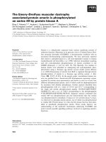
ing by plotting the distribution of chest compressio n
depth for each minute of ALS in a boxplot (Figure 1),
this figure revealed, as demonstrated in our previous
study [17], the great inter-individual variation in c hest
compression depth already evident in the first minute of
ALS. Paramedics were thereafter described and grouped
into different categories based on their initial chest com-
pression depth. The resuscitation attempts were sorted
into t hree different groups (good, bad a nd decay) based
on the development of chest compression depth and
rate over time. The following definitions were used,
based on the recommendations from the 2005 guide-
lines [21,22]:
Good: CPR with average chest compression depth ≥
40 mm for every minute during the 12 minute resuscita-
tion attempt. Average chest compression rate 100-120
for every minute.
Bad: CPR with initial average chest compression depth
< 40 mm. Chest compression rate < 100 or > 120 per
minute at the start of the resuscitation attempt.
Decay: CPR with i nitial average chest compressions
depths ≥ 40 mm which dro pped below 40 mm. Chest
compression rates 100-120 per minute that decreased to
< 100 or increased to > 120 per minute.
The no-flow ratio (NFR) was defined as the time with-
out chest compressions divided by the total time of t he
ALS scenario. The NFR was analysed in three minute
Figure 1 Distribution of chest compression depth.Boxplot
showing the distribution of chest compression depths for each
minute during twelve minutes of advanced life support on a
manikin (n = 19). Centre line indicates median value, boxes indicate
interquartile range and straight lines indicate maximum and
minimum values. The circle denotes an outlier.
Bjørshol et al. Scandinavian Journal of Trauma, Resuscitation and Emergency Medicine 2011, 19:46
/>Page 2 of 7
periods because Norwegian ALS guidelines [23] recom-
mend analysis of rhythm every t hree minutes, as
opposed to international guidelines with their two min-
ute periods [24,25]. The paramedics in the present study
followed the Norwegian guidelines and have been thor-
oughly trained in these guidelines since 2006.
Statistical analyses
We used SPSS version 17.0 (Chicago, IL, USA) for sta-
tistical analyses. Data are presented as mean values for
each minute of ALS. We investigated the overall change
in the NFR in the different three-minute periods using
repeated measures ANOVA. Additionally we tested the
difference between the first and each successive time
interval pairwise using paired t tests. A P value of < 0.05
was regarded as significant. For the pairwise testing we
had to take into account multiple testing effects, i.e. we
adjusted the significance lev el using the Bonferroni cor-
rection. This leads to a significance level of 0.017 (3
pairwise tests).
3. Results
Altogether 20 paramedic teams completed the study.
One regist ration failed due to so ftware failure. Hence, 19
ALS resuscitations were available for this chest compres-
sion quality analysis. In each resuscitation attempt, the
same paramedic performed all the chest compressions,
and 68% of the chest compression providers were male.
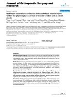
Based on chest compression depth, 26% (5/19) and
47% (9/19) of the ALS resuscitations were classified as
good and bad throughout the 12 minute scenario,
respectively. In these cases no signs of decay or major
changes occurred (Figure 2), except for one among the
bad, where sufficient chest compression depth was
achieved between 3 and 8 minutes (Figure 2B). In 26%
(5/19) of the cases, decay in chest compression depth
was present. Of these five cases, only one paramedic dis-
played chest compression decay to below 40 mm within
the first two minutes, the remainder after 4, 8, 11 and
12 minutes (Figure 2C).
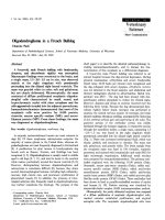
Based on chest compression rate, 32% (6/19) of the
resuscitation attempts were scored as good and 32% (6/
19) as bad. Among the bad, two achieved c orrect rate
after the first minute. Decay was present in 37% (7/19)
of the cases, and only one was evident in the first five
minutes of ALS (Figure 3).
Average NFR for the 19 paramedics was 17%, with a
range from 10 to 32%, and NF R changed significantly
over time (P < 0.001). NFR remained unchanged at 22%
in the 1-3 minute and 4-6 minute periods, but decreased
to 14% from the 1-3 minute period to the 7-9 minute
period (P = 0.002) and further to 10% from the 1-3 min-
ute period to the 10-12 m inute period (P < 0.001) (Fig-
ure 4).
4. Discussion
In this manikin study, where each param edic performed
12 minutes of chest compressions in a realistic ALS sce-
nario, we demonstrated that huge inter-individual differ-
ences in chest compression depth and r ate exist. This is
present already from the initiation of ALS. Decay due to
fatigue seems to be a less frequent problem, as only five
and six out of 19 paramedics d eveloped decay in chest
compression depth and rate, respectively. Noteworthy,
Good
0
10
20
30
40
50
60
123456789101112
Time (min)
Chest compression depth (mm)
Bad
0
10
20
30
40
50
60
123456789101112
Time (min)
Chest compression depth (mm)
Decay
0
10
20
30
40
50
60
123456789101112
Time (min)
Chest compression depth (mm)
A
B
C
Figure 2 Development of chest compression dep th.
Development of chest compression depth for each of 19
resuscitation attempts, the good are illustrated in A (5/19, 26%), the
bad in B (9/19, 47%) and those with decay in C (5/19, 26%). Arrows
indicate when each paramedic first developed decay in chest
compression depth to < 40 mm. See text for definition of groups.
Bjørshol et al. Scandinavian Journal of Trauma, Resuscitation and Emergency Medicine 2011, 19:46
/>Page 3 of 7
only one paramedic showed decay in chest compression
depth within the initial two minutes, and only one
showed decay in compression rate within the initi al fiv e
minutes.
A manikin study by Hightower et al. from 1995, where
11 nursing a ssistants performed chest compressions for
five minutes [9], described a significant and steady
decline in the percentage of correct compressions
already evident in the second minute. The authors spec-
ulate that fatigue might be the reason for this compres-
sion quality decay without specifying whether the
incorrect compressions were due to incomplete com-
pression depth or wrong hand placement. Later manikin
studies showed similar results with a decline in chest
compression depth after the initial minutes of the CPR
attempt [10,11,13,14,26]. A clinical study on in-hospital
cardiac arrested patients [27] described a decay in chest
compres sion depth that was statistically significant after
only 90 seconds. However, no correction was made for
different surfaces on which the patients were located.
These previous studies all conclude that decay in mean
chest compression depth is evident after a very short
period of time. Importantly, their data analyses do not
take into account the huge inter-individual differences
among the CPR providers that will influence the results.
We have in a previous BLS manikin study [17], as in the
present ALS manikin study, documented that these
inter-i ndividual differences are present alre ady from the
initiation of CPR. Thus, it was necessary to analyse the
data by sorting the individuals into diffe rent groups
based on their initial chest compression quality, instead
of ca lculating mean values for a large group of
individuals.
In the 2010 guidelines optimal chest compression
quality is even more emphasized than previously, and a
chest compression depth of at least 50 mm is recom-
mended [4,5]. Although our paramedics were trained in
the previous guidelines re commending a compression
depth of 40-50 mm, it is a cause of concern that 47% in
the present study had chest compression depths of less
than 40 mm already from the initiation of CPR. As seen
in Figure 2B, this is not a result of fatigue or chest com-
pression decay, but an inappropriate chest compression
depth alre ady from initiation of CPR. There are several
potential reasons for this deviation from guidelines;
Good
90
95
100
105
110
115
120
125
130
135
140
1 2 3 4 5 6 7 8 9 10 11 12
Time (min)
Chest compression rate (/min)
Bad
90
95
100
105
110
115
120
125
130
135
140
123456789101112
Time (min)
Chest compression rate (/min)
Decay
90
95
100
105
110
115
120
125
130
135
123456789101112
Time (min)
Chest compression rate (/min)
C
B
A
Figure 3 Development of chest compression rate. Development
of chest compression rate for each of 19 resuscitation attempts, the
good are illustrated in A (6/19, 32%), the bad in B (6/19, 32%) and
those with decay in C (7/19, 37%). Arrows indicate when each
paramedic developed decay in chest compression rate to < 100 or
> 120 per minute. See text for definition of groups.
0,00
0,05
0,10
0,15
0,20
0,25
0,30
0,35
0,40
0,45
0,50
Min 1-3 Min 4-6 Min 7-9 Min 10-12
Time (min)
NFR
Figure 4 Development of no-flow ratio. Development of no-flow
ratio measured in three minute periods for all 19 resuscitation
attempts.
Bjørshol et al. Scandinavian Journal of Trauma, Resuscitation and Emergency Medicine 2011, 19:46
/>Page 4 of 7
insufficient muscular power, lack of sufficient body
weight, as weight previously has been correlated with
compression depth [28], an inaccuracy of chest com-
pression depth because no feedback was available, or a
fear of causing serious patient injury [29]. In a question-
naire among Norwegian and UK paramedics, Ø degaard
et al. reported that many paramedics had concerns caus-
ing serious patient injuries if they compressed to the
guidelines’ depth [29]. Thus, it is very relevant to high-
light chest compressions quality, especially compression
depth, in ALS training and practise in the future. The
fear of causing patient injuries must be overcome.
More positive, all paramedics had compression rates
above 100 per minute for the majority of the resuscita-
tion attempts. This is important as higher compression
rates increase cardiac output resulting in increased myo-
cardial and cerebral blood flow [30,31] and improved
short-term survival in humans [32]. Decay in chest com-
pression rate over time was rare and only evident in one
paramedic within the first five minutes. 26% initiated
CPR with chest compression rates above 120 per min-
ute. This is unfavourable as coronary perfusion is
reduced at rates over 120-130 per minute [31], thereby
reducing the p robability of successful resuscitation [33].
A metronome [34,35] or real time feedback [36] c ould
improve the chest compression rate.
NFR did not increase over time in our study but actu-
ally declined, even though the same rescuer provided all
the chest compressions for as long as 12 minutes. One
likely explanation for this positive, continuous decrease
in NFR over time is that the patient in our scenario
developed PEA after t he first shock, and hence there
was no further need for charging the defibrillator and
shocking the patient. On the other hand, an organised
ECG rhythm necessitates pulse checks to differe ntiate
PEA from ROSC in the absence of end-tidal CO
2
-mea-
surement (ETCO
2
), and hence further increases the
NFR. Further, as the patient was intubated after about
five minutes [20], this could have contributed to the
reduced NFR as this allows for simultaneous ventilations
and continuous chest compressions [37]. A clinical
observation study has also shown no increase in NFR
over time [38]. Our paramedics had a NFR of 17% in
the 12 minute study period which is comparable to
recent clinical observation studies [39,40], and far better
than data from the recent US ROC trials with NFR
between 34 and 46% [36,41].
Importantly, based on our findings it seems unwar-
ranted to recommend changing the person providing
chest compressions every two minutes during ALS as
recommend ed in the new resuscitation guidelines. It has
been shown that provider switches account for at least
40% of NFR during CPR [12], and this can be reduced
by avoiding unnecessary switches. Instead of changing
chest compression provider frequently, we recommend
more attention on optimising chest compression quality
already from the initiation of CPR, and that the chest
compression quality should be monitored continuously
with CPR feedback devices or capnography during ALS.
CPR feedback devices have been shown to improve the
quality of CPR, including chest compression depth and
ROSC rate, but still have not led to increased long-term
survival [36,42]. Capnography, with ETCO
2
measure-
ments, predicts cardiac output [43] and is correlated
with both ROSC and survival [44]. However, more stu-
dies are needed to show if CPR feedback devices or cap-
nography can assist in finding the optimal time point
for switching the provider of chest compressions.
There are limitations to this study. As it was a simula-
tion manikin study, we do not know whether the quality
of chest compression is compromised more or less in
real cardiac arrest situations. It has been shown that
paramedics are physically capable of compressing to
guideline depth fo r 5 minu tesevenonamanikinwith
chest stiffness mimicking the upper eighth of chest stiff-
nesses in a patient population [29]. The manikin in our
study does not represent the large variation in stiffness
and damping found in human chests during CPR
[45,46]. Further, our study included paramedics with a
median experience of 8.5 years and frequent refresher
training in ALS. We do not know if chest compression
decay or chest compression quality in general is differ-
ent for less experienced paramedics and other health
care providers. As this is the first study to explo re chest
compression decay by sorting individuals based on com-
pression quality, a power analysis was not performed
and hence we cannot rule out that our results are
caused by insufficient pow er. Finally, we fol lowed the
recommendations from the Norwegian 2005 guidelines
in the present study [23], with 4 cm of chest compres-
sion depth regarded as good. We might speculate that
the 5 cm reco mmendation f rom 2010 would have
caused m ore decay and fatigue, especially if every para-
medic initially compressed to the guidelines depth.
Further studies are indeed warranted.
5. Conclusion
In this simulated cardiac arrest manikin study, only half
of the providers achieved gui deline recommen ded com-
pression depth during prolonged ALS. Large inter-indi-
vidual differences in chest compression quality were
already present from the in itiation of CPR. Chest com-
pression decay and thereby fatigue within the first two
minutes was rare.
6. Competing interests
CAB has a part-time employment as facilitator at Sta-
vanger Acute Medic ine Foundation for Education and
Bjørshol et al. Scandinavian Journal of Trauma, Resuscitation and Emergency Medicine 2011, 19:46
/>Page 5 of 7
Research (SAFER). ES is medical director at SAFER.
CAB and ES have received financial support from the
Laerdal Foundation for Acute Medicine. HM is an
employee of Laerdal Medical. KS and JA have no com-
peting interests.
Acknowledgements
Thanks to Melinda Kay Christensen, Jules Eilledge and Eirik Illguth for
simulation assistance, Kjetil Lønne Nilsen, Joar Eilevstjønn and Sara Brunner
for technical support, to Linda Sivertsen for manuscript revision and to
Stavanger Acute Medicine Foundation for Education and Research (SAFER)
for offering simulation facilities.
CAB has received financial support from the Laerdal Foundation for Acute
Medicine (Bjørn Lind PhD scholarship) and the Regional Centre for
Emergency Medical Research and Development (RAKOS). Thanks to the
paramedics participating in the study and to the Ambulance Department for
allowing this study.
Author details
1
Department of Anaesthesiology and Intensive Care, Stavanger University
Hospital, Stavanger, Norway.
2
Department of Anaesthesiology, Division of
Critical Care, Oslo Universi ty Hospital, Oslo, Norway.
3
Laerdal Medical AS,
Stavanger, Norway.
4
Centre for Clinical Research, Haukeland University
Hospital, Bergen, Norway.
Authors’ contributions
CAB participated in study design, running the simulations, statistical analyses
and manuscript writing, KS and ES in study design and manuscript writing,
HM in study design, running simulations and manuscript writing, and JA in
statistical analyses and manuscript writing. All authors read and approved
the final manuscript.
Received: 15 May 2011 Accepted: 9 August 2011
Published: 9 August 2011
References
1. Gallagher EJ, Lombardi G, Gennis P: Effectiveness of bystander
cardiopulmonary resuscitation and survival following out-of-hospital
cardiac arrest. JAMA 1995, 274:1922-1925.
2. Van Hoeyweghen RJ, Bossaert LL, Mullie A, Calle P, Martens P, Buylaert WA,
Delooz H: Quality and efficiency of bystander CPR. Belgian Cerebral
Resuscitation Study Group. Resuscitation 1993, 26:47-52.
3. Wik L, Steen PA, Bircher NG: Quality of bystander cardiopulmonary
resuscitation influences outcome after prehospital cardiac arrest.
Resuscitation 1994, 28:195-203.
4. Koster RW, Baubin MA, Bossaert LL, Caballero A, Cassan P, Castren M,
Granja C, Handley AJ, Monsieurs KG, Perkins GD, Raffay V, Sandroni C:
European Resuscitation Council Guidelines for Resuscitation 2010
Section 2. Adult basic life support and use of automated external
defibrillators. Resuscitation 2010, 81:1277-1292.
5. Berg RA, Hemphill R, Abella BS, Aufderheide TP, Cave DM, Hazinski MF,
Lerner EB, Rea TD, Sayre MR, Swor RA: Part 5: adult basic life support:
2010 American Heart Association Guidelines for Cardiopulmonary
Resuscitation and Emergency Cardiovascular Care. Circulation 2010, 122:
S685-705.
6. Kramer-Johansen J, Myklebust H, Wik L, Fellows B, Svensson L, Sørebø H,
Steen PA: Quality of out-of-hospital cardiopulmonary resuscitation with
real time automated feedback: a prospective interventional study.
Resuscitation 2006, 71:283-292.
7. Eftestøl T, Sunde K, Steen PA: Effects of interrupting precordial
compressions on the calculated probability of defibrillation success
during out-of-hospital cardiac arrest. Circulation 2002, 105:2270-2273.
8. Edelson DP, Abella BS, Kramer-Johansen J, Wik L, Myklebust H, Barry AM,
Merchant RM, Hoek TL, Steen PA, Becker LB: Effects of compression depth
and pre-shock pauses predict defibrillation failure during cardiac arrest.
Resuscitation 2006, 71:137-145.
9. Hightower D, Thomas SH, Stone CK, Dunn K, March JA: Decay in quality of
closed-chest compressions over time. Ann Emerg Med 1995, 26:300-303.
10. Ochoa FJ, Ramalle-Gomara E, Lisa V, Saralegui I: The effect of rescuer
fatigue on the quality of chest compressions. Resuscitation 1998,
37:149-152.
11. Ashton A, McCluskey A, Gwinnutt CL, Keenan AM: Effect of rescuer fatigue
on performance of continuous external chest compressions over 3 min.
Resuscitation 2002, 55:151-155.
12. Sutton RM, Maltese MR, Niles D, French B, Nishisaki A, Arbogast KB,
Donoghue A, Berg RA, Helfaer MA, Nadkarni V: Quantitative analysis of
chest compression interruptions during in-hospital resuscitation of older
children and adolescents. Resuscitation 2009, 80:1259-1263.
13. Greingor JL: Quality of cardiac massage with ratio compression-
ventilation 5/1 and 15/2. Resuscitation 2002, 55:263-267.
14. Jäntti H, Silfvast T, Turpeinen A, Kiviniemi V, Uusaro A: Quality of
cardiopulmonary resuscitation on manikins: on the floor and in the bed.
Acta Anaesthesiol Scand 2009,
53:1131-1137.
15.
Miles DS, Underwood PD Jr, Nolan DJ, Frey MA, Gotshall RW: Metabolic,
hemodynamic, and respiratory responses to performing
cardiopulmonary resuscitation. Can J Appl Sport Sci 1984, 9:141-147.
16. Shultz JJ, Mianulli MJ, Gisch TM, Coffeen PR, Haidet GC, Lurie KG:
Comparison of exertion required to perform standard and active
compression-decompression cardiopulmonary resuscitation. Resuscitation
1995, 29:23-31.
17. Bjørshol CA, Søreide E, Torsteinbø TH, Lexow K, Nilsen OB, Sunde K: Quality
of chest compressions during 10 min of single-rescuer basic life support
with different compression: ventilation ratios in a manikin model.
Resuscitation 2008, 77:95-100.
18. Ødegaard S, Saether E, Steen PA, Wik L: Quality of lay person CPR
performance with compression: ventilation ratios 15:2, 30:2 or
continuous chest compressions without ventilations on manikins.
Resuscitation 2006, 71:335-340.
19. Neset A, Birkenes TS, Myklebust H, Mykletun RJ, ødegaard S, Kramer-
Johansen J: A randomized trial of the capability of elderly lay persons to
perform chest compression only CPR versus standard 30:2 CPR.
Resuscitation 2010, 81:887-892.
20. Bjørshol CA, Myklebust H, Nilsen KL, Hoff T, Bjørkli C, Illguth E, Soreide E,
Sunde K: Effect of socioemotional stress on the quality of
cardiopulmonary resuscitation during advanced life support in a
randomized manikin study. Crit Care Med 2011, 39:300-304.
21. Handley AJ, Koster R, Monsieurs K, Perkins GD, Davies S, Bossaert L:
European Resuscitation Council guidelines for resuscitation 2005.
Section 2. Adult basic life support and use of automated external
defibrillators. Resuscitation 2005, 67(Suppl 1):S7-23.
22. 2005 American Heart Association Guidelines for Cardiopulmonary
Resuscitation and Emergency Cardiovascular Care. Circulation 2005, 112:
IV1-203.
23. Lexow K, Sunde K: Why Norwegian 2005 guidelines differs slightly from
the ERC guidelines. Resuscitation 2007, 72:490-492.
24. Deakin CD, Nolan JP, Soar J, Sunde K, Koster RW, Smith GB, Perkins GD:
European Resuscitation Council Guidelines for Resuscitation 2010
Section 4. Adult advanced life support. Resuscitation 2010, 81:1305-1352.
25. Neumar RW, Otto CW, Link MS, Kronick SL, Shuster M, Callaway CW,
Kudenchuk PJ, Ornato JP, McNally B, Silvers SM, Passman RS, White RD,
Hess EP, Tang W, Davis D, Sinz E, Morrison LJ: Part 8: adult advanced
cardiovascular life support: 2010 American Heart Association Guidelines
for Cardiopulmonary Resuscitation and Emergency Cardiovascular Care.
Circulation 2010, 122:S729-767.
26. Heidenreich JW, Berg RA, Higdon TA, Ewy GA, Kern KB, Sanders AB: Rescuer
fatigue: standard versus continuous chest-compression cardiopulmonary
resuscitation. Acad Emerg Med 2006, 13:1020-1026.
27. Sugerman NT, Edelson DP, Leary M, Weidman EK, Herzberg DL, Vanden
Hoek TL, Becker LB, Abella BS: Rescuer fatigue during actual in-hospital
cardiopulmonary resuscitation with audiovisual feedback: A prospective
multicenter study. Resuscitation 2009, 80:981-984.
28. Larsen PD, Perrin K, Galletly DC: Patterns of external chest compression.
Resuscitation 2002, 53:281-287.
29. Ødegaard S, Kramer-Johansen J, Bromley A, Myklebust H, Nysaether J, Wik L,
Steen
PA: Chest compressions by ambulance personnel on chests with
variable stiffness: Abilities and attitudes. Resuscitation 2007, 74:127-134.
30. Sunde K, Wik L, Naess PA, Grund F, Nicolaysen G, Steen PA: Improved
haemodynamics with increased compression-decompression rates
during ACD-CPR in pigs. Resuscitation 1998, 39:197-205.
Bjørshol et al. Scandinavian Journal of Trauma, Resuscitation and Emergency Medicine 2011, 19:46
/>Page 6 of 7
31. Wolfe JA, Maier GW, Newton JR Jr, Glower DD, Tyson GS Jr, Spratt JA,
Rankin JS, Olsen CO: Physiologic determinants of coronary blood flow
during external cardiac massage. The Journal of thoracic and
cardiovascular surgery 1988, 95:523-532.
32. Abella BS, Sandbo N, Vassilatos P, Alvarado JP, O’Hearn N, Wigder HN,
Hoffman P, Tynus K, Vanden Hoek TL, Becker LB: Chest compression rates
during cardiopulmonary resuscitation are suboptimal: a prospective
study during in-hospital cardiac arrest. Circulation 2005, 111:428-434.
33. Paradis NA, Martin GB, Rivers EP, Goetting MG, Appleton TJ, Feingold M,
Nowak RM: Coronary perfusion pressure and the return of spontaneous
circulation in human cardiopulmonary resuscitation. JAMA 1990,
263:1106-1113.
34. Jäntti H, Silfvast T, Turpeinen A, Kiviniemi V, Uusaro A: Influence of chest
compression rate guidance on the quality of cardiopulmonary
resuscitation performed on manikins. Resuscitation 2009, 80:453-457.
35. Milander MM, Hiscok PS, Sanders AB, Kern KB, Berg RA, Ewy GA: Chest
compression and ventilation rates during cardiopulmonary resuscitation:
the effects of audible tone guidance. Acad Emerg Med 1995, 2:708-713.
36. Hostler D, Everson-Stewart S, Rea TD, Stiell IG, Callaway CW, Kudenchuk PJ,
Sears GK, Emerson SS, Nichol G: Effect of real-time feedback during
cardiopulmonary resuscitation outside hospital: prospective, cluster-
randomised trial. Bmj 2011, 342:d512.
37. Kramer-Johansen J, Wik L, Steen PA: Advanced cardiac life support before
and after tracheal intubation–direct measurements of quality.
Resuscitation 2006, 68:61-69.
38. Wik L, Kramer-Johansen J, Myklebust H, Sørebø H, Svensson L, Fellows B,
Steen PA: Quality of cardiopulmonary resuscitation during out-of-
hospital cardiac arrest. JAMA 2005, 293:299-304.
39. Olasveengen TM, Wik L, Kramer-Johansen J, Sunde K, Pytte M, Steen PA: Is
CPR quality improving? A retrospective study of out-of-hospital cardiac
arrest. Resuscitation 2007, 75:260-266.
40. Olasveengen TM, Sunde K, Brunborg C, Thowsen J, Steen PA, Wik L:
Intravenous drug administration during out-of-hospital cardiac arrest: a
randomized trial. JAMA 2009, 302:2222-2229.
41. Christenson J, Andrusiek D, Everson-Stewart S, Kudenchuk P, Hostler D,
Powell J, Callaway CW, Bishop D, Vaillancourt C, Davis D, Aufderheide TP,
Idris A, Stouffer JA, Stiell I, Berg R: Chest compression fraction determines
survival in patients with out-of-hospital ventricular fibrillation. Circulation
2009, 120:1241-1247.
42. Edelson DP, Robertson-Dick BJ, Yuen TC, Eilevstjønn J, Walsh D, Bareis CJ,
Vanden Hoek TL, Abella BS: Safety and efficacy of defibrillator charging
during ongoing chest compressions: a multi-center study. Resuscitation
2010, 81:1521-1526.
43. Weil MH, Bisera J, Trevino RP, Rackow EC: Cardiac output and end-tidal
carbon dioxide. Crit Care Med
1985, 13:907-909.
44. Grmec S, Klemen P: Does the end-tidal carbon dioxide (EtCO2)
concentration have prognostic value during out-of-hospital cardiac
arrest? Eur J Emerg Med 2001, 8:263-269.
45. Nysaether JB, Dorph E, Rafoss I, Steen PA: Manikins with human-like chest
properties–a new tool for chest compression research. IEEE transactions
on bio-medical engineering 2008, 55:2643-2650.
46. Tomlinson AE, Nysaether J, Kramer-Johansen J, Steen PA, Dorph E:
Compression force-depth relationship during out-of-hospital
cardiopulmonary resuscitation. Resuscitation 2007, 72:364-370.
doi:10.1186/1757-7241-19-46
Cite this article as: Bjørshol et al.: Decay in chest compression quality
due to fatigue is rare during prolonged advanced life support in a
manikin model. Scandinavian Journal of Trauma, Resuscitation and
Emergency Medicine 2011 19:46.
Submit your next manuscript to BioMed Central
and take full advantage of:
• Convenient online submission
• Thorough peer review
• No space constraints or color figure charges
• Immediate publication on acceptance
• Inclusion in PubMed, CAS, Scopus and Google Scholar
• Research which is freely available for redistribution
Submit your manuscript at
www.biomedcentral.com/submit
Bjørshol et al. Scandinavian Journal of Trauma, Resuscitation and Emergency Medicine 2011, 19:46
/>Page 7 of 7