Báo cáo y học: " Abdominal Compartment Syndrome: pathophysiology and definitions" ppt
Bạn đang xem bản rút gọn của tài liệu. Xem và tải ngay bản đầy đủ của tài liệu tại đây (472.18 KB, 11 trang )
BioMed Central
Page 1 of 11
(page number not for citation purposes)
Scandinavian Journal of Trauma,
Resuscitation and Emergency Medicine
Open Access
Review
Abdominal Compartment Syndrome: pathophysiology and
definitions
Michael L Cheatham
Address: Department of Surgical Education, Orlando Regional Medical Center, Orlando, Florida 32806, USA
Email: Michael L Cheatham -
Abstract
"Intra-abdominal hypertension", the presence of elevated intra-abdominal pressure, and "abdominal
compartment syndrome", the development of pressure-induced organ-dysfunction and failure, have
been increasingly recognized over the past decade as causes of significant morbidity and mortality
among critically ill surgical and medical patients. Elevated intra-abdominal pressure can cause
significant impairment of cardiac, pulmonary, renal, gastrointestinal, hepatic, and central nervous
system function. The significant prognostic value of elevated intra-abdominal pressure has
prompted many intensive care units to adopt measurement of this physiologic parameter as a
routine vital sign in patients at risk. A thorough understanding of the pathophysiologic implications
of elevated intra-abdominal pressure is fundamental to 1) recognizing the presence of intra-
abdominal hypertension and abdominal compartment syndrome, 2) effectively resuscitating
patients afflicted by these potentially life-threatening diseases, and 3) preventing the development
of intra-abdominal pressure-induced end-organ dysfunction and failure. The currently accepted
consensus definitions surrounding the diagnosis and treatment of intra-abdominal hypertension and
abdominal compartment syndrome are presented.
Review
Although initially recognized over 150 years ago, the
pathophysiologic implications of elevated intra-abdomi-
nal pressure (IAP) have essentially been rediscovered only
within the past two decades [1-3]. An explosion of scien-
tific investigation and accumulation of clinical experience
has confirmed the significant detrimental impact of both
"intra-abdominal hypertension" (IAH) (see figure 1), the
presence of elevated intra-abdominal pressure, and
"abdominal compartment syndrome" (ACS), the devel-
opment of IAH-induced organ-dysfunction and failure,
among the critically ill [4,5]. IAH has been identified as a
continuum of pathophysiologic changes beginning with
regional blood flow disturbances and culminating in
frank end-organ failure and the development of ACS. ACS
has been identified to be a cause of significant morbidity
and mortality among critically ill surgical, medical, and
pediatric patients. Previously present, but significantly
under-appreciated, IAH and ACS are now recognized as
common occurrences in the intensive care unit (ICU) set-
ting [6-16]. Elevated IAP has been identified as an inde-
pendent predictor of mortality during critical illness and
likely plays a major role in the development of multiple
system organ failure, a syndrome which has plagued ICU
patients and physicians for decades [8,17,18].
Recently, evidence-based consensus definitions and rec-
ommendations for the resuscitation and rehabilitation of
patients with IAH and ACS have been published [19,20].
Central to this evolving strategy are the use of early serial
Published: 2 March 2009
Scandinavian Journal of Trauma, Resuscitation and Emergency Medicine 2009, 17:10 doi:10.1186/1757-7241-17-10
Received: 8 February 2009
Accepted: 2 March 2009
This article is available from: />© 2009 Cheatham; licensee BioMed Central Ltd.
This is an Open Access article distributed under the terms of the Creative Commons Attribution License ( />),
which permits unrestricted use, distribution, and reproduction in any medium, provided the original work is properly cited.
Scandinavian Journal of Trauma, Resuscitation and Emergency Medicine 2009, 17:10 />Page 2 of 11
(page number not for citation purposes)
IAP measurements to detect the presence of IAH, applica-
tion of comprehensive medical management strategies to
reduce elevated IAP and restore end-organ perfusion,
timely surgical abdominal decompression for refractory
organ dysfunction, and early attempts at fascial closure
once physiologically appropriate [21,22]. Such a strategy
has been demonstrated to significantly improve patient
survival, reduce complications (such as enteroatmos-
pheric fistula), and decrease resource utilization [23,24].
The following review addresses both the pathophysiologic
impact of elevated IAP on the various organ systems as
well as the currently accepted definitions surrounding
IAH and ACS. The diagnosis, prevention, and treatment of
IAH/ACS have been addressed in a number of recent pub-
lications [6,10,12,13,19-22,24-29].
History
The impact of elevated IAP upon respiratory function was
first documented by Marey in 1863 and subsequently by
Burt in 1870 [30]. In 1890, Henricius identified in an ani-
mal model that an IAP between 27 and 46 cm H
2
O signif-
icantly impaired diaphragmatic excursion leading to
elevated intrathoracic pressure, respiratory failure, and
death [30]. The theory that respiratory failure is the cause
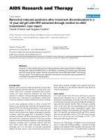
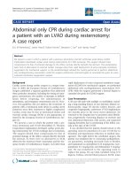
Pathophysiologic Implications of Intra-abdominal HypertensionFigure 1
Pathophysiologic Implications of Intra-abdominal Hypertension. The effects of intra-abdominal hypertension are not
limited just to the intra-abdominal organs, but rather have an impact either directly or indirectly on every organ system in the
body. ICP – intracranial pressure; CPP – cerebral perfusion pressure; ITP – intrathoracic pressure; IVC – inferior vena cava;
SMA – superior mesenteric artery; pHi – gastric intramuscosal pH; APP – abdominal perfusion pressure; PIP- peak inspiratory
pressure; Paw – mean airway pressure; PaO
2
– oxygen tension; PaCO
2
– carbon dioxide tension; Qs/Qt – intrapulmonary
shunt; Vd/Vt – pulmonary dead space ; CO – cardiac output; SVR – systemic vascular resistance; PVR – pulmonary vascular
resistance; PAOP – pulmonary artery occlusion pressure; CVP – central venous pressure; GFR – glomerular filtration rate.
Scandinavian Journal of Trauma, Resuscitation and Emergency Medicine 2009, 17:10 />Page 3 of 11
(page number not for citation purposes)
of death in severe IAH persisted until 1911 when Emerson
demonstrated in cat, dog, and rabbit models that elevated
IAP causes death by cardiovascular collapse rather than by
respiratory failure [30]. The detrimental effect of elevated
IAP on renal function and urinary output was first identi-
fied by Wendt in 1876 and the restoration of urinary out-
put through abdominal decompression by Thorington
and Schmidt in 1923 [31-33]. Overholt extensively stud-
ied the properties of the abdominal wall and confirmed
that normal IAP is subatmospheric and that procedures
which restrict movement of the abdominal wall or disten-
tion of the stomach or colon all result in an increase in IAP
[34]. He postulated that IAP is governed by both the pres-
sure induced by the abdominal contents and the "flexibil-
ity" (compliance) of the abdominal wall. Investigation
into the physiologic effects of IAP on renal function in
humans essentially began in 1947 with the work of Brad-
ley [35]. The experiences of surgeons treating infants with
gastroschisis or omphalocele further contributed to our
understanding of both the concept of "loss of abdominal
domain" as well as the life-threatening cardiac, pulmo-
nary, and gastrointestinal complications which can occur
when abdomens are primarily closed without considera-
tion of elevated IAP [36-39]. Gross, in 1948, first
described the use of a "staged abdominal repair" in the
management of such infants unknowingly pioneering the
open abdomen techniques which have now become
standard in the treatment of IAH and ACS [36].
Although surrogate measurement of IAP via measurement
of intravesicular, intragastric, and intracolonic pressure in
animal models was commonplace in the 1920's and
1930's, it was Söderberg who, in 1970, first described the
strong correlation between IAP and intravesicular pressure
during laparoscopy in humans [40]. The landmark work
of Harman, Kron, and Richards in the early 1980's "redis-
covered" IAH as a cause of unexplained oliguria and sub-
sequent renal failure in post-operative patients with
abdominal distention [32,41,42]. They further reported
the benefits of open abdominal decompression in restor-
ing renal function and improving patient outcome in
patients with an IAP in excess of 25 mmHg [32,41]. The
introduction of laparoscopic techniques into mainstream
surgical practice in the late 1980's and early 1990's led to
numerous experimental and clinical studies which further
advanced our understanding of the injurious effects of ele-
vated IAP on cardiac, pulmonary, renal, gastrointestinal,
hepatic, and cerebral function. Increased appreciation of
these effects by both anesthesiologists and surgeons set
the stage for recognition of both IAH and ACS in the crit-
ically ill patient population.
Pathophysiology
An increasing body of literature has identified the signifi-
cant physiologic derangements that occur as a result of
elevated IAP. The effects of IAH are not limited just to the
intra-abdominal organs, but rather have an impact either
directly or indirectly on every organ system in the body. As
a result, patients with prolonged, untreated IAH com-
monly manifest significant malperfusion and subsequent
organ failure. Pre-existing comorbidities, such as chronic
renal failure, pulmonary disease, or cardiomyopathy, play
an important role in aggravating the effects of elevated IAP
and may reduce the threshold of IAH that causes the clin-
ical manifestations of ACS. The etiology for the patient's
IAH is similarly of vital importance and may be deter-
mined as being either intra-abdominal, as occurs in surgi-
cal or trauma patients following damage control
laparotomy, or extra-abdominal, as occurs in medical
patients with sepsis or burn patients who require aggres-
sive fluid resuscitation [6,7,43-46].
Cardiovascular
As originally described over 80 years ago by Emerson, ris-
ing IAP increases intrathoracic pressure through cephalad
deviation of the diaphragm [30]. Increased intrathoracic
pressure significantly reduces venous return resulting in
reduced cardiac output [33,47-57]. Such reductions have
been demonstrated to occur at an IAP of only 10 mmHg
[18,57]. Hypovolemic patients appear to sustain reduc-
tions in cardiac output at lower levels of IAP than do nor-
movolemic patients [50,53]. Hypervolemic patients
demonstrate increased venous return in the presence of
mild to moderate elevations in IAP suggesting that vol-
ume resuscitation may have a protective effect [53]. Dia-
phragmatic elevation and increased intrathoracic pressure
have also been postulated to cause direct cardiac compres-
sion reducing ventricular compliance and contractility
[49]. Systemic vascular resistance (afterload) is increased
through compression of both the aorta and systemic vas-
culature and pulmonary vascular resistance through com-
pression of the pulmonary parenchyma [33,48,51-56,58].
As a result, in the absence of severe IAH, mean arterial
pressure typically remains stable despite a decrease in
venous return and cardiac output. Such increases in after-
load may be poorly tolerated by those with marginal car-
diac contractility or inadequate intravascular volume.
Preload augmentation through volume administration
appears to ameliorate, at least partially, the injurious
effects of IAH-induced increases in afterload
[18,33,48,53,56,58,59].
Paradoxically, intracardiac filling pressures such as pul-
monary artery occlusion ("wedge") pressure (PAOP) and
central venous pressure (CVP) typically increase with ris-
ing IAP despite the reduced venous return and cardiac out-
put [47-49,51,53,56,57,59-64]. This apparent deviation
from Starling's Law of the heart is due to the fact that both
PAOP and CVP are measured relative to atmospheric pres-
sure and are actually the sum of both intravascular pres-
sure and intrathoracic pressure [63,64]. In the presence of
IAH-induced elevations in intrathoracic pressure, PAOP
Scandinavian Journal of Trauma, Resuscitation and Emergency Medicine 2009, 17:10 />Page 4 of 11
(page number not for citation purposes)
and CVP tend to be erroneously elevated and no longer
reflective of true intravascular volume status [47-
49,57,59-61,63,64]. Such alterations in PAOP and CVP
have been demonstrated with an IAP of only 10 mmHg
[57]. Attempts to correct for this measurement error
through use of transmural pressures (i.e., PAOP minus
intrathoracic pressure) has confirmed that transmural
PAOP decreases with rising IAP correctly reflecting the
decreased venous return and cardiac preload [59]. Several
studies have demonstrated that volumetric parameters,
such as right ventricular end-diastolic volume (RVEDV),
global end-diastolic volume (GEDV), or stroke volume
variation (SVV) are superior predictors of intravascular
volume status whose accuracy is unaffected by changes in
intrathoracic pressure [63-66]. When traditional intracar-
diac filling pressures must be used, transmural pressures
may be estimated as follows [63,64]:
Transmural PAOP = PAOP - 0.5*IAP
Transmural CVP = CVP - 0.5*IAP
IAH also reduces venous return from the lower extremities
functionally obstructing inferior vena caval blood flow by
two mechanisms. First, inferior vena caval pressure
increases significantly in the presence of IAH and has been
demonstrated to parallel changes in IAP [18,33,53,56].
Second, cephalad deviation of the diaphragm causes a
mechanical narrowing of the vena cava at the diaphrag-
matic crura further reducing venous return to the heart
[54,67]. Femoral vein pressures are markedly increased
and venous blood flow and pulsatility dramatically
reduced [68,69]. The resulting increases in extremity
venous hydrostatic pressure promote the formation of
peripheral edema. These changes place the patient with
IAH at risk for development of deep venous thrombosis
[69-71]. Reduction of IAP restores femoral venous blood
flow, but has anecdotally been reported to result in pul-
monary embolism [71].
Pulmonary
The pulmonary effects of elevated IAP have been recog-
nized for many years [30,33,49,51,59,68,72-74]. IAP is
transmitted to the thorax both directly and through
cephalad deviation of the diaphragm. This significantly
increases intrathoracic pressure resulting in extrinsic com-
pression of the pulmonary parenchyma and development
of pulmonary dysfunction [18,47,48,57,59,68]. Com-
pression of the pulmonary parenchyma appears to begin
with an IAP of 16–30 mmHg and is accentuated by the
presence of hemorrhagic shock and hypotension [57,75].
Parenchymal compression results in alveolar atelectasis,
decreased oxygen transport across the pulmonary capil-
lary membrane, and an increased intrapulmonary shunt
fraction (Qsp/Qt). IAH-induced atelectasis has been dem-
onstrated to cause an increase in the rate of pulmonary
infection [76]. Parenchymal compression also reduces
pulmonary capillary blood flow leading to decreased car-
bon dioxide excretion and an increased alveolar dead
space (Vd/Vt) [57]. Both peak inspiratory and mean air-
way pressures are significantly increased and may result in
alveolar volutrauma [57,75]. Spontaneous tidal volumes
and dynamic pulmonary compliance are reduced result-
ing in further ventilation-perfusion mismatching [57,75].
In combination, these effects lead to the arterial hypox-
emia and hypercarbia that, in part, characterize ACS
[18,33,48,51,59,73].
Renal
IAH-induced reductions in renal blood flow and function
have been demonstrated in both animal and human
models [33,35,42,51,77]. These changes occur in direct
response to increasing IAP with oliguria developing at an
IAP of 15 mmHg and anuria at 30 mmHg [32,33,42].
Renal artery blood flow has been demonstrated to be pref-
erentially diminished in comparison to both celiac and
superior mesenteric artery blood flow [68]. Renal vein
pressure and renal vascular resistance are both signifi-
cantly elevated [35,42,48]. All of these changes shunt
blood away from the renal cortex and functioning glomer-
uli leading to impaired glomerular and tubular function
and significant reductions in urinary output
[32,33,35,41,42,48,49,51,73,77-80].
Several mechanisms have been proposed as the etiology
for IAH-induced renal dysfunction and failure. Harman et
al. negated direct ureteral compression as a cause through
studies utilizing ureteral stents [42]. Other authors have
suggested that direct parenchymal compression and
development of a "renal compartment syndrome" results
in renal ischemia and subsequent failure [70,81]. Stone
demonstrated in traumatically injured patients that incis-
ing the renal capsule could reverse renal failure if per-
formed early and prior to development of severe renal
dysfunction [81]. Recent studies suggest that compression
of the renal vein likely plays the primary role in the devel-
opment of renal dysfunction with reduced cardiac output
playing a secondary role [32,33,48,81].
IAH decreases glomerular filtration rate causing a rise in
both blood urea nitrogen and serum creatinine and a
reduction in creatinine clearance [33,35,42,48,51,79].
Osmolar clearance is similarly decreased and fractional
excretion of sodium increased [79]. Urinary sodium and
chloride concentrations decrease and urinary potassium
concentrations increase [33]. Plasma renin activity and
aldosterone levels increase significantly [33,48]. Antidiu-
retic hormone levels have been demonstrated to increase
to more than twice basal levels [82]. All of these patho-
physiologic changes appear to be potentially reversible if
Scandinavian Journal of Trauma, Resuscitation and Emergency Medicine 2009, 17:10 />Page 5 of 11
(page number not for citation purposes)
the patient's IAH is recognized and treated appropriately
before significant organ dysfunction has developed
[32,48].
Gastrointestinal
Of all the organ systems, the gut appears to be one of the
most sensitive to elevations in IAP. Such reductions in
mesenteric blood flow may appear with an IAP of only 10
mmHg [83]. Caldwell et al. has demonstrated decreased
blood flow to virtually all intra-abdominal and retroperi-
toneal organs as a result of elevated IAP [56]. The sole
exception was adrenal blood flow which appears to be
preserved and has been postulated to be a survival mech-
anism by which to support catecholamine release in the
face of ongoing shock [56]. Celiac artery blood flow is
reduced by up to 43% and superior mesenteric artery
blood flow by as much as 69% in the presence of intra-
abdominal pressures of 40 mmHg [68,83,84]. The nega-
tive effects of IAP on mesenteric perfusion are augmented
by the presence of hypovolemia or hemorrhage
[8,50,68,83,85]. Reintam et al. have recently validated a
grading system for predicting mortality due to gastrointes-
tinal dysfunction among patients with IAH/ACS [86].
In addition to reducing arterial blood flow, IAP com-
presses thin walled mesenteric veins promoting venous
hypertension and intestinal edema. Visceral swelling fur-
ther increases IAP initiating a vicious cycle which results in
worsening malperfusion, bowel ischemia, decreased
intramucosal pH, feeding intolerance, systemic metabolic
acidosis, and significantly increased patient mortality
[8,13,50,86,87]. Intestinal mucosal perfusion is dimin-
ished by levels of IAP as low as 20 mmHg as demonstrated
using gastric or colonic tonometry and by laser flow probe
[8,50,84,87]. Sugrue et al. found that patients with IAH
were over 11 times more likely to have abnormal gastric
intramucosal pH measurements than were those without
IAH [87]. Djavani et al have recently reported a similar
significant correlation between abnormal colonic intra-
mucosal pH and IAH [85]. They have further confirmed a
high risk of colonic ischemia in post-abdominal aortic
aneurysmectomy patients with IAP > 20 mmHg [88].
Malperfusion of the gut as a result of elevated IAP has
been speculated as a possible mechanism for loss of the
mucosal barrier and subsequent development of bacterial
translocation, sepsis, and multiple system organ failure
[84,89,90]. Gargiulo et al. demonstrated bacterial translo-
cation to mesenteric lymph nodes in the presence of hem-
orrhage and an IAP of only 10 mmHg [90].
Hepatic
Hepatic artery, hepatic vein, and portal vein blood flow
are all reduced by the presence of IAH [50,52,54,77,91].
Hepatic artery flow is directly affected by decreases in car-
diac output. Hepatic and portal venous flow are dimin-
ished as a result of both extrinsic compression of the liver
as well as anatomic narrowing of the hepatic veins as they
pass through the diaphragm [67]. Increased hepatic vein
pressures have been demonstrated to result in increased
azygos vein blood flow suggesting a compensatory
increase in gastroesophageal collateral blood flow in
response to hepatic venous congestion [54]. On a micro-
scopic level, hepatic microcirculatory blood flow is
decreased resulting in a reduction in hepatic mitochon-
drial function and production of energy substrates
[50,91]. Lactic acid clearance by the liver appears to be
compromised potentially confounding its use as a marker
of resuscitation adequacy [92]. Of particular importance
is that these changes have been documented with IAP ele-
vations of only 10 mmHg and in the presence of both nor-
mal cardiac output and mean arterial blood pressure [50].
Central Nervous System
Cerebral perfusion and function are also directly affected
by the presence of IAH. According to the Monroe-Kellie
doctrine, the brain consists of four discrete compart-
ments: parenchymal, vascular, osseous, and cerebrospinal
fluid. An increase in the pressure within one compartment
results in a reciprocal increase in the pressure within each
of the other non-osseous compartments. Whereas
chronic, slowly developing increases in intracranial pres-
sure (ICP) may allow time for compensation, the acute
increases in ICP characteristic of both traumatic injury
and acute illness commonly result in rapidly escalating
intracranial pressures. Elevations in intra-abdominal and
intrathoracic pressure may also directly impact the pres-
sures within the cranium. Coughing, defecating, emesis,
and other common causes of increased intra-abdominal
and intrathoracic pressure are well known to transiently
increase ICP [48,93,94]. IAH can induce similar increases
in ICP, but these elevations are sustained as long as the
IAH is present and can result in significant reductions in
cerebral perfusion pressure (CPP) [47,48,61,94-96]. The
mechanism by which IAH causes elevations in ICP has
long been a subject of debate [47,48,94,97,98]. Proposed
mechanisms have included decreased lumbar venous
plexus blood flow (leading to increased CSF pressure),
increased PaCO
2
(resulting in increased cerebral blood
flow), and decreased cerebral venous outflow
[47,48,94,97,98]. Luce et al. in a series of animal experi-
ments and Bloomfield et al. in clinical studies involving
humans have confirmed that increased intrathoracic pres-
sure impairs venous return from the cranium and
decreases cerebral venous blood flow [48,97]. This
increases intracranial venous blood volume in a manner
similar to that encountered with the use of both PEEP and
military anti-shock trousers [97-99]. Intracerebral venous
pooling can markedly worsen pre-existing cerebral per-
fusion abnormalities due to trauma, chronic intracranial
hypertension, or other causes of decreased cerebral com-
Scandinavian Journal of Trauma, Resuscitation and Emergency Medicine 2009, 17:10 />Page 6 of 11
(page number not for citation purposes)
pliance [96,98]. Sugerman et al. have demonstrated that
normal cerebral compliance appears to be protective
against intrathoracic pressure-induced increases in ICP
[96]. Decreased pulmonary compliance as a result of
severe pulmonary dysfunction, as occurs in IAH, also
appears to have a protective effect on ICP [61,95]. Hypo-
volemia, on the other hand, may worsen already marginal
cerebral perfusion [79,95].
Abdominal wall
Although commonly overlooked, the abdominal wall is
also subject to the effects of elevated IAP. Visceral edema,
abdominal packs, and free intraperitoneal fluid all dis-
tend the abdomen and reduce abdominal wall compli-
ance [67,100]. Abdominal wall edema secondary to shock
and fluid resuscitation also decreases abdominal compli-
ance. Previous pregnancy, morbid obesity, cirrhosis, and
other conditions associated with increased abdominal
wall compliance all appear to be protective, to an extent,
against the development of IAH [87,96]. Diebel et al. have
demonstrated that IAH dramatically reduces abdominal
wall blood flow [101]. Rectus sheath blood flow decreases
to 58% of baseline at an IAP of only 10 mmHg and to
20% of baseline at 40 mmHg [101]. These findings may
explain the impaired wound healing, high rate of fascial
dehiscence, and predilection to development of necrotiz-
ing fasciitis identified in patients whose abdomens are
closed under tension [70,101].
Definitions
In 2004, a consensus conference was convened by the
World Society of the Abdominal Compartment Syndrome
(WSACS)
consisting of European,
Australasian, and North American surgical, trauma, and
medical critical care specialists. Recognizing the lack of
accepted definitions, and the resulting confusion and dif-
ficulty in comparing studies published in this area, the
WSACS tasked these specialists to create evidence-based
definitions for IAH and ACS. After extensively reviewing
the existing literature, the authors suggested a conceptual
framework for standardizing the definitions of IAH and
ACS as well as a general technique for IAP monitoring
based upon the current understanding of the pathophysi-
ology of these two syndromes [19]. A brief summary of
these definitions follows (Table 1).
Intra-abdominal pressure (IAP)
The abdomen may be considered as a closed box with
walls that are either rigid (costal arch, spine, and pelvis) or
flexible (abdominal wall and diaphragm). The compli-
ance of these walls and the volume of the organs con-
tained within determine the pressure within the abdomen
at any given time [102-104] IAP is defined as the steady-
state pressure concealed within the abdominal cavity,
increasing with inspiration (diaphragmatic contraction)
and decreasing with expiration (diaphragmatic relaxa-
tion). IAP is directly affected by the volume of the solid
organs or hollow viscera (which may be either empty or
filled with air, liquid or fecal matter), the presence of
ascites, blood or other space-occupying lesions (such as
tumors or a gravid uterus), and the presence of conditions
that limit expansion of the abdominal wall (such as burn
eschars or third-space edema) [19].
Abdominal perfusion pressure (APP)
Analogous to the widely utilized concept of cerebral per-
fusion pressure, abdominal perfusion pressure (APP),
defined as MAP minus IAP, has been demonstrated to be
an accurate predictor of visceral perfusion and an end-
point for resuscitation [64,105,106]. APP, by considering
both arterial inflow (MAP) and restrictions to venous out-
flow (IAP), is statistically superior to either parameter
alone in predicting patient survival from IAH and ACS
[64,105,106]. APP is also superior to other common
resuscitation endpoints such as arterial pH, base deficit,
arterial lactate, and hourly urinary output. Failure to
maintain an APP of at least 60 mmHg by day 3 of critical
illness has been demonstrated to predict survival from
IAH and ACS [64,105,106]. APP thus figures prominently
in the resuscitation strategy recommended by the WSACS.
Filtration Gradient
As described above, oliguria is one of the first visible signs
of IAH. Inadequate renal perfusion pressure and renal fil-
tration gradient (FG) have been proposed as key factors in
the development of IAP-induced renal failure [107,108].
The FG is the mechanical force across the glomerulus and
equals the difference between the glomerular filtration
pressure (GFP) and the proximal tubular pressure (PTP).
In the presence of IAH, GFP may be approximated as MAP
minus IAP (or APP) while PTP may be assumed to equal
IAP. The FG is thus defined as MAP minus two times the
IAP, illustrating that changes in IAP have a greater impact
upon renal function and urine production than do
changes in MAP.
IAP measurement
The sensitivity of both clinical judgement and physical
examination have been demonstrated to be very poor in
predicting a patient's IAP [109,110]. Early, serial IAP
measurements are therefore essential to both diagnosing
the presence of IAH as well as guiding resuscitative ther-
apy [111]. While a variety of methods for IAP measure-
ment have been described, intravesicular or "bladder"
pressure has achieved the most widespread adoption
worldwide due to its simplicity, minimal cost, and low
risk of complications [103,112-115]. Several key points
must be considered to ensure accurate and reproducible
IAP measurements. Early IAH studies utilized water
manometers to determine IAP with results reported in cm
H
2
O while subsequent studies using electronic pressure
transducers reported IAP in mmHg (1 mmHg = 1.36 cm
Scandinavian Journal of Trauma, Resuscitation and Emergency Medicine 2009, 17:10 />Page 7 of 11
(page number not for citation purposes)
H
2
O). This led to confusion and difficulty in comparing
studies. A point of further confusion has been the appro-
priate zero reference point for the abdomen. Changes in
body position (i.e., supine, prone, head of bed elevated)
can have a significant impact upon the measured IAP.
While head of bed elevation is now commonly performed
to reduce the incidence of ventilator-associated pneumo-
nia, the clinical studies that determined the threshold IAP
values that lead to organ dysfunction were determined in
the supine position. Further, the presence of both abdom-
inal and bladder detrusor muscle contractions have been
demonstrated to impact the accuracy of IAP measure-
ments. Perhaps the greatest point of contention has been
the proper priming-volume to be instilled into the blad-
der to ensure a conductive fluid column between bladder
wall and transducer. Large instillation volumes, as com-
monly utilized in years past, have been demonstrated to
result in artificial increases in IAP that could lead to inap-
propriate therapy. In an attempt to address these issues
and ensure both the accuracy and reproducibility of IAP
measurements, the WSACS has recommended that IAP be
expressed in mmHg and measured at end-expiration in
the complete supine position after ensuring that abdomi-
nal muscle contractions are absent and with the trans-
ducer zeroed at the level of the mid-axillary line [20].
Further, IAP should be measured via the bladder with a
maximal instillation volume of 25 mL of sterile saline
[20].
Normal and Pathologic IAP values
Normal IAP ranges from sub-atmospheric to zero mmHg
[109,113,116]. In the typical intensive care unit patient,
however, IAP is commonly elevated to a range of 5–7
mmHg while patients with recent abdominal surgery, sep-
sis, organ failure, or need for volume resuscitation may
demonstrate IAPs of 10–20 mmHg [11,15]. Prolonged
elevation in IAP to such levels can result in organ dysfunc-
tion and failure while pressures above 25 mmHg are asso-
ciated with significant potential mortality [65,80,105].
Intra-Abdominal Hypertension (IAH)
Pathological IAP is a continuum ranging from mild IAP
elevations without clinically significant adverse effects to
substantial increases in IAP with grave consequences to
Table 1: Definitions
Definition 1 IAP is the steady-state pressure concealed within the abdominal cavity.
Definition 2 APP = MAP - IAP
Definition 3 FG = GFP - PTP = MAP - 2 * IAP
Definition 4 IAP should be expressed in mmHg and measured at end-expiration in the complete supine position after ensuring that abdominal
muscle contractions are absent and with the transducer zeroed at the level of the mid-axillary line.
Definition 5 The reference standard for intermittent IAP measurement is via the bladder with a maximal instillation volume of 25 mL of sterile
saline.
Definition 6 Normal IAP is approximately 5–7 mmHg in critically ill adults.
Definition 7 IAH is defined by a sustained or repeated pathologic elevation of IAP ≥ 12 mmHg.
Definition 8 IAH is graded as follows:
• Grade I: IAP 12–15 mmHg
• Grade II: IAP 16–20 mmHg
• Grade III: IAP 21–25 mmHg
• Grade IV: IAP > 25 mmHg
Definition 9 ACS is defined as a sustained IAP > 20 mmHg (with or without an APP < 60 mmHg) that is associated with new organ dysfunction/
failure.
Definition 10 Primary ACS is a condition associated with injury or disease in the abdomino-pelvic region that frequently requires early surgical or
interventional radiological intervention.
Definition 11 Secondary ACS refers to conditions that do not originate from the abdomino-pelvic region.
Definition 12 Recurrent ACS refers to the condition in which ACS redevelops following previous surgical or medical treatment of primary or
secondary ACS.
Consensus definitions as proposed by the International Conference of Experts on Intra-abdominal Hypertension and Abdominal Compartment
Syndrome.
Scandinavian Journal of Trauma, Resuscitation and Emergency Medicine 2009, 17:10 />Page 8 of 11
(page number not for citation purposes)
virtually all organ systems in the body. The exact IAP that
defines IAH has long been debated. Burch et al. defined an
early grading system for IAH (in cm H
2
O) as follows:
Grade I, 7.5–11 mmHg (10–15 cm H
2
0); Grade II, 11–18
mmHg (15–25 cm H
2
0); Grade III, 18–25 mmHg (25–35
cm H
2
0); and Grade IV, > 25 mmHg (> 35 cm H
2
0) [117].
Burch suggested that most patients with Grade III and all
patients with Grade IV should undergo abdominal
decompression. The deleterious effects of elevated IAP on
renal, cardiac, and gastrointestinal function, however,
may be witnessed at IAP levels as low as 10–15 mmHg
which would be classified as Grade I in the Burch system
[11,44,87,104,118-124]. In recognition of the pathophys-
iologic impact of these lower levels of IAP, the WSACS has
defined IAH as a sustained or repeated pathologic eleva-
tion of IAP ≥ 12 mmHg. The WSACS has also modified the
Burch system to increase its clinical sensitivity as follows:
Grade I: IAP 12–15 mmHg; Grade II: IAP 16–20 mmHg;
Grade III: IAP 21–25 mmHg; and Grade IV: IAP > 25
mmHg [19,20]. In this scenario, medical intervention is
appropriate for any grade of IAH while surgical decom-
pression is typically reserved for Grade IV IAH.
Abdominal compartment syndrome (ACS)
Among the majority of patients, critical IAP appears to be
10–15 mmHg. It is at this pressure that reductions in
microcirculatory blood flow occur and the initial signs of
organ dysfunction and failure are witnessed. ACS is the
natural progression of these pressure-induced end-organ
changes and develops if IAH is not recognized and treated
in a timely manner. Failure to recognize and appropriately
treat ACS is commonly fatal while prevention and/or
timely intervention is associated with marked improve-
ments in organ function and patient survival
[8,11,23,44,125-127].
In contrast to IAH, ACS is not graded, but rather consid-
ered an "all or nothing" phenomenon. The WSACS
defines ACS as a sustained IAP > 20 mmHg (with or with-
out an APP < 60 mmHg) that is associated with new organ
dysfunction or failure (Appendix 1) [19,20]. ACS may be
further classified as either primary, secondary, or recurrent
based upon the duration and etiology of the patient's IAH.
Primary ACS is characterized by IAH of relatively brief
duration occurring as a result of an intra-abdominal etiol-
ogy such as abdominal trauma, ruptured abdominal aor-
tic aneurysm, hemoperitoneum, acute pancreatitis,
secondary peritonitis, retroperitoneal haemorrhage, or
liver transplantation. Primary ACS is therefore defined as
a condition associated with injury or disease in the
abdomino-pelvic region that frequently requires early sur-
gical or interventional radiological intervention. It is most
commonly encountered in the traumatically injured or
post-operative surgical patient. Secondary ACS is charac-
terized by IAH that develops as a result of an extra-abdom-
inal etiology such as sepsis, capillary leak, major burns, or
other conditions requiring massive fluid resuscitation. It
is most commonly encountered in the medical or burn
patient [43,104,128,129]. Recurrent ACS represents a
redevelopment of ACS symptoms following resolution of
an earlier episode of either primary or secondary ACS. It is
most commonly associated with the development of
acute IAH in a patient who is recovering from IAH/ACS
and therefore represents a "second-hit" phenomenon. It
may occur despite the presence of an open abdomen or as
a new ACS episode following definitive closure of the
abdominal wall. Recurrent ACS, due to the patient's cur-
rent or recent critical illness, is associated with significant
morbidity and mortality.
Conclusion
Elevated IAP commonly causes marked deficits in both
regional and global perfusion that, when unrecognized,
result in significant organ failure and patient morbidity
and mortality. Significant progress has been made over
the past decade with regard to understanding the etiology
of IAH and ACS as well as implementing appropriate
resuscitative therapy. Routine measurement of IAP in
patients at risk is essential to both recognizing the pres-
ence of IAH/ACS and guiding effective treatment. Adop-
tion of the proposed consensus definitions and
recommendations has been demonstrated to significantly
improve patient survival from IAH/ACS and will facilitate
future investigation in this area.
Abbreviations
IAP: intra-abdominal pressure; IAH: intra-abdominal
hypertension; ACS: abdominal compartment syndrome;
MAP: mean arterial pressure; APP: abdominal perfusion
pressure; FG: filtration gradient; GFP: glomerular filtra-
tion pressure; PTP: proximal tubular pressure; PIP: peak
inspiratory pressure; FiO
2
: fraction of inspired oxygen;
PEEP: positive end-expiratory pressure; ICP: intracranial
pressure; PAOP: pulmonary artery occlusion pressure;
CVP: central venous pressure.
Competing interests
Financial competing interests
• Dr. Cheatham has served as a consultant for Kinetic
Concepts, Inc., Wolfe-Tory Medical, Inc., and Bard Medi-
cal, Inc.
Non-financial competing interests
• Dr. Cheatham is a member of the World Society of the
Abdominal Compartment Syndrome Executive Commit-
tee.
Authors' contributions
MLC is the sole contributor to this manuscript.
Scandinavian Journal of Trauma, Resuscitation and Emergency Medicine 2009, 17:10 />Page 9 of 11
(page number not for citation purposes)
Appendix 1 – Signs of Abdominal Compartment
Syndrome
Abdominal distention
Elevated IAP
Oliguria refractory to volume administration
Elevated PIP
Hypercarbia
Hypoxemia refractory to increasing FiO2 and PEEP
Refractory metabolic acidosis
Elevated ICP
Legend: These represent the most common organ dys-
functions associated with the development of severe intra-
abdominal hypertension and a diagnosis of abdominal
compartment syndrome.
References
1. Schein M: Abdominal Compartment Syndrome:Historical
Background. In Abdominal Compartment Syndrome Edited by: Ivatury
R, Cheatham M, Malbrain M, Sugrue M. Georgetown, Texas: Landes
Biosciences; 2006:1-7.
2. Van Hee R: Historical highlights in concept and treatment of
abdominal compartment syndrome. Acta Clin Belg Suppl
2007:9-15.
3. Cheatham ML: Intra-abdominal hypertension and abdominal
compartment syndrome. New Horizons 1999, 7:96-115.
4. Cheatham M, Ivatury R, Malbrain M, Sugrue M: Epilogue: Options
and Challenges for the Future. In Abdominal Compartment Syn-
drome Edited by: Ivatury R, Cheatham M, Malbrain M, Sugrue M. Geor-
getown, Texas: Landes Biosciences; 2006:297-302.
5. Malbrain ML: Abdominal compartment syndrome: it is time.
Acta Clin Belg Suppl 2007, 62:1-5.
6. Ball CG, Kirkpatrick AW, McBeth P: The secondary abdominal
compartment syndrome: not just another post-traumatic
complication. Can J Surg 2008, 51:399-405.
7. Daugherty EL, Hongyan L, Taichman D, Hansen-Flaschen J, Fuchs BD:
Abdominal compartment syndrome is common in medical
intensive care unit patients receiving large-volume resusci-
tation. J Intensive Care Med 2007, 22:294-299.
8. Ivatury RR, Porter JM, Simon RJ, Islam S, John R, Stahl WM: Intra-
abdominal hypertension after life-threatening penetrating
abdominal trauma: prophylaxis, incidence, and clinical rele-
vance to gastric mucosal pH and abdominal compartment
syndrome. J Trauma 1998, 44:1016-1021.
9. Kimball EJ, Kim W, Cheatham ML, Malbrain ML: Clinical awareness
of intra-abdominal hypertension and abdominal compart-
ment syndrome in 2007. Acta Clin Belg Suppl 2007, 1:66-73.
10. Madigan MC, Kemp CD, Johnson JC, Cotton BA: Secondary
abdominal compartment syndrome after severe extremity
injury: are early, aggressive fluid resuscitation strategies to
blame? J Trauma 2008, 64:280-285.
11. Malbrain ML, Chiumello D, Pelosi P, Wilmer A, Brienza N, Malcangi
V, et al.: Prevalence of intra-abdominal hypertension in criti-
cally ill patients: a multicentre epidemiological study. Inten-
sive Care Med 2004, 30:822-829.
12. Parsak CK, Seydaoglu G, Sakman G, Acarturk TO, Karakoc E, Hanta
I, et al.: Abdominal compartment syndrome: current prob-
lems and new strategies. World J Surg 2008, 32:13-19.
13. Reintam A, Parm P, Kitus R, Kern H, Starkopf J: Primary and sec-
ondary intra-abdominal hypertension – different impact on
ICU outcome. Intensive Care Med 2008, 34:1624-1631.
14. Serpytis M, Ivaskevicius J: The influence of fluid balance on intra-
abdominal pressure after major abdominal surgery. Medicina
(Kaunas) 2008, 44:421-427.
15. Malbrain ML, Chiumello D, Pelosi P, Bihari D, Innes R, Ranieri VM, et
al.: Incidence and prognosis of intraabdominal hypertension
in a mixed population of critically ill patients: a multiple-
center epidemiological study. Crit Care Med 2005, 33:315-322.
16. Deeren D, Malbrain M: Prevalence and incidence of intra-
abdominal hypertension. In Abdominal Compartment Syndrome
Edited by: Ivatury R, Cheatham M, Malbrain M, Sugrue M. Georget-
own, Texas: Landes Biosciences; 2006:82-88.
17. Schein M, Ivatury R: Intra-abdominal hypertension and the
abdominal compartment syndrome. Br J Surg 1998,
85:1027-1028.
18. Richardson JD, Trinkle JK: Hemodynamic and respiratory alter-
ations with increased intra-abdominal pressure. J Surg Res
1976, 20:401-404.
19. Malbrain ML, De laet I, Cheatham M: Consensus conference defi-
nitions and recommendations on intra-abdominal hyperten-
sion (IAH) and the abdominal compartment syndrome
(ACS) – the long road to the final publications, how did we
get there? Acta Clin Belg Suppl 2007:44-59.
20. Cheatham ML, Malbrain ML, Kirkpatrick A, Sugrue M, Parr M, De
Waele JJ, et al.: Results from the International Conference of
Experts on Intra-abdominal Hypertension and Abdominal
Compartment Syndrome. II. Recommendations. Intensive
Care Med 2007, 33:951-962.
21. Cheatham ML: Non-operative management of intra-abdomi-
nal hypertension and abdominal compartment syndrome.
World J Surg 2009 in press.
22. Cheatham ML: Abdominal Compartment Syndrome. Curr Opin
Crit Care
2009 in press.
23. Cheatham ML, Safcsak K: Is the evolving management of IAH/
ACS improving survival? Acta Clinica Belgica 2007, 62(suppl
1):268. Abstract
24. Ennis JL, Chung KK, Renz EM, Barillo DJ, Albrecht MC, Jones JA, et al.:
Joint Theater Trauma System implementation of burn
resuscitation guidelines improves outcomes in severely
burned military casualties. J Trauma 2008, 64:S146-S151.
25. Chen H, Li F, Sun JB, Jia JG: Abdominal compartment syndrome
in patients with severe acute pancreatitis in early stage.
World J Gastroenterol 2008, 14:3541-3548.
26. De I, Malbrain M: Current insights in intra-abdominal hyper-
tension and abdominal compartment syndrome. Med Inten-
siva 2007, 31:88-99.
27. Kirkpatrick AW, Balogh Z, Ball CG, Ahmed N, Chun R, McBeth P, et
al.: The secondary abdominal compartment syndrome: iatro-
genic or unavoidable? J Am Coll Surg 2006, 202:668-679.
28. Maerz L, Kaplan LJ: Abdominal compartment syndrome. Crit
Care Med 2008, 36:S212-S215.
29. Cheatham ML: Intraabdominal pressure monitoring during
fluid resuscitation. Curr Opin Crit Care 2008, 14:327-333.
30. Coombs H: The mechanism of the regulation of intra-abdom-
inal pressure. Am J Physiol 1922, 61:159-170.
31. Diebel LN, Dulchavsky SA, Wilson RF: Effect of increased intra-
abdominal pressure on mesenteric arterial and intestinal
mucosal blood flow. J Trauma 1992, 33:45-48.
32. Richards WO, Scovill W, Shin B, Reed W: Acute renal failure
associated with increased intra-abdominal pressure. Ann Surg
1983, 197:183-187.
33. Shenasky JH: The renal hemodynamic and functional effects of
external counterpressure. Surg Gynecol Obstet 1972,
134:
253-258.
34. Overholt R: Intraperitoneal pressure. Arch Surg 1931,
22:691-703.
35. Bradley S, Bradley G: The effect of increased intra-abdominal
pressure on renal function in man. J Clin Invest 1947,
26:1010-1015.
36. Gross R: A new method for surgical treatment of large
omphaloceles. Surgery 1948, 24:277-292.
37. Lacey SR, Carris LA, Beyer AJ III, Azizkhan RG: Bladder pressure
monitoring significantly enhances care of infants with
abdominal wall defects: a prospective clinical study. J Pediatr
Surg 1993, 28:1370-1374.
38. Gongaware RD, Marino BL, Smith RM, Sacks LM, Morrison JV Jr:
Management of gastroschisis. Am Surg 1987, 53:468-471.
Scandinavian Journal of Trauma, Resuscitation and Emergency Medicine 2009, 17:10 />Page 10 of 11
(page number not for citation purposes)
39. Schwartz MZ, Tyson KR, Milliorn K, Lobe TE: Staged reduction
using a Silastic sac is the treatment of choice for large con-
genital abdominal wall defects. J Pediatr Surg 1983, 18:713-719.
40. Soderberg G, Westin B: Transmission of rapid pressure
increase from the peritoneal cavity to the bladder. Scand J
Urol Nephrol 1970, 4:155-156.
41. Kron IL, Harman PK, Nolan SP: The measurement of intra-
abdominal pressure as a criterion for abdominal re-explora-
tion. Ann Surg 1984, 199:28-30.
42. Harman PK, Kron IL, McLachlan HD, Freedlender AE, Nolan SP: Ele-
vated intra-abdominal pressure and renal function. Ann Surg
1982, 196:594-597.
43. Kirkpatrick AW, Balogh Z, Ball CG, Ahmed N, Chun R, McBeth P, et
al.: The secondary abdominal compartment syndrome: iatro-
genic or unavoidable? J Am Coll Surg 2006, 202:668-679.
44. Balogh Z, McKinley BA, Holcomb JB, Miller CC, Cocanour CS, Kozar
RA, et al.: Both primary and secondary abdominal compart-
ment syndrome can be predicted early and are harbingers of
multiple organ failure. J Trauma 2003, 54:848-859.
45. Balogh Z, McKinley BA, Cocanour CS, Kozar RA, Holcomb JB, Ware
DN, et al.: Secondary abdominal compartment syndrome is
an elusive early complication of traumatic shock resuscita-
tion. Am J Surg 2002, 184:538-543.
46. Ivy ME, Atweh NA, Palmer J, Possenti PP, Pineau M, D'Aiuto M: Intra-
abdominal hypertension and abdominal compartment syn-
drome in burn patients. J Trauma 2000, 49:387-391.
47. Bloomfield GL, Ridings PC, Blocher CR, Marmarou A, Sugerman HJ:
Effects of increased intra-abdominal pressure upon intracra-
nial and cerebral perfusion pressure before and after volume
expansion. J Trauma 1996, 40:936-941.
48. Bloomfield GL, Blocher CR, Fakhry IF, Sica DA, Sugerman HJ: Ele-
vated intra-abdominal pressure increases plasma renin
activity and aldosterone levels. J Trauma 1997,
42:997-1004.
49. Cullen DJ, Coyle JP, Teplick R, Long MC: Cardiovascular, pulmo-
nary, and renal effects of massively increased intra-abdomi-
nal pressure in critically ill patients. Crit Care Med 1989,
17:118-121.
50. Diebel LN, Wilson RF, Dulchavsky SA, Saxe J: Effect of increased
intra-abdominal pressure on hepatic arterial, portal venous,
and hepatic microcirculatory blood flow. J Trauma 1992,
33:279-282.
51. Iberti TJ, Lieber CE, Benjamin E: Determination of intra-abdom-
inal pressure using a transurethral bladder catheter: clinical
validation of the technique. Anesthesiology 1989, 70:47-50.
52. Ishizaki Y, Bandai Y, Shimomura K, Abe H, Ohtomo Y, Idezuki Y: Safe
intraabdominal pressure of carbon dioxide pneumoperito-
neum during laparoscopic surgery. Surgery 1993, 114:549-554.
53. Kashtan J, Green JF, Parsons EQ, Holcroft JW: Hemodynamic
effect of increased abdominal pressure. J Surg Res 1981,
30:249-255.
54. Luca A, Cirera I, Garcia-Pagan JC, Feu F, Pizcueta P, Bosch J, et al.:
Hemodynamic effects of acute changes in intra-abdominal
pressure in patients with cirrhosis. Gastroenterology 1993,
104:222-227.
55. Westerband A, Van De WJ, Amzallag M, Lebowitz PW, Nwasokwa
ON, Chardavoyne R, et al.: Cardiovascular changes during
laparoscopic cholecystectomy. Surg Gynecol Obstet 1992,
175:535-538.
56. Caldwell CB, Ricotta JJ: Changes in visceral blood flow with ele-
vated intraabdominal pressure. J Surg Res 1987, 43:14-20.
57. Simon RJ, Friedlander MH, Ivatury RR, DiRaimo R, Machiedo GW:
Hemorrhage lowers the threshold for intra-abdominal
hypertension-induced pulmonary dysfunction. J Trauma 1997,
42:398-403.
58. Smith PK, Tyson GS Jr, Hammon JW Jr, Olsen CO, Hopkins RA,
Maier GW, et al.: Cardiovascular effects of ventilation with pos-
itive expiratory airway pressure. Ann Surg 1982, 195:121-130.
59. Ridings PC, Bloomfield GL, Blocher CR, Sugerman HJ: Cardiopul-
monary effects of raised intra-abdominal pressure before
and after intravascular volume expansion. J Trauma 1995,
39:1071-1075.
60. Bendahan J, Coetzee CJ, Papagianopoulos C, Muller R: Abdominal
compartment syndrome. J Trauma 1995, 38:152-153.
61. Bloomfield GL, Dalton JM, Sugerman HJ, Ridings PC, DeMaria EJ, Bul-
lock R: Treatment of increasing intracranial pressure second-
ary to the acute abdominal compartment syndrome in a
patient with combined abdominal and head trauma. J Trauma
1995, 39:1168-1170.
62. Diamant M, Benumof JL, Saidman LJ: Hemodynamics of increased
intra-abdominal pressure: Interaction with hypovolemia and
halothane anesthesia. Anesthesiology 1978, 48:23-27.
63. Cheatham M, Malbrain M: Intra-abdominal hypetension and the
cardiovascular system. In Abdominal Compartment Syndrome
Edited by: Ivatury R, Cheatham M, Malbrain M, Sugrue M. Georget-
own, Texas: Landes Biosciences; 2006:89-104.
64. Cheatham M, Malbrain M: Cardiovascular implications of
abdominal compartment syndrome. Acta Clin Belg Suppl 2007,
1:98-112.
65. Cheatham ML, Safcsak K, Block EF, Nelson LD: Preload assess-
ment in patients with an open abdomen. J Trauma 1999,
46:16-22.
66. Malbrain ML, De laet I: Functional haemodynamics during intra-
abdominal hypertension: what to use and what not use. Acta
Anaesthesiol Scand 2008, 52:576-577.
67. Schein M, Wittmann DH, Aprahamian CC, Condon RE: The abdom-
inal compartment syndrome: the physiological and clinical
consequences of elevated intra-abdominal pressure. J Am Coll
Surg 1995, 180:745-753.
68. Barnes GE, Laine GA, Giam PY, Smith EE, Granger HJ: Cardiovascu-
lar responses to elevation of intra-abdominal hydrostatic
pressure. Am J Physiol 1985,
248:R208-R213.
69. Goodale RL, Beebe DS, McNevin MP, Boyle M, Letourneau JG,
Abrams JH, et al.: Hemodynamic, respiratory, and metabolic
effects of laparoscopic cholecystectomy. Am J Surg 1993,
166:533-537.
70. Watson RA, Howdieshell TR: Abdominal compartment syn-
drome. South Med J 1998, 91:326-332.
71. MacDonnell SP, Lalude OA, Davidson AC: The abdominal com-
partment syndrome: the physiological and clinical conse-
quences of elevated intra-abdominal pressure. J Am Coll Surg
1996, 183:419-420.
72. Baxter JN, O'Dwyer PJ: Pathophysiology of laparoscopy. Br J
Surg 1995, 82:1-2.
73. Fietsam R Jr, Villalba M, Glover JL, Clark K: Intra-abdominal com-
partment syndrome as a complication of ruptured abdomi-
nal aortic aneurysm repair. Am Surg 1989, 55:396-402.
74. Mertens zur Borg I, Verbrugge S, Olvera C: Intra-abdominal
hypertension and the respiratory system. In Abdominal Com-
partment Syndrome Edited by: Ivatury R, Cheatham M, Malbrain M,
Sugrue M. Georgetown, Texas: Landes Biosciences; 2006:105-118.
75. Obeid F, Saba A, Fath J, Guslits B, Chung R, Sorensen V, et al.:
Increases in intra-abdominal pressure affect pulmonary
compliance. Arch Surg 1995, 130:544-547.
76. Aprahamian C, Wittmann DH, Bergstein JM, Quebbeman EJ: Tem-
porary abdominal closure (TAC) for planned relaparotomy
(etappenlavage) in trauma. J Trauma 1990, 30:719-723.
77. Cade R, Wagemaker H, Vogel S, Mars D, Hood-Lewis D, Privette M,
et al.: Hepatorenal syndrome. Studies of the effect of vascular
volume and intraperitoneal pressure on renal and hepatic
function. Am J Med 1987, 82:427-438.
78. Platell CF, Hall J, Clarke G, Lawrence-Brown M: Intra-abdominal
pressure and renal function after surgery to the abdominal
aorta. Aust N Z J Surg 1990, 60:213-216.
79. Savino JA, Cerabona T, Agarwal N, Byrne D:
Manipulation of
ascitic fluid pressure in cirrhotics to optimize hemodynamic
and renal function. Ann Surg 1988, 208:504-511.
80. Sugrue M, Buist MD, Hourihan F, Deane S, Bauman A, Hillman K:
Prospective study of intra-abdominal hypertension and renal
function after laparotomy. Br J Surg 1995, 82:235-238.
81. Stone HH, Fulenwider JT: Renal decapsulation in the prevention
of post-ischemic oliguria. Ann Surg 1977, 186:343-355.
82. Le Roith D, Bark H, Nyska M, Glick SM: The effect of abdominal
pressure on plasma antidiuretic hormone levels in the dog. J
Surg Res 1982, 32:65-69.
83. Friedlander MH, Simon RJ, Ivatury R, DiRaimo R, Machiedo GW:
Effect of hemorrhage on superior mesenteric artery flow
during increased intra-abdominal pressures. J Trauma 1998,
45:433-489.
84. Diebel LN, Myers T, Dulchavsky S: Effects of increasing airway
pressure and PEEP on the assessment of cardiac preload. J
Trauma 1997, 42:585-590.
Scandinavian Journal of Trauma, Resuscitation and Emergency Medicine 2009, 17:10 />Page 11 of 11
(page number not for citation purposes)
85. Djavani K, Wanhainen A, Valtysson J, Bjorck M: Colonic ischemia
and intra-abdominal hypertension following open surgery
for ruptured abdominal aortic aneurysm: A prospective
study. Br J Surg 2009 in press.
86. Reintam A, Parm P, Kitus R, Starkopf J, Kern H: Gastrointestinal
Failure score in critically ill patients: a prospective observa-
tional study. Crit Care 2008, 12:R90.
87. Sugrue M, Jones F, Janjua KJ, Deane SA, Bristow P, Hillman K: Tem-
porary abdominal closure: a prospective evaluation of its
effects on renal and respiratory physiology. J Trauma 1998,
45:914-921.
88. Djavani K, Wanhainen A, Bjorck M: Intra-abdominal hyperten-
sion and abdominal compartment syndrome following sur-
gery for ruptured abdominal aortic aneurysm. Eur J Vasc
Endovasc Surg 2006, 31:581-584.
89. Diebel LN, Dulchavsky SA, Brown WJ: Splanchnic ischemia and
bacterial translocation in the abdominal compartment syn-
drome. J Trauma 1997, 43:852-855.
90. Gargiulo NJ III, Simon RJ, Leon W, Machiedo GW: Hemorrhage
exacerbates bacterial translocation at low levels of intra-
abdominal pressure. Arch Surg 1998, 133:1351-1355.
91. Nakatani T, Sakamoto Y, Kaneko I, Ando H, Kobayashi K: Effects of
intra-abdominal hypertension on hepatic energy metabo-
lism in a rabbit model. J Trauma 1998, 44:446-453.
92. Burchard KW, Ciombor DM, McLeod MK, Slothman GJ, Gann DS:
Positive end expiratory pressure with increased intra-
abdominal pressure. Surg Gynecol Obstet 1985, 161:313-318.
93. Hopgood P, Moody P, Nelson RA, Edwards P: The abdominal
compartment syndrome: the physiological and clinical con-
sequences of elevated intra-abdominal pressure. J Am Coll Surg
1996, 183:420-422.
94. Josephs LG, Este-McDonald JR, Birkett DH, Hirsch EF: Diagnostic
laparoscopy increases intracranial pressure. J Trauma 1994,
36:815-818.
95. Irgau I, Koyfman Y, Tikellis JI: Elective intraoperative intracranial
pressure monitoring during laparoscopic cholecystectomy.
Arch Surg 1995, 130:1011-1013.
96. Sugerman HJ, DeMaria EJ, Felton WL III, Nakatsuka M, Sismanis A:
Increased intra-abdominal pressure and cardiac filling pres-
sures in obesity-associated pseudotumor cerebri. Neurology
1997, 49:507-511.
97. Luce JM, Huseby JS, Kirk W, Butler J: Mechanism by which posi-
tive end-expiratory pressure increases cerebrospinal fluid
pressure in dogs. J Appl Physiol 1982, 52:231-235.
98. Burchiel KJ, Steege TD, Wyler AR: Intracranial pressure changes
in brain-injured patients requiring positive end-expiratory
pressure ventilation. Neurosurgery 1981, 8:443-449.
99. Gardner SR, Maull KI, Swensson EE, Ward JD: The effects of the
pneumatic antishock garment on intracranial pressure in
man: a prospective study of 12 patients with severe head
injury. J Trauma 1984, 24:896-900.
100. Mutoh T, Lamm WJ, Embree LJ, Hildebrandt J, Albert RK: Volume
infusion produces abdominal distension, lung compression,
and chest wall stiffening in pigs. J Appl Physiol 1992, 72:575-582.
101. Diebel L, Saxe J, Dulchavsky S: Effect of intra-abdominal pres-
sure on abdominal wall blood flow. Am Surg 1992, 58:573-575.
102. Ivatury RR, Sugerman HJ, Peitzman AB: Abdominal compartment
syndrome: recognition and management. Adv Surg 2001,
35:251-269.
103. Malbrain ML: Different techniques to measure intra-abdomi-
nal pressure (IAP): time for a critical re-appraisal. Intensive
Care Med 2004, 30:357-371.
104. Malbrain ML: Is it wise not to think about intraabdominal
hypertension in the ICU? Curr Opin Crit Care 2004, 10:132-145.
105. Cheatham ML, White MW, Sagraves SG, Johnson JL, Block EF:
Abdominal perfusion pressure: a superior parameter in the
assessment of intra-abdominal hypertension. J Trauma 2000,
49:621-626.
106. Cheatham M, Malbrain M: Abdominal perfusion pressure. In
Abdominal Compartment Syndrome Edited by: Ivatury R, Cheatham M,
Malbrain M, Sugrue M. Georgetown, Texas: Landes Biosciences;
2006:69-81.
107. Sugrue M, Hallal A, D'Amours S: Intra-abdominal hypertension
and the kidney. In Abdominal Compartment Syndrome Edited by:
Ivatury RR, Cheatham ML, Malbrain MLNG, Sugrue M. Georgetown,
Texas: Landes Biosciences; 2006:119-128.
108. De laet I, Malbrain M, Jadoul J, Rogiers P, Sugrue M: Renal implica-
tions of increased intra-abdominal pressure: Are the kidneys
the canary for abdominal hypertension? Acta Clin Belg Suppl
2007, 1:119-130.
109. Kirkpatrick AW, Brenneman FD, McLean RF, Rapanos T, Boulanger
BR: Is clinical examination an accurate indicator of raised
intra-abdominal pressure in critically injured patients? Can J
Surg 2000, 43:207-211.
110. Sugrue M, Bauman A, Jones F, Bishop G, Flabouris A, Parr M, et al.:
Clinical examination is an inaccurate predictor of intraab-
dominal pressure. World J Surg 2002, 26:1428-1431.
111. Cheatham ML: Resuscitation end points in severe sepsis: cen-
tral venous pressure, mean arterial pressure, mixed venous
oxygen saturation, and. intra-abdominal pressure. Crit Care
Med 2008, 36:1012-1014.
112. Cheatham ML, Safcsak K: Intraabdominal pressure: a revised
method for measurement. J Am Coll Surg 1998, 186:594-595.
113. De Waele JJ, De l I, Malbrain ML: Rational intraabdominal pres-
sure monitoring: how to do it? Acta Clin Belg Suppl 2007:16-25.
114. Malbrain ML, Jones F: Intra-abdominal pressure monitoring
techniques. In Abdominal Compartment Syndrome Edited by: Ivatury
RR, Cheatham ML, Malbrain MLNG, Sugrue M. Georgetown, Texas:
Landes Biosciences; 2006:19-68.
115. Cheatham ML, Sagraves SG, Johnson JL, White MW: Intravesicular
pressure monitoring does not cause urinary tract infection.
Intensive Care Med 2006, 32:1640-1643.
116. Sanchez NC, Tenofsky PL, Dort JM, Shen LY, Helmer SD, Smith RS:
What is normal intra-abdominal pressure? Am Surg 2001,
67:243-248.
117. Burch JM, Moore EE, Moore FA, Franciose R: The abdominal com-
partment syndrome. Surg Clin North Am 1996, 76:
833-842.
118. Hong JJ, Cohn SM, Perez JM, Dolich MO, Brown M, McKenney MG:
Prospective study of the incidence and outcome of intra-
abdominal hypertension and the abdominal compartment
syndrome. Br J Surg 2002, 89:591-596.
119. Ivy ME, Possenti PP, Kepros J, Atweh NA, D'Aiuto M, Palmer J, et al.:
Abdominal compartment syndrome in patients with burns.
J Burn Care Rehabil 1999, 20:351-353.
120. Loftus IM, Thompson MM: The abdominal compartment syn-
drome following aortic surgery. Eur J Vasc Endovasc Surg 2003,
25:97-109.
121. McNelis J, Marini CP, Jurkiewicz A, Fields S, Caplin D, Stein D, et al.:
Predictive factors associated with the development of
abdominal compartment syndrome in the surgical intensive
care unit. Arch Surg 2002, 137:133-136.
122. Meldrum DR, Moore FA, Moore EE, Franciose RJ, Sauaia A, Burch JM:
Prospective characterization and selective management of
the abdominal compartment syndrome. Am J Surg 1997,
174:667-672.
123. Offner PJ, de Souza AL, Moore EE, Biffl WL, Franciose RJ, Johnson JL,
et al.: Avoidance of abdominal compartment syndrome in
damage-control laparotomy after trauma. Arch Surg 2001,
136:676-681.
124. Raeburn CD, Moore EE, Biffl WL, Johnson JL, Meldrum DR, Offner PJ,
et al.: The abdominal compartment syndrome is a morbid
complication of postinjury damage control surgery. Am J Surg
2001, 182:542-546.
125. Balogh Z, McKinley BA, Cocanour CS, Kozar RA, Valdivia A, Sailors
RM, et al.: Supranormal trauma resuscitation causes more
cases of abdominal compartment syndrome. Arch Surg 2003,
138:637-642.
126. Cheatham ML, Safcsak K, Llerena LE, Morrow CE Jr, Block EF: Long-
term physical, mental, and functional consequences of
abdominal decompression. J Trauma 2004, 56:237-241.
127. Cheatham ML, Safcsak K: Longterm impact of abdominal
decompression: a prospective comparative analysis.
J Am Coll
Surg 2008, 207:573-579.
128. Malbrain ML, Deeren D, De Potter TJ: Intra-abdominal hyperten-
sion in the critically ill: it is time to pay attention. Curr Opin
Crit Care 2005, 11:156-171.
129. Sugrue M: Abdominal compartment syndrome. Curr Opin Crit
Care 2005, 11:333-338.