Báo cáo y học: " Vascular injuries after blunt chest trauma: diagnosis and management" potx
Bạn đang xem bản rút gọn của tài liệu. Xem và tải ngay bản đầy đủ của tài liệu tại đây (1.58 MB, 10 trang )
BioMed Central
Page 1 of 10
(page number not for citation purposes)
Scandinavian Journal of Trauma,
Resuscitation and Emergency Medicine
Open Access
Review
Vascular injuries after blunt chest trauma: diagnosis and
management
James V O'Connor
†1
, Christopher Byrne
†2
, Thomas M Scalea
1
,
Bartley P Griffith
2
and David G Neschis*
2
Address:
1
Program in Ttauma, R. Adams Cowley Shock Trauma Center, Baltimore, USA and
2
Department of Surgery, University of Maryland
School of Medicine, Baltimore, USA
Email: James V O'Connor - ; Christopher Byrne - ; Thomas M Scalea - ;
Bartley P Griffith - ; David G Neschis* -
* Corresponding author †Equal contributors
Abstract
Background: Although relatively rare, blunt injury to thoracic great vessels is the second most
common cause of trauma related death after head injury. Over the last twenty years, the paradigm
for management of these devastating injuries has changed drastically. The goal of this review is to
update the reader on current concepts of diagnosis and management of blunt thoracic vascular
trauma.
Methods: A review of the medical literature was performed to obtain articles pertaining to both
blunt injuries of the thoracic aorta and of the non-aortic great vessels in the chest. Articles were
chosen based on authors' preference and clinical expertise.
Discussion: Blunt thoracic vascular injury remains highly lethal, with most victims dying prior to
reaching a hospital. Those arriving in extremis require immediate intervention, which may include
treatment of other associated life threatening injuries. More stable injuries can often be medically
temporized in order to optimize definitive management. Endovascular techniques are being
employed with increasing frequency and can often significantly simplify management in otherwise
very complex patient scenarios.
Introduction
Blunt thoracic great vessel trauma is relatively rare; repre-
senting less than 5% of traumatic vascular injuries, with
penetrating mechanism predominating [1]. The true inci-
dence is likely underestimated, as many victims die prior
to arriving at the hospital for definitive treatment[2]. Of
those alive on hospital admission, traumatic aortic rup-
ture accounts for the vast majority of blunt thoracic vascu-
lar injuries [3]. With an estimated incidence of 7,500 -
8,000 cases per year in the United States, blunt thoracic
aortic trauma is the second most common cause of
trauma related death after head injury[4]. Most traumatic
aortic injuries are fatal at the scene of the accident in up to
80-90% of cases [5]. Thoracic aortic rupture accounts for
nearly 18% of all deaths in motor vehicle collisions[6].
Patients often sustain injuries to multiple organ systems
including head, pulmonary, and abdominal injury.
Regardless of location, however, blunt injuries to the tho-
racic vasculature are highly lethal injuries requiring timely
diagnosis and life saving intervention. This review will
Published: 14 September 2009
Scandinavian Journal of Trauma, Resuscitation and Emergency Medicine 2009, 17:42 doi:10.1186/1757-7241-17-42
Received: 22 June 2009
Accepted: 14 September 2009
This article is available from: />© 2009 O'Connor et al; licensee BioMed Central Ltd.
This is an Open Access article distributed under the terms of the Creative Commons Attribution License ( />),
which permits unrestricted use, distribution, and reproduction in any medium, provided the original work is properly cited.
Scandinavian Journal of Trauma, Resuscitation and Emergency Medicine 2009, 17:42 />Page 2 of 10
(page number not for citation purposes)
focus on diagnosis and management of blunt injuries of
both the non-aortic thoracic vessels as well as blunt injury
of the thoracic aorta.
The number of patients with blunt thoracic vascular inju-
ries, not including those with traumatic aortic rupture, is
quite small. The analysis of this patient subset is further
hampered by the fact that most of the published reports
consists of case series [7-11], studies combining both
blunt and penetrating trauma [12-15], and those combin-
ing subclavian and axillary injuries [16-18]. There are few
large series limited to blunt injury of the thoracic great
vessels [19,20].
In addition to thoracic aortic injury, the thoracic great ves-
sels to be discussed are the innominate artery and veins,
subclavian artery and veins, left common carotid artery,
pulmonary artery and veins, azygous vein, intra-thoracic
vena cavae, and combined airway and vascular trauma.
The innominate artery accounts for approximately half of
the injuries with subclavian and left common carotid
arteries accounting for almost all the remainder [19]. Pul-
monary vessels, azygous vein, and caval injuries are quite
rare.
Comparing those patients with penetrating versus blunt
thoracic great vessel injury is illustrative. In general, pene-
trating injuries result in higher mortality, more combined
arterial and venous injures, and lower morbidity than
those presenting with blunt trauma [12-14,17]. Mortality
for blunt injury has been reported between zero and
24%[8,12,14,19]. Associated extra-thoracic injures, espe-
cially abdominal and cerebral, are common and may con-
tribute to mortality [2,12,13,15]. Morbidity, including
amputation and brachial plexus injury, is frequent, and
may result in long term disability[8,9,17,19,21].
Mechanism of Injury
Descending thoracic aortic injuries are associated with
high speed motor vehicle collisions (>60 miles per hour)
(100 km/hr), high injury severity scores, and often cou-
pled with significant associated injuries. A prospective
study of blunt aortic injury admissions showed that most
occurred after head on collisions (72%), while side
impact (24%) and rear impact (4%) collisions accounted
less often [22]. Blunt thoracic aortic injury is strongly cor-
related with a change in velocity of 20 mph (32 km/h) or
more, near-side impact, and significant vehicle damage
with intrusion of the wall into the passenger compart-
ment of 15 inches (40 cm) or more, and is not correlated
with use of seat belts or airbags [23]. The overall incidence
of blunt aortic injury has remained the same over the past
12 years despite advances in vehicle restraint systems [24].
Blunt aortic injury is thought to occur after sudden decel-
eration and tearing of the aorta at the transition from
mobile to fixed thoracic aorta, usually at the aortic isth-
mus distal to the origin of the left subclavian artery (liga-
mentum arteriosum). A landmark study by Parmley
described 45% of the blunt thoracic aortic injuries
occurred at this location [5]. Shear forces and stretching of
the aorta are likely mechanisms of injury. "Pinch injury"
as an alternative or additional cause has also been sug-
gested[25]. In this scenario the aortic isthmus is violently
compressed by the first rib. A theoretical sequence of
injury involves rupture of the inner intimal and medial
layers with subsequent delayed rupture of the adventitia.
This window prior to complete rupture is the rationale for
timely diagnosis and treatment.
Similar mechanisms are implicated in the injury of the
non-aortic great vessels as well. Hyperextension and trac-
tion on blood vessels have been postulated as additional
mechanisms[7]. Regardless of the mechanism or mecha-
nisms, the result is vessel wall disruption, occlusion, or
avulsion. Shearing can result in all of these and compres-
sion more often results in occlusion. A small intimal dis-
ruption can lead to thrombus formation and occlusion. If
the mechanism of injury results in vessel avulsion the
patient may die prior to arriving at the hospital[2], or may
not survive operation [19,26]. More commonly, thoracic
trauma results in arterial wall disruption with pseudoan-
eurysm formation, which may not become symptomatic
until years later [27].
Innominate artery and left carotid injuries almost always
occur proximally at the vessel origin[19,20,28]. In con-
trast, blunt subclavian injuries tend to be more dis-
tal[8,12]. While several theories have been postulated, the
exact mechanism remains unknown.
Evaluation and Imaging
The history may be obtained from the patient but more
likely will be provided to the medical staff by pre-hospital
personnel. If the patient was involved in a motor vehicle
collision, information about restraint use, airbag deploy-
ment, occupant compartment intrusion, and injures or
death of other vehicle occupants can provide clues to
crash severity. If the mechanism was a fall from height, the
distance the victim fell, the surface struck, and position on
landing may yield valuable information.
The clinical picture of patients with blunt great vessel
injury varies from asymptomatic to profound shock. On
inspection, signs of chest wall trauma may be absent. In
one series admission hypotension was common [19],
while in others it occurred infrequently but was an omi-
nous finding [20]. The physical findings related to arterial
occlusion include an absent or diminished upper extrem-
Scandinavian Journal of Trauma, Resuscitation and Emergency Medicine 2009, 17:42 />Page 3 of 10
(page number not for citation purposes)
ity pulse and differential upper extremity blood pressures.
The presence of a palpable pulse does not exclude an arte-
rial injury since there is excellent collateral flow around
the shoulder. Although uncommon, the presence of a
thrill or bruit should alert the physician to the presence of
a vascular injury. A thorough neurologic examination is
essential as it may help guide therapy and predict long
term limb function. Specific evaluation of the brachial
plexus is mandatory as there is a strong correlation
between a brachial plexopathy and thoracic vascular inju-
ries, especially the subclavian artery[21,29]. Additionally,
hemispheric neurologic findings may alert the clinician to
injury of the innominate or carotid arteries. Associated
injures are common, need to be fully evaluated, and may
impact survival[8,9,19]. A portable chest radiograph pro-
vides essential information as it may demonstrate a pneu-
mothorax, hemothorax, rib fractures, or a widened
mediastinum. Mediastinal widening is the most common
radiographic finding with blunt great vessel injury and
warrants further investigation [8,19,20]. Unlike the evalu-
ation for a descending thoracic aortic rupture there is only
a minimal role for transesophageal echocardiography in
the assessment of blunt great vessel injury. Similarly,
while color flow Doppler has been advocated it has not
been widely employed [16].
Blunt aortic injury should be considered when mecha-
nism is appropriate (fall, high speed MVC, pedestrian
struck by auto). Symptoms include interscapular pain,
dyspnea, dysphagia, signs of chest wall trauma (steering
wheel imprint), new cardiac or interscapular murmur, left
supraclavicular hematoma, or relative upper extremity
hypertension ("pseudo-coarctation"). Signs of aortic rup-
ture on plain radiography include mediastinal widening
(>8 cm), loss of aortico-pulmonary window, tracheal
deviation to the right, nasogastric shifting to right, left api-
cal cap, depression of the left mainstem bronchus, left
sided pleural effusions, or scapular, sternal, thoracic spine
or multiple rib fractures[30].
Historically bi-planar angiography has been the diagnos-
tic modality of choice for evaluating blunt great vessel and
aortic injury based on the landmark study by Parmley [5].
However, aortography is invasive and requires a special
team for its performance and is therefore not a good
screening study. In the past, the risk of a missed injury in
these cases had been considered too great by some and
routine screening by aortography had been suggested[4].
This dilemma is now largely only of historical interest
since the advent of modern computed tomagraphy (CT)
technology[31,32]. CT has sensitivities of 97-99.3% and
specificities of 87.1-99.8% and routine use before angiog-
raphy resulted in cost savings of greater than $365,000
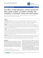
over a four year period[31]. CT is now the diagnostic test
of choice (Figure 1) [31,33]. The same can not be said for
the use of CT scanning for the diagnosis of blunt injury to
aortic branch vessels. The small number of patients under-
going CT for aortic branch vessel trauma and questions as
to its accuracy has limited its use as the diagnostic test of
choice [34,35]. New generation, multiple detector CT
technology, however, has clearly improved diagnostic
quality and reduced the need for catheter based angiogra-
phy. Our practice is similar to others as we use CT as a
screening test and angiography as needed [35,36]. Mag-
netic resonance imaging, transesophageal echocardiogra-
phy, and intravascular ultrasonography are alternative
modalities in particular for diagnosis of blunt aortic
injury.
Initial Management
The initial management of patients with suspected blunt
great vessel injury is similar to that of any trauma patient.
While a comprehensive discussion of the assessment of
the trauma patient is beyond the scope of this article, a few
salient points require mentioning. The primary survey of
airway, breathing, circulation, disability and exposure
(ABCDE) with concomitant treatment of life-threatening
injuries remains the cornerstone in evaluating these
patients. A more detailed examination during the second-
ary survey, chest and pelvic plain radiographs, and the use
of Focused Assessment with Sonography for Trauma
(FAST) allows the formulation of an initial plan. The over-
all plan depends on the clinical situation, constellation of
injuries, and hemodynamic stability. Treatment may be
immediate operation, further imaging studies, or expect-
ant/non-operative management. Clinical judgment is par-
amount and life threatening injuries take precedence.
While associated injuries are common, often the great ves-
Reconstructed computed tomography with contrast depict-ing aortic injury with pseudoaneurysm (arrow)Figure 1
Reconstructed computed tomography with contrast
depicting aortic injury with pseudoaneurysm (arrow).
Arrowhead indicates proximal left subclavian artery.
Scandinavian Journal of Trauma, Resuscitation and Emergency Medicine 2009, 17:42 />Page 4 of 10
(page number not for citation purposes)
sel injury takes priority[8,9,19,20]. The need for emergent
surgery is based on hemodynamics; as the unstable hypo-
tensive patient may need rapid control of hemorrhage
[19,20]. In particular for aortic injuries, timing of repair is
based both on the extent of the patient's coexisting inju-
ries as well as the extent of injury to the thoracic aorta.
Small pseudoaneurysms and intimal injuries that don't
appear to penetrate the outer wall of the aorta can gener-
ally be managed expectantly, reserving treatment for
lesions that do not spontaneously resolve. Lesions with
evidence of significant mediastinal hematoma need to be
managed more aggressively. It should be noted however,
that there is evidence that as many as 50% of minimal
injury lesions (defined as an intimal flap of less than 1 cm
with no or minimal periaortic hematoma) can develop
into pseuoaneuysms at 8 week follow-up [37]. It is likely
that the less invasive nature of endograft repair will allow
more options for patients who were previously managed
non-operatively.
The initial management of hemodynamically stable
patients may include the use of β-blockers to lower the
mean arterial pressure and to decrease aortic shear force
(dP/dt). The target mean arterial pressure is between 60
and 70 mmHg. This approach has been extrapolated from
the initial treatment of traumatic aortic rupture for which
a delayed approach is being employed with increasing fre-
quency[28,38]. A prospective study used beta-blockers
with or without vasodilators to keep systolic blood pres-
sures near 100 mm Hg, and a heart rate below 100 beats
per minute in selected patients with blunt aortic injury
and either concomitant head injury, pulmonary injury or
cardiac insufficiency. There were no treatment failures
prior to delayed aortic repair[39].
However, if there is a significant associated cerebral
injury, even mild hypotension may worsen the neurologic
outcome and normal blood pressure should be main-
tained. The data on hypotensive resuscitation is mixed,
and a good review of this interesting topic is available[40].
In a randomized study of 598 patients with penetrating
trauma, there was a significant survival benefit in the
group which did not receive fluid, especially among those
with cardiac injury[41]. This study has several important
limitations; it is limited to penetrating injury and cardiac
injuries represent a special subset of patients where sur-
vival may be more a function of time to surgery and the
presence of tamponade. A randomized study from our
institution showed no difference in mortality between
those patients treated with normotensive versus hypoten-
sive fluid resuscitation[42].
The concept of damage control surgery for penetrating
abdominal trauma was introduced in 1993 and has
expanded to other cavitary injuries[43,44]. If shock, coag-
ulapathy, and hypothermia are not arrested, death will
ensue. These same principles can be applied to vascular
and thoracic trauma. With regard to vascular surgery, tem-
porary arterial shunts allow distal perfusion and delayed
vascular reconstruction. They are easy to insert and have
an excellent patency rate, especially for proximal extrem-
ity vessels[45,46]. Another technique is the use of pros-
thetic grafts, even in contaminated wounds, as a
temporizing maneuver prior to revascularization with
autogenous conduit [47]. The principles of thoracic dam-
age control are not as straight forward. In addition to
hemorrhage, hypoxia and hypercarbia can also be lethal.
The surgical approach consists of an abbreviated opera-
tion using non-anatomic pulmonary resection and tem-
porary chest closure with delayed definitive
closure[48,49].
Definitive Treatment
Definitive treatment can be divided into operative proce-
dures and the placement of endoluminal stent grafts.
Some general principles will be discussed followed by the
treatment of specific vessel injury. Several incisions have
been used to obtain exposure of the great vessels. There is
agreement that median sternotomy, with clavicular or
neck extension if needed, is it the preferred approach for
the majority of great vessel trauma, including the right
subclavian artery. There is still some debate as to optimal
exposure of the proximal left subclavian artery with some
advocating a high antero-lateral thoracotomy combined
with a clavicular incision[3,50]. Others, our group
included, prefer to approach the proximal left subclavian
using a sternotomy with extension if needed, as it pro-
vides excellent exposure[51,52]. Division of the innomi-
nate vein will greatly improve exposure. Regardless of the
operative approach, intra-operative blood salvage, large
bore intravenous access, and communication with the
anesthesia team are essential.
While there are various techniques to manage vessel
injury it is imperative to adhere to the general principles
and techniques of vascular surgery. Given the arterial
diameter of the great vessels, most will require prosthetic
graft interposition and less commonly the injury is ame-
nable to autogolous vein or primary repair. Ligation of the
subclavian artery should be considered as a life-saving
procedure in the moribund patient. With the exception of
the cavae, most large veins can be ligated. In stable
patients lateral venorrophy should be employed if it does
not result in stenosis.
While there has been a substantial increase in the number
of traumatic aortic ruptures treated with endovascular
intervention, this technique has limited utility in the treat-
ment of aortic branch vessel injury. There are several fac-
tors which limit the use of endovascular techniques to
Scandinavian Journal of Trauma, Resuscitation and Emergency Medicine 2009, 17:42 />Page 5 of 10
(page number not for citation purposes)
aortic branch vessels. As with traumatic aortic rupture,
hemodynamic unstable patients will undergo surgery thus
limiting transcather therapy to those who are hemody-
namically stable. With the exception of the subclavian
artery, most great vessel injuries are proximal at the origin
of the artery from the aortic arch[19,20,28]. This anatomic
location often precludes the use of the adjacent vessel as a
landing zone since it does not have adequate length and,
it may not be possible to preserve adjacent vessels[53,54].
Specific Injuries
Innominate artery
This is the second most commonly injured great vessel,
with the proximal descending aorta the most common.
Most innominate artery injuries occur at the vessel origin
[7,19,20,28]. These are surgically repaired by placing a
graft end-to-side from the ascending aorta and end-to-end
to the distal innominate. Only after the graft is in place is
the proximal innominate artery closed with pledgeted
polypropylene sutures. An interposition graft or stent
placement may be employed if the injury is in the mid
portion of the vessel. More distal injuries (Figure 2) may
require more complex reconstruction [20]. Generally all
these injuries can be repaired without cardiopulmonary
bypass or shunts although some authors recommend
monitoring stump pressure [19].
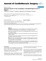
Left carotid artery
Similar to innominate injuries, left carotid injuries almost
always occur at the origin[19,20,28]. (Figure 3, 4) Graft
interposition or, less frequently, primary repair are used to
Reconstructed computed tomography with contrast demon-strating a pseudoaneurysm at the junction of the right subcla-vian and common carotid arteries (arrow)Figure 2
Reconstructed computed tomography with contrast
demonstrating a pseudoaneurysm at the junction of
the right subclavian and common carotid arteries
(arrow).
Reconstructed computed tomography with contrast demon-strating a pseudoaneurysm at the origin of the left common carotid artery (arrow)Figure 3
Reconstructed computed tomography with contrast
demonstrating a pseudoaneurysm at the origin of the
left common carotid artery (arrow).
"Three dimensional" rendering of injury depicted in Figure 3Figure 4
"Three dimensional" rendering of injury depicted in
Figure 3.
Scandinavian Journal of Trauma, Resuscitation and Emergency Medicine 2009, 17:42 />Page 6 of 10
(page number not for citation purposes)
repair these injuries. (Figure 5) As with innominate artery
injuries, bypass and arterial shunts are rarely necessary.
Subclavian artery
Injury to this vessel often necessitates an interposition
graft. Sternotomy with clavicular extension may be
required to obtain optimal exposure of this difficult area,
as blunt subclavian injuries tend to be more distal [8,12].
(Figure 6, 7) While interposition grafting is the most com-
mon method of reconstruction, primary repair may be
feasible. Arterial ligation is uncommonly performed but
may be life saving. Abundant collaterals prevent acute
limb ischemia and, if it were to develop, re-vasculariza-
tion can be performed. Interestingly, some authors have
advocated not acutely re-establishing flow if the limb is
not threatened and there is concomitant severe brachial
plexus injury[8,13,55].
Pulmonary artery and vein
These are very rare injuries and the overwhelming major-
ity of the injured die prior to hospitalization. The mortal-
ity of those who are alive on hospital admission is
prohibitive[2,3].
Venous injuries
Thoracic caval injuries are exceedingly rare and highly
lethal, especially if there is disruption of the atrio-caval
junction. While superior vena caval injuries can rarely be
repaired, injuries to the shorter intrathoracic inferior vena
cava are almost uniformly fatal. Isolated azygous injury is
exceedingly rare, limited to case reports and carries a sig-
nificant mortality[56].
Special Circumstances
Isolated venous injuries are uncommon but may carry a
higher mortality than those to arteries[57]. More com-
monly they are associated with arterial trauma and, in
some series, combined injuries are more lethal [16].
Another combination is trauma to the tracheo-bronchial
tree with a great vessel injury[20,58-60]. These injuries
require immediate operation with meticulous attention to
airway management. Another unusual situation is blunt
innominate injury in the setting where the left common
carotid artery originates off of, or shares a common origin
with, the innominate artery. While this anatomic variant
is relatively common, it may complicate treatment. Sev-
eral reports have described the successful management of
this condition[61,62].
Excluding aortic trauma, blunt injury to the thoracic great
vessels is infrequent and presents several challenges to the
treating physicians and surgeons. On admission most
patients are hemodynamically stable, have extra-thoracic
injuries, may or may not have signs of limb ischemia, and
often have a brachial plexus injury. An abnormal chest
radiograph, especially a widened mediastinum, should
prompt further imaging to precisely define the location
and extent of the vascular injury. Unstable patients
require immediate operation. Among stable patients,
treatment options include operative management and
endovascular intervention. This decision depends on the
specific anatomy and availability of specialized person-
nel. Although these injures are associated with significant
mortality and morbidity, rapid diagnosis and prompt
intervention can yield gratifying results.
Intra-operative photograph of and end-to side anastomosis of the left common carotid to the innominate arteryFigure 5
Intra-operative photograph of and end-to side anas-
tomosis of the left common carotid to the innomi-
nate artery.
Intra-operative photograph of a thrombosed right subclavian artery (arrow)Figure 6
Intra-operative photograph of a thrombosed right
subclavian artery (arrow).
Scandinavian Journal of Trauma, Resuscitation and Emergency Medicine 2009, 17:42 />Page 7 of 10
(page number not for citation purposes)
Thoracic Aorta
Open repair requires single lung ventilation and a thora-
cotomy at the left fourth intercostal space. Today the aorta
is rarely repaired with a clamp and sew technique due to
the risk of paraplegia. Generally, the distal circulation
beyond the proximal aortic clamp is perfused with oxy-
genated blood from an extracorporeal circuit. The circuit
can lack an oxygenator and thus draw its blood from the
left atrium via the inferior pulmonary vein or left atrial
appendage. Our group has popularized use of femoral
venous to femoral arterial cardiopulmonary bypass (CPB)
as an alternative[63]. The advantage to the full CPB is the
use of an integral pump sucker to rapidly deal with unex-
pected and life threatening bleeding encountered during
the operation. Usually an interpostition graft is necessary
as the aorta recoil results in defects of 2-3 inches, and sur-
geons strive to reduce tension on suture lines. However
primary repair is prudent in certain cases. Despite major
advances in surgical technique and adjunctive protective
measures including spinal drainage, and distal aortic per-
fusion, open repair has significant morbidity and mortal-
ity. Rates of 18-28% operative mortality have been
reported with paraplegia occurring in 2.3-14% of
cases[64,65]. To date, our use of full CPB has not been
associated with paraplegia. The associated injuries often
seen with blunt aortic injury often preclude the necessary
measures for open repair. Hypotension and anticoagula-
tion in the setting of closed head injury is ill advised. Sim-
ilarly, single lung ventilation can lead to hypoxia in the
patient with pulmonary contusions.
Endovascular stent grafts were initially described for treat-
ment of abdominal aortic aneurysms by Parodi in 1991
[66]. The first reported case of endovascular stent graft
repair of the thoracic aorta was reported by Dake and col-
leagues in for a patient with an enlarging pseudoaneu-
rysm of the descending thoracic aorta[67]. Subsequent
reports show successful placement and favorable out-
comes for endovascular repair of aneurysms, traumatic
injury and dissection[68]. Endovascular stent grafts are
usually placed via a femoral artery cutdown. Iliac artery
injury is a known complication, especially when these ves-
sels are small [69]. A guide wire is placed under fluoro-
scopic guidance across the injury and the stent graft is
deployed after angiography confirms the location of the
injured segment. (Figures 8 and 9) The stent graft is a com-
bination of metal stents providing radial force outwards
with covered graft material that excludes flow from the
injury. The advantages to endovascular stent grafting
include minimal physiologic insult with access and
deployment. There is no need for lateral decubitus posi-
tioning as in open thoracotomy which is advantageous in
Arch aortogram depicting thrombosed left subclavian artery (arrow) distal to the left vertebral artery (arrowhead)Figure 7
Arch aortogram depicting thrombosed left subcla-
vian artery (arrow) distal to the left vertebral artery
(arrowhead).
Aortogram depicting aortic injury (arrow) with undeployed endovascular graft in position (arrowhead)Figure 8
Aortogram depicting aortic injury (arrow) with
undeployed endovascular graft in position (arrow-
head).
Scandinavian Journal of Trauma, Resuscitation and Emergency Medicine 2009, 17:42 />Page 8 of 10
(page number not for citation purposes)
the head injured patient or pelvic fracture requiring exter-
nal fixation. Finally, the ability to deploy the stent graft
with no thorocotomy, aortic clamping, single lung venti-
lation, nor heparinization allows treatment of even the
most critically injured or frail patients.
The American Association for the Surgery of Trauma
(AAST) prospectively studied the treatment of blunt tho-
racic aortic injury at multiple institutions (AAST2) [70].
There have been significant changes since the landmark
prospective AAST study from 1999 (AAST1) [22]. In 1999
there were no patients in the study treated with endograft-
ing, whereas 64.8% of patients underwent endografting in
the 2008 study. Excluding patients in extremis, mortality
decreased significantly form 22% to 13% and procedure-
related paraplegia decreased from 8.7 to 1.6%. Of those
patients who underwent endografting, the mortality was
7.2% (23.5% open mortality) and procedure related par-
aplegia was 0.8% (2.9% open paraplegia rate). However,
the improvements in mortality and morbidity came at a
price of 20% device related complication rate. The
endoleak rate was 14.4% (18 patients) of which 6 under-
went open repair.
At our institution endograft repair has become the pri-
mary treatment option for blunt aortic injury[71]. In our
first 45 patients the mortality was 11% (none device
related). There were no cases of paraplegia. However, sim-
ilar to AAST2, the endoleak rate was 13.3% (6 patients) of
which 3 patients had to undergo open repair.
It is clear that currently available devices were not
designed for the small, sharply angulated aortic arches of
young patients. The three thoracic endografts currently
available in the US were designed for and approved by the
FDA for nonruptured thoracic aortic aneurysms. The first
available graft was the Gore TAG device. The Medtronic
Talent device and the Cook Zenith TX2 device were later
approved with an additional indication for penetrating
aortic ulcers. There a variety of pitfalls that can lead to
device failure, particularly in the treatment of blunt tho-
racic traumatic injuries. Over sizing of the stent or place-
ment along the arch increases the risk of graft
collapse[72,73]. To prevent graft failure, small diameter,
short abdominal aortic cuffs from abdominal systems
have been successfully used for these injuries [74-76]. The
use of abdominal cuffs has several disadvantages: these
cuffs tend to be short and require several grafts overlap-
ping to cover the appropriate length. Short cuffs tend to be
inflexible and are not well suited for conforming to the
curve of the distal arch.
Devices that address these technical pitfalls are clearly
needed. Home-made fenestrated devices designed to
extend the area of coverage while maintaining patency of
arch vessels have been used with success[77]. Multi-insti-
tutional trials designed to evaluate more flexible grafts are
scheduled to start in the United States soon.
Despite their limitations, currently available endoluminal
stent grafts have been used with promising results. A
recent meta-analysis of seventeen retrospective cohort
studies, demonstrated a significantly lower procedure
related mortality, overall 30 day mortality and postopera-
tive paraplegia in patients treated with endografts vs. open
repair[78,79].
Fortunately, in our experience, there have been no mid-
term graft failures or need for intervention. However, the
durability and long term outcomes for endovascular stent
grafts are unknown in these typically young patients. Long
term follow-up will be required. Follow-up can be diffi-
cult in this group of patients. Additionally the use of radi-
ologic imaging over a long period of time carries with it a
tangible risk of future malignancy [80].
The treatment of blunt aortic injury has undergone a rad-
ical paradigm shift with the introduction of endovascular
stent grafts. With the evolution of graft design and succes-
sive models conforming to the curve of the aortic arch and
produced in smaller diameter sizes, it is likely that
endovascular repair will become the primary treatment in
the majority of blunt aortic injury with improved morbid-
ity and mortality rates for these often challenging injuries.
Aortogram following endograft deployment with successful exclusion of the pseudoaneurysmFigure 9
Aortogram following endograft deployment with
successful exclusion of the pseudoaneurysm.
Scandinavian Journal of Trauma, Resuscitation and Emergency Medicine 2009, 17:42 />Page 9 of 10
(page number not for citation purposes)
Competing interests
The authors declare that they have no competing interests.
Authors' contributions
JO contributed directly to drafting of the manuscript. CB
contributed directly to drafting of the manuscript. TS con-
tributed directly to drafting of the manuscript and partici-
pated in its organization. BG contributed directly to
drafting of the manuscript. DN contributed directly to
drafting of the manuscript, conceived the work, and coor-
dinated its design. All authors read and approved the final
manuscript.
References
1. Mattox KL, Feliciano DV, Burch J, Beall AC Jr, Jordan GL Jr, De Bakey
ME: Five thousand seven hundred sixty cardiovascular inju-
ries in 4459 patients. Epidemiologic evolution 1958 to 1987.
Ann Surg 1989, 209:698-705. discussion 706-697
2. Dosios TJ, Salemis N, Angouras D, Nonas E: Blunt and penetrating
trauma of the thoracic aorta and aortic arch branches: an
autopsy study. J Trauma 2000, 49:696-703.
3. Mattox KL: Thoracic great vessel injury. Surg Clin North Am 1988,
68:693-703.
4. Smith RS, Chang FC: Traumatic rupture of the aorta: still a
lethal injury. Am J Surg 1986, 152:660-663.
5. Parmley LF, Mattingly TW, Manion WC, Jahnke EJ Jr: Nonpenetrat-
ing traumatic injury of the aorta. Circulation 1958, 17:1086-1101.
6. Greendyke RM: Traumatic rupture of aorta; special reference
to automobile accidents. Jama 1966, 195:527-530.
7. Weiman DS, McCoy DW, Haan CK, Pate JW, Fabian TC: Blunt inju-
ries of the brachiocephalic artery. Am Surg 1998, 64:383-387.
8. Costa MC, Robbs JV: Nonpenetrating subclavian artery
trauma. J Vasc Surg 1988, 8:71-75.
9. Posner MP, Deitrick J, McGrath P, Mendez-Picon G, Sobel M, Lower
RR, Lee HM: Nonpenetrating vascular injury to the subclavian
artery. J Vasc Surg 1988, 8:611-617.
10. Johnston RH Jr, Wall MJ Jr, Mattox KL: Innominate artery
trauma: a thirty-year experience. J Vasc Surg 1993, 17:134-139.
discussion 139-140
11. Katras T, Baltazar U, Rush DS, Davis D, Bell TD, Browder IW, Comp-
ton RP, Stanton PE Jr: Subclavian arterial injury associated with
blunt trauma. Vasc Surg 2001, 35:43-50.
12. Cox CS Jr, Allen GS, Fischer RP, Conklin LD, Duke JH, Cocanour CS,
Moore FA: Blunt versus penetrating subclavian artery injury:
presentation, injury pattern, and outcome. J Trauma 1999,
46:445-449.
13. Hyre CE, Cikrit DF, Lalka SG, Sawchuk AP, Dalsing MC: Aggressive
management of vascular injuries of the thoracic outlet. J Vasc
Surg 1998, 27:880-884. discussion 884-885
14. Hoff SJ, Reilly MK, Merrill WH, Stewart J, Frist WH, Morris JA Jr:
Analysis of blunt and penetrating injury of the innominate
and subclavian arteries. Am Surg 1994, 60:151-154.
15. McCoy DW, Weiman DS, Pate JW, Fabian TC, Walker WA: Subcla-
vian artery injuries. Am Surg 1997, 63:761-764.
16. Demetriades D, Asensio JA: Subclavian and axillary vascular
injuries. Surg Clin North Am 2001, 81:1357-1373. xiii
17. Aksoy M, Tunca F, Yanar H, Guloglu R, Ertekin C, Kurtoglu M: Trau-
matic injuries to the subclavian and axillary arteries: a 13-
year review. Surg Today 2005, 35:561-565.
18. McKinley AG, Carrim AT, Robbs JV: Management of proximal
axillary and subclavian artery injuries. Br J Surg 2000, 87:79-85.
19. Rosenberg JM, Bredenberg CE, Marvasti MA, Bucknam C, Conti C,
Parker FB Jr: Blunt injuries to the aortic arch vessels. Ann Thorac
Surg 1989, 48:508-513.
20. Karmy-Jones R, DuBose R, King S: Traumatic rupture of the
innominate artery. Eur J Cardiothorac Surg 2003, 23:782-787.
21. Johnson SF, Johnson SB, Strodel WE, Barker DE, Kearney PA: Bra-
chial plexus injury: association with subclavian and axillary
vascular trauma. J Trauma 1991, 31:1546-1550.
22. Fabian TC, Richardson JD, Croce MA, Smith JS Jr, Rodman G Jr, Kear-
ney PA, Flynn W, Ney AL, Cone JB, Luchette FA, et al.: Prospective
study of blunt aortic injury: Multicenter Trial of the Ameri-
can Association for the Surgery of Trauma.
J Trauma 1997,
42:374-380. discussion 380-373
23. Horton TG, Cohn SM, Heid MP, Augenstein JS, Bowen JC, McKenney
MG, Duncan RC: Identification of trauma patients at risk of
thoracic aortic tear by mechanism of injury. J Trauma 2000,
48:1008-1013. discussion 1013-1004
24. Schulman CI, Carvajal D, Lopez PP, Soffer D, Habib F, Augenstein J:
Incidence and crash mechanisms of aortic injury during the
past decade. J Trauma 2007, 62:664-667.
25. Cohen AM, Crass JR, Thomas HA, Fisher RG, Jacobs DG: CT evi-
dence for the "osseous pinch" mechanism of traumatic aor-
tic injury. AJR Am J Roentgenol 1992, 159:271-274.
26. Carter YM, Karmy-Jones RC, Oxorn DC, Aldea GS: Traumatic dis-
ruption of the aortic arch. Eur J Cardiothorac Surg 2001, 20:1231.
27. Hirose H, Moore E: Delayed presentation and rupture of a
posttraumatic innominate artery aneurysm: case report and
review of the literature. J Trauma 1997, 42:1187-1195.
28. Symbas JD, Halkos ME, Symbas PN: Rupture of the innominate
artery from blunt trauma: current options for management.
J Card Surg 2005, 20:455-459.
29. Shaw AD, Milne AA, Christie J, Jenkins AM, Murie JA, Ruckley CV:
Vascular trauma of the upper limb and associated nerve inju-
ries. Injury 1995, 26:515-518.
30. Mirvis SE, Bidwell JK, Buddemeyer EU, Diaconis JN, Pais SO, Whitley
JE, Goldstein LD: Value of chest radiography in excluding trau-
matic aortic rupture. Radiology 1987, 163:487-493.
31. Mirvis SE, Shanmuganathan K, Miller BH, White CS, Turney SZ:
Traumatic aortic injury: diagnosis with contrast-enhanced
thoracic CT five-year experience at a major trauma center.
Radiology 1996, 200:413-422.
32. Demetriades D, Gomez H, Velmahos GC, Asensio JA, Murray J,
Cornwell EE 3rd, Alo K, Berne TV: Routine helical computed
tomographic evaluation of the mediastinum in high-risk
blunt trauma patients. Arch Surg 1998, 133:1084-1088.
33. Melton SM, Kerby JD, McGiffin D, McGwin G, Smith JK, Oser RF,
Cross JM, Windham ST, Moran SG, Hsia J, Rue LW 3rd: The evolu-
tion of chest computed tomography for the definitive diag-
nosis of blunt aortic injury: a single-center experience. J
Trauma 2004, 56:243-250.
34. Chen MY, Regan JD, D'Amore MJ, Routh WD, Meredith JW, Dyer
RB: Role of angiography in the detection of aortic branch ves-
sel injury after blunt thoracic trauma. J Trauma 2001,
51:1166-1171. discussion 1172
35. Fishman JE: Imaging of blunt aortic and great vessel trauma. J
Thorac Imaging 2000, 15:97-103.
36. Chen MY, Miller PR, McLaughlin CA, Kortesis BG, Kavanagh PV, Dyer
RB: The trend of using computed tomography in the detec-
tion of acute thoracic aortic and branch vessel injury after
blunt thoracic trauma: single-center experience over 13
years. J Trauma 2004, 56:783-785.
37. Malhotra AK, Fabian TC, Croce MA, Weiman DS, Gavant ML, Pate
JW: Minimal aortic injury: a lesion associated with advancing
diagnostic techniques. J Trauma 2001, 51:1042-1048.
38. Goaley TJ, Dente CJ, Feliciano DV: Torso vascular trauma at an
urban level I trauma center. Perspect Vasc Surg Endovasc Ther
2006, 18:102-112.
39. Fabian TC, Davis KA, Gavant ML, Croce MA, Melton SM, Patton JH
Jr, Haan CK, Weiman DS, Pate JW: Prospective study of blunt
aortic injury: helical CT is diagnostic and antihypertensive
therapy reduces rupture. Ann Surg 1998, 227:666-676. discussion
676-667
40. Bickell WH, Wall MJ Jr, Pepe PE, Martin RR, Ginger VF, Allen MK,
Mattox KL: Immediate versus delayed fluid resuscitation for
hypotensive patients with penetrating torso injuries. N Engl J
Med 1994, 331:1105-1109.
41. Dutton RP, Mackenzie CF, Scalea TM: Hypotensive resuscitation
during active hemorrhage: impact on in-hospital mortality. J
Trauma 2002, 52:1141-1146.
42. Stern SA: Low-volume fluid resuscitation for presumed hem-
orrhagic shock: helpful or harmful? Curr Opin Crit Care
2001,
7:422-430.
43. Rotondo MF, Schwab CW, McGonigal MD, Phillips GR 3rd, Fruchter-
man TM, Kauder DR, Latenser BA, Angood PA: 'Damage control':
an approach for improved survival in exsanguinating pene-
Publish with BioMed Central and every
scientist can read your work free of charge
"BioMed Central will be the most significant development for
disseminating the results of biomedical research in our lifetime."
Sir Paul Nurse, Cancer Research UK
Your research papers will be:
available free of charge to the entire biomedical community
peer reviewed and published immediately upon acceptance
cited in PubMed and archived on PubMed Central
yours — you keep the copyright
Submit your manuscript here:
/>BioMedcentral
Scandinavian Journal of Trauma, Resuscitation and Emergency Medicine 2009, 17:42 />Page 10 of 10
(page number not for citation purposes)
trating abdominal injury. J Trauma 1993, 35:375-382. discussion
382-373
44. Shapiro MB, Jenkins DH, Schwab CW, Rotondo MF: Damage con-
trol: collective review. J Trauma 2000, 49:969-978.
45. Rasmussen TE, Clouse WD, Jenkins DH, Peck MA, Eliason JL, Smith
DL: The use of temporary vascular shunts as a damage con-
trol adjunct in the management of wartime vascular injury.
J Trauma 2006, 61:8-12. discussion 12-15
46. Clouse WD, Rasmussen TE, Peck MA, Eliason JL, Cox MW, Bowser
AN, Jenkins DH, Smith DL, Rich NM: In-theater management of
vascular injury: 2 years of the Balad Vascular Registry. J Am
Coll Surg 2007, 204:625-632.
47. Vertrees A, Fox CJ, Quan RW, Cox MW, Adams ED, Gillespie DL:
The use of prosthetic grafts in complex military vascular
trauma: a limb salvage strategy for patients with severely
limited autologous conduit. J Trauma 2009, 66:980-983.
48. Rotondo MF, Bard MR: Damage control surgery for thoracic
injuries. Injury 2004, 35:649-654.
49. O'Connor J, Kells A, Henry S, Scalea T: Vacuum-assisted closure
for the treatment of complex chest wounds. Ann Thorac Surg
2005, 79:1196-1200.
50. Wall MJ Jr, Granchi T, Liscum K, Mattox KL: Penetrating thoracic
vascular injuries. Surg Clin North Am 1996, 76:749-761.
51. Hajarizadeh H, Rohrer MJ, Cutler BS: Surgical exposure of the left
subclavian artery by median sternotomy and left supracla-
vicular extension. J Trauma 1996, 41:136-139.
52. Oconnor J, Scalea TM: Penetrating Great Vessel Injury: Impact
of Admission Hemodynamics and Pre-Operative Imaging. J
Trauma 2009 in press.
53. Arthurs ZM, Sohn VY, Starnes BW: Vascular trauma: endovascu-
lar management and techniques. Surg Clin North Am 2007,
87:1179-1192.
54. Hoffer EK: Endovascular intervention in thoracic arterial
trauma. Injury 2008, 39:1257-1274.
55. Hawthorn IE, Rochester J, Beard JD: Treatment of combined bra-
chial plexus and subclavian artery trauma. Injury 1993,
24:377-379.
56. Wall MJ Jr, Mattox KL, Debakey ME: Injuries to the azygous
venous system. J Trauma 2006, 60:357-362.
57. Baumgartner FJ, Rayhanabad J, Bongard FS, Milliken JC, Donayre C,
Klein SR: Central venous injuries of the subclavian-jugular and
innominate-caval confluences. Tex Heart Inst J 1999, 26:177-181.
58. Katz RI, Briggs JN: Traumatic ruptured bronchus and injury of
major thoracic vessels. Ann Thorac Surg 1967, 3:235-238.
59. Goldfaden D, Seifert P, Milloy F, Thomas P, Levitsky S: Combined
tracheal transection and innominate artery disruption from
blunt chest trauma. Ann Thorac Surg 1986, 41:213-215.
60. Hemmila MR, Hirschl RB, Teitelbaum DH, Austin E, Geiger JD: Tra-
cheobronchial avulsion and associated innominate artery
injury in blunt trauma: case report and literature review. J
Trauma 1999, 46:505-512.
61. Moise MA, Hsu V, Braslow B, Woo YJ: Innominate artery
transection in the setting of a bovine arch. J Thorac Cardiovasc
Surg 2004, 128:632-634.
62. Al-Khaldi A, Robbins RC: Successful repair of blunt injury of aor-
tic arch branches in the setting of bovine arch. J Vasc Surg
2006, 43:396-398.
63. Cardarelli MG, McLaughlin JS, Downing SW, Brown JM, Attar S, Grif-
fith BP: Management of traumatic aortic rupture: a 30-year
experience. Ann Surg 2002, 236:465-469. discussion 469-470
64. von Oppell UO, Dunne TT, De Groot MK, Zilla P: Traumatic aor-
tic rupture: twenty-year metaanalysis of mortality and risk
of paraplegia. Ann Thorac Surg 1994, 58:585-593.
65. Cowley RA, Turney SZ, Hankins JR, Rodriguez A, Attar S, Shankar BS:
Rupture of thoracic aorta caused by blunt trauma. A fifteen-
year experience. J Thorac Cardiovasc Surg 1990, 100:652-660. dis-
cussion 660-651
66. Parodi JC, Palmaz JC, Barone HD: Transfemoral intraluminal
graft implantation for abdominal aortic aneurysms. Annals of
Vascular Surgery 1991, 5:491-499.
67. Semba CP, Kato N, Kee ST, Lee GK, Mitchell RS, Miller DC, Dake
MD: Acute rupture of the descending thoracic aorta: repair
with use of endovascular stent-grafts. J Vasc Interv Radiol 1997,
8:337-342.
68. Dake MD, Miller DC, Semba CP, Mitchell RS, Walker PJ, Liddell RP:
Transluminal placement of endovascular stent-grafts for the
treatment of descending thoracic aortic aneurysms. N Engl J
Med 1994, 331:1729-1734.
69. Bavaria JE, Appoo JJ, Makaroun MS, Verter J, Yu ZF, Mitchell RS:
Endovascular stent grafting versus open surgical repair of
descending thoracic aortic aneurysms in low-risk patients: a
multicenter comparative trial. J Thorac Cardiovasc Surg 2007,
133:369-377.
70. Demetriades D, Velmahos GC, Scalea TM, Jurkovich GJ, Karmy-Jones
R, Teixeira PG, Hemmila MR, O'Connor JV, McKenney MO, Moore
FO, et al.: Operative repair or endovascular stent graft in
blunt traumatic thoracic aortic injuries: results of an Ameri-
can Association for the Surgery of Trauma Multicenter
Study. J Trauma 2008, 64:561-570. discussion 570-561
71. Neschis DG, Scalea TM, Flinn WR, Griffith BP: Blunt aortic injury.
N Engl J Med 2008, 359:1708-1716.
72. Idu MM, Reekers JA, Balm R, Ponsen KJ, de Mol BA, Legemate DA:
Collapse of a stent-graft following treatment of a traumatic
thoracic aortic rupture. J Endovasc Ther 2005, 12:503-507.
73. Rodd CD, Desigan S, Hamady MS, Gibbs RG, Jenkins MP: Salvage
options after stent collapse in the thoracic aorta. J Vasc Surg
2007, 46:780-785.
74. Wellons ED, Milner R, Solis M, Levitt A, Rosenthal D: Stent-graft
repair of traumatic thoracic aortic disruptions. J Vasc Surg
2004, 40:1095-1100.
75. Neschis DG, Moaine S, Gutta R, Charles K, Scalea TM, Flinn WR,
Griffith BP: Twenty consecutive cases of endograft repair of
traumatic aortic disruption: lessons learned. J Vasc Surg 2007,
45:487-492.
76. Peterson BG, Matsumura JS, Morasch MD, West MA, Eskandari MK:
Percutaneous endovascular repair of blunt thoracic aortic
transection. J Trauma 2005, 59:1062-1065.
77. Kurimoto Y, Asai Y, Nara S, Mori K, Hase M, Ohori S, Ito T, Baba T,
Kawaharada N, Higami T: Fenestrated stent-graft facilitates
emergency endovascular therapy for blunt aortic injury. J
Trauma 2009, 66:974-978. discussion 978-979
78. Xenos ES, Abedi NN, Davenport DL, Minion DJ, Hamdallah O, Sorial
EE, Endean ED: Meta-analysis of endovascular vs open repair
for traumatic descending thoracic aortic rupture. J Vasc Surg
2008, 48:1343-1351.
79. Hoffer EK, Forauer AR, Silas AM, Gemery JM: Endovascular stent-
graft or open surgical repair for blunt thoracic aortic
trauma: systematic review. J Vasc Interv Radiol 2008,
19:1153-1164.
80. Brenner DJ, Hall EJ: Computed tomography an increasing
source of radiation exposure. N Engl J Med 2007, 357:2277-2284.