Báo cáo y học: "Endogenous plasma activated protein C levels and the effect of enoxaparin and drotrecogin alfa (activated) on markers of coagulation activation and fibrinolysis in pulmonary embolism." ppt
Bạn đang xem bản rút gọn của tài liệu. Xem và tải ngay bản đầy đủ của tài liệu tại đây (452.06 KB, 10 trang )
RESEARCH Open Access
Endogenous plasma activated protein C levels
and the effect of enoxaparin and drotrecogin alfa
(activated) on markers of coagulation activation
and fibrinolysis in pulmonary embolism
Carl-Erik H Dempfle
1*
, Elif Elmas
1
, Andreas Link
2
, Nenad Suvajac
1
, Volker Liebe
1
, Jonathan Janes
3
,
Martin Borggrefe
1
Abstract
Introduction: There are no published data on the status of endogenous activated protein C (APC) in pulmonary
embolism (PE), and no data on the effect of drotrecogin alfa (activated) (DAA) given in addition to therapeutic
dose enoxaparin.
Methods: In this double-blind clinical trial, 47 patients with computed tomography (CT) -confirmed acute
submassive PE treated with 1 mg/kg body weight of enoxaparin twice daily were randomized to groups receiving
a 12-hour intravenous infusion of 6, 12, 18, or 24 μg/kg/hour of DAA or a placebo. Blood samples were drawn
before starting DAA infusion, after 4, 8 and 12 hours (at the end of the infusion period), and on treatment days 2,
3, 4, 5 and 6.
Results: Initial endogenous plasma activated protein C (APC) levels were 0.36 ± 0.48 ng/ml (<0.10 to 1.72 ng/ml)
and remained in the same range in the placebo group. APC levels in patients treated with DAA were 13.67 ± 3.57
ng/ml, 32.71 ± 8.76 ng/ml, 36.13 ± 7.60 ng/ml, and 51.79 ± 15.84 ng/ml in patients treated with 6, 12, 18, and
24 μg/kg/hour DAA, respectively. In patients with a D-dimer level >4 mg/L indicating a high level of acute fibrin
formation and dissolution, DAA infusion resulted in a more rapid drop in soluble fibrin, D-dimer, and fibrinogen/
fibrin degradation products (FDP) levels, compared to enoxaparin alone. There was a parallel decline of soluble
fibrin, D-dimer, FDP, and plasmin-plasmin inhibitor complex (PPIC) in response to treatment with enoxaparin ±
DAA, with no evidence of a systemic profibrinolytic effect of the treatment.
Conclusions: In patients with acute submassive PE endogenous APC levels are low. DAA infusion enhances the
inhibition of fibrin formation.
Trial registration: ClinicalTrials.gov: NCT00191724
Introduction
Activated protein C inhibits blood coagulation by inacti-
vating factors Va and VIIIa [1]. Inactivation of factor
VIIIa reduces the acti vity of the tenase complex and the
production of factor Xa. Inactivation of factor Va
reduces the activity of the prothrombinase complex and
the production of thrombin. Both mechanisms reduce
the amount of thrombin and fibrin generated. In vivo,
protein C is activated by the thrombin-thrombomodulin
complex, which forms when thrombin binds to throm-
bomodulin on i ntact endothelium. Binding of thrombin
to thrombomodulin also changes the specificity of
thrombin from a procoagulant to an anticoagulant
enzyme [2]. In addition to its effects on blood coagula-
tion activation, activated protein C when bound to
the endothelial protein C receptor (EPCR), activa-
tes protease-activated receptors (PARs), inducing a
variety of cytoprotective cellular responses, including
* Correspondence:
1
I. Department of Medicine, University Medical Center Mannheim, Theodor
Kutzer Ufer, Mannheim, D-68167, Germany
Full list of author information is available at the end of the article
Dempfle et al. Critical Care 2011, 15:R23
/>© 2011 Dempfle et a l.; licensee BioMed Central Ltd. This is an open access article distributed under the terms of the Creative Commons
Attribution License (http://crea tivecommons.org/licenses/by/2.0), which pe rmits unrestricted use, distribution, and re prod uction in
any medium, provide d the original work is properly cited.
alteration of gene expression profiles, anti-inflammatory
activities, anti-apo ptotic activity, and endothelia l barrier
stabilization [3].
A high level of thrombin in a patient with a localized
coagulation event such as venou s thrombosis, and other-
wise intact endothelium would be expected to result in
elevated levels of activated protein C (APC), similar to
what is observed in primates receiving an infusion of
thrombin [4]. APC influences coagulation activation and
organ dysfunction in animal models of sepsis [5]. No data
have been published on the actual plasma levels of endo-
genous activated protein C in patients with acute PE.
Drotrecogin alfa (activated) (DAA) [6] is a rec ombi-
nant form of hu man APC. Whereas endogenous pro-
duction of activated protein C is dependent upon an
ongoi ng coagulation process leading to the formation of
thrombin, DAA levels achieved with infusion of DAA
are independent of endogenous thrombin. Enoxaparin
[7], a low m olecular weight heparin commonly used for
treatment of patients with acute deep vein thrombosis
and pulmonary embolism, binds to antithrombin and
changes its conformation to yield an effective inhibitor
primarily of factor Xa. If enoxaparin and DAA are co m-
bined, this might result in a summation of anticoagulant
effects. Alternatively, it is possible that the anticoagulant
effect of therapeutic dose enoxaparin is maximal and
cannot be enhanced by additional DAA therapy.
The reduction of thrombin-induced fibrin generation
may lead to a drop in plasminogen activation by tPA,
which is dependent upon the cofactor activity of fibrin
[8,9] thus resulting in reduced fibrinolytic activity.
On the other hand, the antico agulant effect of both
drugs may result in an enhancement of fibrinolysis by
reducing the amount of activated thrombin-activated
fibrinolysis inhibitor (TAFIa) generated in the course of
coa gulation activation [10]. DAA may al so have a profi-
brinolytic effect by binding PAI-1 and thus reducing
PAI-1-capacity to inhibit tPA [11]. In fact, lower PAI-1
activity was detected in blood samples from patients
treated with DAA compared to samples from patients
treated with a placebo [12].
In the present study, we investigated the effect of thera-
peutic dose enoxaparin and four doses of DAA on blood
coagulation status and markers of fibrin formation, acti-
vation of fibrinolysis, and fibrin dissolution in acute PE.
This is the first clinical trial on the combination of a low
molecular weight heparin at a therapeutic dose in combi-
nation with DAA, and the first study reporting endogen-
ous APC levels in patients with acute PE.
Materials and methods
Inclusion and exclusion criteria
This was an exploratory, multicenter, randomized, paral-
lel, double-blind, placebo-controlled phase II dose
escalation study comparing a standard therapy for sub-
massive pulmonary embolism (enoxaparin 1 mg/kg body
weight twice daily by subcutaneous injection) to a com-
bined therapy of DAA with enoxaparin. Patients were
randomized according to a blinded randomization list
held by the study coordinator. Patient identification
numbers were obtained telephonically by the study phy-
sicians from the study coordinator. The trial was regis-
teredatClinicalTrials.gov as NCT00191724. The study
was started September 2004 and completed January
2008. The study was conducted in accordance with
applicable laws and regulations, and ethica l principles
that have their origin in the Declaration of Helsinki.
The institutional review boards of University Medical
Center Mannheim and the other participating centers
approved the study protocol, and all patients gave w rit-
ten informed consent.
The study was supported by Eli Lilly UK, Windlesham,
Surrey, United Kingdom. This included funding for a
study nurse; data management and statistics services pro-
vided b y Koordinierungszentrum Klinische Studien
(KKS) Heidelberg; trial medication and laboratory assays.
Co-author Jonathan Janes is an employee of the Lilly
Research Center, Windlesham, Surrey, United Kingdom.
Inclusion criteria were diagnosis of PE by spiral CT,
clinical symptoms of acute PE for less than 48 hours, no
massive PE judged as an indication for thrombolytic
therapy, evidence of right ventricular dysfunction
defined as right ventricular end-diastolic area/left ventri-
cular end-diastolic area (RVEDA/LVEDA) ratio in the
long axis greater than 0.6 associated with septal dyskine-
sia in the short axis [13], and age of ≥18 years. Exclu-
sion criteria were: beginning of infusion of the study
drug anticipated to be more than 24 hours after
PE diagnosis by spiral CT, treatment with vitamin K
antagonists in previous 5 days, pregnant or nursing
women, major surgery within previou s 24 hours, history
of severe head trauma, intracranial surgery or stroke
within the previous 3 months, evidence of intracerebral
arteriovenous malformations or cerebral aneurysm, evi-
dence of central nervous system mass lesion, neoplasm,
or cerebral herniation, history of inherited or acquired
chronic bleeding disorder, clinically significant gastroin-
testinal or genitourinary bleeding within the previous
6 weeks, clinical or laboratory evidence of hepatic fail-
ure, known esophageal varices, contraindications to
enoxaparin for treatment of PE, history of heparin-
induced thrombocytopenia type 2, femoral artery or
subclavian artery puncture within the previous 48 hours,
moribund patients expected to live not more than
24 hours, participation in another experimental inter-
ventional clinical trial within the previous 30 days, plate-
let count below lower limit of normal at inclusion, and
creatinin clearance <30 ml/minute.
Dempfle et al. Critical Care 2011, 15:R23
/>Page 2 of 10
Study treatment
Aft er wri tten informed consent and in addition to stan-
dard treatment with enoxaparin 1 mg/kg body weight
twice daily, patients received a 12-hour continuo us
intravenous infusion of the study drug. A 12-hour infu-
sion period was selected in order to limit the exposure
to DAA because of safety concerns, since there was no
prior experience with the combination of therapeutic
dose enoxaparin or any other low molecular weight
heparin, with DAA. Also, it was decided to start with a
low dose of DAA and gradually increase the dose of
DAA up to 24 μg/kg /hr, corresponding to the dose used
in patients with severe sepsis.
Warfarin anticoagulation was initiated after Day 3.
Enoxaparin treatment was terminated when therapeutic
INR values of >2 were reached in response to warfarin.
Patients were randomly assigned to receiving DAA
or a placebo as a study drug infusion. The study drug
was prepared by a study pharmacist not involved in
patient care and provided to the study physician in an
infusion syringe labeled with the pa tient number and
study identification. Group 1 included six patients trea-
tedwithDAAatadoseof6μg/kg/hour and six
patients receiving the placebo; group 2 included nine
patients receiving DAA at a dose of 12 μg/kg/hour and
three patients receiving the placebo; group 3 included
nine patients treated with DAA at a dose of 18 μg/kg/
hour and three patients receiving the placebo; and
group 4 included eight patients treated with DAA at a
dose of 24 μg/kg/h ou r and three patients receiving the
placebo. Patients receiving the placebo from all phases
of the study were combined for evaluation. After com-
pletion of each dose group, treatment and adverse
event documentation were reviews and the safety eval-
uated by an independent data safety monitoring board
(DSMB) before proceeding to the next dose of DAA.
The study was terminated after inclusion of 47 of the
originally planned 48 patients due to delays related to
DSMB analysis and slow enrollment caused by com-
peting t rials.
The sample size was calculated to evaluate major
bleeding. Assuming an approximate 5% rate with Enoxa-
parin there would be a >50% probability of detec ting an
additional event.
Safety analyses
Safety analyses were based on the data from all 47
patients included. Hematology parameters (erythrocytes,
hemoglobin level, leukocytes, platelets), prothrombin
time (PT) and activated partial thromboplastin time
(aPTT) were measured in fresh blood samples within
four hours after blood sampling by the local laboratories
of the participa ting centers. PT results were reported as
Quick% and INR.
Safety endpoints included life-threatening bleeding,
defined as fatal hemorrhage, reduction of hemoglobin
level by >5 g/dl, hypotension caused by bleeding requir-
ing inotro pic support, intracranial hemorrhage, transfu-
sion of >4 units of packed red blood cells, maj or
bleeding defined as a decrease in hemoglobin levels of 2
to 5 g/dl, transfusion of two to four units of packed red
blood cells, retroperitoneal bleeding, bleeding requiring
surgical intervention, or development of hematomas
requiring prolonged hospitalization, and minor bleeding
defined as a decrease in hemoglobin of <2 g/dl, develop-
ment of hematomas not requiring prolonged hospitaliza-
tion, or blood transfusion o f less than two units of
packed red blood cells. Further safety endpoints were an
aPTT more than three-fold the upper cutoff of normal
range, recurrent pulmonary embolism or worsening of
symptoms of pulmonary embolism requiring treatment
with thrombolytic drugs, surgical or catheter embolect-
omy, occurrence of allergic reactions, diagnosis or
heparin-induced thrombocytopenia type 2 (HIT-2),
other types of thrombocytopenia, worsening of symp-
toms leading t o endotracheal intubation and artificial
ventilation, cardiopulmonary resuscitation, and death.
Blood samples and laboratory analyses
Blood samples for preparation of citrated plasma were
drawn immediately before starting the study drug infu-
sion, 4, 8, and 12 hours after the start of the study drug
infusion, and once daily on days 2, 3, 4, 5, and 6 of
treatment. Special blood samples containing benzami-
dine for m easurement of APC were drawn before the
study drug infusion, and after 4, 8, and 12 hours.
A sufficient set of plasma and serum samples for the
batch labor atory analyses was available from 12 patients
treated with enoxaparin alone, all 6 patients of the DAA
6 μg/kg/hour group, 7 patients of the DAA 12 μ g/kg/
hour group, all 9 patients with the DAA 18 μg/kg/hour
group, and 7 patients of the DAA 24 μg/kg/hour group,
resulting in a total of 41 evaluable patients for the analy-
sis of laboratory markers of coagulation and fibrinolysis
activation. Samples were lost in two cases, and could
not be used for laboratory analysis due to pre-analytical
and handling mistakes in four cases.
The laboratory assays included prothrombin t ime
(PT), aPTT, an d anti-factor Xa chromogenic assay,
using reagents and equipment from DadeBehring Diag-
nostics, Marburg, Germany. Fibrinogen was measured
by turbidimetric immunoassay from Dako, Hamburg,
Germany, using a Hitachi 904 autoanalyzer. Photometric
immunoassays using antibody-coated latex particles
were also performed o n a Hitachi 904 autoanalyzer
(Roche Diagnostics, Mannheim, Ger many). The FDP-P
assay for fibrinogen and fibrin degradation products was
from Iatron Laboratories, Chiba, Japan. The Sekusui SF
Dempfle et al. Critical Care 2011, 15:R23
/>Page 3 of 10
assay for measurement of soluble fibrin was from
Daiichi Pure Chemicals, Ibaraki, Japan, and was also
performed on the Hitachi 904 autoanalyzer. Plasmin-
plasmin inhibitor complexes (PPIC, PAP) were mea-
sured using a 96-well microtiter plate ELISA from DRG
Instruments GmbH, Marburg, Germany.
APC was measured using the enzyme capture assay of
Gruber and Griffin [14] with minor modifications. The
lower limit of detection of the assay was 0.5 ng/mL.
Statistical analyses
Statistical analyses involved calculation of means, stan-
dard deviations, media ns, interquartile ranges. In order
to minimize the effect of outliers and distribution effects
in view of the small number of patients, medians were
used rather than mean values for the line graphs. All
group comparisons were performed using Wilcoxon’s
signed rank sum test. For correlation graphs, coefficients
of correlation R were calculated, using a linear regres-
sion model.
Results
Table 1 contains the baseline characteristics of patients
enrolled in the study by treatment group. There were
imbalances in baseline characterist ics that are likely due
to the small number of patients enrolled in each treat-
ment group. Patients who were enrolled earlier and
received the lower dosages of DAA tended to be older
than patients enrol led later in the study. Right ventricu-
lar dysfunction was present at admission in all patients,
as this was an entry criterion. Right ventricular end-dia-
stolic area divided by left ventricular end-diastolic area
(RVEDA/LVEDA) was used as an indicator of right ven-
tricular dysfunction. A v alue of >0.6 was considered to
be pathologic. Mean and median values of RVEDA/
LVEDA ratio decreased during treatment in all groups,
with no obvious differences between patients receiving
DAA or placebo (Table 2). RVEDA/LVEDA ratios were
calculated on the basis of echocardiography examina-
tions performed at admission, after 6 days, and after
90 days.
All patients were treated with therapeutic dose enoxa-
parin, which led to elevated anti-factor Xa ac tivity levels
within the therapeutic range for enoxaparin, with no sig-
nificant differences between DAA treatment groups.
Mean value was 0.66 ± 0.16 aXa U/mL during DAA
treatment phase (range 0.38 to 1.06 aXa U/mL).
Laboratory results of the samples drawn before the
startofthestudydruginfusionareshowninTable3.
All patients displayed abnormal D-dimer levels, as well
as elevated levels of soluble fibrin, fibrinogen/fibrin
degradation products and PPIC.
Despite the high level of coagulation activation present
in patients with acute pulmonary embolism, levels of
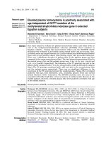
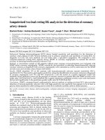
endogenous APC were low (Table 3, and Figure 1).
Values were below the detection limit of 0.5 ng/mL in
the majority of patients. In the patients treated with
enoxaparin alone, values did not change. Infusion of
DAA led to a dose-dependent increase in APC levels
(Figure 1). The APC levels attained were above the phy-
siological range in all dose groups and remained con-
stant during the i nfusion period of 12 hours. APC levels
in patients treated with DAA were 13.67 ± 3.57 ng/ml,
32.71 ± 8.76 ng/ml, 36.13 ± 7 .60 ng/ml, and 51.79 ±
15.84 ng/ml in patients treated with 6, 12, 18, and
24 μg/kg/hour DAA, respectively.
Three patients of the 6 μg/kg group and one patient
of the 12 μg/kg group had been treated with a bolus
dose of unfractionat ed heparin (UFH) initially, which
caused prolongation of aPTT in the pre-DAA-treatment
Table 1 Baseline characteristics of patients included (means ± standard deviation, range)
Variable DAA 6 μg/kg/h DAA 12 μg/kg/h DAA 18 μg/kg/h DAA 24 μg/kg/h Placebo
n699815
Age (years) 70.8 ± 4.4 (66.0 to
78.0)
65.7 ± 8.9 (46.0 to
74.0)
51.9 ± 16.5 (30.0 to
72.0)
45.6 ± 22.6 (18.0 to
78.0)
60.7 ± 21.9 (22.0 to
84.0)
Sex (Female) 3/6 3/9 5/9 3/8 11/15
Body weight (kg) 76.0 ± 9.7 (60.0 to
85.0)
93.9 ± 13.3 (79.0 to
120.0)
85.2 ± 17.6 (66.0 to
114.0)
86.4 ± 13.8 (62.0 to
104.0)
85.7 ± 18.6 (50.0 to
113.0)
Systolic blood pressure
(mmHg)
115.0 ± 15.8 (100.0 to
140.0)
129.1 ± 20.4 (95.0 to
160.0)
129.1 ± 30.7 (95.0 to
188.0)
119.5 ± 16.7 (80.0 to
132.0)
133.7 ± 25.4 (100.0 to
181.0)
Diastolic blood pressure
(mmHg)
66.4 ± 10.4 (57.0 to
80.0)
86.4 ± 17.5 (62.0 to
120.0)
82.4 ± 12.7 (70.0 to
104.0)
68.9 ± 12.0 (50.0 to
85.0)
76.6 ± 14.3 (60.0 to
110.0)
Highest heart rate (1/
minute)
83.5 ± 14.2 (60.0 to
100.0)
95.2 ± 19.4 (80.0 to
140.0)
94.4 ± 17.3 (75.0 to
127.0)
105.4 ± 21.6 (70.0 to
130.0)
106.6 ± 9.9 (95.0 to
130.0)
Earlier PE 1/6 2/9 0 1/8 0
Earlier DVT 1/6 2/9 1/9 2/8 2/15
Earlier ischemic stroke 1/6 1/9 0 0 3/15
DAA, Drotrecogin alfa (activated); DVT, deep vein thrombosis; PE, pulmonary embolism.
Dempfle et al. Critical Care 2011, 15:R23
/>Page 4 of 10
plasma sample. For analysis of the effect of DAA on
aPTT, but not for all other analyses; these patient s were
excluded.
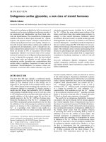
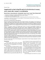
Infusion of DAA caused a transient increase in pro-
thrombin time (resulting in a reduced Quick percent
ratio) and aPTT. Figure 2 shows the results of the
12-hour sample drawn at the end of DAA infusion.
Median maximal aPTT levels were approximately 115%
of the initial value at the highest DAA dose. After termi-
nation of DAA infusion, PT and aPTT returned to pre-
DAA treatment l evels. These results indicate a detect-
able additional anticoagulant effect induced by DAA
given in addition to therapeutic dose enoxaparin in
patients with acute PE. Since conventional citrated
plasma was used for these analyses, the actual in vivo
effect is expected to be greater, d ue to the short in vitro
half-life of DAA.
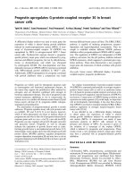
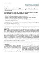
The distribution of D-dimer levels of all patients is
shown in Figure 3. Three of 12 patients in the placebo
group, 2 of 6 patients in the 6 μg/kg BW group, 2 of 7
patients in the 12 μg/kgBWgroup,2of9patientsin
the 18 μg/kg BW group, and 3 of 7 pat ients in the 24
μg/kg BW group displayed TINAquant D-dimer values
of <4 mg/L in the baseline plasma samples. For analysis
of the effect of DAA on fibrin formation and fibrinoly-
sis, these patients were excluded, because calculation of
a relative decrease (percent of initial value) led to a dis-
proportional effect of low initial values on the final
results. For the analysis, patients treated with DAA were
combined in one group. This resulted in a popu lation of
9 patients in the placebo group and 20 patients tre ated
with DAA.
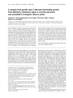
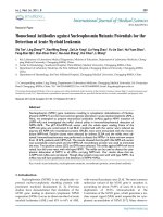
Treatment of patients with acute submassive PE with
enoxaparin caused a rapid decrease in markers of fibrin
formation and fibrin dissolution (Figure 4). There is no
obvious profibrinolytic effect, as soluble fibrin, D-dimer,
and fibrinogen/fibrin degradation products decrease in
parallel.
Addition of DAA to enoxaparin in the initial treat-
ment phase resulted in a more rapid decline in soluble
fibrin, D-dimer, and fibrinogen/fibrin degradation pro-
ducts, compared to enoxaparin alone, in patients with
an initial D-dimer level of >4.0 mg/L (Figure 4). As
Table 2 RVEDA/LVEDA ratio
Variable DAA 6 μg/kg/h DAA 12 μg/kg/h DAA 18 μg/kg/h DAA 24 μg/kg/h Placebo
RVEDA/LVEDA Day 0 0.8 ± 0.3 (0.6 to 1.4) 1.0 0.3 (0.7 to 1.6) 1.0 0.4 (0.7 to 1.7) 0.9 ± 0.4 (0.6 to 1.6) 1.1 0.5 (0.6 to 2.8)
N =6 N =9 N =9 N =8 N =15
RVEDA/LVEDA Day 6 0.7 ± 0.3 (0.5 to 1.2) 0.7 ± 0.2 (0.5 to 1.0) 0.8 ± 0.3 (0.5 to 1.5) 0.6 ± 0.2 (0.4 to 1.0) 0.7 ± 0.2 (0.5 to 1.1)
N =5 N =8 N =8 N =7 N =15
RVEDA/LVEDA Day 90 0.5 ± 0.1 (0.4 to 0.7) 0.5 ± 0.1 (0.4 to 0.6) 0.6 ± 0.1 (0.4 to 0.6) 0.5 ± 0.1 (0.4 to 0.6) 0.6 ± 0.2 (0.3 to 0.9)
N =5 N =8 N =8 N =5 N =11
DAA, Drotrecogin alfa (activated); LVEDA, levt ventricular enddiastolic area; RVEDA, right ventricular enddiastolic area.
Table 3 Laboratory values before start of study
medication
Parameter Mean SD Median Min Max
APC (ng/mL) 0.36 0.48 0.00 0.00 1.72
PT Quick (%) 92 11 93 75 119
INR 1.05 0.08 1.00 0.90 1.20
aPTT (sec) 30 13 26 22 89
Fibrinogen (g/L) 3.10 1.01 3.08 1.54 6.61
TINAquant D-dimer (mg/L) 7.19 4.25 6.80 0.76 15.53
Sekisui SF (mg/L) 33.73 20.84 33.75 11.10 125.10
Iatron FDP-P (mg/L) 23.02 20.78 18.80 4.30 105.10
PPIC (μg/L) 1,022 731 777 219 3,217
APC, activated protein C; aPTT, activated partial thromboplastin time; FDP-P,
fibrinogen/fibrin degradation products in plasma; INR, international
normalized ratio; PPIC, plasmin plasmin inhibitor complex; PT, prothrombin
time; SF, soluble fibrin.
Figure 1 APC act ivity at 0, 4 , 8 and 12 hours (end of study
drug infusion). Course of APC activity at inclusion, after 4 hours, 8
hours and after 12 hours (end of the study drug infusion). Patients
receiving placebo as the study drug infusion displayed low APC
activity levels. DAA infusion results in a dose-dependent increase in
APC activity levels.
Dempfle et al. Critical Care 2011, 15:R23
/>Page 5 of 10
shown in Figure 5, the difference is statistically signifi-
cant for the 12-h sampl e drawn at the en d of the DAA
infusion period.
Plasmin-plasmin inhibitor complexes (PPIC) decline in
parallel to soluble fibrin, and the fibrin degradation pro-
ducts, with no obvious effect of DAA (Figure 6).
There were no significant changes in hemoglobin,
hematocrit, or leukocyte count during enoxaparin ther-
apy. DAA treatment also had no effect on these
parameters.
Bleeding complications were within the expected
range for full-dose enoxaparin therapy. No patient
experienced life-threatening bleeding. Two patien ts
experienced major bleeding after infusion of DAA: One
patient in the 6 μg/kg/hour DAA group suffered from
intracranial hemorrhage on Day 4 of treatment, asso-
ciated with a drop in hemoglobin level >2 g/L. One
patient in the placebo group showed a drop in hemoglo-
bin level by >5 g/L. DAA treatment did not appear to
increase the risk of bleeding in any of the dose groups
studied.
Discussion
In healthy persons, APC levels are in the range of 1 to 3
ng/ml [ 14]. Patients with systemic coagulation activation
but normal endothelial function display APC levels as
high as 50 to 80 ng/ml. In patients with severe sepsis,
which also have strongly elevated levels of D-dimer and
other fibrin-related markers, APC levels are generally in
the range of 10 to 20 ng/ml [15]. In the patients with
acute submassive P E, the intravascu lar fibrin formation
is not associated with elevated levels of endogenous
APC. Baseline endogenous APC levels are low, and
remain in the low range during treatment with
enoxaparin.
In patents with severe sepsis, treatment with activated
protein C may improve clinical outcome, by reducing
organ dysfunction due to microvascular occlusion and
other mechanisms [16,17]. In view of the impaired pro-
tein C system present in many patients with PE, we
hypothesized that treatment with DAA might lead to
more effective anticoagulation and improved activation of
fibrinolysis, compared to therapy with enoxaparin alone.
In the present investigation, DAA treatment leading to
supraphysiological levels of APC had an additional
anticoagulant effect, associated with a prolongation
of prothrombin time and aPTT during the 12 hours of
infusion. Given the short plasma half-life of APC of
approximately 25 minutes, the ability to show an antic-
oagulant effect will be dependent on the speed of sam-
ple preparation and analysis and the actual in vivo effect
might be greater. Petäjä et al. described a synergistic
Figure 2 Prothrombin time (PT Quick percent) and aP TT 12 hours after start of infusion. Prothrombin time (P T Quick percent) and aPTT
12 hours after the start of the study drug infusion. DAA infusion caused a prolongation of PT (reduction in Quick percent ratio) and aPTT.
Dempfle et al. Critical Care 2011, 15:R23
/>Page 6 of 10
effect of unfractionated heparin and APC con cerning
aPTT [18]. As th erapeutic range enoxaparin has only
minimal effect on prothrombin time and aPTT; the
effect found i n the present study can be attributed to
DAA alone.
The in vivo effects of anticoagulants on coagulation
are reflected by markers of fibrin formation and fibrin
dissolution. The Sekisui SF assay specifically detects
non-plasmin degraded fibrin monomer complexes [19].
TINAquant D-dimer is specific for plasmin-degraded
crosslinked f ibrin [20]. In addition, we used a quantita-
tive fibrinogen/fibrin degradation product assay for ana-
lysis of the status of intravascular fibrin formation and
fibrin dissolution.
Anticoagulant therapy with enoxaparin blunted intra-
vascular fibrin formation, leading to a decline in soluble
fibrin levels. D-dimer and fibrinogen/fibrin degradation
product levels declined in parallel, indicating a close
association between intravascular fibrin formation and
fibrin dissolution in the patients with submassive PE.
DAA enhances the anticoagulant eff ect of enoxaparin,
leading to more rapid decline in soluble fibrin and the
other fibrin-related m arkers in patients with high levels
of these fibrin-related markers.
The currently approved therapeutic dose of 24 μg/kg/
hr (for 96 hours) of DAA for the treatment of severe
sepsis patients partially (reduced by about 25%) blunted
thrombin formation, as evidenced by reduction of D-
dimer levels, prothrombin fragment F1.2, and thrombin-
antithrombin complex [12,16].
In a human model of low dose endotoxemia, DAA
alone at a dose of 24 μg/kg/hr was unable to reduce
coagulation activation [21,22]. In this same model of
human low dose endotoxemia, low molecular weight
heparin and unfractionated heparin almost totally sup-
pressed coagulation activation [23,24]. The combination
Figure 3 TINAquant D-dimer levels in sample drawn
immediately before study drug infusion. Distribution of
TINAquant D-dimer levels (0 h sample drawn immediately before
the start of the study drug infusion).
Figure 4 Course of Sekisui soluble fibrin, Tinaquant D-dimer, and Iatron FDP-P. Sekisui soluble fibrin, Tinaquant D-dimer, and Iatron FDP-P
before the study drug infusion, after 4, 8 and 12 hours, and on days 2, 3, 4, 5 and 6 days for all patients with an initial (0 h) TINAquant D-dimer
level of >4 mg/L. Individual initial values were set at 100% to compensate for individual differences in levels. Initiation of anticoagulant therapy
results in a drop in all fibrin-related markers, DAA infusion accelerates the decline of the fibrin-related markers.
Dempfle et al. Critical Care 2011, 15:R23
/>Page 7 of 10
of DAA and therapeutic dose enoxaparin has not been
investigated in this model.
Therewerenosignsofasystemicprofibrinolytic
effect of enoxaparin, or the combination of enoxaparin
with DAA. The lack of a profibrinolytic effect of enoxa-
parin or DAA combined with enoxaparin was also
obvious in the resul ts of the PPIC assay. Levels of PPIC
dropped in parallel to the soluble fibrin levels, empha-
sizing the role of soluble fibrin as cofactor in plasmino-
gen activation [25,26].
Apart from the safety aspect, the aim of the study was
to detect short-term effects of DAA on markers of fibrin
formation and fibrin dissolution in patients with acute
submassive PE. The study was not intended to show
clinical efficacy, and clinical evaluatio n was focused pri-
marily on safety issues such as occurrence of bleeding.
The incidence of major blee ding was low and within the
expected range for therapeutic dose enoxaparin alone.
One of the two cases of severe bleeding occurred in th e
group receiving no DAA.
Conclusions
Coagulation acti vation in acute submassive PE does not
lead to a systemic activation of protein C. Treatment
with enoxaparin causes a parallel reduction in soluble
Figure 5 Sekisui soluble fibrin, TINAquant D-dimer, and Iatron FDP-P at end of study drug infusion. Sekisui soluble fibrin, Tinaquant D-
dimer, and Iatron FDP-P: comparison of the results of the 12-hour sample for all patients with an initial TINAquant D-dimer level of >4 mg/L
Patients receiving DAA displayed significantly lower levels of fibrin-related markers at the end of the study drug infusion.
Figure 6 Plasmin-plasmin inhibitor complex (PPIC). Plasmin-
plasmin inhibitor complex (PPIC) levels before the study drug
infusion, after 4, 8 and 12 hours, and on days 2, 3, 4, 5 and 6 days.
Individual initial values were set at 100% to compensate for
individual differences in levels. Initiation of anticoagulant therapy
results in a reduction in PPIC values, indicating a lower plasmin
generation compared to initial levels.
Dempfle et al. Critical Care 2011, 15:R23
/>Page 8 of 10
fibrin and fibrin degradation products, with no obvious
profibrinolytic effect. Addition of DAA causes a more
rapid decline in fibrin-related markers, but does n ot
change PPIC levels, or the relationship between markers
of fibrin formation and fibrin dissolution. A profibrino-
lytic effect of anticoagulants in PE leading to more rapid
clot dissolution appears to be a local effect at the site of
the embolus rather than a systemic phenomenon.
Further studies are needed to investigate a potential
clinical benefit related to application of DAA in acute
thromboembolic events. A longer time frame of DAA
application might result in more pronounced effects.
Key messages
• Patients with an acute submassive pulmonary embo-
lism do not display elevated levels of endogenous
activated protein C, despite a high level of coagulation
activation and presence of intact endothelium.
• Treatment with therapeuticdoseenoxaparin
reduces the level of coagulation activation, with a
drop in the levels of soluble fibrin complexes,
D-dimer antigen, and fibrinogen/fibrin degradation
products.
• Recombinant human activated protein C (Drotre-
cogin alfa (activated)) accelerates suppres sion of coa-
gulation activation in patients with high levels of
intravascular fibrin.
• Neither enoxaparin, nor the combination of enoxa-
parin with Drotrecogin alfa (activated) induces a sys-
temic profibrinolytic response.
Abbreviations
APC: Activated protein C; aPTT: Activated partial thromboplastin time; BW:
Body weight; CT: Computerized tomography; DAA: Drotrecogin alfa
(activated); recombinant activated protein C; DSMB: Data safety monitoring
board; EPCR: Endothelial protein C receptor; FDP: Fibrinogen/fibrin
degradation products; HIT-2: Heparin-induced thrombocytopenia type 2; KKS:
Koordinierungszentrum klinische Studien (coord inating center for clinical
trials); LVEDA: Left ventricular enddiastolic area; PAI-1: Plasminogen activator
inhibitor-1; PAP: Plasmin-Antiplasmin complex; PAR: Protease- activated
receptor; PE: Pulmonary embolism; PPIC: Plasmin-plasmin inhibitor-complex;
PT: Prothrombin time; RVEDA: Right ventricular enddiastolic area; SF: Soluble
fibrin; tPA: Tissue plasminogen activator.
Acknowledgements
We would like to acknowledge the tremendous support of the study by Dr
Johannes Huesing, who was responsible for data entry and statistical
evaluation, and Mrs Almaz Desta, the study monitor, both of the
coordinating center for clinical trials (KKS) Heidelberg, of Mrs Cheryl Link, the
study nurse who was responsible for managing the patient files and for
coordinating tasks within the study, and Mrs Anja Kirchner, Mrs Natascha
Heim and Mrs Cornelia Kehl for the extensive laboratory analyses.
We would also like to express our gratitude to the members of the DSMB,
Professor Dieter L Heene, Professor Silvia Haas, and Professor Michael
Quintel.
The study was supported by Eli Lilly UK, Windlesham, Surrey, United Kingdom.
Author details
1
I. Department of Medicine, University Medical Center Mannheim, Theodor
Kutzer Ufer, Mannheim, D-68167, Germany.
2
III. Department of Medicine,
University Hospital of Homburg/Saar, Kirrberger Strasse, Homburg/Saar, D-
66424, Germany.
3
Lilly Research Centre, London Road, Windlesham, GU20
6PH, UK.
Authors’ contributions
CED developed the trial design, recruited and treated patients, supervised
the laboratory analyses and statistical evaluation, and wrote the manuscript.
EE, AL, NS, and VL recruited and treated study patients. JJ was involved in
data evaluation and interpretation, and writing of the manuscript. MB
supervised trial performance. All authors read and approved the final
manuscript.
Competing interests
Co-author Jonathan Janes is an employee of the Lilly Research Center,
Windlesham, Surrey, United Kingdom. All other authors declare that they
have no competing interests.
Received: 15 July 2010 Revised: 23 October 2010
Accepted: 17 January 2011 Published: 17 January 2011
References
1. Griffin JH, Fernandez JA, Gale AJ, Mosnier LO: Activated protein C. J
Thromb Haemost 2007, 5(Suppl 1):73-80.
2. Jakubowski HV, Kline MD, Owen WG: The effect of bovine
thrombomodulin on the specificity of bovine thrombin. J Biol Chem 1986,
261:3876-3882.
3. Mosnier LO, Zlokovic BV, Griffin JH: The cytoprotective protein C pathway.
Blood 2007, 109:3161-3172.
4. Hanson SR, Griffin JH, Harker LA, Kelly AB, Esmon CT, Gruber A:
Antithrombotic effects of thrombin-induced activation of endogenous
protein C in primates. J Clin Invest 1993, 92:2003-2012.
5. Taylor FB Jr, Chang A, Esmon CT, D’ Angelo A, Vigano-D’Angelo S, Blick KE:
Protein C prevents the coagulopathic and lethal effects of Escherichia
coli infusion in the baboon. J Clin Invest 1987, 79:918-925.
6. Lyseng-Williamson KA, Perry CM: Drotrecogin alfa (activated). Drugs 2002,
62:617-630, discussion 631-612.
7. Turpie AG, Le vine MN, Hirsh J, Carter CJ, Jay RM, Powers PJ, Andrew M,
Hull RD, Gent M: A randomized controlled trial of a low-molecular-
weight heparin (enoxaparin) to prevent deep-vein thrombosis in
patients undergoing elective hip surgery. NEnglJMed1986,
315:925-929.
8. Halvorsen S, Skjonsberg OH, Godal HC: The stimulatory capacity of soluble
fibrin prepared from high and low molecular weight fibrinogen on
plasminogen activation. Blood Coagul Fibrinolysis 1993, 4:133-137.
9. Lijnen HR, Van Hoef B, De Cock F, Collen D: Effect of fibrin-like stimulators
on the activation of plasminogen by tissue-type plasminogen activator
(t-PA)–studies with active site mutagenized plasminogen and plasmin
resistant t-PA. Thromb Haemost 1990, 64:61-68.
10. Mosnier LO, Bouma BN: Regulation of fibrinolysis by thrombin activatable
fibrinolysis inhibitor, an unstable carboxypeptidase B that unites the
pathways of coagulation and fibrinolysis. Arterioscler Thromb Vasc Biol
2006, 26:2445-2453.
11. de Fouw NJ, de Jong YF, Haverkate F, Bertina RM: Activated protein C
increases fibrin clot lysis by neutralization of plasminogen activator
inhibitor–no evidence for a cofactor role of protein S. Thromb Haemost
1988, 60:328-333.
12. Dhainaut JF, Yan SB, Margolis BD, Lorente JA, Russell JA, Freebairn RC,
Spapen HD, Riess H, Basson B, Johnson G, Kinasewitz GT: Drotrecogin alfa
(activated) (recombinant human activated protein C) reduces host
coagulopathy response in patients with severe sepsis. Thromb Haemost
2003, 90:642-653.
13. Vieillard-Baron A, Page B, Augarde R, Prin S, Qanadli S, Beauchet A,
Dubourg O, Jardin F: Acute cor pulmonale in massive pulmonary
embolism: incidence, echocardiographic pattern, clinical implications
and recovery rate. Intensive Care Med 2001, 27:1481-1486.
14. Gruber A, Griffin JH:
Direct detection of activated protein C in blood from
human
subjects. Blood 1992, 79:2340-2348.
15. Yan SB, Dhainaut JF: Activated protein C versus protein C in severe
sepsis. Crit Care Med 2001, 29:S69-74.
16. Bernard GR, Vincent JL, Laterre PF, LaRosa SP, Dhainaut JF, Lopez-
Rodriguez A, Steingrub JS, Garber GE, Helterbrand JD, Ely EW, Fisher CJ Jr:
Dempfle et al. Critical Care 2011, 15:R23
/>Page 9 of 10
Efficacy and safety of recombinant human activated protein C for severe
sepsis. N Engl J Med 2001, 344:699-709.
17. Dhainaut JF, Yan SB, Joyce DE, Pettila V, Basson B, Brandt JT, Sundin DP,
Levi M: Treatment effects of drotrecogin alfa (activated) in patients with
severe sepsis with or without overt disseminated intravascular
coagulation. J Thromb Haemost 2004, 2:1924-1933.
18. Petaja J, Fernandez JA, Gruber A, Griffin JH: Anticoagulant synergism of
heparin and activated protein C in vitro. Role of a novel anticoagulant
mechanism of heparin, enhancement of inactivation of factor V by
activated protein C. J Clin Invest 1997, 99:2655-2663.
19. Suzuki A, Ebinuma H, Matsuo M, Miyazaki O, Yago H: The monoclonal
antibody that recognizes an epitope in the C-terminal region of the
fibrinogen alpha-chain reacts with soluble fibrin and fibrin monomer
generated by thrombin but not with those formed as plasmin
degradation products. Thromb Res 2007, 121:377-385.
20. Matsuda M, Terukina S, Yamazumi K, Maekawa H, Soe G: A monoclonal
antibody that recognizes the NH2-terminal conformation of fragment D.
In Fibrinogen 4: current Basic and Clinical Aspects. Volume 892. Edited by:
Matsuda M, Iwanaga S, Takada A, Henshen A. Amsterdam: Excerpta Medica;
1990:43-48.
21. Derhaschnig U, Reiter R, Knobl P, Baumgartner M, Keen P, Jilma B:
Recombinant human activated protein C (rhAPC, drotrecogin alfa
activated) has minimal effect on markers of coagulation, fibrinolysis and
inflammation in acute human endotoxemia. Blood 2003, 102:2093-2098.
22. Kalil AC, Coyle SM, Um JY, LaRosa SP, Turlo MA, Calvano SE, Sundin DP,
Nelson DR, Lowry SF: Effects of drotrecogin alfa (activated) in human
endotoxemia. Shock 2004, 21:222-229.
23. Pernerstorfer T, Hollenstein U, Hansen J, Knechtelsdorfer M, Stohlawetz P,
Graninger W, Eichler HG, Speiser W, Jilma B: Heparin blunts endotoxin-
induced coagulation activation. Circulation 1999, 100:2485-2490.
24. Hollenstein UM, Pernerstorfer T, Homoncik M, Hansen JB, Finzen H,
Handler S, Jilma B: Effect of factor x inhibition on coagulation activation
and cytokine induction in human systemic inflammation. J Infect Dis
2002, 186:1270-1276.
25. Ranby M: Studies on the kinetics of plasminogen activation by tissue
plasminogen activator. Biochim Biophys Acta 1982, 704:461-469.
26. Mosesson MW, Siebenlist KR, Voskuilen M, Nieuwenhuizen W: Evaluation of
the factors contributing to fibrin-dependent plasminogen activation.
Thromb Haemost 1998, 79:796-801.
doi:10.1186/cc9968
Cite this article as: Dempfle et al.: Endogenous plasma activated protein
C levels and the effect of enoxaparin and drotrecogin alfa (activated)
on markers of coagulation activation and fibrinolysis in pulmonary
embolism. Critical Care 2011 15:R23.
Submit your next manuscript to BioMed Central
and take full advantage of:
• Convenient online submission
• Thorough peer review
• No space constraints or color figure charges
• Immediate publication on acceptance
• Inclusion in PubMed, CAS, Scopus and Google Scholar
• Research which is freely available for redistribution
Submit your manuscript at
www.biomedcentral.com/submit
Dempfle et al. Critical Care 2011, 15:R23
/>Page 10 of 10