Báo cáo y học: " Bioinformatics and Computational Biology, University of North Carolin" ppsx
Bạn đang xem bản rút gọn của tài liệu. Xem và tải ngay bản đầy đủ của tài liệu tại đây (3.01 MB, 17 trang )
Genome Biology 2007, 8:R76
comment reviews reports deposited research refereed research interactions information
Open Access
2007Herschkowitzet al.Volume 8, Issue 5, Article R76
Research
Identification of conserved gene expression features between
murine mammary carcinoma models and human breast tumors
Jason I Herschkowitz
¤
*†
, Karl Simin
¤
‡
, Victor J Weigman
§
, Igor Mikaelian
¶
,
Jerry Usary
*¥
, Zhiyuan Hu
*¥
, Karen E Rasmussen
*¥
, Laundette P Jones
#
,
Shahin Assefnia
#
, Subhashini Chandrasekharan
¥
, Michael G Backlund
†
,
Yuzhi Yin
#
, Andrey I Khramtsov
**
, Roy Bastein
††
, John Quackenbush
††
,
Robert I Glazer
#
, Powel H Brown
‡‡
, Jeffrey E Green
§§
, Levy Kopelovich,
Priscilla A Furth
#
, Juan P Palazzo, Olufunmilayo I Olopade,
Philip S Bernard
††
, Gary A Churchill
¶
, Terry Van Dyke
*¥
and
Charles M Perou
*¥
Addresses:
*
Lineberger Comprehensive Cancer Center.
†
Curriculum in Genetics and Molecular Biology, University of North Carolina at Chapel
Hill, Chapel Hill, NC 27599, USA.
‡
Department of Cancer Biology, University of Massachusetts Medical School, Worcester, MA 01605, USA.
§
Department of Biology and Program in Bioinformatics and Computational Biology, University of North Carolina at Chapel Hill, Chapel Hill,
NC 27599, USA.
¶
The Jackson Laboratory, Bar Harbor, ME 04609, USA.
¥
Department of Genetics, University of North Carolina at Chapel Hill,
Chapel Hill, NC 27599, USA.
#
Department of Oncology, Lombardi Comprehensive Cancer Center, Georgetown University, Washington, DC
20057, USA.
**
Department of Pathology, University of Chicago, Chicago, IL 60637, USA.
††
Department of Pathology, University of Utah School
of Medicine, Salt Lake City, UT 84132, USA.
‡‡
Baylor College of Medicine, Houston, TX 77030, USA.
§§
Transgenic Oncogenesis Group,
Laboratory of Cancer Biology and Genetics. Chemoprevention Agent Development Research Group, National Cancer Institute, Bethesda, MD
20892, USA. Department of Pathology, Thomas Jefferson University, Philadelphia, PA 19107, USA. Section of Hematology/Oncology,
Department of Medicine, Committees on Genetics and Cancer Biology, University of Chicago, Chicago, IL 60637, USA. Department of
Pathology and Laboratory Medicine, University of North Carolina at Chapel Hill, Chapel Hill, NC 27599, USA.
¤ These authors contributed equally to this work.
Correspondence: Charles M Perou. Email:
© 2007 Herschkowitz, et al., licensee BioMed Central Ltd.
This is an open access article distributed under the terms of the Creative Commons Attribution License ( which
permits unrestricted use, distribution, and reproduction in any medium, provided the original work is properly cited.
Breast cancer-model expression<p>Comparison of mammary tumor gene-expression profiles from thirteen murine models using microarrays and with that of human breast tumors showed that many of the defining characteristics of human subtypes were conserved among mouse models.</p>
Abstract
Background: Although numerous mouse models of breast carcinomas have been developed, we
do not know the extent to which any faithfully represent clinically significant human phenotypes.
To address this need, we characterized mammary tumor gene expression profiles from 13 different
murine models using DNA microarrays and compared the resulting data to those from human
breast tumors.
Results: Unsupervised hierarchical clustering analysis showed that six models (TgWAP-Myc,
TgMMTV-Neu, TgMMTV-PyMT, TgWAP-Int3, TgWAP-Tag, and TgC3(1)-Tag) yielded tumors with
distinctive and homogeneous expression patterns within each strain. However, in each of four
other models (TgWAP-T
121
, TgMMTV-Wnt1, Brca1
Co/Co
;TgMMTV-Cre;p53
+/-
and DMBA-induced),
Published: 10 May 2007
Genome Biology 2007, 8:R76 (doi:10.1186/gb-2007-8-5-r76)
Received: 29 August 2006
Revised: 18 January 2007
Accepted: 10 May 2007
The electronic version of this article is the complete one and can be
found online at />R76.2 Genome Biology 2007, Volume 8, Issue 5, Article R76 Herschkowitz et al. />Genome Biology 2007, 8:R76
tumors with a variety of histologies and expression profiles developed. In many models, similarities
to human breast tumors were recognized, including proliferation and human breast tumor subtype
signatures. Significantly, tumors of several models displayed characteristics of human basal-like
breast tumors, including two models with induced Brca1 deficiencies. Tumors of other murine
models shared features and trended towards significance of gene enrichment with human luminal
tumors; however, these murine tumors lacked expression of estrogen receptor (ER) and ER-
regulated genes. TgMMTV-Neu tumors did not have a significant gene overlap with the human
HER2+/ER- subtype and were more similar to human luminal tumors.
Conclusion: Many of the defining characteristics of human subtypes were conserved among the
mouse models. Although no single mouse model recapitulated all the expression features of a given
human subtype, these shared expression features provide a common framework for an improved
integration of murine mammary tumor models with human breast tumors.
Background
Global gene expression analyses of human breast cancers
have identified at least three major tumor subtypes and a nor-
mal breast tissue group [1]. Two subtypes are estrogen recep-
tor (ER)-negative with poor patient outcomes [2,3]; one of
these two subtypes is defined by the high expression of
HER2/ERBB2/NEU (HER2+/ER-) and the other shows
characteristics of basal/myoepithelial cells (basal-like). The
third major subtype is ER-positive and Keratin 8/18-positive,
and designated the 'luminal' subtype. This subtype has been
subdivided into good outcome 'luminal A' tumors and poor
outcome 'luminal B' tumors [2,3]. These studies emphasize
that human breast cancers are multiple distinct diseases, with
each of the major subtypes likely harboring different genetic
alterations and responding distinctly to therapy [4,5]. Fur-
ther similar investigations may well identify additional sub-
types useful in diagnosis and treatment; however, such
research would be accelerated if the relevant disease proper-
ties could be accurately modeled in experimental animals.
Signatures associated with specific genetic lesions and biolo-
gies can be causally assigned in such models, potentially
allowing for refinement of human data.
Significant progress in the ability to genetically engineer mice
has led to the generation of models that recapitulate many
properties of human cancers [6]. Mouse mammary tumor
models have been designed to emulate genetic alterations
found in human breast cancers, including inactivation of
TP53, BRCA1, and RB, and overexpression of MYC and
HER2/ERBB2/NEU. Such models have been generated
through several strategies, including transgenic overexpres-
sion of oncogenes, expression of dominant interfering pro-
teins, targeted disruption of tumor suppressor genes, and by
treatment with chemical carcinogens [7]. While there are
many advantages to using the mouse as a surrogate, there are
also potential caveats, including differences in mammary
physiologies and the possibility of unknown species-specific
pathway differences. Furthermore, it is not always clear
which features of a human cancer are most relevant for dis-
ease comparisons (for example, genetic aberrations, histolog-
ical features, tumor biology). Genomic profiling provides a
tool for comparative cancer analysis and offers a powerful
means of cross-species comparison. Recent studies applying
microarray technology to human lung, liver, or prostate car-
cinomas and their respective murine counterparts have
reported commonalities [8-10]. In general, each of these
studies focused on a single or few mouse models. Here, we
used gene expression analysis to classify a large set of mouse
mammary tumor models and human breast tumors. The
results provide biological insights among and across the
mouse models, and comparisons with human data identify
biologically and clinically significant shared features.
Results
Murine tumor analysis
To characterize the diversity of biological phenotypes present
within murine mammary carcinoma models, we performed
microarray-based gene expression analyses on tumors from
13 different murine models (Table 1) using Agilent microar-
rays and a common reference design [1]. We performed 122
microarrays consisting of 108 unique mammary tumors and
10 normal mammary gland samples (Additional data file 1).
Using an unsupervised hierarchical cluster analysis of the
data (Additional data file 2), murine tumor profiles indicated
the presence of gene sets characteristic of endothelial cells,
fibroblasts, adipocytes, lymphocytes, and two distinct epithe-
lial cell types (basal/myoepithelial and luminal). Grouping of
the murine tumors in this unsupervised cluster showed that
some models developed tumors with consistent, model-spe-
cific patterns of expression, while other models showed
greater diversity and did not necessarily group together. Spe-
cifically, the TgWAP-Myc, TgMMTV-Neu, TgMMTV-PyMT,
TgWAP-Int3 (Notch4), TgWAP-Tag and TgC3(1)-Tag
tumors had high within-model correlations. In contrast,
tumors from the TgWAP-T
121
, TgMMTV-Wnt1, Brca1
Co/
Co
;TgMMTV-Cre;p53
+/-
, and DMBA-induced models showed
diverse expression patterns. The p53
-/-
transplant model
tended to be homogenous, with 4/5 tumors grouping
together, while the Brca1
+/-
;p53
+/-
ionizing radiation (IR) and
Genome Biology 2007, Volume 8, Issue 5, Article R76 Herschkowitz et al. R76.3
comment reviews reports refereed researchdeposited research interactions information
Genome Biology 2007, 8:R76
p53
+/-
IR models showed somewhat heterogeneous features
between tumors; yet, 6/7 Brca1
+/-
;p53
+/-
IR and 5/7 p53
+/-
IR
were all present within a single dendrogram branch.
As with previous human tumor studies [1,3], we performed an
'intrinsic' analysis to select genes consistently representative
of groups/classes of murine samples. In the human studies,
expression variation for each gene was determined using bio-
logical replicates from the same patient, and the 'intrinsic
genes' identified by the algorithm had relatively low variation
within biological replicates and high variation across individ-
uals. In contrast, in this mouse study we applied the algo-
rithm to groups of murine samples defined by an empirically
determined correlation threshold of > 0.65 using the dendro-
gram from Additional data file 2. This 'intrinsic' analysis
yielded 866 genes that we then used in a hierarchical cluster
analysis (Figure 1 and Additional data file 3 for the complete
cluster diagram). This analysis identified ten potential groups
containing five or more samples each, including a normal
mammary gland group (Group I) and nine tumor groups
(designated Groups II-X).
In general, these ten groups were contained within four main
categories that included (Figure 1b, left to right): the normal
mammary gland samples (Group I) and tumors with mesen-
chymal characteristics (Group II); tumors with basal/myoep-
ithelial features (Groups III-V); tumors with luminal
characteristics (Groups VI-VIII); and tumors containing
mixed characteristics (Groups IX and X). Group I contained
all normal mammary gland samples, which showed a high
level of similarity regardless of strain, and was characterized
by the high expression of basal/myoepithelial (Figure 1e) and
mesenchymal features, including vimentin (Figure 1g). Group
II samples were derived from several models (2/10 Brca1
Co/
Co
;TgMMTV-Cre;p53
+/-
, 3/11 DMBA-induced, 1/5 p53
-/-
transplant, 1/7 p53
+/-
IR, 1/10 TgMMTV-Neu and 1/7
TgWAP-T
121
) and also showed high expression of mesenchy-
mal features (Figure 1g) that were shared with the normal
samples in addition to a second highly expressed mesenchy-
mal-like cluster that contained snail homolog 1 (a gene impli-
cated in epithelial-mesenchymal transition [11]), the latter of
which was not expressed in the normal samples (Figure 1f).
Two TgWAP-Myc tumors at the extreme left of the dendro-
gram, which showed a distinct spindloid histology, also
expressed these mesenchymal-like gene features. Further evi-
dence for a mesenchymal phenotype for Group II tumors
came from Keratin 8/18 (K8/18) and smooth muscle actin
(SMA) immunofluorescence (IF) analyses, which showed that
most spindloid tumors were K8/18-negative and SMA-posi-
tive (Figure 2l).
The second large category contained Groups III-V, with
Group III (4/11 DMBA-induced and 5/11 Wnt1), Group IV (7/
7 Brca1
+/-
;p53
+/-
IR, 4/10 Brca1
Co/Co
;TgMMTV-Cre;p53
+/-
, 4/
6 p53
+/-
IR and 3/11 Wnt1) and Group V (4/5 p53
-/-
transplant
and 1/6 p53
+/-
IR), showing characteristics of basal/myoepi-
thelial cells (Figure 1d, e). These features were encompassed
within two expression patterns. One cluster included Keratin
14, 17 and LY6D (Figure 1d); Keratin 17 is a known human
basal-like tumor marker [1,12], while LY6D is a member of
Table 1
Summary of mouse mammary tumor models
Tumor model No. of tumors Specificity of lesions Experimental oncogenic lesion(s) Strain Reference
TgWAP-Myc 13 WAP* cMyc overexpression FVB [60]
TgWAP-Int3 7 WAP Notch4 overexpression FVB [61]
TgWAP-T
121
5 WAP pRb, p107, p130 inactivation B6D2 [37]
TgWAP-T
121
2 WAP pRb, p107, p130 inactivation BALB/cJ [37]
TgWAP-Tag 5 WAP SV40 L-T (pRb, p107, p130, p53, p300 inactivation,
others); SV40 s-t
C57Bl/6 [62]
TgC3(1)-Tag 8 C3(1)
†
SV40 L-T (pRb, p107, p130, p53, p300 inactivation,
others); SV40 s-t
FVB [63]
TgMMTV-Neu 10 MMTV
‡
Unactivated rat Her2 overexpression FVB [64]
TgMMTV-Wnt1 11 MMTV Wnt 1 overexpression FVB [65]
TgMMTV-PyMT 7 MMTV Py-MT (activation of Src, PI-3' kinase, and Shc) FVB [66]
TgMMTV-Cre;Brca1
Co/Co
;p53
+/-
10 MMTV Brca1 truncation mutant; p53 heterozygous null C57Bl/6 [67]
p53
-/-
transplanted 5 None p53 inactivation BALB/cJ [68]
Medroxyprogesterone-
DMBA-induced
11 None Random DMBA-induced FVB [69]
p53
+/-
irradiated 7 None p53 heterozygous null, random IR induced BALB/cJ [70]
Brca1
+/-
;p53
+/-
irradiated 7 None Brca1 and p53 heterozygous null, random IR induced BALB/cJ [1]
*WAP, whey acidic protein promoter, commonly restricted to lactating mammary gland luminal cells.
†
C3(1), 5' flanking region of the C3(1)
component of the rat prostate steroid binding protein, expressed in mammary ductal cells.
‡
MMTV, mouse mammary tumor virus promoter, often
expressed in virgin mammary gland epithelium, induced with lactation; often expressed at ectopic sites (for example, lymphoid cells, salivary gland,
others).
R76.4 Genome Biology 2007, Volume 8, Issue 5, Article R76 Herschkowitz et al. />Genome Biology 2007, 8:R76
Figure 1 (see legend on next page)
NALP10
Heme binding protein 2
Laminin, beta 3
Laminin, gamma 2
Laminin, alpha 3
RIKEN cDNA 5730559C18
RIKEN cDNA 3110079O15
TRPV6
Naked cuticle 2 homolog
CELSR1
Envoplakin
KCNK7
RIKEN cDNA 2310007B03
LY 6 D
Keratin 17
RIKEN cDNA C130090K23
TAC ST D 2
RIKEN cDNA 2310061G07
Keratin 14
RIKEN cDNA 1200016G03
Plakophilin 1
Retinoic acid induced 3
Desmoplakin
(c)
(d)
(e)
(f)
GST, theta 3
Transferrin
ENPP3
Aldolase 3, C isoform
Aldolase 3, C isoform
AU040576
Procollagen, type IX, alpha 1
C630011I23
TIM2
X-box binding protein 1
L-amino acid oxidase 1
Folate receptor 1 (adult)
Alanyl aminopeptidase
RIKEN cDNA 4632417N05
ECHDC3
SREBF1
RIKEN cDNA D730039F16
CDNA sequence BC004728
1:1 >2 >4 >6>2>4>6
Relative to median expression
RIKEN cDNA A930027K05
NG_001368
Cadherin 3
Jagged 2
BMP7
Keratin 5
TP63
Tripartite motif protein 29
COL17A1
ADP-ribosyltransferase 4
Inhibitor of DNA binding 4
Ectodysplasin-A receptor
Iroquois related homeobox 4
AU040377
FVB/N WapMyc CA02-540Brep spindloid
FVB/N WapMyc CA02-540B spindloid
FVB/N WapMyc CA02-550A spindloid
BALB/c NORMAL 100992
BALB/c NORMAL 100989
FVB/N NORMAL CA02-450A
FVB/N NORMAL CA04-679A
FVB/N NORMAL CA02-489A
FVB/N NORMAL CA04-678A
FVB/N NORMAL CA04-677A
BALB/c NORMAL 100993
BALB/c NORMAL 100991
BALB/c NORMAL 100990
C57BL6 MMTV Cre BRCA1CoCo p53het 88a2
FVB/N DMBA 13 Spindle
FVB/N DMBA 11 Spindle
C57BL6 MMTV Cre BRCA1CoCo p53het 108b
BALB/c p53 null TRANSPLANT 2657R
FVB/N DMBA 12 Spindle
BALB/c p53het IR C1301.4
FVB/N MMTV Neu #404
B6D2F1 Wap T121 KS580
FVB/N DMBA 8 Squa
FVB/N DMBA 6 Squa
FVB/N DMBA 5 Squa
C57BL6 MMTV Cre BRCA1CoCo p53het 88c1
BALB/c Wap T121 KS556
BALB/c Wap T121 KS555
FVB/N Wap Int3 CA02-575A
FVB/N MMTV Wnt1 CA02-506A
FVB/N DMBA 2 Adeno
FVB/N MMTV PyMT '91
FVB/N DMBA 9rep Adenosqua
FVB/N DMBA 9 Adenosqua
FVB/N MMTV Wnt1 CA02-493A
FVB/N MMTV Wnt1 CA02-486A
FVB/N MMTV Wnt1 CA02-478A
FVB/N MMTV Wnt1 CA03-634A
FVB/N MMTV Wnt1 CA03-587A
FVB/N DMBA 1 Adeno
FVB/N DMBA 4 Adeno
FVB/N DMBA 3 Adeno
BALB/c BRCA1het p53het IR B9965.1
C57BL6 MMTV Cre BRCA1CoCo p53het 172d
BALB/c p53het IR A2989.7
C57BL6 MMTV Cre BRCA1CoCo p53het 106c1
BALB/c p53het IR 10915.7
BALB/c BRCA1het p53het IR B9964.6
BALB/c BRCA1het p53het IR C0912.12
BALB/c p53het IR C0323.4
BALB/c BRCA1het p53het IR C0912.13
BALB/c BRCA1het p53het IR C0379.5
BALB/c p53het IR C1301.1
C57BL6 MMTV Cre BRCA1CoCo p53het 145a2
C57BL6 MMTV Cre BRCA1CoCo p53het 100a
BALB/c BRCA1het p53het C0917.4
BALB/c BRCA1het p53het B1129.4
FVB/N MMTV Wnt1 CA02-467A
FVB/N MMTV Wnt1 CA04-683A
FVB/N MMTV Wnt1 CA04-676A
FVB/N MMTV Wnt1 CA02-570B
BALB/c p53 null TRANSPLANT 4304R
BALB/c p53 null TRANSPLANT 3941R
BALB/c p53 null TRANSPLANT 3939R
BALB/c p53het IR a5824.7
BALB/c p53 null TRANSPLANT 1634R
C57BL6 MMTV Cre BRCA1CoCo p53het 113a
C57BL6 MMTV Cre BRCA1CoCo p53het 129
BALB/c p53het IR A1446.1
FVB/N MMTV Wnt1 CA02-570A
FVB/N MMTV PyMT 430
FVB/N MMTV Neu CA01-431A
FVB/N MMTV Neu 69331
FVB/N MMTV Neu CA01-416C
FVB/N MMTV Neu CA01-432A
FVB/N MMTV Neu CA01-416A
FVB/N MMTV Neu 8-2-99
FVB/N MMTV Neu CA05-875A
FVB/N MMTV Neu CA05-861A
FVB/N MMTV Neu 7-6-99
FVB/N MMTV PyMT '89
FVB/N MMTV PyMT '91#3
FVB/N MMTV PyMT '91#2
FVB/N MMTV PyMT '31
FVB/N MMTV PyMT 575
FVB/N WapMyc CA02-569A
FVB/N WapMyc CA02-545A
FVB/N WapMyc CA02-567C
FVB/N WapMyc CA05-867A
FVB/N WapMyc CA02-548A
FVB/N WapMyc CA02-579C
FVB/N WapMyc CA02-549A
FVB/N WapMyc CA02-579F
FVB/N WapMyc CA02-540A
FVB/N WapMyc CA02-544A
FVB/N WapMyc CA05-869A
FVB/N Wap Int3 CA02-566A
FVB/N Wap Int3 CA01-434B
FVB/N Wap Int3 CA01-434A
FVB/N Wap Int3 CA02-437A
FVB/N Wap Int3 CA01-426A
FVB/N Wap Int3 CA01-433Arep
FVB/N Wap Int3 CA01-433Arep
FVB/N Wap Int3 CA01-433A
C57BL6 MMTV Cre BRCA1CoCo p53het 96b
B6D2F1 Wap T121 KS150
B6D2F1 Wap T121 KS644
B6D2F1 Wap T121 KS643
B6D2F1 Wap T121 p53het KS581
C57BL6 Wap Tag CA-215A
C57BL6 Wap Tag CA-213A
C57BL6 Wap Tag CA-226A
C57BL6 Wap Tag CA-226B
C57BL6 Wap Tag CA-224A
FVB/N C3(1) Tag #84
FVB/N C3(1) Tag E29-5A-645
FVB/N C3(1) Tag #86
FVB/N C3(1) Tag E29-2A-632
FVB/N C3(1) Tag E29-1A-614
FVB/N C3(1) Tag #76
FVB/N C3(1) Tag #74
FVB/N C3(1) Tag #72
(a)
(b)
Rho GTPase activating 22
Snail homolog 1
RIKEN cDNA C330012H03
TIMP1
Diphtheria toxin receptor
AKR1B8
(g)
Vimentin
RAS p21 protein activator 3
Laminin B1 subunit 1
RCN3
FK506 binding protein 10
FK506 binding protein 7
Peptidylprolyl isomerase C
RIKEN cDNA 1200009F10
LGALS1
EMP3
Protease, serine, 11
PDGFA
PCOLCE
I II III IV V VI VII VIII IX X
Genome Biology 2007, Volume 8, Issue 5, Article R76 Herschkowitz et al. R76.5
comment reviews reports refereed researchdeposited research interactions information
Genome Biology 2007, 8:R76
the Ly6 family of glycosylphosphatidylinositol (GPI)-
anchored proteins that is highly expressed in head and neck
squamous cell carcinomas [13]. This cluster also contained
components of the basement membrane (for example, Lam-
inins) and hemidesmosomes (for example, Envoplakin and
Desmoplakin), which link the basement membrane to cyto-
plasmic keratin filaments. A second basal/myoepithelial clus-
ter highly expressed in Group III and IV tumors and a subset
of DMBA tumors with squamous morphology was character-
ized by high expression of ID4, TRIM29, and Keratin 5 (Fig-
ure 1e), the latter of which is another human basal-like tumor
marker [1,12]. This gene set is expressed in a smaller subset of
models compared to the set described above (Figure 1d), and
is lower or absent in most Group V tumors. As predicted by
gene expression data, most of these tumors stained positive
for Keratin 5 (K5) by IF (Figure 2g-k).
The third category of tumors (Groups VI-VIII) contained
many of the 'homogenous' models, all of which showed a
potential 'luminal' cell phenotype: Group VI contained the
majority of the TgMMTV-Neu (9/10) and TgMMTV-PyMT
(6/7) tumors, while Groups VII and VIII contained most of
the TgWAP-Myc tumors (11/13) and TgWAP-Int3 samples
(6/7), respectively. A distinguishing feature of these tumors
(in particular Group VI) was the high expression of XBP1
(Figure 1c), which is a human luminal tumor-defining gene
[14-17]. These tumors also expressed tight junction structural
component genes, including Occludin, Tight Junction Pro-
tein 2 and 3, and the luminal cell K8/18 (Additional data file
2). IF for K8/18 and K5 confirmed that these tumors all exclu-
sively expressed K8/18 (Figure 2b-f).
Finally, Group IX (1/10 Brca1
Co/Co
;TgMMTV-Cre;p53
+/-
, 4/7
TgWAP-T
121
tumors and 5/5 TgWAP-Tag tumors) and Group
X (8/8 TgC3(1)-Tag) tumors were present at the far right and
showed 'mixed' characteristics; in particular, the Group IX
tumors showed some expression of luminal (Figure 1c), basal
(Figure 1d) and mesenchymal genes (Figure 1f), while Group
X tumors expressed basal (Figure 1e,f) and mesenchymal
genes (Figure 1f,g).
IF analyses showed that, as in humans [12,18], the murine
basal-like models tended to express K5 while the murine
luminal models expressed only K8/18. However, some of the
murine basal-like models developed tumors that harbored
nests of cells of both basal (K5+) and luminal (K8/18+) cell
lineages. For example, in some TgMMTV-Wnt1 [19], DMBA-
induced (Figure 2g,i), and Brca1-deficient strain tumors, dis-
tinct regions of single positive K5 and K8/18 cells were
observed within the same tumor. Intriguingly, in some
Brca1
Co/Co
;TgMMTV-Cre;p53
+/-
samples, nodules of double-
positive K5 and K8/18 cells were identified, suggestive of a
potential transition state or precursor/stem cell population
(Figure 2j), while in some TgMMTV-Wnt1 (Figure 2h) [19]
and Brca1-deficient tumors, large regions of epithelioid cells
were present that had little to no detectable K5 or K8/18
staining (data not shown).
The reproducibility of these groups was evaluated using 'con-
sensus clustering' (CC) [20]. CC using the intrinsic gene list
showed strong concordance with the results sown in Figure 1
and supports the existence of most of the groups identified
using hierarchical clustering analysis (Additional data file 4).
However, our further division of some of the CC-defined
groups appears justified based upon biological knowledge.
For instance, hierarchical clustering separated the normal
mammary gland samples (Group I) and the histologically dis-
tinct spindloid tumors (Group II), which were combined into
a single group by CC. Groups VI (TgMMTV-Neu and PyMT)
and VII (TgWAP-Myc) were likewise separated by
hierarchical clustering, but CC placed them into a single cate-
gory. CC was also performed using all genes that were
expressed and varied in expression (taken from Additional
data file 2), which showed far less concordance with the
intrinsic list-based classifications, and which often separated
tumors from individual models into different groups (Figure
3c, bottom most panel); for example, the TgMMTV-Neu
tumors were separated into two or three different groups,
whereas these were distinct and single groups when analyzed
using the intrinsic list. This is likely due to the presence or
absence of gene expression patterns coming from other cell
types (that is, lymphocytes, fibroblasts, and so on) in the 'all
genes' list, which causes tumors to be grouped based upon
qualities not coming from the tumor cells [1].
Mouse-human combined unsupervised analysis
The murine gene clusters were reminiscent of gene clusters
identified previously in human breast tumor samples. To
more directly evaluate these potential shared characteristics,
we performed an integrated analysis of the mouse data pre-
sented here with an expanded version of our previously
reported human breast tumor data. The human data were
derived from 232 microarrays representing 184 primary
breast tumors and 9 normal breast samples also assayed on
Agilent microarrays and using a common reference strategy
(combined human datasets of [21-23] plus 58 new patients/
arrays). To combine the human and mouse datasets, we first
used the Mouse Genome Informatics database to identify
Mouse models intrinsic gene set cluster analysisFigure 1 (see previous page)
Mouse models intrinsic gene set cluster analysis. (a) Overview of the complete 866 gene cluster diagram. (b) Experimental sample associated dendrogram
colored to indicate ten groups. (c) Luminal epithelial gene expression pattern that is highly expressed in TgMMTV-PyMT, TgMMTV-Neu, and TgWAP-myc
tumors. (d) Genes encoding components of the basal lamina. (e) A second basal epithelial cluster of genes, including Keratin 5. (f) Genes expressed in
fibroblast cells and implicated in epithelial to mesenchymal transition, including snail homolog 1. (g) A second mesenchymal cluster that is expressed in
normals. See Additional data file 2 for the complete cluster diagram with all gene names.
R76.6 Genome Biology 2007, Volume 8, Issue 5, Article R76 Herschkowitz et al. />Genome Biology 2007, 8:R76
well-annotated mouse and human orthologous genes. We
then performed a distance weighted discrimination correc-
tion, which is a supervised analysis method that identifies
systematic differences present between two datasets and
makes a global correction to compensate for these global
biases [24]. Finally, we created an unsupervised hierarchical
cluster of the mouse and human combined data (Figure 3 and
Additional data file 5 for the complete cluster diagram).
This analysis identified many shared features, including clus-
ters that resemble the cell-lineage clusters described above.
Specifically, human basal-like tumors and murine Brca1
+/-
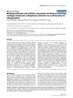
Immunofluorescence staining of mouse samples for basal/myoepithelial and luminal cytokeratinsFigure 2
Immunofluorescence staining of mouse samples for basal/myoepithelial and luminal cytokeratins. (a) Wild-type (wt) mammary gland stained for Keratins 8/
18 (red) and Keratin 5 (green) shows K8/18 expression in luminal epithelial cells and K5 expression in basal/myoepithelial cells. (b-f) Mouse models that
show luminal-like gene expression patterns stained with K8/18 (red) and K5 (green). (g-k) Tumor samples that show basal-like, or mixed luminal and basal
characteristics by gene expression, stained for K8/18 (red) and K5 (green). (j) A subset of Brca1
Co/Co
;TgMMTV-Cre;p53
+/-
tumors showing nodules of K5/
K8/18 double positive cells. (l) A splindloid tumor stained for K8/18 (red) and smooth muscle actin (green).
FVB_Wap_Int3_CA02_575A
wt duct
FVB_DMBA_5_Squa
BDF1_TgWAPT121_KS644
FVB_MMTV_Wnt1_CA03_634A
FVB_DMBA_13_Spindle
FVB_DMBA_9_AdenoSqua
FVB_MMTV_PYVT_'31
FVB_MMTV_Neu_CA01_432A
BALB_BRCA1het_p53het_IR_C0
379_5
FVB_Wap_Myc_CA02_540A
C57Bl6_MMTV_Cre_BRCA1
Co/Co
_
p53het_100a
(a)
(d)
(e) (f)
(i)
(k) (l)
(h)
(g)
(j)
(b)
(c)
Genome Biology 2007, Volume 8, Issue 5, Article R76 Herschkowitz et al. R76.7
comment reviews reports refereed researchdeposited research interactions information
Genome Biology 2007, 8:R76
;p53
+/-
;IR, Brca1
Co/Co
;TgMMTV-Cre;p53
+/-
, TgMMTV-Wnt1,
and some DMBA-induced tumors were characterized by the
high expression of Laminin gamma 2, Keratins 5, 6B, 13, 14,
15, TRIM29, c-KIT and CRYAB (Figure 3b), the last of which
is a human basal-like tumor marker possibly involved in
resistance to chemotherapy [25]. As described above, the
Brca1
+/-
;p53
+/-
;IR, some Brca1
Co/Co
;TgMMTV-Cre;p53
+/
,
DMBA-induced, and TgMMTV-Wnt1 tumors stained positive
for K5 by IF, and human basal-like tumors tend to stain posi-
tive using a K5/6 antibody [1,12,18,26], thus showing that
basal-like tumors from both species share K5 protein expres-
sion as a distinguishing feature.
The murine and human 'luminal tumor' shared profile was
not as similar as the shared basal profile, but did include the
high expression of SPDEF, XBP1 and GATA3 (Figure 3c), and
both species' luminal tumors also stained positive for K8/18
(Figure 2 and see [18]). For many genes in this luminal clus-
ter, however, the relative level of expression differed between
the two species. For example, some genes were consistently
high across both species' tumors (for example, XBP1, SPDEF
and GATA3), while others, including TFF, SLC39A6, and
FOXA1, were high in human luminal tumors and showed
lower expression in murine tumors. Of note is that the human
luminal epithelial gene cluster always contains the Estrogen-
Receptor (ER) and many estrogen-regulated genes, including
TFF1 and SLC39A6 [22]; since most murine mammary
tumors, including those profiled here, are ER-negative, the
apparent lack of involvement of ER and most ER-regulated
genes could explain the difference in expression for some of
the human luminal epithelial genes that show discordant
expression in mice.
Several other prominent and noteworthy features were also
identified across species, including a 'proliferation' signature
that includes the well documented proliferation marker Ki-67
(Figure 3e) [1,27,28] and an interferon-regulated pattern
(Figure 3f) [27]. The proliferation signature was highest in
human basal-like tumors and in the murine models with
impaired pRb function (that is, Group IX and X tumors). Cur-
rently, the growth regulatory impact of interferon-signaling
in human breast tumors is not understood, and murine mod-
els that share this expression feature (TgMMTV-Neu,
TgWAP-Tag, p53
-/-
transplants, and spindloid tumors) may
provide a model for future studies of this pathway. A fibro-
blast profile (Figure 3g) that was highly expressed in murine
samples with spindloid morphology and in the TgWAP-Myc
'spindloid' tumors was also observed in many human luminal
and basal-like tumors; however, on average, this profile was
expressed at lower levels in the murine tumors, which is con-
sistent with the relative epithelial to stromal cell proportions
seen histologically.
Through these analyses we also discovered a potential new
human subtype (Figure 3, top line-yellow group, and Addi-
tional data file 6). This subtype, which was apparent in both
the human only and mouse-human combined dataset, is
referred to as the 'claudin-low' subtype and is characterized
by the low expression of genes involved in tight junctions and
cell-cell adhesion, including Claudins 3, 4, 7, Occludin, and E-
cadherin (Figure 3d). These human tumors (n = 13) also
showed low expression of luminal genes, inconsistent basal
gene expression, and high expression of lymphocyte and
endothelial cell markers. All but one tumor in this group was
clinically ER-negative, and all were diagnosed as grade II or
III infiltrating ductal carcinomas (Additional data file 7 for
representative hematoxylin and eosin images); thus, these
tumors do not appear to be lobular carcinomas as might be
predicted by their low expression of E-cadherin. The
uniqueness of this group was supported by shared mesenchy-
mal expression features with the murine spindloid tumors
(Figure 3g), which cluster near these human tumors and also
lack expression of the Claudin gene cluster (Figure 3d). Fur-
ther analyses will be required to determine the cellular origins
of these human tumors.
A common region of amplification across species
The murine C3(1)-Tag tumors and a subset of human basal-
like tumors showed high expression of a cluster of genes,
including Kras2, Ipo8, Ppfibp1, Surb, and Cmas, that are all
located in a syntenic region corresponding to human chromo-
some 12p12 and mouse chromosome 6 (Figure 3h). Kras2
amplification is associated with tumor progression in the
C3(1)-Tag model [29], and haplo-insufficiency of Kras2
delays tumor progression [30]. High co-expression of Kras2-
linked genes prompted us to test whether DNA copy number
changes might also account for the high expression of Kras2
among a subset of the human tumors. Indeed, 9 of 16 human
basal-like tumors tested by quantitative PCR had increased
genomic DNA copy numbers at the KRAS2 locus; however, no
mutations were detected in KRAS2 in any of these 16 basal-
like tumors. In addition, van Beers et al. [31] reported that
this region of human chromosome 12 is amplified in 47% of
BRCA1-associated tumors by comparative genomic hybridi-
zation analysis; BRCA1-associated tumors are known to
exhibit a basal-like molecular profile [3,32]. In cultured
human mammary epithelial cells, which show basal/myoepi-
thelial characteristics [1,33], both high oncogenic H-ras and
SV40 Large T-antigen expression are necessary for transfor-
mation [34]. Taken together, these findings suggest that
amplification of KRAS2 may either influence the cellular phe-
notype or define a susceptible target cell type for basal-like
tumors.
Mouse-human shared intrinsic features
To simultaneously classify mouse and human tumors, we
identified the gene set that was in common between a human
breast tumor intrinsic list (1,300 genes described in Hu et al.
[21]) and the mouse intrinsic list developed here (866 genes).
The overlap of these two lists totaled 106 genes, which when
used in a hierarchical clustering analysis (Figure 4) identifies
four main groups: the leftmost group contains all the human
R76.8 Genome Biology 2007, Volume 8, Issue 5, Article R76 Herschkowitz et al. />Genome Biology 2007, 8:R76
Figure 3 (see legend on next page)
Lamc2; laminin, gamma 2
Lamb3; laminin, beta 3
Klf5; Kruppel-like factor 5
Ndrg2; N-myc downstream regulated gene 2
Vsx1; visual system homeobox 1 homolog (zebrafish)
Krt1-23; keratin complex 1, acidic, gene 23
Nfib; nuclear factor I/B
Prom1; prominin 1
Cdh3; cadherin 3
Idb4; inhibitor of DNA binding 4
Krt1-14; keratin complex 1, acidic, gene 14
Trim29; tripartite motif protein 29
Krt2-5; keratin complex 2, basic, gene 5
Col17a1; procollagen, type XVII, alpha 1
Cryab; crystallin, alpha B
Sfrp1; secreted frizzled-related sequence protein 1
Mia1; melanoma inhibitory activity 1
1110030O19Rik; RIKEN cDNA 1110030O19 gene
Prss19; protease, serine, 19 (neuropsin)
Prss18; protease, serine, 18
Klk10; kallikrein 10
Foxc1; forkhead box C1
Krt2-6b; keratin complex 2, basic, gene 6b
Trim2; tripartite motif protein 2
Krt1-15; keratin complex 1, acidic, gene 15
Krt1-13; keratin complex 1, acidic, gene 13
Tcf3; transcription factor 3
Kit; kit oncogene
BC031353; cDNA sequence BC031353
5330417C22Rik; RIKEN cDNA 5330417C22 gene
Spdef
4930504E06Rik; RIKEN cDNA 4930504E06 gene
Statip1
Slc39a6
Dncl2b; dynein, cytoplasmic, light chain 2B
Rnf103; ring finger protein 103
Stard10; START domain containing 10
Maged2; melanoma antigen, family D, 2
Pte2b; peroxisomal acyl-CoA thioesterase 2B
2310044D20Rik; RIKEN cDNA 2310044D20 gene
Dnali1; dynein, axonemal, light intermediate polypeptide 1
Slc7a8; solute carrier family 7, member 8
4933406E20Rik; RIKEN cDNA 4933406E20 gene
Xbp1; X-box binding protein 1
Gata3; GATA binding protein 3
Tff3; trefoil factor 3, intestinal
Agr2; anterior gradient 2 (Xenopus laevis)
Foxa1; forkhead box A1
Dnajc12; DnaJ (Hsp40) homolog, subfamily C, member 12
1110003E01Rik; RIKEN cDNA 1110003E01 gene
Scube2; signal peptide, CUB domain, EGF-like 2
Tmem25; transmembrane protein 25
Wwp1; WW domain containing E3 ubiquitin protein ligase 1
Inpp4b; inositol polyphosphate-4-phosphatase, type II
Chchd5
Sytl2; synaptotagmin-like 2
Cxxc5; CXXC finger 5
Tjp2; tight junction protein 2
Krt1-18; keratin complex 1, acidic, gene 18
Krt2-8; keratin complex 2, basic, gene 8
Marveld3
Ddr1; discoidin domain receptor family, member 1
Irf6; interferon regulatory factor 6
Tcfap2c; transcription factor AP-2, gamma
Fxyd3; FXYD domain-containing ion transport regulator 3
Ocln; occludin
Tcfcp2l2; transcription factor CP2-like 2
A030007D23Rik; RIKEN cDNA A030007D23 gene
Spint1; serine protease inhibitor, Kunitz type 1
Pkp3; plakophilin 3
Tcfcp2l3; transcription factor CP2-like 3
Bspry; B-box and SPRY domain containing
Arhgef16; Rho guanine nucleotide exchange factor (GEF) 16
Crb3; crumbs homolog 3 (Drosophila)
1810019J16Rik; RIKEN cDNA 1810019J16 gene
Ap1m2; adaptor protein complex AP-1, mu 2 subunit
Cldn7; claudin 7
Spint2; serine protease inhibitor, Kunitz type 2
St14; suppression of tumorigenicity 14 (colon carcinoma)
Lisch7; liver-specific bHLH-Zip transcription factor
Tacstd1; tumor-associated calcium signal transducer 1
9530027K23Rik; RIKEN cDNA 9530027K23 gene
Cldn3; claudin 3
Prss8; protease, serine, 8 (prostasin)
1810017F10Rik; RIKEN cDNA 1810017F10 gene
Ptprf; protein tyrosine phosphatase, receptor type, F
BC037006; cDNA sequence BC037006
AW049765; expressed sequence AW049765
Rhpn2; rhophilin, Rho GTPase binding protein 2
Cdh1; cadherin 1
Mal2; mal, T-cell differentiation protein 2
Mybl2; myeloblastosis oncogene-like 2
Trip13; thyroid hormone receptor interactor 13
Stk6; serine/threonine kinase 6
Ube2c; ubiquitin-conjugating enzyme E2C
Chek1; checkpoint kinase 1 homolog (S. pombe)
Mki67; antigen identified by monoclonal antibody Ki 67
Prc1; protein regulator of cytokinesis 1
Ttk; Ttk protein kinase
Cdca8; cell division cycle associated 8
Racgap1; Rac GTPase-activating protein 1
Ccnb2; cyclin B2
Nek2
2700084L22Rik; RIKEN cDNA 2700084L22 gene
Kntc2; kinetochore associated 2
Cenpf; centromere autoantigen F
Calmbp1; calmodulin binding protein 1
Bub1; budding uninhibited by benzimidazoles 1 homolog
Cdca1; cell division cycle associated 1
Cdca5; cell division cycle associated 5
Melk; maternal embryonic leucine zipper kinase
Cenpe; centromere protein E
Kif20a; kinesin family member 20A
Exo1; exonuclease 1
2600017H08Rik; RIKEN cDNA 2600017H08 gene
Rad51; RAD51 homolog (S. cerevisiae)
Pbk; PDZ binding kinase
Cenpa; centromere autoantigen A
Tpx2; TPX2, microtubule-associated protein homolog
Nusap1; nucleolar and spindle associated protein 1
Blm; Bloom syndrome homolog (human)
Cdc20; cell division cycle 20 homolog (S. cerevisiae)
6720460F02Rik; RIKEN cDNA 6720460F02 gene
Ifi35; interferon-induced protein 35
Lgals3bp
Epsti1; epithelial stromal interaction 1 (breast)
Psmb8; proteosome subunit, beta type 8
B2m; beta-2 microglobulin
H2-Q10; histocompatibility 2, Q region locus 10
Zbp1; Z-DNA binding protein 1
Stat2; signal transducer and activator of transcription 2
Oas2; 2’-5’ oligoadenylate synthetase 2
Gbp4; guanylate nucleotide binding protein 4
Phf11; PHD finger protein 11
Bst2; bone marrow stromal cell antigen 2
Isgf3g
Ddx58; DEAD (Asp-Glu-Ala-Asp) box polypeptide 58
Ifih1; interferon induced with helicase C domain 1
Ifit2
Oasl1; 2’-5’ oligoadenylate synthetase-like 1
G1p2; interferon, alpha-inducible protein
Ifi44; interferon-induced protein 44
Ifit3
Mx2; myxovirus (influenza virus) resistance 2
Usp18; ubiquitin specific protease 18
5830458K16Rik; RIKEN cDNA 5830458K16 gene
Parp9; poly (ADP-ribose) polymerase family, member 9
Ube1l; ubiquitin-activating enzyme E1-like
Prkr
Cklfsf3; chemokine-like factor super family 3
Col6a3; procollagen, type VI, alpha 3
Col5a1; procollagen, type V, alpha 1
Srpx2; sushi-repeat-containing protein, X-linked 2
Loxl1; lysyl oxidase-like 1
Col1a1; procollagen, type I, alpha 1
Fn1; fibronectin 1
Prss11; protease, serine, 11 (Igf binding)
Ctsk; cathepsin K
Lum; lumican
Cdh11; cadherin 11
Fbn1; fibrillin 1
Fap; fibroblast activation protein
Sparc; secreted acidic cysteine rich glycoprotein
Col1a2; procollagen, type I, alpha 2
Col5a2; procollagen, type V, alpha 2
Thbs2; thrombospondin 2
Col12a1; procollagen, type XII, alpha 1
Col6a1; procollagen, type VI, alpha 1
Col6a2; procollagen, type VI, alpha 2
Postn; periostin, osteoblast specific factor
Sulf1; sulfatase 1
Nid2; nidogen 2
Serpinf1
Dcn; decorin
2610001E17Rik; RIKEN cDNA 2610001E17 gene
Fstl1; follistatin-like 1
Adamts2
2310061A22Rik; RIKEN cDNA 2310061A22 gene
Recql; RecQ protein-like
2010012C16Rik; RIKEN cDNA 2010012C16 gene
Strap; serine/threonine kinase receptor associated protein
4933424B01Rik; RIKEN cDNA 4933424B01 gene
Mrps35; mitochondrial ribosomal protein S35
Surb7; SRB7 (supressor of RNA polymerase B) homolog
Stk38l; serine/threonine kinase 38 like
BC027061; cDNA sequence BC027061
Kras2; Kirsten rat sarcoma oncogene 2, expressed
Ppfibp1; PTPRF interacting protein, binding protein 1
Tm7sf3; transmembrane 7 superfamily member 3
(a)
(b)
(c)
(d)
(e)
(f)
(g)
(h)
1:1 >2 >4 >6>2>4>6
Relative to median expression
WAP Int3
Human subtype
MMTV PyMT
MMTV NeuMMTV Neu
WAP Myc
p53-/- transplant
DMBA
MMTV Wnt1
p53+/- IR
BRCA1+/- p53+/- IR
MMTV Cre BRCA1 p53+/-
WAP Tag
C3(1) Tag
WAP T121
Normal
HER2 status
ER status
Genome Biology 2007, Volume 8, Issue 5, Article R76 Herschkowitz et al. R76.9
comment reviews reports refereed researchdeposited research interactions information
Genome Biology 2007, 8:R76
basal-like, 'claudin-low', and 5/44 HER2+/ER- tumors, and
the murine C3(1)-Tag, TgWAP-Tag, and spindloid tumors.
The second group (left to right) contains the normal samples
from both humans and mice, a small subset (6/44) of human
HER2+/ER- and 10/92 luminal tumors, and a significant
portion of the remaining murine basal-like models. By clini-
cal criteria, nearly all human tumors in these two groups were
clinically classified as ER-negative.
The third group contains 33/44 human HER2+/ER- tumors
and the murine TgMMTV-Neu, MMTV-PyMT and TgWAP-
Myc samples. Although the human HER2+/ER- tumors are
predominantly ER-negative, this comparative genomic anal-
ysis and their keratin expression profiles as assessed by
immunohistochemistry, suggests that the HER2+/ER-
human tumors are 'luminal' in origin as opposed to showing
basal-like features [18]. The fourth and right-most group is
composed of ER-positive human luminal tumors and, lastly,
the mouse TgWAP-Int3 (Notch4) tumors were in a group by
themselves. These data show that although many mouse and
human tumors were located on a large dendrogram branch
that contained most murine luminal models and human
HER2+/ER- tumors, none of the murine models we tested
showed a strong human 'luminal' phenotype that is character-
ized by the high expression of ER, GATA3, XBP1 and FOXA1.
These analyses suggest that the murine luminal models like
MMTV-Neu showed their own unique profile that was a rela-
tively weak human luminal phenotype that is missing the ER-
signature. Presented at the bottom of Figure 4 are biologically
important genes discussed here, genes previously shown to be
human basal-like tumor markers (Figure 4c), human luminal
tumor markers, including ER (Figure 4d), and HER2/
ERBB2/NEU (Figure 4e).
A comparison of gene sets defining human tumors and
murine models
We used a second analysis method called gene set enrichment
analysis (GSEA) [35] to search for shared relationships
between human tumor subtypes and murine models. For this
analysis, we first performed a two-class unpaired significance
analysis of microarray (SAM) [36] analysis for each of the ten
murine groups defined in Figure 1, and obtained a list of
highly expressed genes that defined each group. Next, we per-
formed similar analyses using each human subtype versus all
other human tumors. Lastly, the murine lists were compared
to each human subtype list using GSEA, which utilizes both
gene list overlap and gene rank (Table 2). We found that the
murine Groups IX (p = 0.004) and X (p = 0.001), which com-
prised tumors from pRb-deficient/p53-deficient models,
shared significant overlap with the human basal-like subtype
and tended to be anti-correlated with human luminal tumors
(p = 0.083 and 0.006, respectively). Group III murine tumors
(TgMMTV-Wnt1 mostly) significantly overlapped human
normal breast samples (p = 0.008), possibly due to the
expression of both luminal and basal/myoepithelial gene
clusters in both groups. Group IV (Brca1-deficient and Wnt1)
showed a significant association (p = 0.058) with the human
basal-like profile. The murine Group VI (TgMMTV-Neu and
TgMMTV-PyMT) showed a near significant association (p =
0.078) with the human luminal profile and were anti-corre-
lated with the human basal-like subtype (p = 0.04). Finally,
the murine Group II spindloid tumors showed significant
overlap with human 'claudin-low' tumors (p = 0.001), which
further suggests that this may be a distinct and novel human
tumor subtype.
We also performed a two-class unpaired SAM analysis using
each mouse model as a representative of a pathway perturba-
tion using the transgenic 'event' as a means of defining
groups. Models that yielded a significant gene list (false dis-
covery rate (FDR) = 1%) were compared to each human sub-
type as described above (Additional data file 8). The models
based upon SV40 T-antigen (all C3(1)-Tag and WAP-Tag
tumors) shared significant overlap with the human basal-like
tumors (p = 0.002) and were marginally anti-correlated with
the human luminal class. The BRCA1 deficient models (all
Brca1
+/-
;p53
+/-
IR and Brca1
Co/Co
;TgMMTV-Cre;p53
+/-
tumors) were marginally significant with human basal-like
tumors (p = 0.088). The TgMMTV-Neu tumors were nomi-
nally significant (before correction for multiple comparisons)
with human luminal tumors (p = 0.006) and anti-correlated
with human basal-like tumors (p = 0.027).
The two most important human breast tumor biomarkers are
ER and HER2; therefore, we also analyzed these data relative
to these two markers. Of the 232 human tumors assayed here,
137 had ER and HER2 data assessed by immunohistochemis-
try and microarray data. As has been noted before [3,18,21],
there is a very high correlation between tumor intrinsic sub-
type and ER and HER2 clinical status (p < 0.0001): for exam-
ple, 81% of ER+ tumors were of the luminal phenotype, 63%
of HER2+ tumors were classified as HER2+/ER-, and 80% of
ER- and HER2- tumors were of the basal-like subtype. Using
GSEA, we compared the ten mouse classes as defined in Fig-
Unsupervised cluster analysis of the combined gene expression data for 232 human breast tumor samples and 122 mouse mammary tumor samplesFigure 3 (see previous page)
Unsupervised cluster analysis of the combined gene expression data for 232 human breast tumor samples and 122 mouse mammary tumor samples. (a) A
color-coded matrix below the dendrogram identifies each sample; the first two rows show clinical ER and HER2 status, respectively, with red = positive,
green = negative, and gray = not tested; the third row includes all human samples colored by intrinsic subtype as determined from Additional data file 6;
red = basal-like, blue = luminal, pink = HER2+/ER-, yellow = claudin-low and green = normal breast-like. The remaining rows correspond to murine
models indicated at the right. (b) A gene cluster containing basal epithelial genes. (c) A luminal epithelial gene cluster that includes XBP1 and GATA3. (d) A
second luminal cluster containing Keratins 8 and 18. (e) Proliferation gene cluster. (f) Interferon-regulated genes. (g) Fibroblast/mesenchymal enriched
gene cluster. (h) The Kras2 amplicon cluster. See Additional data file 5 for the complete cluster diagram.
R76.10 Genome Biology 2007, Volume 8, Issue 5, Article R76 Herschkowitz et al. />Genome Biology 2007, 8:R76
Figure 4 (see legend on next page)
Wap T121
MMTV Cre BRCA1 p53+/-
DMBADMBA
MMTV Wnt1
Wap M yc
MMTV Neu
p53-/- transplant
p53+/- IR
MMTV PyMT
BRCA1+/- p53+/- IR
Wap Tag
C3(1) Tag
Wap I nt3
Normal
RIKEN cDNA C530044N13
Ak3l1
Echdc1
epoxide hydrolase 2
Ppp2r5a
phytanoyl-CoA hydroxylase
RIKEN cDNA 2810439K08
Srcasm
CXXC finger 5
Igfals
Srebf1
Dnajc12
X-box binding protein 1
RIKEN cDNA 4922503N01
Acox2
cytochrome b-5
cyclin D1
Pbx3
Bcas1
forkhead box P1
myeloblastosis oncogene
Celsr1
Sema3b
sal-like 2 (Drosophila)
laminin, alpha 3
cDNA sequence BC010304
catenin alpha 1
Hipk2
Ribosomal protein L18A
Galnt14
Eif4ebp1
diazepam binding inhibitor
Ilf2
Efs
RIKEN cDNA 4732452J19
Ppfibp2
claudin 3
Tcfcp2l2
Bspry
Mal2
Traf4
Grb7
procollagen, type IX, alpha 1
folate receptor 1 (adult)
Padi2
Echdc3
absent in melanoma 1
D6Wsu176e
inhibin beta-B
aryl-hydrocarbon receptor
Te r a
RIKEN cDNA 5730559C18
drebrin 1
syndecan 1
kit oncogene
Ly6d
laminin, beta 3
cadherin 3
protease, serine, 18
keratin 14
keratin 6b
keratin 15
nuclear factor I/B
Iroquois related homeobox 4
Wnt6
inhibitor of DNA binding 4
Gpr125
Bmp7
procollagen, type IX, alpha 3
prion protein
desmoplakin
Bambi
nebulette
RIKEN cDNA B830028P19
RIKEN cDNA 1500011H22
Trp53bp2
Nfe2l3
claudin 23
Asf1a
RIKEN cDNA 4921532K09
B-cell translocation gene 3
Ctps
breast cancer 1
RIKEN cDNA 2410004L22
sperm associated antigen 5
Mcm2
retroviral integration site 2
AW209059
stathmin 1
Gpsm2
RAD51 associated protein 1
RIKEN cDNA 2810417H13
Cdc2a
Mad2l1
Racgap1
centromere autoantigen F
Nek2
PDZ binding kinase
Chaf1b
timeless homolog
cell division cycle 6 homolog
Casp3
RIKEN cDNA E130303B06
Wwp2
sorting nexin 7
Gtf2f2
ERBB2/HER2/Neu
Keratin 6b
KRAS2
Keratin 5
CRYAB
KIT
EGFR
FOXA1
RERG
G ATA 3
Keratin 18
Keratin 8
XBP1
ESR1
(a)
(b)
(c)
(d)
(e)
LUMINAL HUMANBASAL HUMAN
INT3MYCBRCA1+WNT1 NEUPYVTNORMALSPINDLET-antigen
HER2 HUMAN
1:1 >2 >4 >6>2>4
>6
Relative to median expression
Genome Biology 2007, Volume 8, Issue 5, Article R76 Herschkowitz et al. R76.11
comment reviews reports refereed researchdeposited research interactions information
Genome Biology 2007, 8:R76
ure 1 (Additional data file 9) and the mouse model-based gene
lists (Additional data file 10) to the human data/gene lists
that were obtained by performing supervised analyses based
upon human ER and HER2 status (please note that analyses
using HER2 status alone (that is, HER2+ versus HER2-), and
ER+ and HER2+ versus others were not included as human
classes because HER2 status alone yielded genes on only the
HER2 amplicon, and the ER+ and HER2+ classification did
not yield a significant gene list). We found that the murine
Groups IX (p = 0.009) and X (p = 0.003) tumors shared
significant overlap with ER- HER2- human tumors and were
significantly anti-correlated with human ER+ tumors (p =
0.024 and 0.043, respectively). Group VI murine samples
(TgMMTV-Neu and TgMMTV-PyMT) likewise showed the
same trend of enrichment with ER+ human tumors and anti-
correlation with the ER- HER2- class. Although not perfect,
these GSEA results are consistent with our observations from
Figures 1 and 3 and again demonstrate that the basal-like pro-
file is robustly shared between humans and mice, while the
luminal profile shows some shared and some distinct features
across species.
Discussion
Gene expression profiling of murine tumors and their com-
parison to human tumors identified characteristics relevant
to individual murine models, to murine models in general,
and to cancers of both species. First was the discovery that
some murine models developed highly similar tumors within
models, while others showed heterogeneity in expression and
histological phenotypes. For the homogenous models, the
study of progression or response to therapy is simplified
Cluster analysis of mouse and human tumors using the subset of genes common to both species intrinsic lists (106 total genes)Figure 4 (see previous page)
Cluster analysis of mouse and human tumors using the subset of genes common to both species intrinsic lists (106 total genes). (a) Experimental sample
associated dendrogram color coded according to human tumor subtype and with a matrix below showing murine tumor origins. (b) The complete 106
gene cluster diagram. (c) Close-up of genes known to be important for human basal-like tumors. (d) Close-up of genes known to be important for human
luminal tumors, including ER. (e) Expression pattern of HER2/ERBB2/NEU.
Table 2
Gene set enrichment analysis of the ten murine groups versus five human subtypes
Basal-like Luminal HER2+/ER- Normal Claudin-low
Mouse class No. of genes p value p value p value p value p value p value p value p value p value p value
Is class
I 1,882 - - 0.4625 0.8755 0.5388 0.9137 0.1659 0.5628 0.0048 0.1028
II 912 - - - - 0.5867 0.9609 - - 0.0021 0.001
III 143 0.5289 0.9048 - - 0.5285 0.9047 0 0.008
IV 1,019 0 0.0581
V 34 - - 0.8492 0.998 0.9324 0.999 - - 0.0427 0.09274
VI 820 - - 0.0062 0.0783 0.3536 0.7864 0.8653 0.9769 - -
VII 851 0.1258 0.3768 - - 0.5616 0.9137 - - - -
VIII 236 0.1449 0.6098 0.3483 0.8205 - - 0.01878 0.2349 - -
IX 462 0.0019 0.004 0.560.9509
X33800.001 - - 0.9275 0.998 - - - -
Is not class
I 1,882 0.0128 0.1662 - - - - - - - -
II 912 0.3996 0.8348 0.8601 0.999 - - 0.3602 0.7655 - -
III 143 - - 0.3178 0.7259 - - - - 0.7628 0.991
IV 1,019 - - 0.1833 0.6516 0.398 0.8427 0.2241 0.7255 0.1453 0.6116
V 34 0.86 1 - - - - 0.0656 0.1653 - -
VI 820 0 0.04 - - - - - - 0.1043 0.4444
VII 851 - - 0.1733 0.5151 - - 0.5403 0.9128 0.1628 0.5215
VIII 236 - - - - 0.1131 0.5305 - - 0.6427 0.961
IX 462 - - 0.04305 0.0833 - - 0.022 0.037 0.2612 0.5936
X 338 - - 0.02236 0.0682 - - 0.1313 0.3717 0.5437 0.9489
Statistically significant findings are highlighted in bold. NOM = nominal.
R76.12 Genome Biology 2007, Volume 8, Issue 5, Article R76 Herschkowitz et al. />Genome Biology 2007, 8:R76
because confounding variation across individuals is low. An
example of this consistency even extended to secondary
events that occurred within the TgC3(1)-Tag model, where
many tumors shared the amplification and high expression of
Kras2 (Figure 3h) - a feature also evident in a subset of
human basal-like tumors.
In contrast to the 'homogenous' models are models such as
TgWAP-T
121
, DMBA-induced and Brca1
Co/Co
;TgMMTV-
Cre;p53
+/-
, where individual tumors within a given model
often showed different gene expression profiles and histolo-
gies. It is likely that these models fall into one of three scenar-
ios that could explain their heterogeneity: the first,
represented by the TgWAP-T
121
model [37], is that the trans-
gene is responsible only for initiating tumorigenesis, leaving
progression events to evolve stochastically and with longer
latency periods. Such a model would likely give rise to differ-
ent tumor subtypes depending on the subsequent pathways
that are disrupted during tumor progression. A second possi-
bility is that the initiating event generates genomic instability
such that multiple distinct pathways can be affected by the
experimental causal event, which may be the mechanism in
the Brca1-inactivation tumors. The third scenario is that the
target cell of transformation is a multi-potent progenitor with
the ability to undergo differentiation into multiple epithelial
lineages, or even mesenchymal lineages (for example, DMBA-
induced and Brca1
Co/Co
;TgMMTV-Cre;p53
+/-
); support for
this hypothesis comes from Keratin IF analyses in which,
even within a histologically homogenous tumor, two types of
epithelial cells are present (Figures 2g-k). The presence of
subsets of individual cells positive for markers of two epithe-
lial cell types also supports this possibility (Figure 2j). Alter-
native hypotheses include the possibility that multiple cell
types sustain transforming events, and also that extensive
non-cell-autonomous tissue responses occur. Regardless of
the paradigm of transformation for these heterogeneous
models, the study of progression or therapeutic response will
best be accomplished by first sub-setting by subtype, and then
focusing on biological phenotypes.
There are at least two major applications for genomic com-
parisons between human tumors and their potential murine
counterparts. First, such studies should identify those models
that contain individual and/or global characteristics of a par-
ticular class of human tumors. Examples of important global
characteristics identified here include the classification of
murine and human tumors into basal and luminal groups. It
appears as if four murine models developed potential lumi-
nal-like tumors (TgMMTV-Neu, TgMMTV-PyMT, TgWAP-
Myc, and TgWAP-Int3), which is not surprising since both
MMTV and WAP are thought to direct expression in differen-
tiated alveolar/luminal cells [38,39]; however, it should be
noted that the luminal profile across species was not statisti-
cally significant, likely due to the lack of ER and ER-regulated
genes in the murine luminal tumors. Several murine models
did show expression features consistent with human basal-
like tumors, including the TgC3(1)-Tag, TgWAP-Tag and
Brca1-deficient models. The SV40 T-antigen used in the
TgC3(1)-Tag and TgWAP-Tag models inactivates p53 and
RB, which also appear to be two likely events that occur in
human basal-like tumors because these tumors are known to
harbor p53 mutations [2], have high mitotic grade and the
highest expression of proliferation genes (Figure 3) [2,3],
which are known E2F targets [40]. The proliferation signa-
ture in human breast cancers is itself prognostic [41], and is
also predictive of response to chemotherapy [42]. These data
suggest that human basal-like tumors might have impair-
ment of RB function and highlight an important shared fea-
ture of murine and human mammary carcinomas.
The finding that Brca1 loss (coincident with p53 mutation) in
mice gives rise to tumors with a basal-like phenotype is nota-
ble because humans carrying BRCA1 germline mutations also
develop basal-like tumors [3,32], and most human BRCA1
mutant tumors are p53-deficient [43,44]. These data suggest
a conserved predisposition of the basal-like cell type, or its
progenitor cell, to transform as a result of BRCA1, TP53, and
RB-pathway loss. Most DMBA-induced carcinomas also
showed basal-like cell lineage features, suggesting that this
cell type is also susceptible to DMBA-mediated tumorigene-
sis. Finally, some TgMMTV-Wnt1 tumors showed a combina-
tion of basal-like and luminal characteristics by gene
expression, which is consistent with the observation that
tumors of this model generally contain cells from both mam-
mary epithelial lineages [45].
The second major purpose of comparative studies is to deter-
mine the extent to which analyses of murine models can
inform the human disease and guide further discovery. An
example of murine models informing the human disease is
encompassed by the analysis of the new potential human sub-
type discovered here (that is, claudin-low subtype). Further
analysis will be necessary to confirm whether this is a bona
fide subtype; however, the statistically significant gene over-
lap with a histologically distinct subset of murine tumors sug-
gests it is a distinct biological entity. A second example of the
murine tumors guiding discovery in humans was the common
association of a K-Ras containing amplicon in a subset of
human basal-like tumors and in the murine basal-like
TgC3(1)-Tag strain tumors.
An important caveat to all comparative studies is that there
are clear biological differences between mice and humans,
which may or may not directly impact disease mechanisms. A
potential example of inherent species difference could be the
aforementioned biology associated with ER and its down-
stream pathway. In humans, ER is highly expressed in lumi-
nal tumors [1], with the luminal phenotype being
characterized by the high expression of some genes that are
ER-regulated like PR and RERG [22], and other luminal
genes that are likely GATA3-regulated, including AGR2 and
K8/18 [46]. In mice, ER expression is low to absent in all the
Genome Biology 2007, Volume 8, Issue 5, Article R76 Herschkowitz et al. R76.13
comment reviews reports refereed researchdeposited research interactions information
Genome Biology 2007, 8:R76
tumors we tested, as is the expression of most human ER-
responsive genes. This finding is consistent with previous
reports that most late-stage murine mammary tumors are
ER-negative ([47] and references within). However, it should
be noted that two human luminal tumor-defining genes
(XBP1 and GATA3 [46], were both highly expressed in
murine luminal tumors (Additional data file 2). Taken
together, these data suggest that the human 'luminal' profile
may actually be a combination of at least two profiles, one of
which is ER-regulated and another of which is GATA3-regu-
lated; support for a link between GATA3 and luminal cell ori-
gins comes from GATA3 loss studies in mice where the
selective loss of GATA3 in the mammary gland resulted in
either a lack of luminal cells, or a significant decrease in the
number and/or maturation of luminal cells [48,49]. These
results suggest that, in the mouse models tested here, the ER-
regulated gene cassette that is present in human luminal
tumors is missing, and that the GATA3-mediated luminal sig-
nature remains. Due to the partial luminal tumor signature in
mice, we believe that the murine luminal models, including
TgMMTV-Neu profiled here, best resemble human luminal
tumors and more specifically possibly luminal B tumors,
which are luminal tumors that express low amounts of ER
and show a poor outcome [2,3,21]. While human HER2+/ER-
subtype tumors and the murine TgMMTV-Neu, TgMMTV-
PyMT, and TgWAP-Myc fall next to each other in the intrin-
sic-shared cluster (Figure 4), all of the other data argue
against this association. A few murine ER-positive mammary
tumor models have been developed [50-53]; however, none of
these models were analyzed here.
Of note, many expression patterns detected in this study were
observed in only one species (Additional data file 5), and it is
possible that some of these differences may arise from techni-
cal limitations rather than reflect important biological differ-
ences. Comparison between two expression datasets,
especially when derived from different species, remains a
technical challenge. Thus, we acknowledge the possibility
that artifacts may have been introduced depending on the
data analysis methodology. However, we are confident that
the analyses described here identified many common and
biologically relevant clusters, including a proliferation, basal
epithelial, interferon-regulated and fibroblast signature, thus
showing that the act of data combining across species did
retain important features present within the individual data-
sets. There are many murine models of breast cancer that we
did not look at in this study and many more will be developed.
Like the 13 models we discussed here, we would expect that
some of these models will have overlapping gene expression
patterns with human subtypes while others will not. We
believe that additional studies with larger numbers of sam-
ples, including more diversity from each species, is war-
ranted. These analyses do confirm the notion that there is not
a single murine model that perfectly represents a human
breast cancer subtype; however, the murine models do show
shared features with specific human subtypes and it is these
commonalties that will lay the groundwork for many future
studies.
Materials and methods
Murine and human tumors
The murine tumor samples were obtained from multiple par-
ticipating investigators, who all maintained the mice and har-
vested the murine tumors in the 0.5-1 cm stage following
internationally recognized guidelines. The details concerning
strain background, promoter, transgene, and specific alleles,
and so on, are provided in Additional data file 1. All human
tumor samples were collected from fresh frozen primary
breast tumors using Institutional Review Board (IRB)-
approved protocols and were profiled as described in [21-23].
The clinical and pathological information for these human
samples can be obtained at the University of North Carolina
Microarray Database (UMD) [54].
Microarray experiments
Total RNA was collected from murine tumors, and wild-type
mammary samples of both FVB and BALB/c inbred strains.
RNA was purified using the RNeasy Mini Kit (Qiagen Inc.,
Valencia, CA, USA) according to the manufacturer's protocol
using 20-30 mg tissue. RNA integrity was assessed using the
RNA 6000 Nano LabChip kit followed by analysis using a Bio-
analyzer (Agilent Technologies Inc., Santa Clara, CA, USA).
Total RNA (2.5 μg) was reverse transcribed, amplified and
labeled with Cy5 using a Low RNA Input Amplification kit
(Agilent). The common reference RNA sample for these
experiments consisted of total RNA harvested from equal
numbers of C57Bl6/J and 129 male and female day 1 pups (a
gift from Dr Cam Patterson, UNC). The reference RNA was
reverse transcribed, amplified, and labeled with Cy3. The
amplified sample and reference were co-hybridized overnight
to Agilent Mouse Oligo Microarrays (G4121A). They were
then washed and scanned on a GenePix 4000B scanner
(Molecular Devices Corporation, Sunnyvale, CA, USA), ana-
lyzed using GenePix 4.1 software and uploaded into our data-
base where a Lowess normalization is automatically
performed.
Microarray data analysis
All primary microarray data are available from the UMD [54],
and at the Gene Expression Omnibus under the series
GSE3165 (mouse and new human data), GSE1992, GSE2740
and GSE2741 (previously published human data) [55]. The
genes for all analyses were filtered by requiring the Lowess
normalized intensity values in both channels to be > 30. The
log
2
ratio of Cy5/Cy3 was then reported for each gene. In the
final dataset, only genes that reported values in 70% or more
of the samples were included. The genes were median
centered and then hierarchical clustering was performed
using Cluster v2.12 [56]. For the murine unsupervised analy-
sis, and human-mouse unsupervised cluster analyses, we fil-
tered for genes that varied at least three-fold or more, in at
R76.14 Genome Biology 2007, Volume 8, Issue 5, Article R76 Herschkowitz et al. />Genome Biology 2007, 8:R76
least three or more samples. Average linkage clustering was
performed on genes and arrays and cluster viewing and dis-
play was performed using JavaTreeview v1.0.8 [57].
Mouse Intrinsic gene set analysis
Intrinsic 'groups' of experimental samples were chosen based
upon having a Pearson correlation value of 0.65 or greater
from the unsupervised clustering analysis of the 122 murine
samples. The analysis was performed using the Intrinsic Gene
Identifier v1.0 by Max Diehn/Stanford University [1]. Techni-
cal replicates were removed from the file and the members of
every highly correlated node were given identical class num-
bers, giving every sample that fell outside the 0.65 correlation
cut-off a class of their own. Using these criteria, 16 groups of
samples were identified (see Additional data file 1 for these
groups) and a list of 866 'intrinsic' genes was selected using
the criteria of one standard deviation below the mean intrin-
sic gene value. A human intrinsic list of 1,300 genes was cre-
ated using a subset of 146 of the 232 samples used here, and
is described in Hu et al. [21].
Consensus clustering
CC [20] was performed locally using Gene Pattern 1.3.1 (built
Jan 6, 2005), which was downloaded from the Broad Institute
distribution website [58]. Analyses were performed on the
mouse dataset with all genes, and just with intrinsic genes
separately. Ranges for the number of K clusters (or the
focused number of classes) were from 2 to 15 to evaluate a
wide range of possible groups. Using a Euclidian distance
measure with average linkage, we re-sampled 1,000 times
with both column and row normalization.
Combining murine and human expression datasets
Orthologous genes were reported by Mouse Genome Infor-
matics (MGI 3.1) of The Jackson Laboratory. For both the
human and murine datasets, Locus Link IDs assigned to Agi-
lent oligo probe ID numbers were used to assign to MGI ID
numbers. In cases where a single gene was represented by
multiple probes, the median value of the redundant probes
was used. This led to a total of orthologous pairings of 14,680
Agilent probes. Prior to combining the two datasets, each was
column standardized to N(0,1), row median centered, and
probe identifiers were converted to MGI IDs. The intersection
of mouse and human MGI identifiers from genes that passed
filters (same as used above) in both datasets yielded 7,907
orthologous genes in the total combined dataset. This dataset
was next corrected for systemic biases using distance
weighted discrimination [24]. Finally, the combined dataset
was used for an average linkage hierarchical clustering
analysis.
Gene set enrichment analysis
We took the 232 human samples and classified them as basal-
like, luminal, HER2+/ER-, claudin-low, and normal breast-
like according to a clustering analysis of the human dataset
only (Additional data file 6), using the new intrinsic/UNC
human gene list developed in Hu et al. [21]. Second, the
murine samples were also classified based upon their cluster-
ing pattern in Figure 1 that used the mouse intrinsic gene list,
and were assigned to Groups I-X. Two-class unpaired SAM
analysis was performed for each murine class separately ver-
sus all other classes using an FDR of 1% [36], resulting in 10
class-specific gene lists. Using only the set of highly expressed
genes that were associated with each analysis (and ignoring
the genes whose low expression correlated with a given class),
GSEA [35] was performed in R (v. 2.0.1) using the GSEA R
package [59]. The ten murine gene sets were then compared
to each human subtype-ranked gene set and significant
enrichments reported. For statistical strength of these enrich-
ments, GSEA uses family wise error rate (FWER) to correct
for multiple testing and FDR to reduce false positive report-
ing. The parameters used for all GSEA were: nperm = 1,000,
weighted.score.type = 1, nom.p.val.threshold = -1,
fwer.p.val.threshold = -1, fdr.q.val.threshold = 0.25, topgs =
12, adjust.FDR.q.val = FALSE, gs.size.threshold.min = 25,
gs.size.threshold.max = 2,000, reverse.sign = FALSE, pre-
proc.type = 0, random.seed = 3,338, perm.type = 0, fraction
= 1, replace = FALSE.
Immunofluorescence
Paraffin-embedded sections (5 μm thick) were processed
using standard immunostaining methods. The antibodies
and their dilution were α-cytokeratin 5 (K5, 1:8,000, PRB-
160P, Covance, Berkeley, CA, USA), and α-cytokeratins 8/18
(Ker8/18, 1:450, GP11, Progen Biotecknik, Heidelberg, Ger-
many). Briefly, slides were deparaffinized and hydrated
through a series of xylenes and graded ethanol steps. Heat-
mediated epitope retrieval was performed in boiling citrate
buffer (pH 6.0) for 15 minutes, then samples cooled to room
temperature for 30 minutes. Secondary antibodies for
immunofluorescence were conjugated with Alexa Fluor-488
or -594 fluorophores (1:200, Molecular Probes, Invitrogen,
Carlsbad, CA, USA). IF samples were mounted with VectaSh-
ield Hardset with DAPI mounting media (Vector, Burlin-
game, CA, USA).
Human KRAS2 amplification assay
We performed real-time quantitative PCR and fluorescent
melting curve analyses using genomic DNAs from 16 basal-
like tumors, a normal breast tissue sample, 2 leukocyte DNA,
and 3 luminal tumors. DNA was extracted using the DNAeasy
kit (Qiagen) and amplification was performed on the LightCy-
cler using the following temperature parameters: 95°C, 8
minutes; 50 cycles of 57°C, 6 s; 72°C, 6 s; 95°C, 2 s; followed
by cooling to 60°C and a 0.1°C/s ramp to 97°C. Each PCR
reaction contained 7.5 ng template DNA in a 10 μl reaction
using the LightCycler Faststart DNA Master SYBR Green I kit
(Roche Applied Science, Indianapolis, IN, USA). Relative
DNA copy number for each gene was determined by import-
ing an external efficiency curve and using a 'normal' breast
sample for a within-run calibrator. For each sample, the copy
number for KRAS2 was divided by the average copy number
Genome Biology 2007, Volume 8, Issue 5, Article R76 Herschkowitz et al. R76.15
comment reviews reports refereed researchdeposited research interactions information
Genome Biology 2007, 8:R76
of ACTB and G1P3. Amplification in any tumor was called if
the relative fold change was greater than three standard devi-
ations above the average of five control samples (two normal
leukocyte samples and three luminal tumors).
Additional data files
The following additional data are available with the online
version of this paper. Additional data file 1 is a table listing
mouse tumor and normal sample associated data, including
source, transgene and promoter information. Additional data
file 2 is a complete unsupervised cluster diagram of all mouse
tumors. Samples are colored according to mouse model from
which they were derived, and the genes were selected using a
variation filter of three-fold or more on three or more sam-
ples. Additional data file 3 ia a complete mouse models clus-
ter diagram using the 866 gene murine intrinsic gene list.
Additional data file 4 provides CC analyses applied to the
mouse models. (a) CC matrices generated using the 866 gene
mouse intrinsic list, by cluster numbers K = 2 through K = 15.
(b) Empirical cumulative distribution (CDF) plot corre-
sponding to the consensus matrices in the range K = 2 to 15.
(c) CC directly compared to the hierarchical clustering-based
results. The dendrogram from Figure 1 (using the intrinsic
gene set) is shown and immediately below is a colored matrix
showing sample assignments based upon the various number
of K clusters from the CC. By comparison, the analysis per-
formed on the mouse dataset using all genes (bottom matrix)
is presented. Additional data file 5 is a complete unsupervised
cluster diagram of the combined gene expression patterns of
232 human breast tumor samples and 122 mouse mammary
tumor samples. This unsupervised cluster analysis is based
upon the orthologous gene overlap between the human and
mouse microarrays, and then we selected for the subset of
genes that varied three-fold or more on three or more arrays.
Additional data file 6 shows a cluster analysis of the 232
human samples using the human intrinsic/UNC gene set
from Hu et al. [21]. This analysis was used to determine a
human samples subtype (basal-like, luminal, HER2+/ER-,
and so on), which was then used in the various SAM and
GSEA analyses. Samples are colored according to their sub-
type: red = basal-like, blue = luminal, pink = HER2+/ER-,
yellow = claudin-low and green = normal breast-like. Addi-
tional data file 7 shows a histological characterization of six
different human 'claudin-low' tumors using hematoxylin and
eosin sections. Additional data file 8 shows GSEA of murine
pathway models versus five human subtypes. Additional data
file 9 shows GSEA of ten murine classes versus clinical ER
status and HER2 status in ER negative patients. Additional
data file 10 shows GSEA of murine pathway models versus
clinical ER status and HER2 status in ER negative patients.
Additional data file 1Mouse tumor and normal sample associated data including source, transgene and promoter informationMouse tumor and normal sample associated data including source, transgene and promoter information.Click here for fileAdditional data file 2Complete unsupervised cluster diagram of all mouse tumorsSamples are colored according to mouse model from which they were derived, and the genes were selected using a variation filter of three-fold or more on three or more samples.Click here for fileAdditional data file 3Complete mouse models cluster diagram using the 866 gene murine intrinsic gene listComplete mouse models cluster diagram using the 866 gene murine intrinsic gene list.Click here for fileAdditional data file 4CC analyses applied to the mouse models(a) CC matrices generated using the 866 gene mouse intrinsic list, by cluster numbers K = 2 through K = 15. (b) Empirical cumulative distribution (CDF) plot corresponding to the consensus matrices in the range K = 2 to 15. (c) CC directly compared to the hierarchical clustering-based results. The dendrogram from Figure 1 (using the intrinsic gene set) is shown and immediately below is a colored matrix showing sample assignments based upon the various number of K clusters from the CC. By comparison, the analysis per-formed on the mouse dataset using all genes (bottom matrix) is presented.Click here for fileAdditional data file 5Complete unsupervised cluster diagram of the combined gene expression patterns of 232 human breast tumor samples and 122 mouse mammary tumor samplesThis unsupervised cluster analysis is based upon the orthologous gene overlap between the human and mouse microarrays, and then we selected for the subset of genes that varied three-fold or more on three or more arrays.Click here for fileAdditional data file 6Cluster analysis of the 232 human samples using the human intrin-sic/UNC gene set from [21]This analysis was used to determine a human samples subtype (basal-like, luminal, HER2+/ER-, and so on), which was then used the various SAM and GSEA analyses. Samples are colored accord-ing to their subtype: red = basal-like, blue = luminal, pink = HER2+/ER-, yellow = claudin-low and green = normal breast-like.Click here for fileAdditional data file 7Histological characterization of six different human 'claudin-low' tumors using hematoxylin and eosin sectionsHistological characterization of six different human 'claudin-low' tumors using hematoxylin and eosin sections.Click here for fileAdditional data file 8GSEA of murine pathway models versus five human subtypesGSEA of murine pathway models versus five human subtypes.Click here for fileAdditional data file 9GSEA of ten murine classes versus clinical ER status and HER2 sta-tus in ER negative patientsGSEA of ten murine classes versus clinical ER status and HER2 sta-tus in ER negative patients.Click here for fileAdditional data file 10GSEA of murine pathway models versus clinical ER status and HER2 status in ER negative patientsGSEA of murine pathway models versus clinical ER status and HER2 status in ER negative patients.Click here for file
Acknowledgements
CMP was supported by funds from the NCI Breast SPORE program to
UNC-CH (P50-CA58223-09A1), by NCI (RO1-CA-101227-01), by the
Breast Cancer Research Foundation and by HHSN-261200433008C (N01-
CN43308). KS was supported by a grant from NIEHS (T32 ES07017) and
TVD was supported by NCI (RO1-CA046283-16). RG was supported by
NO1-CN15044, and PAF was supported by NO1-CN-05024/CN/NCI.
This research was supported in part by a grant from the Susan G Komen
Breast Cancer Foundation (LPJ and SA). PHB was supported by funds from
RO1-CA101211. We thank Beverly H Koller and Daniel Medina for gener-
ously providing tumor samples, and Ronald Lubet for assistance in obtaining
murine tumor samples.
References
1. Perou CM, Sørlie T, Eisen MB, van de Rijn M, Jeffrey SS, Rees CA, Pol-
lack JR, Ross DT, Johnsen H, Akslen LA, et al.: Molecular portraits
of human breast tumours. Nature 2000, 406:747-752.
2. Sorlie T, Perou CM, Tibshirani R, Aas T, Geisler S, Johnsen H, Hastie
T, Eisen MB, van de Rijn M, Jeffrey SS, et al.: Gene expression pat-
terns of breast carcinomas distinguish tumor subclasses with
clinical implications. Proc Natl Acad Sci USA 2001, 98:10869-10874.
3. Sorlie T, Tibshirani R, Parker J, Hastie T, Marron JS, Nobel A, Deng S,
Johnsen H, Pesich R, Geisler S, et al.: Repeated observation of
breast tumor subtypes in independent gene expression
datasets. Proc Natl Acad Sci USA 2003, 100:8418-8423.
4. Rouzier R, Perou CM, Symmans WF, Ibrahim N, Cristofanilli M,
Anderson K, Hess KR, Stec J, Ayers M, Wagner P, et al.: Breast can-
cer molecular subtypes respond differently to preoperative
chemotherapy. Clin Cancer Res 2005, 11:5678-5685.
5. Troester MA, Hoadley KA, Sorlie T, Herbert BS, Borresen-Dale AL,
Lonning PE, Shay JW, Kaufmann WK, Perou CM: Cell-type-specific
responses to chemotherapeutics in breast cancer. Cancer Res
2004, 64:4218-4226.
6. Van Dyke T, Jacks T: Cancer modeling in the modern era:
progress and challenges. Cell 2002, 108:135-144.
7. Hennighausen L: Mouse models for breast cancer. Oncogene
2000, 19:966-967.
8. Ellwood-Yen K, Graeber TG, Wongvipat J, Iruela-Arispe ML, Zhang J,
Matusik R, Thomas GV, Sawyers CL: Myc-driven murine prostate
cancer shares molecular features with human prostate
tumors. Cancer Cell 2003, 4:223-238.
9. Lee JS, Chu IS, Mikaelyan A, Calvisi DF, Heo J, Reddy JK, Thorgeirsson
SS: Application of comparative functional genomics to iden-
tify best-fit mouse models to study human cancer. Nat Genet
2004, 36:1306-1311.
10. Sweet-Cordero A, Mukherjee S, Subramanian A, You H, Roix JJ, Ladd-
Acosta C, Mesirov J, Golub TR, Jacks T: An oncogenic KRAS2
expression signature identified by cross-species gene-
expression analysis. Nat Genet 2005, 37:48-55.
11. Cano A, Perez-Moreno MA, Rodrigo I, Locascio A, Blanco MJ, del Bar-
rio MG, Portillo F, Nieto MA: The transcription factor snail con-
trols epithelial-mesenchymal transitions by repressing E-
cadherin expression. Nat Cell Biol 2000, 2:76-83.
12. van de Rijn M, Perou CM, Tibshirani R, Haas P, Kallioniemi O,
Kononen J, Torhorst J, Sauter G, Zuber M, Kochli OR, et al.: Expres-
sion of cytokeratins 17 and 5 identifies a group of breast car-
cinomas with poor clinical outcome. Am J Pathol 2002,
161:1991-1996.
13. Colnot DR, Nieuwenhuis EJ, Kuik DJ, Leemans CR, Dijkstra J, Snow
GB, van Dongen GA, Brakenhoff RH: Clinical significance of
micrometastatic cells detected by E48 (Ly-6D) reverse tran-
scription-polymerase chain reaction in bone marrow of head
and neck cancer patients. Clin Cancer Res 2004, 10:7827-7833.
14. Gruvberger S, Ringner M, Chen Y, Panavally S, Saal LH, Borg A, Ferno
M, Peterson C, Meltzer PS: Estrogen receptor status in breast
cancer is associated with remarkably distinct gene expres-
sion patterns. Cancer Res 2001, 61:5979-5984.
15. Sotiriou C, Neo SY, McShane LM, Korn EL, Long PM, Jazaeri A, Mar-
tiat P, Fox SB, Harris AL, Liu ET: Breast cancer classification and
prognosis based on gene expression profiles from a popula-
tion-based study. Proc Natl Acad Sci USA 2003, 100:10393-10398.
16. van 't Veer LJ, Dai H, van de Vijver MJ, He YD, Hart AA, Mao M,
Peterse HL, van der Kooy K, Marton MJ, Witteveen AT, et al.: Gene
expression profiling predicts clinical outcome of breast
cancer. Nature 2002, 415:530-536.
17. West M, Blanchette C, Dressman H, Huang E, Ishida S, Spang R, Zuzan
H, Olson JA Jr, Marks JR, Nevins JR: Predicting the clinical status
of human breast cancer by using gene expression profiles.
Proc Natl Acad Sci USA 2001, 98:11462-11467.
R76.16 Genome Biology 2007, Volume 8, Issue 5, Article R76 Herschkowitz et al. />Genome Biology 2007, 8:R76
18. Livasy CA, Karaca G, Nanda R, Tretiakova MS, Olopade OI, Moore
DT, Perou CM: Phenotypic evaluation of the basal-like subtype
of invasive breast carcinoma. Mod Pathol 2005, 19:264-271.
19. Li Y, Hively WP, Varmus HE: Use of MMTV-Wnt-1 transgenic
mice for studying the genetic basis of breast cancer. Oncogene
2000, 19:1002-1009.
20. Monti S, Tamayo P, Mesirov J, Golub TR: Consensus clustering: a
resampling-based method for class discovery and visualiza-
tion of gene expression microarray data. Machine Learning
2003, 52:91-118.
21. Hu Z, Fan C, Oh DS, Marron JS, He X, Qaqish BF, Livasy C, Carey LA,
Reynolds E, Dressler L, et al.: The molecular portraits of breast
tumors are conserved across microarray platforms. BMC
Genomics 2006, 7:96.
22. Oh DS, Troester MA, Usary J, Hu Z, He X, Fan C, Wu J, Carey LA,
Perou CM: Estrogen-regulated genes predict survival in hor-
mone receptor-positive breast cancers. J Clin Oncol 2006,
24:1656-1664.
23. Weigelt B, Hu Z, He X, Livasy C, Carey LA, Ewend MG, Glas AM,
Perou CM, Van't Veer LJ: Molecular portraits and 70-gene prog-
nosis signature are preserved throughout the metastatic
process of breast cancer. Cancer Res 2005, 65:9155-9158.
24. Benito M, Parker J, Du Q, Wu J, Xiang D, Perou CM, Marron JS:
Adjustment of systematic microarray data biases. Bioinformat-
ics 2004, 20:105-114.
25. Moyano JV, Evans JR, Chen F, Lu M, Werner ME, Yehiely F, Diaz LK,
Turbin D, Karaca G, Wiley E, et al.: alphaB-Crystallin is a novel
oncoprotein that predicts poor clinical outcome in breast
cancer. J Clin Invest 2006, 116:261-270.
26. Nielsen TO, Hsu FD, Jensen K, Cheang M, Karaca G, Hu Z, Hernan-
dez-Boussard T, Livasy C, Cowan D, Dressler L, et al.:
Immunohis-
tochemical and clinical characterization of the basal-like
subtype of invasive breast carcinoma. Clin Cancer Res 2004,
10:5367-5374.
27. Perou CM, Jeffrey SS, van de Rijn M, Rees CA, Eisen MB, Ross DT,
Pergamenschikov A, Williams CF, Zhu SX, Lee JC, et al.: Distinctive
gene expression patterns in human mammary epithelial
cells and breast cancers. Proc Natl Acad Sci USA 1999,
96:9212-9217.
28. Whitfield ML, Sherlock G, Saldanha AJ, Murray JI, Ball CA, Alexander
KE, Matese JC, Perou CM, Hurt MM, Brown PO, Botstein D: Identi-
fication of genes periodically expressed in the human cell
cycle and their expression in tumors. Mol Biol Cell 2002,
13:1977-2000.
29. Liu ML, Von Lintig FC, Liyanage M, Shibata MA, Jorcyk CL, Ried T,
Boss GR, Green JE: Amplification of Ki-ras and elevation of
MAP kinase activity during mammary tumor progression in
C3(1)/SV40 Tag transgenic mice. Oncogene 1998, 17:2403-2411.
30. Liu ML, Shibata MA, Von Lintig FC, Wang W, Cassenaer S, Boss GR,
Green JE: Haploid loss of Ki-ras delays mammary tumor pro-
gression in C3 (1)/SV40 Tag transgenic mice. Oncogene 2001,
20:2044-2049.
31. van Beers EH, van Welsem T, Wessels LF, Li Y, Oldenburg RA, Dev-
ilee P, Cornelisse CJ, Verhoef S, Hogervorst FB, van't Veer LJ, Ned-
erlof PM: Comparative genomic hybridization profiles in
human BRCA1 and BRCA2 breast tumors highlight
differential sets of genomic aberrations. Cancer Res 2005,
65:822-827.
32. Foulkes WD, Stefansson IM, Chappuis PO, Begin LR, Goffin JR, Wong
N, Trudel M, Akslen LA: Germline BRCA1 mutations and a
basal epithelial phenotype in breast cancer. J Natl Cancer Inst
2003, 95:1482-1485.
33. Ross DT, Perou CM: A comparison of gene expression signa-
tures from breast tumors and breast tissue derived cell lines.
Dis Markers 2001, 17:99-109.
34. Elenbaas B, Spirio L, Koerner F, Fleming MD, Zimonjic DB, Donaher
JL, Popescu NC, Hahn WC, Weinberg RA: Human breast cancer
cells generated by oncogenic transformation of primary
mammary epithelial cells. Genes Dev 2001, 15:50-65.
35. Subramanian A, Tamayo P, Mootha VK, Mukherjee S, Ebert BL, Gil-
lette MA, Paulovich A, Pomeroy SL, Golub TR, Lander ES, Mesirov JP:
Gene set enrichment analysis: a knowledge-based approach
for interpreting genome-wide expression profiles. Proc Natl
Acad Sci USA 2005, 102:15545-15550.
36. Tusher VG, Tibshirani R, Chu G: Significance analysis of micro-
arrays applied to the ionizing radiation response. Proc Natl
Acad Sci USA 2001, 98:5116-5121.
37. Simin K, Wu H, Lu L, Pinkel D, Albertson D, Cardiff RD, Van Dyke T:
pRb inactivation in mammary cells reveals common mecha-
nisms for tumor initiation and progression in divergent
epithelia. PLoS Biol 2004, 2:E22.
38. Hennighausen LG, Sippel AE: Mouse whey acidic protein is a
novel member of the family of 'four-disulfide core' proteins.
Nucleic Acids Res 1982, 10:2677-2684.
39. Munoz B, Bolander FF Jr: Prolactin regulation of mouse mam-
mary tumor virus (MMTV) expression in normal mouse
mammary epithelium. Mol Cell Endocrinol 1989, 62:23-29.
40. Rhodes DR, Kalyana-Sundaram S, Mahavisno V, Barrette TR, Ghosh
D, Chinnaiyan AM: Mining for regulatory programs in the can-
cer transcriptome. Nat Genet 2005, 37:579-583.
41. Perreard L, Fan C, Quackenbush JF, Mullins M, Gauthier NP, Nelson
E, Mone M, Hansen H, Buys SS, Rasmussen K, et al.: Classification
and risk stratification of invasive breast carcinomas using a
real-time quantitative RT-PCR assay. Breast Cancer Res 2006,
8:R23.
42. Paik S, Tang G, Shak S, Kim C, Baker J, Kim W, Cronin M, Baehner
FL, Watson D, Bryant J, et al.: Gene expression and benefit of
chemotherapy in women with node-negative, estrogen
receptor-positive breast cancer. J Clin Oncol 2006, 24:3726-34.
43. Crook T, Brooks LA, Crossland S, Osin P, Barker KT, Waller J, Philp
E, Smith PD, Yulug I, Peto J, et al.: p53 mutation with frequent
novel codons but not a mutator phenotype in BRCA1- and
BRCA2-associated breast tumours. Oncogene 1998,
17:1681-1689.
44. Phillips KA, Nichol K, Ozcelik H, Knight J, Done SJ, Goodwin PJ,
Andrulis IL: Frequency of p53 mutations in breast carcinomas
from Ashkenazi Jewish carriers of BRCA1 mutations.
J Natl
Cancer Inst 1999, 91:469-473.
45. Li Y, Welm B, Podsypanina K, Huang S, Chamorro M, Zhang X, Row-
lands T, Egeblad M, Cowin P, Werb Z, et al.: Evidence that trans-
genes encoding components of the Wnt signaling pathway
preferentially induce mammary cancers from progenitor
cells. Proc Natl Acad Sci USA 2003, 100:15853-15858.
46. Usary J, Llaca V, Karaca G, Presswala S, Karaca M, He X, Langerod A,
Karesen R, Oh DS, Dressler LG, et al.: Mutation of GATA3 in
human breast tumors. Oncogene 2004, 23:7669-7678.
47. Medina D: Mammary developmental fate and breast cancer
risk. Endocr Relat Cancer 2005, 12:483-495.
48. Kouros-Mehr H, Slorach EM, Sternlicht MD, Werb Z: GATA-3
maintains the differentiation of the luminal cell fate in the
mammary gland. Cell 2006, 127:1041-1055.
49. Asselin-Labat ML, Sutherland KD, Barker H, Thomas R, Shackleton M,
Forrest NC, Hartley L, Robb L, Grosveld FG, van der Wees J, et al.:
Gata-3 is an essential regulator of mammary-gland morpho-
genesis and luminal-cell differentiation. Nat Cell Biol 2006,
9:201-9.
50. Wijnhoven SW, Zwart E, Speksnijder EN, Beems RB, Olive KP, Tuve-
son DA, Jonkers J, Schaap MM, van den Berg J, Jacks T, et al.: Mice
expressing a mammary gland-specific R270H mutation in
the p53 tumor suppressor gene mimic human breast cancer
development. Cancer Res 2005, 65:8166-8173.
51. Frech MS, Halama ED, Tilli MT, Singh B, Gunther EJ, Chodosh LA,
Flaws JA, Furth PA: Deregulated estrogen receptor alpha
expression in mammary epithelial cells of transgenic mice
results in the development of ductal carcinoma in situ. Cancer
Res 2005, 65:681-685.
52. Lin SC, Lee KF, Nikitin AY, Hilsenbeck SG, Cardiff RD, Li A, Kang
KW, Frank SA, Lee WH, Lee EY: Somatic mutation of p53 leads
to estrogen receptor alpha-positive and -negative mouse
mammary tumors with high frequency of metastasis.
Cancer
Res 2004, 64:3525-3532.
53. Torres-Arzayus MI, Font de Mora J, Yuan J, Vazquez F, Bronson R,
Rue M, Sellers WR, Brown M: High tumor incidence and activa-
tion of the PI3K/AKT pathway in transgenic mice define
AIB1 as an oncogene. Cancer Cell 2004, 6:263-274.
54. The UNC Microarray Database [ />55. Gene Expression Omnibus (GEO) [http://
www.ncbi.nlm.nih.gov/geo/]
56. Eisen MB, Spellman PT, Brown PO, Botstein D: Cluster analysis
and display of genome-wide expression patterns. Proc Natl
Acad Sci USA 1998, 95:14863-14868.
57. Saldanha AJ: Java Treeview - extensible visualization of micro-
array data. Bioinformatics 2004, 20:3246-3248.
58. Gene Pattern 1.3 [ />genepattern/]
59. Gene Set Enrichment Analysis (GSEA) [http://
Genome Biology 2007, Volume 8, Issue 5, Article R76 Herschkowitz et al. R76.17
comment reviews reports refereed researchdeposited research interactions information
Genome Biology 2007, 8:R76
www.broad.mit.edu/gsea/]
60. Sandgren EP, Schroeder JA, Qui TH, Palmiter RD, Brinster RL, Lee
DC: Inhibition of mammary gland involution is associated
with transforming growth factor alpha but not c-myc-
induced tumorigenesis in transgenic mice. Cancer Res 1995,
55:3915-3927.
61. Gallahan D, Jhappan C, Robinson G, Hennighausen L, Sharp R, Kor-
don E, Callahan R, Merlino G, Smith GH: Expression of a trun-
cated Int3 gene in developing secretory mammary
epithelium specifically retards lobular differentiation result-
ing in tumorigenesis. Cancer Res 1996, 56:1775-1785.
62. Husler MR, Kotopoulis KA, Sundberg JP, Tennent BJ, Kunig SV,
Knowles BB: Lactation-induced WAP-SV40 Tag transgene
expression in C57BL/6J mice leads to mammary carcinoma.
Transgenic Res 1998, 7:253-263.
63. Maroulakou IG, Anver M, Garrett L, Green JE: Prostate and mam-
mary adenocarcinoma in transgenic mice carrying a rat
C3(1) simian virus 40 large tumor antigen fusion gene. Proc
Natl Acad Sci USA 1994, 91:11236-11240.
64. Guy CT, Webster MA, Schaller M, Parsons TJ, Cardiff RD, Muller WJ:
Expression of the neu protooncogene in the mammary epi-
thelium of transgenic mice induces metastatic disease. Proc
Natl Acad Sci USA 1992, 89:10578-10582.
65. Tsukamoto AS, Grosschedl R, Guzman RC, Parslow T, Varmus HE:
Expression of the int-1 gene in transgenic mice is associated
with mammary gland hyperplasia and adenocarcinomas in
male and female mice. Cell 1988, 55:619-625.
66. Guy CT, Cardiff RD, Muller WJ: Induction of mammary tumors
by expression of polyomavirus middle T oncogene: a
transgenic mouse model for metastatic disease. Mol Cell Biol
1992, 12:954-961.
67. Xu X, Wagner KU, Larson D, Weaver Z, Li C, Ried T, Hennighausen
L, Wynshaw-Boris A, Deng CX: Conditional mutation of Brca1
in mammary epithelial cells results in blunted ductal mor-
phogenesis and tumour formation. Nat Genet 1999, 22:37-43.
68. Jerry DJ, Kittrell FS, Kuperwasser C, Laucirica R, Dickinson ES, Bonilla
PJ, Butel JS, Medina D: A mammary-specific model demon-
strates the role of the p53 tumor suppressor gene in tumor
development. Oncogene 2000, 19:1052-1058.
69. Yin Y, Bai R, Russell RG, Beildeck ME, Xie Z, Kopelovich L, Glazer RI:
Characterization of medroxyprogesterone and DMBA-
induced multilineage mammary tumors by gene expression
profiling. Mol Carcinog 2005, 44:42-50.
70. Backlund MG, Trasti SL, Backlund DC, Cressman VL, Godfrey V,
Koller BH: Impact of ionizing radiation and genetic back-
ground on mammary tumorigenesis in p53-deficient mice.
Cancer Res 2001, 61:6577-6582.
71. Cressman VL, Backlund DC, Hicks EM, Gowen LC, Godfrey V, Koller
BH: Mammary tumor formation in p53- and BRCA1-deficient
mice. Cell Growth Differ 1999, 10:1-10.