Báo cáo y học: "Antagonizing Methuselah to extend life span" pot
Bạn đang xem bản rút gọn của tài liệu. Xem và tải ngay bản đầy đủ của tài liệu tại đây (101.38 KB, 3 trang )
Genome Biology 2007, 8:222
Minireview
Antagonizing Methuselah to extend life span
Nazif Alic and Linda Partridge
Address: UCL Centre for Research on Ageing, Department of Biology, Darwin Building, University College London, Gower St, London WC1E
6BT, UK.
Correspondence: Linda Partridge. Email:
Abstract
A recent report describes the identification through the use of in vitro selection of a peptide that
antagonizes Methuselah signaling in Drosophila in vitro and extends fly life span in vivo.
Published: 31 August 2007
Genome Biology 2007, 8:222 (doi:10.1186/gb-2007-8-8-222)
The electronic version of this article is the complete one and can be
found online at />© 2007 BioMed Central Ltd
Like their biblical namesake, methuselah
1
(mth
1
) mutant
Drosophila melanogaster significantly outlive their wild-
type peers. Aging, observed on a population level as a time-
dependent increase in the probability of death, is delayed
in homozygous mth
1
/mth
1
flies, resulting in an
approximately 35% increase in average and maximum life
span of males at 29°C, the basis on which mth was
identified and named [1]. Almost a decade after the first
report of the mth
1
mutant, the function of the mth gene
product and the mechanism by which it extends life span
are still under intensive investigation, with the most recent
study, by Ja and co-workers [2], reporting an antagonist of
mth function that extends life span.
The longevity phenotype of mth
1
has been observed to
depend on the sex of the flies, the temperature at which the
experiment is conducted [3], food source and mating status
[4], and is greatest in males at 29°C on a highly nutritious
medium. Life-span extension is accompanied by a
reduction in reproductive output, sensitivity to cold [3]
and increased resistance to a superoxide-generating drug
(paraquat), heat stress or starvation [1,4]. A clue to the
cellular role of mth comes from observations that mth
controls synaptic efficacy at neuromuscular junctions [5].
Furthermore, mth plays an essential role in the fly, as null
alleles are not viable [1]. How these phenotypes are
connected, as well as the nature of their interaction with
the environment, remain unclear. To answer these
questions, an insight into the molecular and cellular
functioning of the Methuselah protein (Mth) is required.
mth encodes a G-protein coupled receptor (GPCR) [1]. GPCRs
comprise a large family of integral membrane proteins that
respond to extracellular cues by activating intracellular
signaling pathways through the regulation of heterotrimeric G
proteins. GPCRs have a common protein architecture, with an
amino-terminal extracellular domain, which is held to be
mainly responsible for ligand binding, followed by seven
transmembrane domains, linked by three extra- and three
intracellular loops, and an intracellular carboxy-terminal
domain [6]. The native ligand of Mth, the product of the
stunted (sun) gene, has been identified and sun mutants also
display an extension of life span [7]. The signaling pathway
within which Mth functions has not yet been elucidated.
mth is a part of a larger puzzle, as it is now clear that
alterations in many genes encoding signal transduction
proteins result in longevity. Indeed, it appears that several
interacting signal-transduction pathways, including
insulin/insulin-like growth factor, target of rapamycin
(TOR) and Jun N-terminal kinase (JNK) signaling pathways,
control animal physiology in such a way that modulating
them can extend life span [8-11]. Mutations in the
components of these pathways in model organisms such as
D. melanogaster are being used intensively to probe the
biology of aging. Unfortunately, there is often a wide gap
between the wealth of knowledge of the genetics of the
pathways and their biochemical functioning within a given
model organism. In their recent publication in Nature
Chemical Biology, Ja et al. [2] make a significant
contribution to bridging that gap for Drosophila Mth.
Creating a Methuselah antagonist
Ja et al. [2] set off to uncover artificial ligands for Mth,
peptides that would interact with its ectodomain and hence
might modulate the activity of the receptor, using an in vitro
selection method developed previously [12]. The authors
created a DNA library encoding random peptides of 27
amino acids. The library was transcribed in vitro and the
resulting mRNAs fused to a peptide acceptor at their 3’ ends.
During the subsequent in vitro translation of the messages,
the ribosomes were cheated into covalently attaching the
nascent peptide onto its cognate mRNA. Hence, the chimeric
molecules in the library contained both the functional part
(the peptide) that allowed selection based on its capacity to
bind the purified Mth ectodomain, and the informational
part (mRNA) that allowed for amplification of the selected
peptides. The authors started with an initial library containing
approximately 10
13
peptides and performed eight rounds of
enrichment and amplification. To reduce the occurrence of
‘sticky’ peptides that may be nonspecifically retained on the
Mth-binding column, the last four rounds included pre-
clearing of peptides and specific elution with free Mth.
After the last round of amplification, Ja et al. [2] recovered
ten unique peptides with high binding affinities for the Mth
ecodomain (K
d
values as low as 18 nM). Interestingly, all the
peptides contained a simple (R/P)XXWXXR motif and, by
mutation of the tryptophan (W) or the initial arginine (R) in
one of the peptides, the authors demonstrated that this motif
is required for binding. The simplicity of the motif precluded
any meaningful identification of Drosophila proteins as
potential native ligands.
The peptides competed with each other for Mth-ectodomain
binding, indicating that they bind at the same site. The
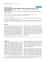
authors obtained the crystal structure of the Mth ecto-
domain bound to an RWR peptide, which revealed the
binding to occur near the carboxyl terminus of the ecto-
domain (Figure 1), at a site different from the ligand-binding
site previously proposed on the basis of the structure of the
Mth ectodomain alone [13]. The RWR peptides competed
with the amino-terminal portion of Sun (N-Sun), indicating
that the native ligand may also bind the carboxy-terminal
part of the ectodomain. Because the competition may have
occurred through allosteric interactions between two
separate sites, further work is required to establish the
binding site of the native ligand. Experiments in cell culture
showed that an RWR peptide can bind the whole Mth
receptor presented on the cell surface and act as an
antagonist of Mth activation by its native ligand, the latter
revealed by a reduction in N-Sun-induced Mth-dependent
calcium mobilization.
As well as providing an important insight into the
molecular functioning of the receptor, the identification of
a peptide antagonist of Mth demonstrates that peptides
with biologically relevant activity towards GPCRs, an
important class of drug targets [14], can be obtained by in
vitro selection of mRNA-peptide fusions. Furthermore, a
multitude of orphan GPCRs are present in animal genomes
[15] and the elucidation of their function will be aided by an
unbiased identification of modulators of their activity. Most
importantly for aging research, Ja and co-workers [2] have
provided us with a biochemically characterized tool for the
examination of the in vivo function of Mth.
Methuselah antagonist and life span
The authors initiated the examination of the effects of the
Mth antagonist in vivo. They expressed an RWR peptide
under the control of the GAL4 transcription factor that was
in turn expressed from the daughterless promoter. This
ubiquitous expression of the antagonist led to extension of
life span of males at 29°C and 25°C. The result indicates that
the RWR peptide can antagonize the activity of Mth in vivo.
Indeed, mutations in the RWR motif, critical for binding to
Mth in vitro, abolished the life span extension. It is still
possible that the peptide may act through another GPCR,
however, as the specificity of the peptide for Mth among
Drosophila GPCRs, including 12 Mth paralogs, is unknown,
making it important to examine the genetic interactions
between mth and its agonist and antagonist. It will also be
reassuring to see that the effect of the antagonist on life span
is robust to placing the transgenes into a standard genetic
background.
The authors carried out the life-span experiment under
conditions in which the effect of modulating Mth activity
should be most pronounced. Because the effect of mth
1
222.2 Genome Biology 2007, Volume 8, Issue 8, Article 222 Alic and Partridge />Genome Biology 2007, 8:222
Figure 1
The location of RWR peptide binding to the Mth ectodomain. The Mth
ectodomain structure [13] was visualized with PyMOL (PyMOL Molecular
Graphics System). The tryptophan (W) residue previously thought to be
positioned at the ligand-binding site is indicated in green, and the region
near the carboxyl terminus to which the RWR peptide was located by Ja
et al. [2] is indicated by an orange circle.
appears to depend on environmental conditions, mating
status and the sex of the flies, it will be interesting to expand
on the initial findings of Ja and co-workers [2], including the
examination of the effects of the antagonist in different
genetic backgrounds. Indeed, the antagonist may prove
useful in establishing mechanistic links between the
environmental conditions and mth
1
phenotypes, as well as
the connections between different phenotypes.
In the system the authors used, the peptide was not targeted
for excretion, while the receptor binding site is extracellular.
This would imply that the peptide engages the receptor
before extracellular presentation of the ectodomain. If this is
the case, the antagonist will only affect the Mth in the cell
that expresses it: it will be acting autonomously. It will be
interesting to know if the secreted version of the peptide can
act in a cell-nonautonomous manner. And conversely, the
system that is described by Ja et al. [2] may provide a tool
for the examination of physiological outcomes of tissue-
restricted Mth inhibition.
At least 30% of currently available drugs act on GPCRs [14].
Ja and co-workers [2] demonstrated that a targeted design
of an artificial modulator of GPCR activity could extend life
span. Although Mth has no homologs in humans, is it likely
that a modulator of a signaling pathway could be used as a
drug to delay aging? It will probably not be as simple as
that. Signaling pathways that affect aging in model
organisms have pleiotropic effects. Many of these would be
undesirable in humans, including the reduced reproductive
output in mth
1
flies, or the diabetic phenotypes in long-lived
flies with reduced production of insulin-like peptides [16].
Furthermore, mth
1
is an interesting example of the fact that
a longer life span need not mean a healthier old age; the
delay in aging, observed in the mth
1
mutants on a
population level, appears to be uncoupled from a delay in
age-related functional decline in olfaction and motor
activity [17,18]. Much further work is required to gain a
detailed understanding of the molecular mechanisms that
underlie ageing, and the reagents developed by Ja and
co-workers [2] will help just that.
Acknowledgements
We acknowledge funding from the Wellcome Trust Functional Genomic
Analysis of Ageing Grant (LP) and European Molecular Biology Organiza-
tion Long-Term Fellowship (NA). We thank M Piper for a critical reading
of the manuscript and WW Ja and RW Roberts for their help in obtaining
the image of the Mth ectodomain.
References
1. Lin YJ, Seroude L, Benzer S: Extended life-span and stress resis-
tance in the Drosophila mutant methuselah. Science 1998, 282:
943-946.
2. Ja WW, West AP Jr, Delker SL, Bjorkman PJ, Benzer S, Roberts RW:
Extension of Drosophila melanogaster life span with a GPCR
peptide inhibitor. Nat Chem Biol 2007, 3:415-419.
3. Mockett RJ, Sohal RS: Temperature-dependent trade-offs
between longevity and fertility in the Drosophila mutant,
methuselah. Exp Gerontol 2006, 41:566-573.
4. Baldal EA, Baktawar W, Brakefield PM, Zwaan BJ: Methuselah life
history in a variety of conditions, implications for the use of
mutants in longevity research. Exp Gerontol 2006, 41:1126-1135.
5. Song W, Ranjan R, Dawson-Scully K, Bronk P, Marin L, Seroude L,
Lin YJ, Nie Z, Atwood HL, Benzer S, et al.: Presynaptic regulation
of neurotransmission in Drosophila by the G protein-coupled
receptor methuselah. Neuron 2002, 36:105-119.
6. Harmar AJ: Family-B G-protein-coupled receptors. Genome Biol
2001, 2:reviews3013.1-3013.10.
7. Cvejic S, Zhu Z, Felice SJ, Berman Y, Huang XY: The endogenous
ligand Stunted of the GPCR Methuselah extends lifespan in
Drosophila. Nat Cell Biol 2004, 6:540-546.
8. Tatar M, Kopelman A, Epstein D, Tu MP, Yin CM, Garofalo RS: A
mutant Drosophila insulin receptor homolog that extends
life-span and impairs neuroendocrine function. Science 2001,
292:107-110.
9. Clancy DJ, Gems D, Harshman LG, Oldham S, Stocker H, Hafen E,
Leevers SJ, Partridge L: Extension of life-span by loss of
CHICO, a Drosophila insulin receptor substrate protein.
Science 2001, 292:104-106.
10. Kapahi P, Zid BM, Harper T, Koslover D, Sapin V, Benzer S: Regula-
tion of lifespan in Drosophila by modulation of genes in the
TOR signaling pathway. Curr Biol 2004, 14:885-890.
11. Wang MC, Bohmann D, Jasper H: JNK signaling confers toler-
ance to oxidative stress and extends lifespan in
Drosophila.
Dev Cell 2003, 5:811-816.
12. Roberts RW, Szostak JW: RNA-peptide fusions for the in vitro
selection of peptides and proteins. Proc Natl Acad Sci USA 1997,
94:12297-12302.
13. West AP Jr, Llamas LL, Snow PM, Benzer S, Bjorkman PJ: Crystal
structure of the ectodomain of Methuselah, a Drosophila G
protein-coupled receptor associated with extended lifespan.
Proc Natl Acad Sci USA 2001, 98:3744-3749.
14. Wise A, Gearing K, Rees S: Target validation of G-protein
coupled receptors. Drug Discov Today 2002, 7:235-246.
15. Howard AD, McAllister G, Feighner SD, Liu Q, Nargund RP, Van der
Ploeg LH, Patchett AA: Orphan G-protein-coupled receptors
and natural ligand discovery. Trends Pharmacol Sci 2001, 22:132-
140.
16. Broughton SJ, Piper MD, Ikeya T, Bass TM, Jacobson J, Driege Y,
Martinez P, Hafen E, Withers DJ, Leevers SJ, et al.: Longer lifespan,
altered metabolism, and stress resistance in Drosophila
from ablation of cells making insulin-like ligands. Proc Natl
Acad Sci USA 2005, 102:3105-3110.
17. Cook-Wiens E, Grotewiel MS: Dissociation between functional
senescence and oxidative stress resistance in Drosophila. Exp
Gerontol 2002, 37:1347-1357.
18. Petrosyan A, Hsieh IH, Saberi K: Age-dependent stability of sen-
sorimotor functions in the life-extended Drosophila mutant
methuselah. Behav Genet 2007, 37:585-594.
Genome Biology 2007, Volume 8, Issue 8, Article 222 Alic and Partridge 222.3
Genome Biology 2007, 8:222